Article

Histological and Molecular Characterisation of Gonadal Phenotypes Confirms Diandry in a Protogynous Wrasse †

1
School of Science, University of Waikato, Tauranga 3110, New Zealand
2
Department of Anatomy, University of Otago, Dunedin 9016, New Zealand
*
Author to whom correspondence should be addressed.
This manuscript includes part of a Master of Science thesis by the first author T. Hamer. Ecology and Biodiversity Program in the University of Waikato.
Fishes 2025, 10(11), 554; https://doi.org/10.3390/fishes10110554 (registering DOI)
Submission received: 31 August 2025 / Revised: 21 October 2025 / Accepted: 23 October 2025 / Published: 2 November 2025

Abstract

Sequentially hermaphroditic fish begin life as one sex and change to another at some stage of their lifecycle. To date, the majority of research into this fascinating process has focused on sex change in sexually mature fish. The current study on the New Zealand spotty wrasse (Notolabrus celidotus) seeks to describe sex change in sexually immature females that are transitioning to become so called initial-phase males. This was achieved through analysis of gonadal histology, and the expression of key genes involved in sex differentiation (amh, dmrt1, and cyp19a1a) and methylation (dnmt1 and dnmt3a). It was found that initial-phase (IP) male spotty wrasse likely reach sexual maturity at a much earlier developmental stage and smaller size than previously realised. This study furthermore shows that all juvenile spotty wrasse are female first, before some individuals undergo pre-maturational sex change to become IP males. Thus, we confirm diandry in the protogynous New Zealand spotty wrasse and provide clarity around the definitions of primary male formation.
Key Contribution: This study reveals that the spotty wrasse, a popular model species for the study of teleost sex change, is diandric rather than monandric, as previously thought. By describing sex change in immature individuals, the research provides a clearer definition for what constitutes primary male formation in diandric species.

1. Introduction

Sex determination and gonadal development are extraordinarily plastic in teleost fish. While the majority of fish have a fixed sex throughout their life, there are differences in the pattern of gonadal development between gonochoristic species. In primary gonochoristic species, early gonadal development proceeds from an undifferentiated gonad to directly yield either an ovary or testis [1]. Alternatively, in undifferentiated gonochorists, the gonads of all individuals initially develop ovarian tissue. This ovarian tissue then degenerates in approximately half of the population, and the gonad is subsequently invaded by additional somatic cells [2,3]. The gonad will transition through an intersexual state before becoming a testis [4]. However, these fish will only functionally reproduce as one sex throughout their life cycle.
Approximately six percent of all fish species show the remarkable ability to reproduce as both sexes at some point in their life history [5]. While some teleost hermaphrodites function simultaneously as male and female, others transition sequentially between sexes [6]. Sequential hermaphroditism exists in three forms: 1. protogynous (female-to-male), as described in species such as bluehead wrasse (Thalassoma bifasciatum) [7], 2. protandrous (male-to-female), as described in barramundi (Lates calcarifer) [8], and 3. serial bidirectional sex change, as seen in Māori coral gobies (Gobidion histrio) [9]. In all cases, functional sex reversal entails radical restructuring of the gonad, alongside changes in morphology and behaviour [10,11].
The evolution of protogynous sex change is thought to be largely driven by a male size advantage. Here, large males use aggressive territorial defence to monopolise matings with females [11]. This disadvantages small males and leads to strong selection for size-based protogyny and reproductive strategy. By initially reproducing as a small female and then changing to become male at a larger size, in order to breed with multiple females, an individual may magnify its reproductive output and success [12]. In contrast, a female-biassed reproductive size advantage favours protandry. This is associated with monogamy or systems without male territorial social structures [13].
However, another less common male pathway also exists; initial-phase (IP) males are relatively small in size and use a sneaker spawning strategy to compete with dominant terminal-phase fish rather than competing for territorial dominance [7]. Very few studies have described sex differentiation in IP males from either tropical or temperate wrasse, and as such, the timing and regulation of sex change in individuals using this strategy remains uncharacterised [13]. The existence of the IP male phenotype in some protogynous species and not in others means that protogyny can be further separated into two divisions based on male reproductive strategy: in monandric protogyny, males are derived exclusively from sexually mature functional females to form a characteristically structured secondary testis. Monandry has been observed in foxfish (Bodianus frenchii) [14], hogfish (Lachnolaimus maximus) [15], and Ballan wrasse (Labrus bergylta) [16]. In diandric protogyny, on the other hand, males develop from one of two developmental pathways: either from sexually mature females, as in monandry, or directly as juvenile fish [17]. Typically, diandric males have two colour morphs; IP males tend to mimic female colour and patterning and are less abundant than terminal-phase (TP) males, which usually have dimorphic markings in comparison. Interestingly, IP males can also transition to assume TP markings upon the death or removal of a dominant TP fish [18]. Diandric IP and TP male fish typically have different testicular structures. While TP males tend to have a remnant ovarian lumen in the centre of the secondary testis, IP males usually lack this lumen, as has been described in T. bifasciatum [19] and saddle wrasse (T. Duperrey) [20]. However, some species have been described as having no morphological testicular differences despite being labelled as diandric [21].
The New Zealand spotty wrasse (Notolabrus celidotus) is a small (<280 mm TL) endemic protogynous hermaphrodite found on temperate reefs around New Zealand. Sexual maturity has been described to occur at approximately 110 mm, and subsequent sex change typically leads to the formation of a secondary testis [22,23,24,25]. In spotty wrasse, TP colour change occurs after sex change. Initial-phase males typically possess a primary testis and mimic female IP patterning to suit their sneaker reproductive tactic.
Despite the fact that spotty wrasse have two male morphs, there is still confusion in the literature as to whether they qualify as a monandric or diandric species. Early research by Jones (1980) [22] classified spotty wrasse as monandric, as all of the male individuals in this study were found to have secondary testes, with a residual ovarian lumen. Additionally, Jones suggests that all spotty wrasse begin as female, which further supports monandry. A similar study by McBride & Johnson (2007) [15] classified the hogfish as monandric based on the same rationale. Terminal-phase male spotty wrasse usually have a remnant ovarian lumen in the testes [25], whereas another testis type, mostly seen in IP males, also exists and hints at diandry. These testes appear solid without a central lumen and are believed to result from the testis rupturing along a seam in the tunicata albuginea and evaginating back on itself [22]. The fact that this species has two different male developmental strategies and morphologies, yet may always start life as female, makes classification of monandry or diandry equivocal. This study seeks to clarify where in the life cycle spotty wrasse first begin to change sex and whether sex change occurs prior to female puberty. This is achieved using histological and molecular analysis of juvenile and sexually mature N. Celidotus.

2. Materials and Methods

2.1. Fish Husbandry

A total of 141 spotty wrasse were captured using baited traps from May to June 2022 in the Tauranga Harbour (37°640411, 176°181424), New Zealand. Fish were housed in recirculating seawater tanks (1600 L) at the Toi Ohomai Institute of Technology Aquaculture Lab. Water quality parameters (dissolved oxygen, salinity, temperature, ammonia, nitrite, and nitrate) were monitored daily, and fish were fed fresh greenshell mussels (Perna canaliculus) three times a week until sampling. Fish were handled in accordance with the New Zealand Animal Welfare Act.

2.2. Gonadal Dissection and Histology

Prior to dissection, fish were heavily sedated in an aerated 10 L seawater bath of 2-phenoxyethanol (0.6 mL L−1). They were then weighed (g), measured (mm, total length), and external body photographs were taken. Fish were subsequently euthanized by rapid decapitation with a sharp knife. After an abdominal incision was made along the ventral surface, gonadal lobes were dissected from the fused posterior region of the gonad. One whole gonad lobe and the fused posterior region were preserved in Bouin’s solution for 24 h and then transferred to 70% ethanol until sectioning. Fixed samples were processed by the Otago University Histology unit as described previously [26]: gonad tissues were serially dehydrated, cleared, infiltrated, embedded in paraffin for histological sectioning at 3–4 μm, and finally stained with hematoxylin and eosin. Histological slides were viewed under an Olympus BX53 compound microscope for close tissue examination, fitted with an Olympus DP27 camera and connected to a laptop with CellSens Entry software (version 4.2). Gonadal histology samples were categorised into one of six stages (Table 1): juvenile female (JF) (<110 mm TL), adult female (AF) (>110 mm TL), early-transitional (ET), mid-transitional (MT), late-transitional (LT), and IP male (IP), in accordance with [24].

2.3. Quantitative PCR

Quantitative polymerase chain reaction (PCR) was performed on gonadal samples from four IPM fish (77–105 mm TL), thirteen JF fish (73–106 mm TL), and five AF fish (128–270 mm TL). These fish showed no signs of sex change and were thus selected to illustrate the difference in gene expression for both a functional and fully formed male and female at each end of the sex change pathway. In addition, seven ET fish (52–95 mm TL) in the earliest detectable stages of transitioning and five MT fish (60–76 mm TL) were also selected and used to examine levels of gene expression once sex change began. One LT individual had tissue available for gene expression analysis. Gene sequences for the following genes were identified within the available genome (fNotCel1.alt; GenBank assembly accession: GCA_009762545.1): glucose-6-phosphate dehydrogenase (g6pd), β-actin 2 (actb2), cytochrome P450 family 19 subfamily A polypeptide 1a (cyp19a1a), anti-Mullerian hormone (amh), doublesex- and mab-3 related transcription factor 1 (dmrt1), DNA methyltransferase 1 (dnmt1), and DNA methyltransferase 3a (dnmt3a).
RNA was extracted from gonad tissue samples using the Direct-zol™ RNA Miniprep kit (Zymo Research, Irvine, CA, USA, Catalogue Number: R2053). Extraction of total RNA from ~100 mg of gonad tissue was carried out following the manufacturer’s instructions. RNA concentration and purity was determined using the DeNovix® DS-11 spectrophotometer (DeNovix, Wilmington, DE, USA). The mRNA was then used immediately for cDNA synthesis and then stored at −20 °C to minimise RNA degradation. To synthesise cDNA, the qScript™ XLT cDNA SuperMix kit (Quanta Biosciences, Beverley, MA, USA) was used, according to the manufacturer’s instructions. qPCR was performed using a 48-well Magnetic Induction Cycler (MIC) qPCR (Bio Molecular Systems, Upper Coomera, QLD, Australia), using PerfeCTa® SYBR® Green FastMix® (Quanta Biosciences, USA) and following the manufacturer’s instruction.
Expression levels of the selected genes were normalised to two housekeeping genes, g6pd and actb2 (Table 2). Expression ratios were calculated using the geometric mean of the two housekeeping genes. Statistical analyses were performed using GraphPad Prism (version 10.6.1). Data were tested for homogeneity of variance using an F test and transformed using the natural logarithm where necessary. Differences in gene expression among gonadal stages were tested using one-way Analysis of Variance (ANOVA) or the Kruskal–Wallis test if the data were not normally distributed (Shapiro–Wilk test). Tukey’s multiple comparisons test was used for post hoc testing of differences between individual gonadal stages. One gonadal stage (LT) was represented by a single individual. Therefore, it was excluded from the statistical analysis but included in figures for descriptive comparison only. Differences were considered statistically significant when p < 0.05.

3. Results

3.1. Gonadal Histology

Histological signs of prepubertal sex change were evident in approximately 21% of the fish sampled, with an additional 5.5% being IP male and the remaining 73.5% being IP female (Table 3). Only sexually mature IP females were found with oocytes beyond the previtellogenic stage (Figure 1). The 75 juvenile female fish had small previtellogenic oocytes and ranged between 64 and 110 mm TL. The ovaries of the 19 ET fish contained a combination of healthy and degenerating previtellogenic oocytes. The ET group also included the smallest transitional fish (52 mm TL). Only five MT fish were identified (60–72 mm TL). The ovaries of these fish harboured many atretic oocytes and gonial cells, as well as putative cysts of spermatocytes. Tissue remodelling was also more evident with the increased presence of stromal cells, eosinophilic granulocytes, and associated cellular debris. The gonads of the four LT fish (72–104 mm TL) were characterised by an increased proportion of spermatogenic germ cells and the early signs of lobular arrangement. Of the eight IP males collected, the smallest was 77 mm, indicating an overlap in length between LT and IPM stages. The IP male testes lacked an obvious central lumen and typically contained various stages of spermatogenic germ cells, including spermatozoa.

3.2. Gene Expression Profiles

3.2.1. Anti-Mullerian Hormone

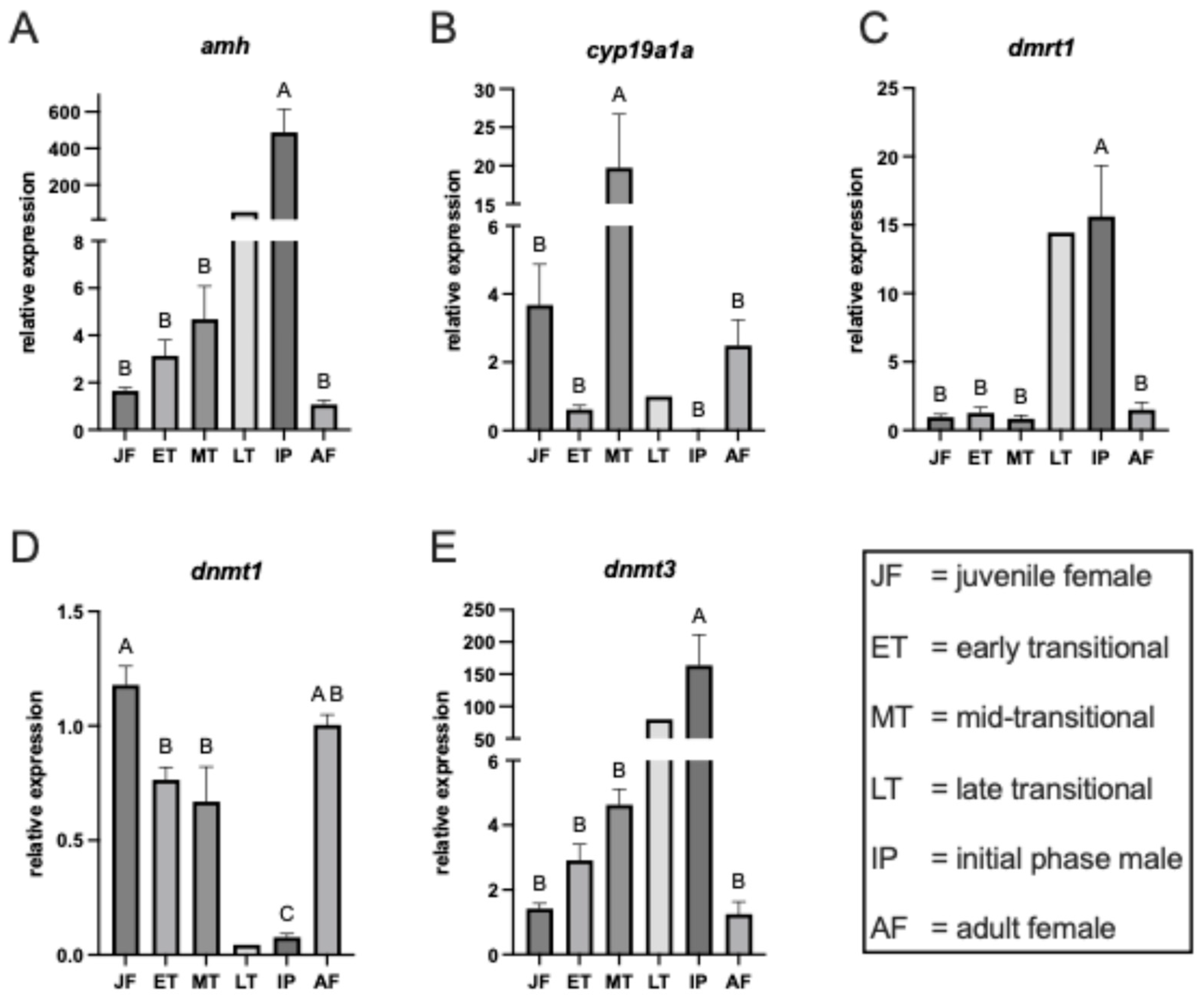
Amh is a key regulator of male sex differentiation, promoting testis development and inhibiting female reproductive structures (4). Amh showed a strong trend of increasing expression across sex change (Figure 2A; F = 25.03, p < 0.0001, ANOVA), ranging from 1.65 ± SE 0.14 in JF to 52.4 in LT. Expression was by far the highest in IP however (488 ± 127).

3.2.2. Gonadal Aromatase

Cyp19a1a encodes aromatase, the enzyme responsible for converting androgens to estrogens, and is therefore critical for ovarian development and maintenance of the female pathway. Gonadal aromatase expression showed a significant change in expression across sex change (Figure 2B; F = 6.67, p < 0.001, ANOVA). This change initially indicates a decreasing trend across sex change, from JF (mean = 3.67 ± 1.20) to nearly zero in IPM (mean = 0.004 ± 0.0019). However, this was punctuated by an unexpected increase in gonadal cyp19a1a expression in MT fish (Mean = 19.65 ± 6.23). Although results suggest initial downregulation, there was no significant decrease in gene expression from JF to ET (Mean = 0.61 ± 0.12). However, there was significant upregulation from JF to MT (p < 0.01), and a further significant increase from ET to MT (p < 0.001).

3.2.3. Doublesex and mab-3 Related Transcription Factor 1

Dmrt1 is a conserved transcription factor essential for testis differentiation and maintenance of male fate across vertebrates. Expression of dmrt1 showed a similar trend to amh, with increasing expression across sex change (Figure 2C; F = 27.5, p < 0.0001, ANOVA), from JF (Mean = 0.95 ± 0.2) to IPM (Mean = 15.62 ± 3.72). Expression was also remarkably high in the LT sample (14.4).

3.2.4. DNA Methyltransferase 1

Dnmt1 maintains established DNA methylation patterns during cell division, contributing to the stable inheritance of gene expression states that regulate sexual phenotype. The expression pattern of dnmt1 was found to be inversely correllated with that of dnmt3a, with significantly different expression levels across sex change (Figure 2D; F = 16.52, p < 0.0001, ANOVA). Expression was highest in JF (1.18 ± 0.1) and gradually decreased across sex change, with the lowest levels found in LT (0.04) and IP (0.08 ± 0.04).

3.2.5. DNA Methyltransferase 3a

Dnmt3a Catalyses de novo DNA methylation, thereby establishing new epigenetic markers that can reprogram gene expression during gonadal differentiation. Dnmt3a expression showed a strong trend of increasing expression across sex change (Figure 2E; F = 20.44, p < 0.0001, ANOVA), ranging from 1.42 ± 0.16 in JF to 164 ± 47.5 in IP. Expression levels were highly similar between JF and AF (1.25 ± 0.4).

4. Discussion

The study of sex change in diandric protogynous hermaphrodites has focused primarily on the transition from female to terminal-phase male [27,28,29] and rarely includes a detailed description of the prepubertal IP male formation. Considering that other species, such as bluehead wrasse and orange-spotted grouper (E. coioides), have two developmental pathways to becoming male, it seems logical to determine any differences in development between IP and TP males. From the current study, it is evident that the developmental pathway from juvenile female to IP male closely resembles the TP pathway in spotty wrasse, albeit with different timing. In the current study, New Zealand spotty wrasse were classified into three different transitional states (ET, MT, and LT) between female and IP male based on gonadal histology. Individuals identified as either initiating sex change or as sexually mature males were found to measure as small as 52 mm TL and 77 mm TL, respectively. This is considerably smaller than previously recorded in the literature [22]. However, studies of further gonadal development shows that the vast majority of IP male spotty wrasse has solid testes with centrally located sperm-collecting ducts and lacks a remnant ovarian lumen, while the opposite is typically true of TP males. Early descriptions in this species suggest that the solid testis structure may arise through evagination of the gonad [22]. Our unpublished observations in adult male spotty wrasse support this theory. Due to the small size of the dissected gonads of the IP males in the current study, we were unable to verify the persistence of an ovarian lumen or the evagination process leading to a solid testis. However, it seems clear that in this species, both the solid evaginated testis and the ‘hollow’ testis with a remnant lumen develop first from an ovary, as either prepubertal or sexually mature fish. Terminal-phase males typically arise from the largest and most dominant female in a social group undergoing sex change. Our laboratory observations also indicate that in the absence of a TP male, comparatively small IP males can assume the TP male markings and role despite the presence of larger IP females. This may result from the advantageous ability that IP males possess to immediately elevate androgen production and thereby rapidly assume TP male secondary sexual characteristics.
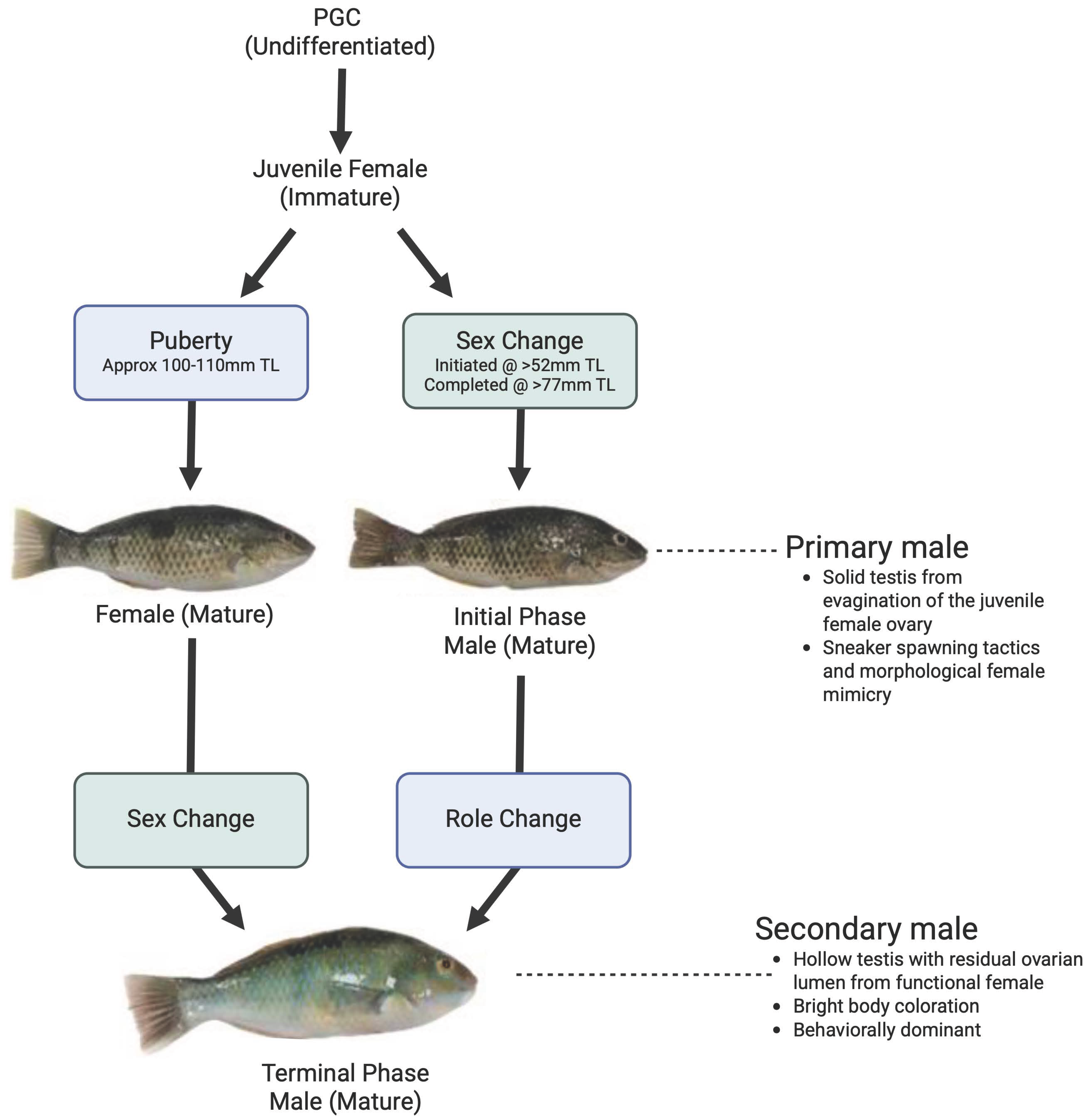
Initial-phase male spotty wrasse undergo puberty earlier than IP females, which may also convey a reproductive advantage to these individuals. Transitional fish were largely categorised according to the relative presence or absence of female and male germ cell types. Mid-transitional fish typically contained cysts of putative meiotic spermatocytes, were not present in ET individuals (Figure 3). The testes of fish at subsequent sexual stages (LT, IPM) also contained cysts of later-stage spermatogenic germ cells. In male fish, puberty is defined as the onset of the first wave of rapid spermatogonial proliferation, which occurs as a shift from the slow self-renewal of type-A spermatogonia into a rapid mitotic proliferation of type-B spermatogonia [30]. These type B spermatogonia will differentiate into meiotic spermatocytes and are already committed to the developmental pathway to become spermatozoa [31]. Based on these criteria, spotty wrasse undergoing IP male sex change are pubertal from MT/LT stages onward and may be at least 30 mm shorter than pubertal IP females (Figure 4). After puberty, IP female spotty wrasse are subject to aggression from larger females, which socially inhibits oocyte ripening [31]. Consequently, by reaching sexual maturity at a smaller size, IP male fish are likely to reproduce sooner, which may also increase their overall reproductive fitness in comparison to IP females.
The gene expression patterns of amh, cyp19a1a, dmrt1, dnmt1, and dnmt3a provided further validation for the histological classifications of early transitioning individuals, as it was often difficult to identify between oogonial and spermatogonial germ cells by morphology alone [25]. All five genes showed significant changes across sex change. Individuals classified through histology as early-transitional and mid-transitional showed significant upregulation in amh expression from juvenile female and greater significance from juvenile female to mid-transitional. Therefore, this data provides supporting evidence for the concept that amh may form part of the molecular trigger that initiates sex change, and its upregulation could be a useful early molecular marker for protogynous species [25,26,27,28,29,30,31,32,33]. Expression of dmrt1, a key gene involved in the male developmental pathway, increased significantly in mid-transitional fish, suggesting a key role in progressing rather than initiating sex change [32,33,34]). While the feminising gene cyp19a1a showed an overall trend of decreasing expression with sex change, results indicated a drastic upregulation in mid-transitional fish. During protogynous sex change, expression profiles of cyp19a1a typically reflect an opposing pattern to the upregulation of male-biassed genes, such as amh and dmrt1 [33]. This unexpected increase in cyp19a1a expression in mid-transitional fish requires further investigation in the absence of an obvious explanation. The expression pattern of dnmt1 and dnmt3a mirrors that observed during sex change in adult spotty wrasse and bluehead wrasse, highlighting the role of epigenetic regulation in both the female and male phenotypes [24,33]. Epigenetic modification of the cyp19a1a promoter region was suggested to regulate sex change in orange-spotted grouper [35]. Collectively, these results indicate that all three sex differentiation genes play a similar role in prepubertal sex change leading to IP male formation as they do in TP formation in adult spotty wrasse.
Throughout the literature on early development in protogynous hermaphrodites, a common theme is a lack of cohesion in the terminology and different definitions used to describe early development. This is likely in part due to the vast diversity and plasticity across different teleost orders. For example, in some species considered to be gonochorists, all juveniles initially develop ovaries. Males form through the degeneration of the ovarian tissue and subsequent development of testes while still immature. These fish remain male for life and have been termed undifferentiated gonochorists by some authors [2,3]). Yet this concept of gonochorism is contradicted by other definitions in the literature, where gonochorists are defined as having separate sexes that are fixed throughout their entire life cycle [4]. Certainly, definitions of sex differentiation based on the presence of specific stages of germ cell development would qualify these juvenile fish as being sexually differentiated females. Alternatively, other authors describe fish similar to undifferentiated gonochorists as non-functional hermaphrodites [36]. This terminology seems to align more closely with the gonadal transformations that occur in fish that have an all-female juvenile stage. In contrast to these fish, functional hermaphrodites have individuals who reproduce as one sex and then the other or both simultaneously. Although it is not necessarily the intent of these definitions, it could be argued that spotty wrasse technically have males that can arise as either non-functional or functional hermaphrodites. This highlights both the diversity of fish sexual strategies and associated terminology in the literature.
It is often argued that fish that have two male morphs with at least one arising after sexual maturity can be considered functional hermaphrodites. Still, a lack of clarity exists around the definitions of primary male formation. These definitions become important when distinguishing between monandry and diandry. Sadovy and Liu (2008) state that primary males arise prior to sexual maturity while secondary males arise from sex change after sexual maturation [5]. In the literature, primary males often possess solid testes that lack an ovarian lumen, in contrast to the testes of secondary males. Robertson, Reinboth, & Bruce (1982) describe the existence of what they term ‘functional analogues’ of primary males among the parrotfish (L. vaigiensis, C. spinidens and C. carolinus) [37]. These fish have males arising from females prior to sexual maturity and retain a remnant ovarian lumen. Liu & Sadovy (2004) first pose the question as to whether sex ‘change’ can be said to occur in pre-maturational individuals based on histological criteria [38]. These authors further suggest that prepubertal development from female to male cannot be classified as sex change due to the lack of sexual function. Instead, the authors consider such pre-maturational transitions between ovary and testis to be an additional form of sex differentiation involving a transitory bisexual gonad. This further contrasts with Robertson & Warner (1979), who suggest that pre-maturational sex change appears common in parrotfish, which are also among the Labridae family [39].
Here, they report female individuals that change sex to become male prior to sexual maturity and then go on to spend their adult lives as males with functional testes. In the current study, IP male formation in spotty wrasse appears to follow a similar pattern to that of both the grouper studied by Liu & Sadovy (2009) [40] and the parrotfish described by Robertson et al. (1982) [37]. The fact that all of these fish have a differentiated ovary prior to IP male formation yet use different descriptors for the same physiological transition highlights the ambiguity of the terminology in the literature. In this study preference is given to the term pre-maturational sex change to describe IP male formation. To this extent, the expression pattern of the molecular markers reported here largely reflects those observed to regulate adult sex change in this species. Based on the results of the current study, the New Zealand spotty wrasse does not seem to meet the requirements for monandry. This is supported by the fact that all individuals appear to first differentiate as female before some undergo pre-maturational sex change to become IP males. The fact that sex change occurs at different ontogenetic stages, often leading to males with different gonadal structures and sexual strategies, adds further weight to the conclusion that spotty wrasse are, in fact, diandric protogynous hermaphrodites.

Author Contributions

Conceptualization, S.M.; methodology, T.E.H., H.A.R., N.M., S.B. and S.M.; formal analysis, T.E.H., H.A.R., N.M., S.B., K.K. and S.M.; investigation, T.E.H., H.A.R., N.M. and S.M.; data curation, T.E.H., H.A.R., N.M., K.K. and S.M.; writing—original draft preparation, T.E.H., H.A.R., K.K. and S.M.; writing—review and editing, K.K. and S.M.; visualisation, T.E.H., K.K. and S.M.; supervision, S.B. and S.M.; project administration, S.M.; funding acquisition, S.M. All authors have read and agreed to the published version of the manuscript.

Funding

This research was funded by the University of Waikato.

Institutional Board Review Statement

The study was approved by the Animal Ethics Committee of Toi Ohomai Insitute of Technology and the University of Waikato. The approval code is AEC 2020_02; the approval date is 6 April 2020.

Data Availability Statement

The original contributions presented in this study are included in the article. Further inquiries can be directed to the corresponding author(s).

Conflicts of Interest

The authors declare no conflicts of interest.

Abbreviations

The following abbreviations are used in this manuscript:
AMHAnti-Mullerian hormone
AtOAtretic oocyte
Cyp19a1aGonadal aromatase
Dmrt1Doublesex- and mab-3 related transcription factor
Dnmt1DNA methyltransferase 1
Dnmt3aDNA methyltransferase 3a
EGrEosinic granulocytes
GonGonial cells
OogOogonia
PvOPrevitellogenic oocyte
SpcSpermatocytes
SpgSpermatogonia
SptSpermatids
StCStromal cells
VitVitellogenic oocyte

References

  1. Yamamoto, T.-O. 3 Sex differentiation. Fish Physiol. 1969, 3, 117–175. [Google Scholar]
  2. Takahashi, H. Juvenile hermaphroditism in the zebrafish, Brachydanio rerio. Bull. Fac. Fish. Hokkaido Univ. 1977, 28, 57–65. [Google Scholar]
  3. Takahashi, H.; Shimizu, M. Juvenile Intersexuality in a Cyprinid Fish, the Sumatra Barb. Bull. Fac. Fish. Hokkaido Univ. 1983, 34, 69–78. [Google Scholar]
  4. Devlin, R.H.; Nagahama, Y. Sex determination and sex differentiation in fish: An overview of genetic, physiological, and environmental influences. Aquaculture 2002, 208, 191–364. [Google Scholar] [CrossRef]
  5. De Mitcheson, Y.S.; Liu, M. Functional hermaphroditism in teleosts. Fish Fish. 2008, 9, 1–43. [Google Scholar] [CrossRef]
  6. Leonard, J.L. Sexual conflict in simultaneous hermaphrodites: Evidence from serranid fishes. Environ. Biol. Fishes 1993, 36, 135–148. [Google Scholar] [CrossRef]
  7. Warner, R.R.; Robertson, D.R.; Leigh, E.G., Jr. Sex Change and Sexual Selection: The reproductive biology of a labrid fish is used to illuminate a theory of sex change. Science 1975, 190, 633–638. [Google Scholar] [CrossRef]
  8. Domingos, J.A.; Budd, A.M.; Banh, Q.Q.; Goldsbury, J.A.; Zenger, K.R.; Jerry, D.R. Sex-specific dmrt1 and cyp19a1 methylation and alternative splicing in gonads of the protandrous hermaphrodite barramundi. PLoS ONE 2018, 13, e0204182. [Google Scholar] [CrossRef]
  9. Todd, E.V.; Liu, H.; Muncaster, S.; Gemmell, N.J. Bending genders: The biology of natural sex change in fish. Sex. Dev. 2016, 10, 223–241. [Google Scholar] [CrossRef]
  10. Godwin, J. Social determination of sex in reef fishes. Semin. Cell Dev. Biol. 2009, 20, 264–270. [Google Scholar] [CrossRef]
  11. Warner, R.R. Mating behavior and hermaphroditism in coral reef fishes. Am. Sci. 1984, 72, 128–136. [Google Scholar]
  12. Charnov, E. Alternative life-histories in protogynous fishes: A general evolutionary theory. Mar. Ecol. Prog. Ser. 1982, 9, 305–307. [Google Scholar] [CrossRef]
  13. Munday, P.L.; Wilson White, J.; Warner, R.R. A social basis for the development of primary males in a sex-changing fish. Proc. R. Soc. B Biol. Sci. 2006, 273, 2845–2851. [Google Scholar] [CrossRef] [PubMed]
  14. Cossington, S.; Hesp, S.; Hall, N.; Potter, I. Growth and reproductive biology of the foxfish Bodianus frenchii, a very long-lived and monandric protogynous hermaphroditic labrid. J. Fish. Biol. 2010, 77, 600–626. [Google Scholar] [CrossRef]
  15. McBride, R.S.; Johnson, M. Sexual development and reproductive seasonality of hogfish (Labridae: Lachnolaimus maximus), an hermaphroditic reef fish. J. Fish. Biol. 2007, 71, 1270–1292. [Google Scholar] [CrossRef]
  16. Muncaster, S.; Andersson, E.; Kjesbu, O.S.; Taranger, G.L.; Skiftesvik, A.B.; Norberg, B. The reproductive cycle of female Ballan wrasse Labrus bergylta in high latitude, temperate waters. J. Fish. Biol. 2010, 77, 494–511. [Google Scholar] [CrossRef]
  17. Sadovy, Y.; Shapiro, D.Y. Criteria for the diagnosis of hermaphroditism in fishes. Copeia 1987, 1987, 136–156. [Google Scholar] [CrossRef]
  18. Kazancıoğlu, E.; Alonzo, S.H. A comparative analysis of sex change in Labridae supports the size advantage hypothesis. Evolution 2010, 64, 2254–2264. [Google Scholar] [CrossRef]
  19. McCaffrey, K.; Hawkins, M.B.; Godwin, J. Sexual phenotype differences in zic2 mRNA abundance in the preoptic area of a protogynous teleost, Thalassoma bifasciatum. PLoS ONE 2011, 6, e23213. [Google Scholar] [CrossRef]
  20. Nakamura, M.; Hourigan, T.F.; Yamauchi, K.; Nagahama, Y.; Grau, E.G. Histological and ultrastructural evidence for the role of gonadal steroid hormones in sex change in the protogynous wrasse Thalassoma duperrey. Environ. Biol. Fishes 1989, 24, 117–136. [Google Scholar] [CrossRef]
  21. Fennessy, S.T.; Sadovy, Y. Reproductive biology of a diandric protogynous hermaphrodite, the serranid Epinephelus andersoni. Mar. Freshw. Res. 2002, 53, 147–158. [Google Scholar] [CrossRef]
  22. Jones, G. Growth and reproduction in the protogynous hermaphrodite Pseudolabrus celidotus (Pisces: Labridae) in New Zealand. Copeia 1980, 1980, 660–675. [Google Scholar] [CrossRef]
  23. Thomas, J.T.; Todd, E.V.; Muncaster, S.; Lokman, P.M.; Damsteegt, E.L.; Liu, H.; Soyano, K.; Gléonnec, F.; Lamm, M.S.; Godwin, J.R. Conservation and diversity in expression of candidate genes regulating socially-induced female-male sex change in wrasses. PeerJ 2019, 7, e7032. [Google Scholar] [CrossRef]
  24. Muncaster, S.; Goikoetxea, A.; Lokman, P.; De Farias e Moraes, C.; Damsteegt, E.; Edgecombe, J.; Gemmell, N.; Todd, E. Genes involved in sex differentiation, epigenetic reprogramming, and cell fate regulate sex change in a wrasse. Rev. Fish Biol. Fish. 2023, 33, 281–294. [Google Scholar] [CrossRef]
  25. Goikoetxea, A.; Muncaster, S.; Todd, E.; Lokman, P.; Robertson, H.; De Farias e Moraes, C.; Damsteegt, E.; Gemmell, N. A new experimental model for the investigation of sequential hermaphroditism. Sci. Rep. 2021, 11, 22881. [Google Scholar] [CrossRef] [PubMed]
  26. Kamstra, K.; van der Burg, C.; Quertermous, H.M.; Muncaster, S.; Todd, E.V.; Jasoni, C.L.; Brown, C.; Gemmell, N.J. Neuroanatomy of a sex changing fish: The New Zealand spotty wrasse (Notolabrus celidotus) brain atlas. N. Z. J. Zool. 2024, 51, 228–239. [Google Scholar] [CrossRef]
  27. Candi, G.; Castriota, L.; Andaloro, F.; Finoia, M.; Marino, G. Reproductive cycle and sex inversion in razor fish, a protogynous labrid in the southern Mediterranean Sea. J. Fish Biol. 2004, 64, 1498–1513. [Google Scholar] [CrossRef]
  28. Shapiro, D.; Rasotto, M.B. Sex differentiation and gonadal development in the diandric, protogynous wrasse, Thalassoma bifasciatum (Pisces, Labridae). J. Zool. 1993, 230, 231–245. [Google Scholar] [CrossRef]
  29. Quertermous, H.M.; Kamstra, K.; van der Burg, C.A.; Muncaster, S.; Todd, E.; Jasoni, C.L.; Brown, C.; Gemmell, N. Behavioural and neural correlates of social hierarchy formation in a sex-changing fish. Proc. B 2025, 292, 20242097. [Google Scholar] [CrossRef]
  30. Taranger, G.L.; Carrillo, M.; Schulz, R.W.; Fontaine, P.; Zanuy, S.; Felip, A.; Weltzien, F.-A.; Dufour, S.; Karlsen, Ø.; Norberg, B. Control of puberty in farmed fish. Gen. Comp. Endocrinol. 2010, 165, 483–515. [Google Scholar] [CrossRef]
  31. Miura, T.; Higuchi, M.; Ozaki, Y.; Ohta, T.; Miura, C. Progestin is an essential factor for the initiation of the meiosis in spermatogenetic cells of the eel. Proc. Natl. Acad. Sci. USA 2006, 103, 7333–7338. [Google Scholar] [CrossRef]
  32. Jones, G.; Thompson, S. Social inhibition of maturation in females of the temperate wrasse Pseudolabrus celidotus and a comparison with the blennioid Tripterygion varium. Mar. Biol. 1980, 59, 247–256. [Google Scholar] [CrossRef]
  33. Todd, E.V.; Ortega-Recalde, O.; Liu, H.; Lamm, M.S.; Rutherford, K.M.; Cross, H.; Black, M.A.; Kardailsky, O.; Marshall Graves, J.A.; Hore, T.A.; et al. Stress, novel sex genes, and epigenetic reprogramming orchestrate socially controlled sex change. Sci. Adv. 2019, 5, eaaw7006. [Google Scholar] [CrossRef] [PubMed]
  34. Piferrer, F. Epigenetics of sex determination and gonadogenesis. Dev. Dyn. 2013, 242, 360–370. [Google Scholar] [CrossRef] [PubMed]
  35. Guo, C.-Y.; Tseng, P.-W.; Hwang, J.-S.; Wu, G.-C.; Chang, C.-F. Potential role of DNA methylation of cyp19a1a promoter during sex change in protogynous orange-spotted grouper, Epinephelus coioides. Gen. Comp. Endocrinol. 2021, 311, 113840. [Google Scholar] [CrossRef]
  36. Asoh, K.; Kasuya, M. Gonadal development and mode of sexuality in a coral-reef damselfish, Dascyllus trimaculatus. J. Zool. 2002, 256, 301–309. [Google Scholar] [CrossRef]
  37. Robertson, R.D.; Reinboth, R.; Bruce, R.W. Gonochorism, protogynous sex-change and spawning in three sparisomatinine parrotfishes from the western Indian Ocean. Bull. Mar. Sci. 1982, 32, 868–879. [Google Scholar]
  38. Liu, M.; Sadovy, Y. Early gonadal development and primary males in the protogynous epinepheline, Cephalopholis boenak. J. Fish Biol. 2004, 65, 987–1002. [Google Scholar] [CrossRef]
  39. Robertson, D.R.; Warner, R.R. Sexual Patterns in the Labroid Fishes of the Western Caribbean, II: The Parrotfishes (Scaridae); Smithsonian Institution Press: Washington, WA, USA, 1978. [Google Scholar]
  40. Liu, M.; Sadovy, Y. Gonad development during sexual differentiation in hatchery-produced orange-spotted grouper (Epinephelus coioides) and humpback grouper (Cromileptes altivelis) (Pisces: Serranidae, Epinephelinae). Aquaculture 2009, 287, 191–192. [Google Scholar] [CrossRef]
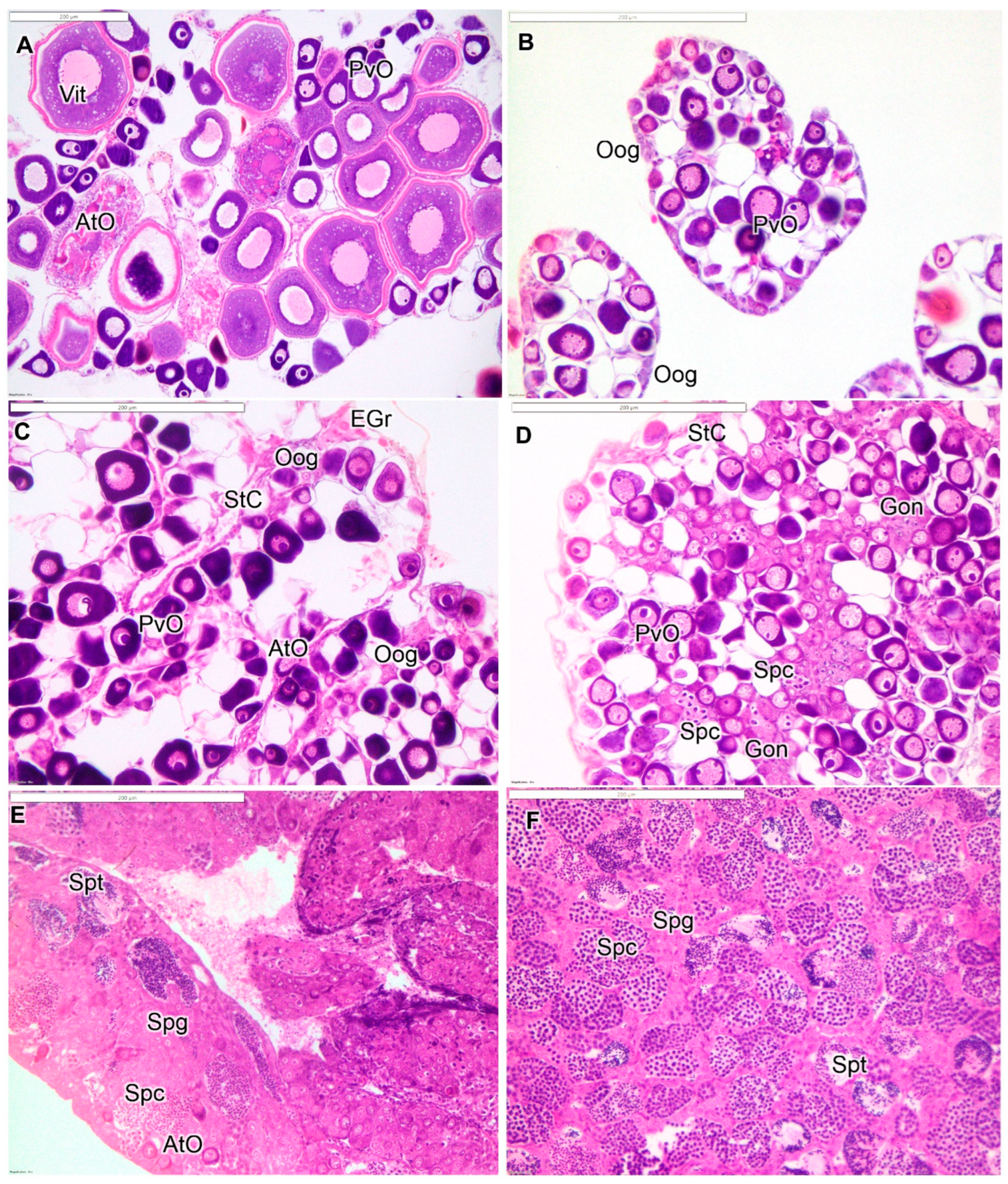
Figure 1. Histological stages of gonadal development in spotty wrasse: (A) adult female; (B) juvenile female; (C) early-transitioning fish; (D) mid-transitioning fish; (E) late-transitioning fish; and (F) initial-phase male. Abbreviations: atretic oocyte (AtO); gonial cells (Gon); eosinic granulocytes (EGr); previtellogenic oocyte (PvO); oogonia (Oog); stromal cells (StC); spermatocytes (Spc); spermatogonia (Spg); spermatids (Spt); vitellogenic oocyte (Vit). Scale bars = 200 µm.
Fishes 10 00554 g001
Figure 2. Relative gene expression profiles of (A) amh; (B) dmrt1; (C) cyp19a1a; (D) dnmt1; and (E) dnmt3. Different letters on top of bars denote statistically significant values.
Fishes 10 00554 g002
Figure 3. Proposed life history of the diandric protogynous New Zealand spotty wrasse.
Fishes 10 00554 g003
Figure 4. Key gonadal germ cell types in spotty wrasse undergoing transition to initial-phase male: (A) early-transitional fish; (B) mid-transitional fish; and (C) late-transitional fish. Abbreviations: atretic oocyte (AtO); gonial cells (Gon); previtellogenic oocyte (PvO); oogonia (Oog); spermatocytes (Spc); spermatogonia (Spg). Black arrowhead indicates Sertoli cell. Scale bars = 50 µm.
Fishes 10 00554 g004
Table 1. Histological description of stages of sex change in prepubertal spotty wrasse.
StageDescription
AF (>110 mm)Clear ovarian lamellar structure with previtellogenic oocytes of varying sizes. May contain vitellogenic and/or maturing oocytes depending on season. No evidence of male structure.
JF (<110 mm)Ovary dominated by small previtellogenic oocytes, includes oogonial nests in lamellar periphery.
ETAtretic previtellogenic oocytes and nests of gonial cells are common. May include presence of eosinophilic granulocytes and cellular debris. Male germ cells not present.
MTDiminished presence of oocytes and those remaining are mostly atretic. Stromal cells, increased connective tissue, and eosinophilic granulocytes are common. Proliferation of gonial cells is evident, and spermatocytes are present.
LTNumber of spermatogenic cysts predominates over oocytes, with some lobular testicular structure becoming evident. Stromal cells are often present.
IPMClear lobular structure is evident, with many cysts of spermatogenic germ cells. May include more advanced stages of development, such as spermatids and spermatozoa.
Table 2. qPCR primer design.
GenePrimerSequence (5′–3′)Annealing Temp. (°C)Tm (°C)Amplicon Size (bp)Efficiency GC (%)
cyp19a1afwTGGACACTGTTGTTGGTGAC6062.51610.91 ± 0.0350
rvAGGTTACTCTAAAGCCCTAGTAGTG6062.244
amhfwGAAGACGTAAAACAAGATCTGCAC6062.21340.90 ± 0.0242
rvGGATTACAGGTGAAGGGAAGAG6062.652
dmrt1fwACCCTCACAACTCACAATAACC6161.22050.91 ± 0.0242
rvAGACCTCCTGGAGAAAAGAG6162.150
g6pdfwCGAGCTCATGGCAAACCA6062.51060.96 ± 0.0250
rvGCACAGCTTCAACCTTTTGT6060.650
actb2fwCCCACTCACATGAAGATTAAGATCA6062.22000.95 ± 0.0340
rvAGTGTGTTTTTGGGGGAGG6064.455
dnmt1fwTGGCCACCTTTGTCCATTTG60581400.96 ± 0.3550
rvCGTTGATGGGTCCAAGCTTC605955
dnmt3afwGGAGAACAGGCTACACCCAG60602092.48 ± 1.5660
rvTTCTCCACGCAAACCACAGA605850
Table 3. Proportion (%) and length (mm TL)of spotty wrasse at each developmental stage.
Stage of Development % Total Mean TL ± SE (mm) Range TL (mm)
Adult female (AF) 21.0% (n = 30)153.6 ± 31.4112–270
Juvenile female (JF) 52.5% (n = 75) 89.3 ± 11.8 64–110
Early-transitional (ET) 14.7% (n = 19)75.9 ± 14.652–107
Mid-transitional (MT) 3.5% (n = 5)70.2 ± 6.060–72
Late-transitional (LT) 2.8% (n = 4)87.8 ± 14.4 72–104
Male (IPM) 5.6% (n = 8)95.5 ± 19.3 77–137
Disclaimer/Publisher’s Note: The statements, opinions and data contained in all publications are solely those of the individual author(s) and contributor(s) and not of MDPI and/or the editor(s). MDPI and/or the editor(s) disclaim responsibility for any injury to people or property resulting from any ideas, methods, instructions or products referred to in the content.