You are currently viewing a new version of our website. To view the old version click .
Fishes
  • Article
  • Open Access

1 November 2025

Morphological and Histological Changes of Experimental Pseudomonas fluorescens Infection in Zebrafish (Danio rerio)

,
,
,
,
,
,
,
and
1
Academic Division of Agricultural Sciences, Juárez Autonomous University of Tabasco, Villahermosa-Teapa Highway, Kilometer 25+2, Rancheria la Huasteca, 2nd Section, Villahermosa 86298, Tabasco, Mexico
2
Man and His Environment Department, Metropolitan Autonomous University Xochimilco Unit, Calzada del Hueso 1100, Coyoacan, Mexico City 04960, Mexico
3
Faculty of Chemistry, Autonomous University of Queretaro, Queretaro 76010, Mexico
4
Hidrobiology Department, Metropolitan Autonomous University Iztapalapa Unit, Av. San Rafael Atlixco 186, Iztapalapa, Mexico City 09340, Mexico
Fishes2025, 10(11), 551;https://doi.org/10.3390/fishes10110551 
(registering DOI)
This article belongs to the Special Issue Infection and Detection of Bacterial Pathogens in Aquaculture

Abstract

Pseudomonas fluorescens is a bacterium that has been identified as a significant pathogen in fish from the aquaculture industry. However, the clinical signs and changes caused by the disease have not been studied well yet. This study aimed to determine the survival, morphological, and histopathological changes in zebrafish (Danio rerio) in an experiment involving 180 fish (0.250 ± 0.05 g). The organisms were inoculated intraperitoneally at four bacterial concentrations 1.5 × 10⁶, 1.5 × 10⁷, 1.5 × 10⁸, and 1.5 × 10⁹ CFU mL−1 with respective control groups. Results showed that cumulative mortality at 96 h post infection (hpi) was 0% (1.5 × 10⁶ CFU mL−1), 0% (1.5 × 10⁷ CFU mL−1), 6.66% (1.5 × 10⁸ CFU mL−1), and 33.33% (1.5 × 10⁹ CFU mL−1). The survival curves were not statistically different (p = 1.000). Corresponding to differences between clinical signs and concentrations for 24 hpi (p = 0.7576) there were no significant differences for 48 hpi statistical differences were obtained (p = 0.0008), while for 72 hpi (p = 0.8922) and 96 hpi (p = 0.333) they were not observed. The predominant clinical signs were anorexia, lethargy, erratic swimming, and peritoneal haemorrhage. The acute septicemic clinical form was determined. Histological analyses showed that the gills, liver, and spleen were infected and had severe pathological alterations. These findings indicate that the zebrafish is susceptible to P. fluorescens infection.
Key Contribution:
This study concludes that the bacterium is capable of causing infection in zebrafish. Describes the survival rate, mortality, and bacterial inoculation for inducing infection, and the timing and frequency of clinical signs and tissue changes. This characterisation provides positive control reference for future research investigating the effect of new treatments on model fish or other species.

1. Introduction

Aquaculture stands out as a rapidly expanding sector and plays a fundamental role in meeting the global demand for animal protein for human consumption, surpassing other food-producing industries [,]. In 2022, aquaculture produced an estimated 94 million tonnes. Inland waters contributed 70 million tonnes, of which 84 percent was derived from aquaculture []. However, this activity is not exempt from problems that limit its growth; the intensive culture had led to outbreaks of several infectious diseases mainly caused by parasites, viruses, fungi, and bacteria [,]. Bacterial diseases in both wild and cultured fish have been identified as a significant contributor to heavy mortality rates and economic losses [,,]. It has been documented that countries such as China, India, and Vietnam have reported production losses of 30 percent due to infectious diseases []. The most commonly occurring pathogenic bacteria in cultured fish are Aeromonas salmonicida, Aeromonas hydrophila, Edwarsiella ictaluri, Edwarsiella tarda, Yersinia ruckery, Piscirickettsia salmonis, Franciella spp., Photobacterium spp., Flavobacterium psychrophilum, Vibrio spp., Streptococcus, Lactococus, Clostridium and Pseudomonas spp. [,,,,]. The species of the genus Pseudomonas that have the greatest impact due to their infectious capacity are Pseudomonas aeruginosa; this is one of the major fish pathogens causing diseases such as ulcerative syndrome and hemorrhagic septicemia. It is a major public health concern due to its potential multidrug resistance and zoonotic risk [,]. Another bacteria, Pseudomonas anguilliseptica, are a significant pathogen in freshwater fish such as tilapia. Pseudomonas fluorescens and Pseudomonas putida are considered opportunistic pathogens in fish and environmental microbiota with occasional disease association but no less important []. Pseudomonas fluorescens has been identified in a variety of ecological niches and although it is used as a biological control agent in agriculture [], in other areas such as aquaculture it is also regarded as an opportunistic, pathogenic, and zoonotic organisms []. It has been demonstrated to cause hemorrhagic septicaemia in humans, fish, birds, and reptiles [,,]. These genera belong to the normal bacterial flora aquaria, hatcheries, fish farms, and ponds of water. Specifically, P. fluorescens is considered common in natural aquatic environments and anthropogenically altered water environments [,,]; however, the activity and abundance of this bacterium in aquatic environments depends on physicochemical parameters such as water temperature, pH, content of mineral substances, and organic compounds, and on biological factors (predation and interspecific competition) []. In a study in a recirculating aquaculture system (RAS) during farming broodstock refers that the counts of this bacterium were strongly correlated with feed dose and total fish biomass, indicating that these farming conditions and the nutrient requirements affect the abundance of the analysed potentially pathogenic bacteria []. The transmission mechanism includes water contamination, bacterial filamentous hemagglutin, motility, biofilm formation, immune evasion and it may even be due to vectors such as microplastics in water [].
Pseudomonas fluorescens is one of the most important species in fish pathology and is very often associated with skin and fin disease [] which cause significant economic losses in fish farming by leading to high mortalities and poor growth. It has been reported in a wide variety of fish species such as grass carp (Ctenopharyngodon idella), common carp (Cyprinus carpio), japanise flounder (Platichthys spp.), (Paralichthys olivaceus), tilapia (Oreochromis spp.), turbot (Scophthalmus maxima), catfish (Silurus asotus), goldfish (Carassius auratus), eel (Anguilla japonica), rainbow trout (Onchorhynchus mykiss), indian major carp (Catla catla), silver carp (Hypophthalmichthys molitrix), and African magur (Clarias gatiepinus) [,,,,,]. The infectious process of P. fluorescens is characterised by the presence of haemorrhages in the skin, fins, and internal organs, which can be attributed to bacterial hemolytic activity and proteolytic properties. Many P. fluorescens strains are able to secrete an extracellular metalloproteases called AprX, yet no AprX-like proteins have been identified in pathogenic P. fluorescens associated with aquaculture. Filamentus Haemagglutinin (FHA), an iron-responsive protein who helps the bacterium attach to host cells, reduced capacity of hemagglutination and survival in host serum [,,,,]. However, the precise mechanism of infection remains poorly understood, and there is a need for more effective and reliable methods for the characterisation of infectious agents [,,]. The study of infection by P. fluorescens is important for understanding and serves as a reference for the development of studies that evaluate alternative treatments for its control []. This is due to the antimicrobial resistance P. fluorescens exhibits. It has a reported resistance to Trimethoprim/Sulfamethoxazole, Fosfomycin, Amoxy/Clavulanic acid, Florfenicol, Amoxycillin, Clindamycin, Ampicillin, Erythromycin, Trimethoprim, Oxolinic acid, Chloramphenicol, Penicillin G, Nitrofurantoin, and Oxytetracycline [,] which makes it difficult to control.
Most studies on P. fluorescens infections in cultured O. niloticus and Oncorhynchus mykiss have focused on characterising the signs and lesions observed in the fish [,], but few have evaluated tissue and internal organ damage through histopathological studies. Therefore, this study will determine the morphological and histological changes caused by experimental Pseudomonas fluorescens infection in zebrafish (Danio rerio) as a valuable reference for future research. The zebrafish has been employed as a biological model in several fields, including developmental biology, genetics, pharmacology, biomedicine, and the characterisation of diseases and infectious processes in humans and fish, principally freshwater species [,,,]. The use of zebrafish in aquaculture research is significant due to its classification within the Cyprinidae family, which represents one of the most prevalent groups of freshwater fish, and its status as an organism that is relatively straightforward to handle []. In aquaculture zebrafish has been employed as a model to investigate fish health including different disease mechanisms, host–pathogen interaction, drug screening, vaccine development, microbiomics, the immunomodulatory properties and distribution of probiotic bacteria, and infection protocol in teleost species []. Bacteria such as Aeromonas hydrophila, Pseudomonas aeruginosa, Edwarsiella tarda, Stratococus iniae, and Vibrio angularirum; viruses such as spring viremia of carp virus, common carp paramyxovirus, common carp orthomyxovirus, chum salmum reovirus, common carp birnavirus, cyprinid herpesvirus, and carp oedema virus [,,,,]. The aim of this study was to determine whether zebrafish could be used as an animal model to study P. fluorescens infection and evaluate dose-dependent responses, mortality, clinical signs, and tissue damage. This study is important for aquatic medicine and aquaculture sustainability.

2. Materials and Methods

2.1. Bacterial Strain and Growth Conditions

Pseudomonas fluorescens (ATCC 13525) was cultured in Brain Heart Infusion Agar (BHIA), (BD Bioxon®, Mexico, Mexico) medium plates (SYM Laboratories®, Puebla, Mexico) at 30 °C for 48 h in a digital incubator (Luzeren®, Mexico city, Mexico) and harvested in 10 mL of PBS (phosphate-buffered saline: NaCL J.T. Baker Inc.® (Philadelphia, PA, USA) 137 mM, KCL J.T. Baker Inc.® (Philadelphia, PA, USA) 2.7 mM, Na2HPO4 J.T. Baker Inc.® (Philadelphia, PA, USA) 10 mM, KH2PO4 J.T. Baker Inc.® (Philadelphia, PA, USA) 1.8 mM, pH 7.4. A total of 1 mL−1 aliquot was dispensed in 100 mL of Brain Heart Infusion following incubation at 30 °C for 24 h. A bacterial pellet was obtained, washed, and resuspended in PBS, pH 7.4. Thereafter, the culture was adjusted to optical density (OD600 = 1.5 ± 0.01 stock solution to 2 × 10⁹ CFU mL−1 []. The bacterial suspensions were diluted with PBS, pH 7.4 to obtain the final bacterial concentrations at 1.5 × 10⁶, 1.5 × 10⁷, 1.5 × 10⁸, and 1.5 × 10⁹ colony forming units CFU mL−1.

2.2. Animal Model and Maintenance

Healthy adult zebrafish (90 days) were used (0.250 ± 0.05 g), cultured at the Laboratory of Chemical Analysis of Live Food of the Metropolitan Autonomous University. The maintenance conditions were as follows: dissolved oxygen of 6 mg L−1, temperature of 26 °C ± 1, with a 12:12 h light-dark photoperiod and 25% water replacement every seven days. Fish were fed a commercial (El Pedregal Silvercup®, Toluca, Mexico), microtek 50-16, 0.4 mm, ad livitum, in two feeding schedules: 50% at 7:00 a.m. and 50% at 5:00 p.m. Prior to the experiment, the fish were acclimatised for three weeks in 20 L aerated water tanks and were shown to be in a healthy condition at the start of the experiments.

2.3. Experimental Design and Infectious Challenge

One hundred and eighty healthy adult zebrafish with an average weight of (0.250 ± 0.05 g) were randomly distributed in twelve 20 L aquaria with constant aeration and filtration which were set up for the experiments. Throughout the experiments the temperature was 25 ± 1 °C, dissolved oxygen 8 ± 0.5 mg L−1, pH 7.38–7.50 and kept with a 12:12 h light-dark photoperiod.
Four 20 L aquaria were used to group control. The negative control with (n = 15 fish each one), including the first control with organisms without manipulation and the second negative control with organisms intraperitoneal inoculated with 7 μL g−1 body weight of PBS per fish. And eight aquaria with (n = 15 fish each one), divided into two groups (a) group pathology and (b) group for quantification and observation of clinical signs and mortality, Table 1. The number of samples were selected attending to []. Feeding was daily ad libitum. Prior to the intraperitoneal inoculation the organisms were anaesthetised with clove oil (Meyer®, Mexico city, Mexico) 40 mg L−1. 7 μL g−1. The body weight of the inoculum were used at concentrations of 1.5 × 10⁶, 1.5 × 10⁷, 1.5 × 10⁸, and 1.5 × 10⁹ CFU mL−1.
Table 1. Designing the bacterial concentrations for different treatments.
Continuous monitoring and mortality recording for each treatment was carried out at 96 h post inoculation (hpi). A sample of three organisms were collected at 24, 48, 72 and, 96 hpi and euthanized with an anaesthetic overdose (40 mg L−1 clove oil) for each experimental and control groups for histopathological analysis []. The use and management of animals were conducted following the national NOM-062-ZOO-1999 and NOM−008-ZOO-1994, the institutional CICUAL-UAM-X of UNIVERSIDAD AUTÓNOMA METROPOLITANA (Sesion 70/20) (Approval Code: UAM-X-CONSEJO DIVISIONAL DE C.B.S 10/20; Approval Date: 10 February 2020), and international bioethical protocols.

2.4. Histopathological Analysis

Due to the size of the fish and method and reference method []. Whole organisms were preserved in Davidson’s solution (7% formaldehyde (HYCEL®,Zapopan, Jalisco, Mexico), 2% methanol (HYCEL®, Jalisco, Mexico), 11.5% glacial acetic acid (Meyer®, Mexico city, Mexico), 33% ethanol (HYCEL®, Jalisco, Mexico, and 46.5% water) for 24 h, placed in plastic cassettes and were processed (dehydrated in ascending series of ethanol (HYCEL®, Jalisco, Mexico) 70–100%, clearing in xylene (HYCEL®, Jalisco, Mexico), and paraffin wax (Merk®, Darmstadt, Germany) embedding at 60 °C (EG 1140h, Leica Microsystem®, Wetzlar, Germany). A series of 5 µm thick sections were taken from the paraffin blocks with rotary microtome (MICROM HM 315, MICROM International®, Walldorf, Germany). The sections were stained with hematoxylin-eosin (H&E) (Merk®, Darmstadt, Germany). The slides were evaluated under a (Carl Zeiss Cx31®, Jena, Germany) light microscope and edited using (Axio Vision Rel Software. 4.8., Jena, Germany) The histological changes examination was performed according to [,]. In terms of external signs, the clinical form of the infection in zebrafish was characterised according to the clinical signs identified: acute septicemic, subacute ascitic, and chronic ulcerative according to the classification by [].

2.5. Statistical Analysis

Survival and mortality were expressed as percentages. Survival rates were calculated and compared among control groups and those infected with bacterial concentrations using the Kaplan–Meier method. Pairwise comparisons used the log-rank test with Bonferroni correction for multiple comparisons []. The tests were performed using SigmaPlot Software version 14.0., (Systat, Palo Alto, CA, USA) [] and were considered significantly different if the p-value was <0.05. Analysis of frequencies of clinical signs was evaluated by Fisher’s exact test []. This analysis was performed using GraphPad Prism software, version 10.4.1. (GraphPad Software Inc., San Diego, CA, USA).

3. Results

3.1. Clinical Findings

At 24 hpi, all organisms showed signs of disease except the negative control groups. Visible clinical signs include anorexia, hyperemia in pectoral and ventral fins, lethargic, bottom dwelling, erratic swimming, ulcerative skin, increased opercular movements, celomic distension, haemorrhages, and mortality. Acute septicaemic, subacute ascitic, and chronic ulcerative clinical forms were observed according to []. Cumulative mortality between hpi at the concentration of 1.5 × 10⁶ UFC mL−1 was 0%, 1.5 × 10⁷ UFC mL−1 was 0%, 1.5 × 10⁸ UFC mL−1 was 6.66%, and 1.5 × 10⁹ UFC mL−1 was 33.33%, as shown on Table 2. For this study the log-rank for the survival curves is greater than would be expected by chance. There is a statistical difference between survival curves (p = 0.035); however, the pairwise multiple comparison procedure post hoc Bonferroni method correction with an overall significance level α = 0.05, which is more conservative and indicates the difference between groups, indicates that there were not enough to be different (p = 1.000). The data does not provide sufficient evidence to support a significant effect between bacterial concentrations and survival.
Table 2. Percentage of the mortalities of zebrafish after experimental infection with P. fluorescens (ATCC 13525).
For controls, 1.5 × 10⁶ UFC mL−1, and 1.5 × 10⁷ UFC mL−1 there is a 100% probability of survival but with 1.5 × 10⁸ UFC mL−1 and 1.5 × 10⁹ UFC mL−1 there is a 50% and 0% probability of survival, respectively, Figure 1.
Figure 1. Survival curve (log-rank) of experimental zebrafish infected by P. fluorescens bacteria (p = 0.035).
Regarding the differences between clinical signs and concentrations evaluated by Fisher’s test, for 24 hpi (p = 0.7576) there were no significant differences, for 48 hpi a statistical difference was obtained (p = 0.0008), while for 72 hpi (p = 0.8922) and 96 hpi (p = 0.333) there were no observed differences, Figure 2.
Figure 2. Frequency of observations of clinical signs of P. fluorescens infection at different inoculated concentrations (A) 24 hpi, (B) 48 hpi, (C) 72 hpi, and (D) 96 hpi.

3.2. Histopathology

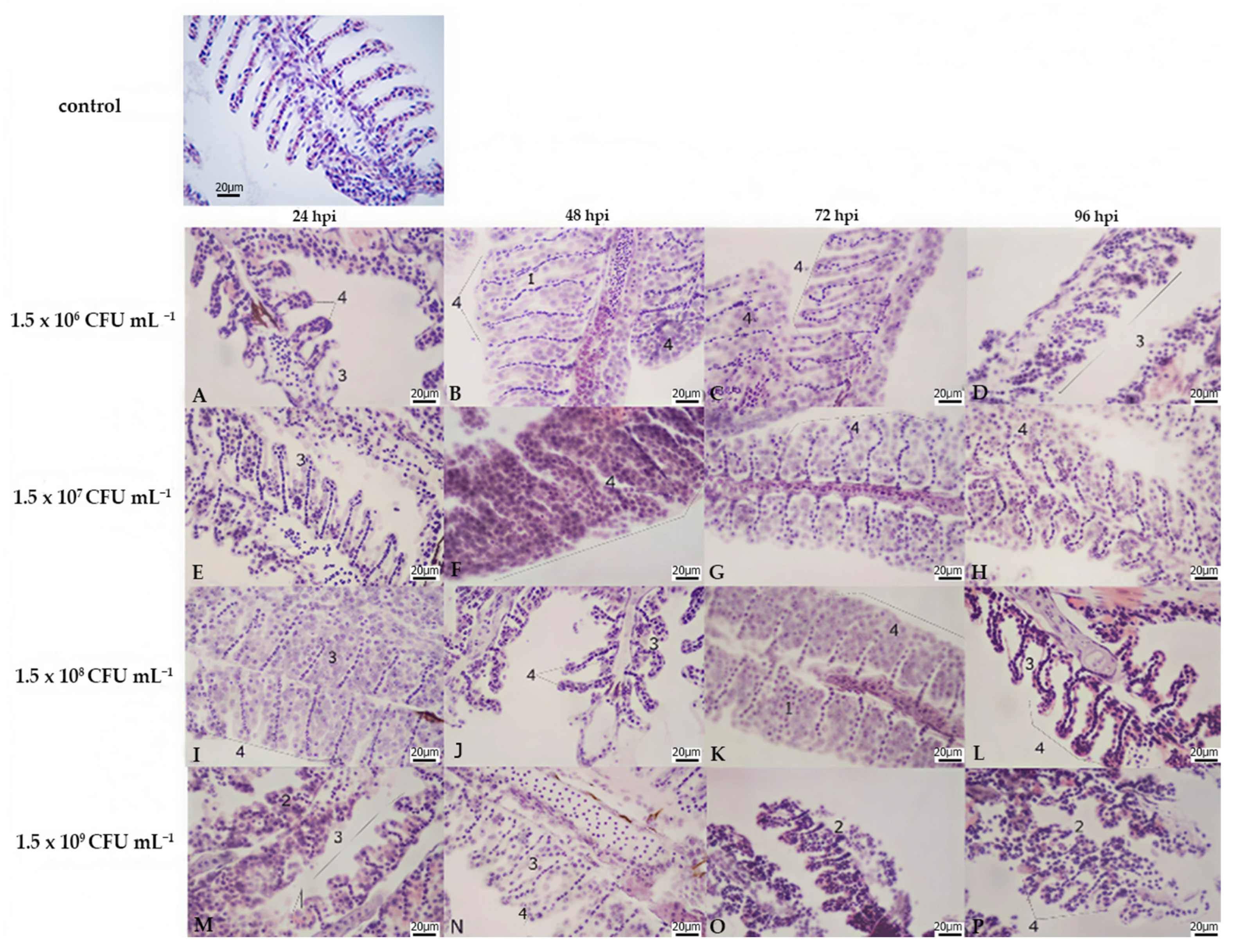
Throughout the experiments, no histological alterations were observed in the tissues of the controls. Tissue analysis of gill tissue showed changes in the structure of secondary and primary gill lamellae, mainly lamellar hypertrophy and fusion, and infiltration of inflammatory cells with extensive changes, Table 3, Figure 3.
Table 3. Classification of changes according to the corresponding pathological category, liver, anterior kidney, gills.
Figure 3. Micrographs of gills of zebrafish (Danio rerio) experimentally infected with Pseudomonas fluorescens 1.5 × 10⁶ (AD), 1.5 × 10⁷ (EH), 1.5 × 10⁸ (IL), 1.5 × 10⁹ (MP), and gills control. Key: (1) Lamellar fusion, (2) Necrosis, (3) Lamellar atrophy, (4) Severe hyperplasia. H&E, X 400.
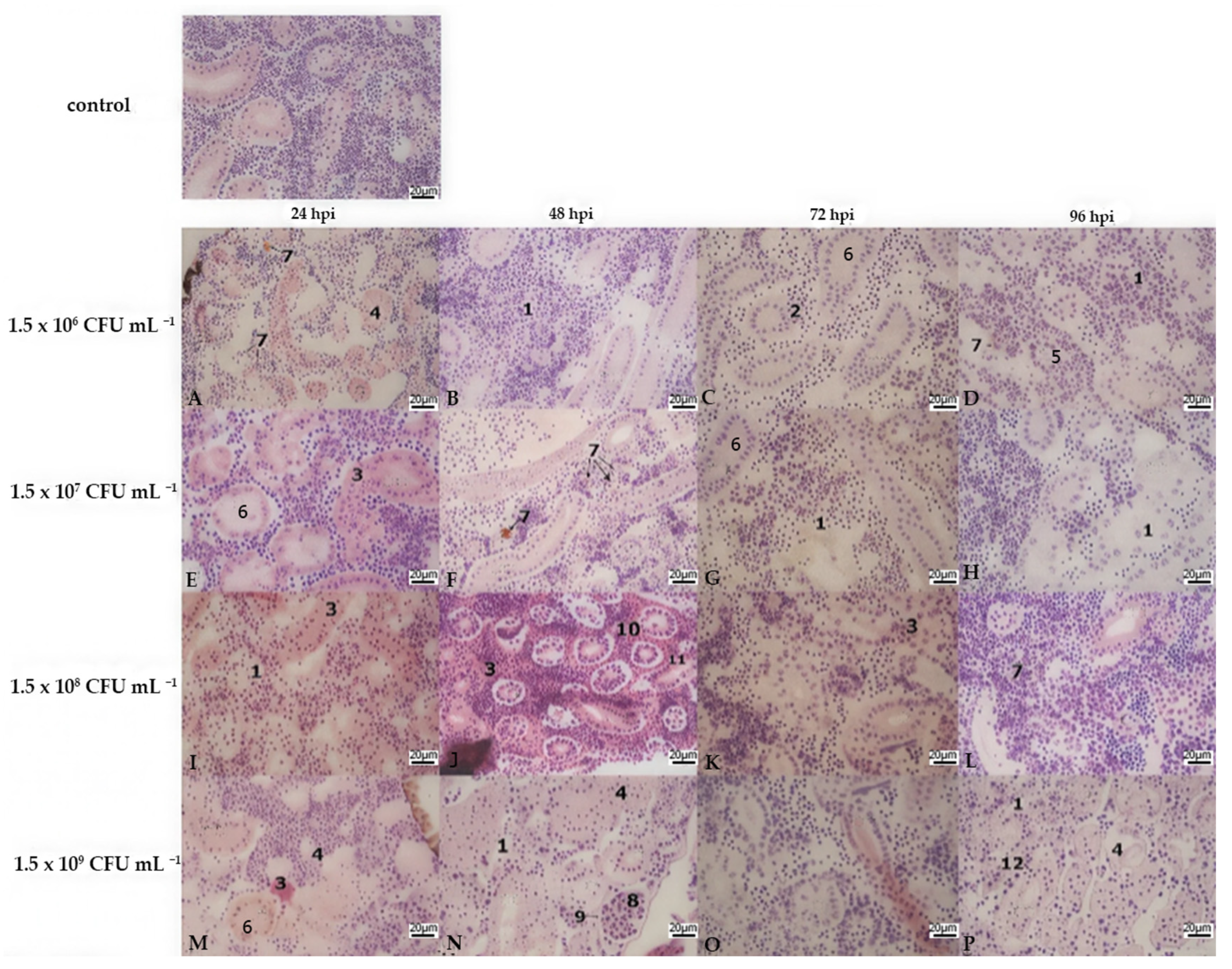
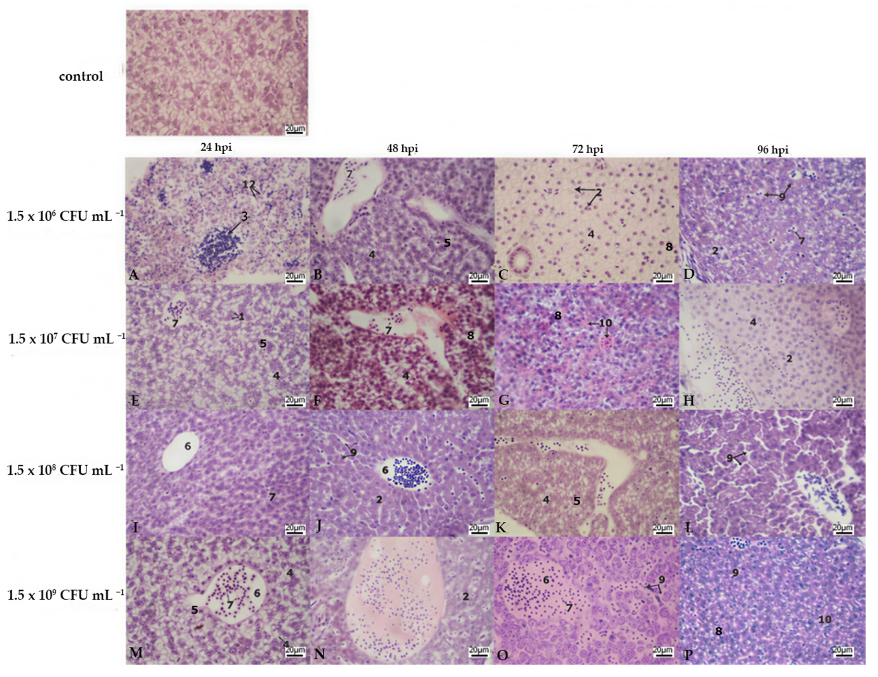
The liver and kidney were also affected, with differences depending on the bacterial concentration used. Such as changes in the liver parenchyma, cell infiltration, foci of necrosis in the liver tissue and necrotic areas in the kidney and loss of renal tubular arrangement. The lesions in these organs showed medium changes and systemic damage to the organisms as show. Figure 4 and Figure 5.
Figure 4. Micrographs of kidney of zebrafish (Danio rerio) experimentally infected with Pseudomonas fluorescens 1.5 × 10⁶ (AD), 1.5 × 10⁷ (EH), 1.5 × 10⁸ (IL), 1.5 × 10⁹ (MP), and kidney control. Key: (1) Loss of renal tubule arrangement, (2) normal renal tubule structure, (3) infiltration of inflammatory cells, (4) acute tubular necrosis, (5) decrease in tubular epithelium height, (6) tubular dilation, (7) melanomacrophage center, (8) glomerulus, (9) Bowman’s capsule, (10) hemosiderosis, (11) erythrocyte infiltration, (12) hyaline droplet. H&E, X 400.
Figure 5. Micrographs of liver of zebrafish (Danio rerio) experimentally infected with Pseudomonas fluorescens 1.5 × 10⁶ (AD), 1.5 × 10⁷ (EH), 1.5 × 10⁸ (IL), 1.5 × 10⁹ (MP), and liver control. Key: (1) Hepatocytes with normal histological structure, (2) loss of hepatocyte cell structure, (3) infiltration of inflammatory cells, (4) advanced vacuolar degeneration, (5) integrity of hepatocytes with onset of vacuolization, (6) vein, (7) nucleated erythrocytes, (8) hepatocytes with pyknotic nuclei, (9) diffuse sinusoidal dilatation, (10) peliosis, H&E, X 400.
At the highest concentration used, 1.5 × 10⁹ CFU mL−1, a mortality rate of 33.33% was observed at 96 h. The clinical picture included the chronic ulcerative form, due to the appearance of skin ulcers and lethargy, and the acute septicemic form, with signs such as anorexia, lethargy, petechial skin haemorrhages, exophthalmia, and erratic swimming. Histologically, this study showed changes in the liver and kidney, with differences depending on the bacterial concentration used. Changes in the liver parenchyma, cellular infiltration, necrotic foci in the liver tissue and multifocal necrotic areas in the kidney, and loss of renal tubular arrangement. The lesions in these organs were considered the cause of death. On haematological examination, infected animals showed increased monocytes, abundant blasts, marked karyorrhexis, hypochromia and anisocytosis characteristic of hypochromic anaemia, and erythroclasia.

4. Discussion

Pseudomonas fluorescens is considered an opportunistic pathogen. Infection begins with an inflammatory response that includes phagocytic activity by macrophages and neutrophils. References [,] refers that zebrafish is relatively resistant to Pseudomonas and that a large inoculum 1 × 10⁹ CFU mL−1 is required to establish infection and host death. However, the validity of this assumption depends on the age of the fish and the method used to induce the infection. The present study was prompted by the need to examine four bacterial concentrations to determine the most effective concentration to produce an infection and mortality. According to our study, all organisms inoculated intraperitoneally with the four tested concentrations of 1.5 × 10⁶, 1.5 × 10⁷, 1.5 × 10⁸, and 1.5 × 10⁹ CFU mL−1 developed P. fluorescens infection, but in the present study, a maximum mortality rate of 33 percent was reported at the maximum bacterial concentration 1.5 × 10⁹ CFU mL−1. In comparison to that reported by [] in Tench (Tinca tinca), the authors describe an elevated death rate up to 40 percent of the total population for the infestation and infection of this pathogen.
There is no previous report demonstrating P. fluorescens infection in zebrafish, but there are other studies using Gram bacteria such as P. aeruginosa; Yersinia ruckeri, A. salmonicida, F. psychrophilum, V. anguillarum in zebrafish [,]. Refs. [,,] P. fluorescens and other species of the genus such as P. putida, P. pseudocaligenes, P. chlororhaphis, P. plecoglossicida, P. aeruginosa and Pseudomonas sp. in fish such as goldfish (Carassius auratus), carp (Cyprinus carpio), tilapia (Oreochromis niloticus), and trout (Oncorhynchus mykiss). Infection was mainly characterised by septicaemia, fin erosion, ulceration, increased pathogenicity at lower temperatures, and frequent antibiotic resistance. In the study developed, the macroscopic changes caused by P. fluorescens infection in O. niloticus are consistent with those reported, but other signs can be added, such as exophthalmia, skin darkening, patchy and nodular lesions in the spleen, liver, kidney and gills, and inflammation of the swim bladder. Ref. [] indicated that the dissemination was systemic. Histopathological evidence showed abscesses in the eyes, liver, and swim bladder and focal necrosis in the spleen, gills, and kidney of some affected animals. In our study, no significant abscesses or granulomas were observed in the liver, only focal necrosis in the liver and kidney. Non-infected hepatocytes in the liver appear atrophied or visible. In the kidney, inflammatory cell infiltration and lesions sometimes involved the renal tubules with necrosis. According to this, hemolysins contribute to erythrocyte infiltration by the lysis of red blood cells through membrane disruption. Specifically, P. fluorescens is described as having slightly haemolytic activity []. This could explain why there were no high mortality rates at the highest concentration used. Inflammatory cell infiltration in response to P. fluorescens infection reflects fish immune system activation. The recruited inflammatory cells produce cytokines and chemokines which are implicated in inflammatory response induction in fish []. In the gills, report invasion of the bacteria into the connective tissue of the gill filaments, causing focal necrosis, inflammatory cell infiltration, and fibrin precipitation, which we also found in D. rerio infection in this study. The clinical pathology of P. fluorescens indicates haemorrhagic septicemia, which may be acute. Haemorrhagic skin lesions are one of the most observed signs and mortality is high; infected organisms can survive for a very short time after the appearance of the lesions. Ref. [] mentions that in cyprinids, fish often show ascites in addition to the characteristics described above. In the present study, ascites was present in all organisms that died in this study. These findings are similar to those reported by [], who reported septicaemia caused by P. fluorescens in coexistence with Aeromonas hydrophila and Vibrio parahemoliticus, and who also reported in a histopathological analysis the acute form of liver infiltration and fatty degeneration in almost 50% of the samples analysed. In the subacute ascitic form, haemorrhages in the peritoneum, degeneration, and necrosis of hepatocytes with bloody ascitic fluid were observed in the necropsies. There are also abscesses in the peritoneum, spleen, liver, and kidneys. In this research, haemorrhage was observed in organisms inoculated with the four bacterial concentrations being more severe at 48 h with a concentration of 1.5 × 10⁸. Tissue analysis of the kidney and liver showed inflammatory cell infiltration and haemosiderosis, mainly in the kidney and gill, but not severe haemorrhaging as seen in other species infected, such as O. niloticus []. In a study by [] O. niloticus fish were inoculated with 4.0 × 10⁶ CFU mL−1. The study did not characterise the signs produced by P. fluorescens infection at the external or microscopic level, but only indicated an increased immune system response in terms of the presence of monocytes, neutrophils, and mortality 60.42 ± 1.36%. Ref. [] observed that O. niloticus infected with P. fluorescens 1.5 × 10⁵ showed hepatocytes with pyknosis, vacuolar degeneration, and dilatation of sinusoids with severe lipid vacuoles. And in the kidney, dilatation of Bowman’s space, haemorrhages, areas of necrosis between the renal tubules, and accumulation of haemosiderin with inflammatory cells in the renal tubules. In other studies, with bacteria of the genus Pseudomonas, infection by Pseudomonas anguilliseptica in carp and goldfish indicates the presence of petechiae around the mouth; operculum and ventral internal macroscopic signs may not be present, but it generally causes high mortality. The microscopic signs observed in our research are similar to those reported by [], who characterised the infectious picture caused by Pseudomonas anguiliseptica in Psetta maxima or sole, in which changes were observed in the kidney. This was extended to haematopoietic cells in the liver vasodilatation and bacterial aggregates; however, this study reports that the organisms infected by immersion showed no mortality due to infection and no external signs, only lethargy in contrast to those infected by intraperitoneal injection, which died without any visible clinical signs of infection. This supports that the inoculation method influences the response, and is in line with the findings which refer that zebrafish is relatively resistant to Pseudomonas and that a large inoculum 1 × 10⁹ CFU mL is required to establish infection and host death []. In this study the authors used the immersion method. This is undoubtedly a topic that still needs to be researched to gain a better understanding of its infectious capacity.
In this study the experiment was stopped at 96 h post-infection (hpi) because the remaining organisms appeared to have overcome the infection process or were showing no visible clinical signs. The surviving organisms overcame the infectious process, becoming solely carriers of the disease. For future studies, we strongly recommend increasing the sample size to improve statistical power and calculating another histopathological index. It is important to consider calculating the LD50 of the Pseudomonas strain for this species in the future. Blood tests should be performed and the immune response evaluated. In addition, extend the observation and measurement time, and consider measuring P. fluorescens levels in the gills, liver or kidneys to confirm that tissue lesions are directly caused by bacterial colonisation.
These findings establish a positive control for future research that investigates the pathogenic disease or the effect of new treatments on model fish or other species. Our dose-dependent mortality, clinical signs, and lesion data could be used as a reference to select the bacterial concentration according with the research interest. Research in this field is ongoing, with recent advances focusing on controlling or preventing bacterial growth and providing effective treatment for infected organisms. These studies require the establishment of experimental challenges to evaluate the effects of antibiotics or herbal natural extracts, immunological responses, and other factors that improve the welfare, health, and rearing conditions of fish.

5. Conclusions

According to the results, we can mention that P. fluorescens showed the ability to cause disease in Danio rerio, presenting signs of disease such as lesions on the skin and at the base of the fins. Signs of septicemia, such as accumulation of ascitic fluid in the peritoneal cavity with petechial haemorrhages and a mortality rate of 33.33% at the highest concentration tested do not have differences between the survival curves. In this study there were statistical differences only at 48 hpi. After 96 h, the surviving organisms overcame the infectious process, becoming solely carriers of the disease. The acute septicemic clinical form was determined.

Author Contributions

Conceptualization, M.G.-R. and M.d.C.M.-D.; methodology, M.G.-R. and M.d.C.M.-D.; software, M.G.-R., J.D.B.-G. and M.A.R.-L.; validation, M.A.R.-L. and J.C.-G.; formal analysis, M.G.-R. and J.D.B.-G.; investigation, M.G.-R., M.d.C.M.-D. and M.A.P.-G.; resources, M.d.C.M.-D. and M.A.R.-L.; data curation, M.G.-R., J.D.B.-G., L.G.-N. and J.C.-G.; writing—original draft preparation, M.G.-R., E.A.Q.-O. and A.Á.-R.; writing—review and editing, M.G.-R., E.A.Q.-O., A.Á.-R., J.C.-G., L.G.-N., M.A.P.-G. and M.d.C.M.-D.; visualisation, M.G.-R., M.d.C.M.-D., L.G.-N., M.A.P.-G. and E.A.Q.-O.; supervision, M.d.C.M.-D., E.A.Q.-O. and M.A.R.-L.; project administration, M.G.-R.; funding acquisition, M.d.C.M.-D. All authors have read and agreed to the published version of the manuscript.

Funding

This research received no external funding.

Institutional Review Board Statement

The experimental work took place in the facilities of the Live Food Production Laboratory and the Live Food Chemical Analysis Laboratory at the Universidad Autónoma Metropolitana, Unidad Xochimilco. For the use of laboratory animals, the university has the Internal Committee for the Care and Use of Laboratory Animals (CICUAL-UAM-X). The study was conducted in accordance with the Declaration of Helsinki and approved by the Institutional Review Board (CICUAL-UAM-X) of UNIVERSIDAD AUTÓNOMA METROPOLITANA (Sesion 70/20) (Approval Code: UAM-X-CONSEJO DIVISIONAL DE C.B.S 10/20; Approval Date: 10 February 2020).

Data Availability Statement

Data are contained within the article.

Acknowledgments

We would like to express our gratitude to Irma Hernández Calderas, Rubén Ortiz Herrera, Eduardo Méndez Tomas; Víctor Alfonso González Renteria and Óscar José Morelos Miñón for their support and collaboration.

Conflicts of Interest

The authors declare no conflicts of interest.

Abbreviations

ATCCAmerican Type Culture Collection
BHIABrain Heart Infusion Agar
CFUColony Forming Units
FHAFilamentus Haemagglutinin
HPIHours Post Inoculation
HHours
ODOptical Density
PBSPhosphate-Buffered Saline
pHPotential of hydrogen

References

  1. FAO. The State of World Fisheries and Aquaculture 2024; FAO: Rome, Italy, 2024; ISBN 9789251387634. [Google Scholar]
  2. Garlock, T.; Asche, F.; Anderson, J.; Ceballos-Concha, A.; Love, D.C.; Osmundsen, T.C.; Pincinato, R.B.M. Aquaculture: The Missing Contributor in the Food Security Agenda. Glob. Food Secur. 2022, 32, 100620. [Google Scholar] [CrossRef]
  3. de Kinkelin, P.; Michel, C.; Ghittino, P. Tratado de Las Enfermedades de Los Peces; Acribia: Zaragoza, Spain, 1991; ISBN 8420006866. [Google Scholar]
  4. Pridgeon, J.W.; Klesius, P.H. Major Bacterial Diseases in Aquaculture and Their Vaccine Development. CAB Rev. Perspect. Agric. Vet. Sci. Nutr. Nat. Resour. 2012, 7, 1–16. [Google Scholar] [CrossRef]
  5. Maldonado-Miranda, J.J.; Castillo-Pérez, L.J.; Ponce-Hernández, A.; Carranza-Álvarez, C. Summary of Economic Losses Due to Bacterial Pathogens in Aquaculture Industry. In Bacterial Fish Diseases; Elsevier: Amsterdam, The Netherlands, 2022; pp. 399–417. ISBN 9780323856249. [Google Scholar]
  6. Vanamala, P.; Sindhura, P.; Sultana, U.; Vasavilatha, T.; Gul, M.Z. Chapter 14—Common Bacterial Pathogens in Fish: An Overview. In Bacterial Fish Diseases; Dar, G.H., Bhat, R.A., Qadri, H., Al-Ghamdy, K.M., Hakeem, K.R.B.T.-B.F.D., Eds.; Academic Press: Cambridge, MA, USA, 2022; pp. 279–306. ISBN 978-0-323-85624-9. [Google Scholar]
  7. Dey, M.M. Strategies and Options for Increasing and Sustaining Fisheries and Aquaculture Production to Benefit Poorer Households in Asia; WorldFish Center: Penang, Malaysia, 2008; ISBN 9789832346661. [Google Scholar]
  8. Attia, M.M.; Abdelsalam, M.; Elgendy, M.Y.; Sherif, A.H. Dactylogyrus extensus and Pseudomonas fluorescens Dual Infection in Farmed Common Carp (Cyprinus carpio). Microb. Pathog. 2022, 173, 105867. [Google Scholar] [CrossRef]
  9. Lee, J.; Zhang, L. The Hierarchy Quorum Sensing Network in Pseudomonas aeruginosa. Protein Cell 2014, 6, 26–41. [Google Scholar] [CrossRef]
  10. Roberts, R.J. Fish Pathology, 4th ed.; Wiley-Blackwell: Chichester, UK, 2012; ISBN 9781444332827. [Google Scholar]
  11. Wang, H.-R.; Hu, Y.-H.; Zhang, W.-W.; Sun, L. Construction of an Attenuated Pseudomonas fluorescens Strain and Evaluation of Its Potential as a Cross-Protective Vaccine. Vaccine 2009, 27, 4047–4055. [Google Scholar] [CrossRef]
  12. Garvey, M.I.; Bradley, C.W.; Holden, E. Waterborne Pseudomonas aeruginosa Transmission in a Hematology Unit? Am. J. Infect. Control 2018, 46, 383–386. [Google Scholar] [CrossRef]
  13. Algammal, A.M.; Mabrok, M.; Sivaramasamy, E.; Youssef, F.M.; Atwa, M.H.; El-kholy, A.W.; Hetta, H.F.; Hozzein, W.N. Emerging MDR-Pseudomonas aeruginosa in Fish Commonly Harbor OprL and ToxA Virulence Genes and Bla TEM, Bla CTX-M, and TetA Antibiotic-Resistance Genes. Sci. Rep. 2020, 10, 15961. [Google Scholar] [CrossRef] [PubMed]
  14. Panpatte, D.G.; Jhala, Y.K.; Shelat, H.N.; Vyas, R.V. Pseudomonas fluorescens: A Promising Biocontrol Agent and PGPR for Sustainable Agriculture. In Microbial Inoculants in Sustainable Agricultural Productivity: Vol. 1: Research Perspectives; Springer: New delhi, India, 2016; pp. 257–270. ISBN 9788132226475. [Google Scholar]
  15. Gołaś, I. The Influence of Aquaculture Efluents on the Prevalence and Biocides Resistance of Opportunistic Pseudomonas fluorescens Bacteria in the Drweca River Protected under the Natura 2000 Network. Water 2020, 12, 1947. [Google Scholar] [CrossRef]
  16. Miyazaki, T.; Kubota, S.; Miyashita, T. A Histopathological Study of Pseudomonas fluorescens Infection in Tilapia. Fish. Pathol. 1984, 19, 161–166. [Google Scholar] [CrossRef]
  17. Picot, L.; Abdelmoula, S.M.; Merieau, A.; Leroux, P.; Cazin, L.; Orange, N.; Feuilloley, M.G.J. Pseudomonas fluorescens as a Potential Pathogen: Adherence to Nerve Cells. Microbes Infect. 2001, 3, 985–995. [Google Scholar] [CrossRef] [PubMed]
  18. Dinçtürk, E.; Tanrıkul, T.T. Yersinia ruckeri and Pseudomonas fluorescens Co-Infection in Rainbow Trout (Oncorhynchus mykiss Walbaum, 1792). Aquac. Res. 2021, 52, 4858–4866. [Google Scholar] [CrossRef]
  19. Darak, O.; Dhondiraj Barde, R. Pseudomonas fluorescens Associated with Bacterial Disease in Catla catla in Marathwada Region of Maharashtra 1. Int. J. Adv. Biotechnol. Res. (IJBR) 2015, 6, 189–195. [Google Scholar]
  20. Gołaś, I.; Szmyt, M.; Potorski, J.; Łopata, M.; Gotkowska-Płachta, A.; GlińZska-Lewczuk, K. Distribution of Pseudomonas fluorescens and Aeromonas hydrophila Bacteria in a Recirculating Aquaculture System during Farming of European Grayling (Thymallus thymallus L.) Broodstock. Water 2019, 11, 376. [Google Scholar] [CrossRef]
  21. Pȩkala-Safińska, A. Contemporary Threats of Bacterial Infections in Freshwater Fish. J. Vet. Res. 2018, 62, 261–267. [Google Scholar] [PubMed]
  22. Foysal, M.J.; Rahman, M.M.; Alam, M. Antibiotic Sensitivity and In Vitro Antimicrobial Activity of Plant Extracts to Pseudomonas fluorescens Isolates Collected from Diseased Fish. Int. J. Nat. Sci. 2011, 1, 82–88. [Google Scholar]
  23. Khalil, S.A.; Khalil, R.H. Studies on Pseudomonas Septicemia among Cultured Oreochromis niloticus. J. Arab. Aquac. Soc. 2010, 5, 55–64. [Google Scholar]
  24. Sierra, E.M.; Espinosa de Los Monteros, A.; Real, F.; Herráez, P.; Rodríguez, P.; Fernández, A. Histología y Patología de Los Peces Parte II: Infecciones Víricas, Bacterianas y Micoticas. Rev. Canar. De Las Cienc. Vet. 2011, 46–57. Available online: https://accedacris.ulpgc.es/bitstream/10553/8168/2/0280574_00002_0008.pdf (accessed on 10 July 2025).
  25. Zhang, W.W.; Hu, Y.H.; Wang, H.L.; Sun, L. Identification and Characterization of a Virulence-Associated Protease from a Pathogenic Pseudomonas fluorescens Strain. Vet. Microbiol. 2009, 139, 183–188. [Google Scholar] [CrossRef] [PubMed]
  26. Sun, Y.Y.; Sun, L. Pseudomonas fluorescens: Iron-Responsive Proteins and Their Involvement in Host Infection. Vet. Microbiol. 2015, 176, 309–320. [Google Scholar] [CrossRef]
  27. Sikandar, A. Histopathology: An Old Yet Important Technique in Modern Science. In Histopathology—An Update; InTech: London, UK, 2018. [Google Scholar]
  28. González-Renteria, M.; del Carmen Monroy-Dosta, M.; Guzmán-García, X.; Hernández-Calderas, I.; Ramos-Lopez, Y.M.A. Antibacterial Activity of Lemna minor Extracts against Pseudomonas fluorescens and Safety Evaluation in a Zebrafish Model. Saudi J. Biol. Sci. 2020, 27, 3465–3473. [Google Scholar] [CrossRef]
  29. Von Gersdorff, J.L. Zebrafish as a Model for Fish Diseases in Aquaculture. Pathogens 2020, 9, 609. [Google Scholar] [CrossRef]
  30. Neely, M.N. The Zebrafish as a Model for Human Bacterial Infections. In Bacterial Pathogenesis: Methods and Protocols, Methods in Molecular Biology; Nordenfelt, P., Ed.; Springer Science: Berlin/Heidelberg, Germany, 2017; Volume 1535, pp. 245–266. [Google Scholar]
  31. Sousa Borges, R.; César Matias Pereira, A.; Custodio de Souza, G.; Carlos Tavares Carvalho, J. Histopathology of Zebrafish (Danio rerio) in Nonclinical Toxicological Studies of New Drugs. In Zebrafish in Biomedical Research; IntechOpen: London, UK, 2020. [Google Scholar] [CrossRef]
  32. Sullivan, C.; Matty, M.A.; Jurczyszak, D.; Gabor, K.A.; Millard, P.J.; Tobin, D.M.; Kim, C.H. Infectious Disease Models in Zebrafish. Methods Cell Biol. 2017, 138, 101–136. [Google Scholar] [CrossRef]
  33. Aleström, P.; Winther-Larsen, H.C. Zebrafish Offer Aquaculture Research Their Services. In Genomics in Aquaculture; Elsevier Inc.: Amsterdam, The Netherlands, 2016; pp. 165–194. ISBN 9780128016909. [Google Scholar]
  34. Ehsannia, S.; Ahari, H.; Kakoolaki, S.; Anvar, S.A.; Yousefi, S. Effects of Probiotics on Zebrafish Model Infected with Aeromonas hydrophila: Spatial Distribution, Antimicrobial, and Histopathological Investigation. BMC Microbiol. 2022, 22, 167. [Google Scholar] [CrossRef]
  35. Nogaret, P.; El Garah, F.; Blanc-Potard, A.B. A Novel Infection Protocol in Zebrafish Embryo to Assess Pseudomonas aeruginosa Virulence and Validate Efficacy of a Quorum Sensing Inhibitor in Vivo. Pathogens 2021, 10, 401. [Google Scholar] [CrossRef]
  36. Rakus, K.; Adamek, M.; Mojżesz, M.; Podlasz, P.; Chmielewska-Krzesińska, M.; Naumowicz, K.; Kasica-Jarosz, N.; Kłak, K.; Rakers, S.; Way, K.; et al. Evaluation of Zebrafish (Danio rerio) as an Animal Model for the Viral Infections of Fish. J. Fish. Dis. 2019, 42, 923–934. [Google Scholar] [CrossRef] [PubMed]
  37. Gao, Y.; Wu, H.; Wang, Q.; Qu, J.; Liu, Q.; Xiao, J.; Zhang, Y. A Live Attenuated Combination Vaccine Evokes Effective Immune-Mediated Protection against Edwardsiella tarda and Vibrio anguillarum. Vaccine 2014, 32, 5937–5944. [Google Scholar] [CrossRef] [PubMed]
  38. Jin, S.; Fu, S.; Li, R.; Dang, H.; Gao, D.; Ye, S.; Jiang, Z. Identification and Histopathological and Pathogenicity Analysis of Aeromonas Salmonicida salmonicida from Goldfish (Carassius auratus) in North China. Aquac. Fish. 2020, 5, 36–41. [Google Scholar] [CrossRef]
  39. Ulucan, A.; Yüksel, H.; Dörtbudak, M.B.; Yüksel, S. Comparative Examination of Commonly Used Some Fixatives with Routine Histochemical Staining’s for The Optimal Histological Appearance in The Gill Tissue of Zebrafish. Kocatepe Vet. J. 2019, 12, 158–167. [Google Scholar] [CrossRef]
  40. Meza, K.; Inami, M.; Dalum, A.S.; Lund, H.; Bjelland, A.M.; Sørum, H.; Løvoll, M. Comparative Evaluation of Experimental Challenge by Intraperitoneal Injection and Cohabitation of Atlantic Salmon (Salmo salar L.) after Vaccination against Piscirickettsia salmonis (EM90-Like). J. Fish Dis. 2019, 42, 1713–1730. [Google Scholar] [CrossRef]
  41. Mitchell, S.O.; Baxter, E.J.; Holland, C.; Rodger, H.D. Development of a Novel Histopathological Gill Scoring Protocol for Assessment of Gill Health during a Longitudinal Study in Marine-Farmed Atlantic Salmon (Salmo salar). Aquac. Int. 2012, 20, 813–825. [Google Scholar] [CrossRef]
  42. Carnevia, D.; Letamendía, M.; Perretta, A.; Delgado, E. Caracterización de Septicemia Hemorrágica Bacteriana (SHB), Diagnosticadas En Peces Ornamentales de Uruguay. Veterinaria 2010, 46, 27–32. [Google Scholar]
  43. Rahmawaty, A.; Cheng, L.W.; Wang, P.C.; Chen, S.C. Comparative Pathogenicity and Histopathological Analysis of Edwardsiella anguillarum Intraperitoneal Infection in Milkfish (Chanos chanos), Nile Tilapia (Oreochromis niloticus) and Asian Seabass (Lates calcarifer). J. Fish Dis. 2024, 47, e13982. [Google Scholar] [CrossRef]
  44. SigmaPlot, version 14.0; Systat Software, Inc.: Palo Alto, CA, USA, 2019. Available online: https://systatsoftware.com (accessed on 5 January 2019).
  45. Kim, H.-Y. Statistical Notes for Clinical Researchers: Chi-Squared Test and Fisher’s Exact Test. Restor. Dent. Endod. 2017, 42, 152. [Google Scholar] [CrossRef] [PubMed]
  46. Clatworthy, A.E.; Lee, J.S.W.; Leibman, M.; Kostun, Z.; Davidson, A.J.; Hung, D.T. Pseudomonas aeruginosa Infection of Zebrafish Involves Both Host and Pathogen Determinants. Infect. Immun. 2009, 77, 1293–1303. [Google Scholar] [CrossRef]
  47. Torraca, V.; Mostowy, S. Zebra Fish Infection: From Pathogenesis to Cell Biology. Trends Cell Biol. 2018, 28, 143–156. [Google Scholar] [CrossRef]
  48. Ahne, W.; Popp, W.; Hoffmann, R. Pseudomonas fluorescens as a Pathogen of Tench (Tinca tinca). Bull. Eur. Assoc. Fish Pathol. 1982, 4, 56–57. [Google Scholar]
  49. Solís, C.J.; Poblete-morales, M.; Cabral, S.; Valdés, J.A.; Reyes, A.E.; Avendaño-Herrera, R.; Feijóo, C.G. Neutrophil Migration in the Activation of the Innate Immune Response to Different Flavobacterium psychrophilum Vaccines in Zebrafish (Danio rerio). J. Immunol. Res. 2015, 2015, 515187. [Google Scholar] [CrossRef]
  50. Thomas, J.; Thanigaivel, S.; Vijayakumar, S.; Acharya, K.; Shinge, D.; Seelan, T.S.J.; Mukherjee, A.; Chandrasekaran, N. Pathogenecity of Pseudomonas aeruginosa in Oreochromis mossambicus and Treatment Using Lime Oil Nanoemulsion. Colloids Surf. B Biointerfaces 2014, 116, 372–377. [Google Scholar] [CrossRef]
  51. Simmons, J.; Gibson, S. Bacterial and Mycotic Diseases of Nonhuman Primates. In Nonhuman Primates in Biomedical Research; Elsevier Inc.: Amsterdam, The Netherlands, 2012; pp. 105–172. ISBN 9780123813664. [Google Scholar]
  52. Mokhtar, D.M.; Zaccone, G.; Alesci, A.; Kuciel, M.; Hussein, M.T.; Sayed, R.K.A. Main Components of Fish Immunity: An Overview of the Fish Immune System. Fishes 2023, 8, 93. [Google Scholar] [CrossRef]
  53. Diab, A.S.; Aly, S.M.; John, G.; Abde-hadi, Y.; Mohamed, F. Effect of Garlic, Black Seed and Biogen as Immunostimulants on the Growth and Survival of Nile Tilapia, Oreochromis niloticus (Teleostei: Cichlidae), and Their Response to Artificial Infection with Pseudomonas fluorescens. Afr. J. Aquat. Sci. 2010, 5914, 63–68. [Google Scholar] [CrossRef]
  54. Hal, A.M.; El-Barbary, M.I. Gene Expression and Histopathological Changes of Nile Tilapia (Oreochromis niloticus) Infected with Aeromonas hydrophila and Pseudomonas fluorescens. Aquaculture 2020, 526, 735392. [Google Scholar] [CrossRef]
  55. Magi, G.E.; Lopez-Romalde, S.; Magariños, B.; Lamas, J.; Toranzo, A.E.; Romalde, J.L. Experimental Pseudomonas anguilliseptica Infection in Turbot Psetta maxima (L.): A Histopathological and Immunohistochemical Study. Eur. J. Histochem. 2009, 53, 73–80. [Google Scholar] [CrossRef] [PubMed]
Disclaimer/Publisher’s Note: The statements, opinions and data contained in all publications are solely those of the individual author(s) and contributor(s) and not of MDPI and/or the editor(s). MDPI and/or the editor(s) disclaim responsibility for any injury to people or property resulting from any ideas, methods, instructions or products referred to in the content.

Article Metrics

Citations

Article Access Statistics

Multiple requests from the same IP address are counted as one view.