Article

Air Nanobubbles Enhance Viable Bacteria Counts, Abundance of Nitrifying Bacteria, and Reduce Nitrite Levels in Marine Recirculation Aquaculture Systems

Tropical Futures Institute, James Cook University, 149 Sims Drive, Singapore 387380, Singapore
*
Author to whom correspondence should be addressed.
Fishes 2025, 10(11), 550; https://doi.org/10.3390/fishes10110550 (registering DOI)
Submission received: 22 August 2025 / Revised: 17 October 2025 / Accepted: 23 October 2025 / Published: 1 November 2025
(This article belongs to the Section Sustainable Aquaculture)

Abstract

Recirculating aquaculture systems (RAS) address pollution, disease, and sustainability in commercial fish farming, but marine RAS are limited by biofilter maturation and nitrification. This study investigated the effects of air nanobubbles on water quality, fish growth, and bacterial communities in marine RAS stocked with juvenile Malabar red snapper, barramundi and saline-tolerant hybrid tilapia. Flow cytometry was evaluated as a rapid management tool for non-culturable microbes, finding viable bacterial counts 30–100 times higher than conventional total plate counts. There were no significant differences in fish growth, survival, or Feed Conversion Ratio between groups, likely due to low stocking densities (<20 kg/m3) and high water exchange rates (>100%/hour), indicating low system stress. Air nanobubbles did not significantly increase dissolved oxygen levels. While bacterial abundance in water was consistently higher in nanobubble-treated RAS (RAS-N), tank walls showed less biofilm. RAS-N also exhibited a higher abundance of nitrifying bacteria like Nitrospira and Marinobacter, leading to improved nitrogenous waste breakdown and lower nitrite levels. Future research should investigate nanobubbles’ benefits at higher stocking densities and longer durations to fully assess their impact on intensive aquaculture.
Key Contribution: Biofilter maturation and nitrification are limiting factors in marine recirculating aquaculture systems. We showed that marine RAS injected with air nanobubbles had a higher abundance of beneficial nitrifying bacteria such as Nitrospira and Marinobacter. resulting in lower nitrite levels. Flow cytometry provides a rapid, high throughput tool for monitoring trends in total viable bacteria counts to enable critical management decisions.

1. Introduction

Recirculating aquaculture systems (RAS) offer a sustainable approach to marine food fish production, addressing the growing global demand for seafood while minimizing environmental impact. The recycling of water within closed-loop RAS is a viable alternative to traditional cage culture in open-water environments and the use of scarce freshwater resources for pond culture. This technology reduces water use through recycling and treatment, and mitigates pollution and disease spread [1]. Although RAS gives greater control over fish and their environment, maintaining optimal water quality remains challenging at high stocking densities and large biomass in grow-out aquaculture [2].
Nanobubble technology has gained much interest in effluent treatment, food production, biomedical engineering, and medical technology [3,4]. Nanobubbles are tiny gaseous bubbles (<200 nm diameter) with a large surface area and high internal pressure, stable in water for extended periods compared to regular gas bubbles [3,5,6]. The longevity of nanobubbles can be up to 3 weeks in distilled water and promotes efficient gaseous exchange [7]. The most stable nanobubbles are generated by oxygen, followed by air and carbon dioxide [4]. Oxygen nanobubbles (30–180 nm diameter) are stable in liquid for up to six days while air nanobubbles (140–350 nm diameter) dissipate in a matter of hours. The stability of gases increases with higher absolute zeta potential level. The zeta potential of oxygen is −45 mv to −35 mv and of air is −20 mv to −17 mv [8,9]. The research studies consistently observed that nanobubbles positively influence shrimp growth performance, though this benefit is often tied to the nanobubbles’ effect on water quality and disease control. These oxygen-enriched nanobubbles improved weight, length, and FCR in whiteleg shrimp (Litopenaeus vannamei) in raceways in comparison to the use of paddlewheels and were attributed to higher dissolved oxygen (DO) [10]. Recent research demonstrates that long-term exposure to oxygen nanobubbles significantly improves the growth, digestive enzyme activity, muscle composition, and overall health of kuruma prawn (Penaeus japonicus) while also enhancing water quality by reducing pathogenic microbes [11] Optimal and stable DO reduces stress on the shrimp, which frees up energy to be directed toward growth. Oxygen nanobubbles improved growth in red tilapia (Oreochromis sp.) at high stocking densities, increased dissolved oxygen and decreased ammonia [12]. Despite increasing dissolved oxygen levels, both short- and long-term exposure to oxygen nanobubbles did not show adverse effects on Nile tilapia (Oreochromis niloticus) survival, growth, or immunity, though minor gill changes and gut microbiome alterations were observed [13].
The health of fish in aquaculture systems is impacted by spikes in organic loads, often leading to rapid changes in microbial communities within the water. In intensive aquaculture systems, it is important to have the tools to be able to identify when these spikes represent a danger to the health of fish before mortality events. Not all microbes are harmful, and heterotrophic and nitrifying bacteria play crucial roles in maintaining healthy aquatic ecosystems. In biofloc aquaculture, heterotrophic bacteria are crucial for converting nitrogenous waste into bacterial biomass which serves as a valuable feed source. Microorganisms play vital roles in maintaining a healthy biofloc ecosystem [14,15]. The abundance of biofilm-forming nitrifying bacteria depends on available nitrogenous compounds [16]. Nitrifying bacteria are slow-growing, obligate autotrophs dependent on CO2 as an energy source and very sensitive to high organic loads [17]. High organic loads favor the rapid growth of heterotrophic bacteria which outcompete the slower-growing nitrifying bacteria [18]. Blooms of commensal aquatic bacteria may act as opportunistic pathogens [19,20,21]. Ubiquitous bacteria such as Vibrio species have been reported to cause high mortality in cultured whiteleg shrimp (Penaeus vannamei) [22].
Reliable monitoring tools are needed to inform timely management decisions in intensive aquaculture systems [23]. Traditionally, conventional total bacteria plate counts grown on solid agar medium are commonly used to monitor bacteria load within intensive aquaculture systems, to help manage water exchange rates [24]. The issue of this method requires serial ten-fold dilutions of water samples to achieve enumerable total bacteria plate counts (<300 colonies), and the results are often obtained after 12–24 h incubation. In addition, the vast majority of bacteria are ‘viable-but-not-culturable’ (VBNC) cells and will not grow on solid culture media [25,26]. Flow cytometry (FCM) offers a good alternative to total plate counts, detects VBNC bacteria using fluorescent dyes and antibodies, and eliminates the need for specialized culture media [27]. Flow cytometry offers a rapid and more accurate assessment of bacteria loads including VBNC [28].
High stocking densities pose challenges in maintaining optimal water quality and beneficial microbial communities in intensive marine RAS. This study evaluated whether air nanobubble technology could address these challenges by investigating its effects on bacterial abundance (flow cytometry and plate counts), community diversity (16S rRNA metagenomics), water quality, and growth performance across three commercially important marine fish species: barramundi, red snapper, and Nile tilapia.

2. Materials and Methods

2.1. Experimental Trial and Animal Husbandry Routine

A six-week experimental trial was conducted at the Aquaculture Research and Teaching Facility, James Cook University Singapore (JCUS), under JCUS Institutional Animal Care and Use Committee (IACUC) approval 2020-A04. The trial focused on Malabar snapper (Lutjanus malabaricus), barramundi (Lates calcarifer), and salt-tolerant hybrid tilapia (Oreochromis niloticus × O. mossambicus). These were sourced from local nurseries and acclimatized for at least one week before the commencement of the trial.
In Figure 1, the fish were housed in two recirculating aquaculture systems (RAS), each consisting of nine units of 350 L conical bottom cylindrical tanks with two air stones for aeration and three unit tanks serviced as a reservoir. The two systems were serviced by a 5000 L underground sump connected by a VREKT-20 water pump (Pentair, Minneapolis, NC, USA), three X100 cartridge filters (Pall Corporation, New York, NY, USA) connected in series and fitted with 100 µm, 50 µm and 25 µm bag filters (Pall Corporation, New York, NY, USA), two RK10AC-FSF fluidized sand filters (RK2 Systems, Escondido, CA, USA) filled with coarse (0.35 mm to 1.2 mm) sand, two RK10AC protein skimmers (RK2 Systems, Escondido, CA, USA), two ASUN-C/S-5. OP heat pumps (Asun International Pte Ltd., Singapore) for temperature control, a degassing tower, and two 150-watt E150S-230 ultraviolet sterilizers (Pentair, Minneapolis, NC, USA) for disinfection. Each of the RAS consisted of two loops, one loop between the sump and the filtration equipment and another loop between the sump and the fish tanks. Each tank was supplied with seawater from the sump (24–26 ppt salinity, 27–30 °C) at approximately 5–6 L per minute (approximate 100% of tank water exchanged per hour). One of the two RAS was outfitted with an air nanobubble generator 1.3 kW aQua + 110M (AquaPro Solutions Pte Ltd., Singapore) with a gas (air) suction rate of 2 L per minute. The machine produces NBs with an average size of 168.9 ± 73.8 nm and a concentration of 1.04 × 10⁹ ± 2.6 × 10⁸ bubbles/mL (AquaPro Solutions Pte Ltd., Singapore) and maintained at an operating pressure of 3.8–4.2 mbar (as per the supplier’s recommendations) to provide the nine experimental fish tanks with air nanobubbles at an approximate rate of 1.5 L per minute. The nanobubble generator was set to switch on and off intermittently for a total of approximately 16 h each day, to avoid overheating.
Fish of each species were randomly distributed into triplicate tanks each housing snappers (n = 15, ABW = 177.92 ± 2.04 g), barramundi (n = 20, ABW = 132.45 ± 1.82 g) or tilapia (n = 40, ABW = 9.37 ± 2.96 g), in experimental RAS with nanobubbles (RAS-N) and control RAS without nanobubbles (RAS-C) (Figure 1). Malabar snapper and barramundi were fed twice a day to satiety with a commercial feed Lucky Star (Taiwan Hung Kuo Industrial Co., Ltd., Singapore), while the smaller-sized hybrid tilapia was fed four times daily. Tanks were routinely cleaned after the first feeding daily, to minimize the disruption of feeding behavior. Individual weight measurements of all experimental fish were taken on days 0, 14, 28 and 42. To facilitate handling, fishes were anesthetized with an Aqui-S (AQUI-S New Zealand Ltd., Lower Hutt, New Zealand) immersion bath (50 ppm), weighed, and recovered in an acriflavine immersion bath (~5 ppm) before being transferred back to their experimental tanks. Any fish mortalities during the six-week experiment and their individual weight were recorded.
Water quality included total ammonia nitrogen (TAN), nitrite, nitrate, and pH, which were analyzed daily using WaterLink® Spin Touch (LaMotte, Washington, DC, USA), salinity was measured using a refractometer, and temperature and dissolved oxygen were measured using a YSI ProSolo Digital Water Quality Meter (Xylem, Washington, DC, USA).

2.2. Flow Cytometry (FCM)

Mid-column water samples were collected into 50 mL Falcon tubes from each 350 L tank containing fish in RAS-N and RAS-C on Monday to Friday over four weeks (Weeks 3–6). The water samples were kept chilled at 4 °C until analysis using flow cytometry on the same day. For consistency, all water samples were collected before the morning feed and routine tank maintenance.
The CyFlow™ Cube 6 V2m (Sysmex, Hyogo, Japan) was primed and calibrated by using duplicate wells of 170 µL of 0.5 µm Calibration BeadsTM, 170 µL of Sheath fluid, or 170 µL of Count Check Beads Green in a 96-well plate as quality controls, according to the manufacturer’s instructions (Sysmex, Japan). The BacCount Viable kit (Sysmex, Japan) contains CyStain Green™ and CyStain Red™. The Cystain Red™ is a membrane-impermeable dye that stains the nucleic acid in dead cells. A total of 20 µL of each CyStain dye solution from the Viable kits was added to 180 µL of each water sample in a 96-well plate and incubated at 37 °C for 13 min in the dark. The plate was analyzed in an autoloader station of CyFlow™ Robby 6 V2m. Total and viable microbial enumeration of water samples were performed in duplicate wells, using the flow cytometer CyFlow™ Cube 6 V2m pre-configured for rapid microbial enumeration using the BacCount Viable kits.
Bacterial abundance or the total bacteria counts per ml is based on gating the Fluorescence Plot (P1). For differentiation of viable and dead cells, the flow cytometric analyses are based on gating red versus green fluorescence dot plots after excluding auto-fluorescence. As each cell is sorted based on cell size and cellular complexity, the high nucleic acid content (HNA) and Low Nucleic Acid content (LNA) bacteria are distinguished by plotting P1 against side-scatter (SSC) after excluding the dead cell fraction. Data acquisition and real-time data analysis were obtained using the CyView™ software (Sysmex, Hyogo, Japan). HNA bacteria with increased scatter and fluorescence are indicators of metabolic activity in an actively growing cell while LNA bacteria are considered inactive cells [29,30].

2.3. Total Bacteria Plate Counts

The water collected also underwent the conventional plate count. Total plate bacterial counts were performed on Tuesday and Thursday each week, over the same four weeks, on marine broth 2216 (Millipore Sigma-Aldrich, Singapore) agar plates. Preparation of medium and incubation period techniques as well as aseptic technique was adapted by previous studies [31]. In this study, 20 µL of each water sample was inoculated onto an agar plate, spread out using a sterilized glass loop and incubated overnight at ambient room temperature (25 °C) for colony counts the next day.

2.4. Collection of Water Samples for 16s rRNA Analysis

Additional one liter water samples were collected from tanks 1 (tilapia), 4 (snapper), and 11 (barramundi) in RAS-C and tanks 1 (snapper), 4 (barramundi), and 10 (tilapia) in RAS-N on Day 33 in Week 6. Water samples were filtered through 0.2 µm glass polyethersulfone (PES) [32] membrane filters (Sterlitech, Washington, DC, USA) in an Airstream Plus Class II Biological Safety Cabinet (Esco, Singapore), and stored at −80 °C until processed for DNA extraction. DNA was extracted from the filters using DNeasy PowerWater kit (Qiagen, Singapore), according to the manufacturer’s instructions. Water samples from these 6 tanks were analyzed for 16S rRNA gene to determine the effects of air nanobubbles on bacterial communities.

2.5. Library Preparation and Illumia Novaseq Sequencing

V3-V4 hypervariable regions of the bacteria 16S rRNA gene were amplified with Forward (5′-CCTACGGRRBGCASCAGKVRVGAAT-3′) and Reverse primers (5′-GGACTACNVGGGTWTCTAATCC-3′) [33]. Subsequent methodology was adapted from a previous study [19]. In brief, indexed adapters were added to the ends of 16S rRNA amplicons during PCR. DNA libraries were cleaned up with Agencourt AMPure XP (Beckman Coulter, Singapore) and quantified by Qubit 2.0 Fluorometer, and size was validated using Agilent 2100 Bioanalyzer (Agilent Technologies, Palo Alto, CA, USA). DNA libraries were loaded on an Illumina Novaseq instrument according to the manufacturer’s instructions (Illumina, San Diego, CA, USA). Sequencing was performed using a 2 × 250 paired-end (PE) configuration, and image analysis and base calling were conducted using the NovaSeq Control Software Version 1.8 (Illumina, San Diego, CA, USA) on the NovaSeq instrument.

2.6. Sequence Analysis

The quantitative Insights into Microbial Ecology (QIIME) package [34] was employed for 16S rRNA data analysis. The forward and reverse reads were joined and assigned to samples based on barcode and truncated by cutting off the barcode and primer sequence. Sequences with length < 200 bp were discarded and the UCHIME algorithm was applied to eliminate chimera sequences. The effective sequences were assembled into Operational taxonomic units (OTUs) with a sequence similarity at 97% by using the VSEARCH (1.9.6) clustering program against the Silva 119 database. The taxonomic information was obtained using the RDP classifier (Version 2.2).

2.7. Statistical Analysis

Statistical analysis was performed with the SPSS software Version 29.0.1.1 (IBM SPSS Statistics). Data was analyzed for homogeneity of variance using Levene’s test and checked for normality using the Shapiro–Wilks test. One Way Analysis of Variance (ANOVA) was performed to test for differences between experimental groups of all three individual species. Water quality parameters between both systems were also tested for differences using Student’s T-test. Data was visualized graphically using GraphPad Prism version 10, and outliers checked using boxplots and QQ plots. Differences were regarded as statistically significant when p < 0.05.

3. Results

3.1. Water Quality

The surfaces of tanks in the experimental system with air nanobubbles were generally cleaner throughout the experiment, whereas control tanks had heavier biofilm formation. A thin yellowish-brown foam layer was occasionally observed on the water surface in experimental RAS-N tanks, but not in control tanks.
Daily monitoring of pH, total ammonia nitrogen (TAN), temperature, and dissolved oxygen (DO) did not show statistical difference between RAS-C and RAS-N, whereas DO was not significant but was consistently much lower in RAS-N (p > 0.05). Nitrate was higher in RAS-N than RAS-C (182.22 vs. 165.63 mg/L, p > 0.05), whereas nitrite was significantly lower in RAS-N than RAS-C (0.99 vs. 1.40 mg/L, p < 0.05) (Table 1).

3.2. Fish Growth Performance

All fish took readily to the experimental diets and fed well for the duration of the experiment. Barramundi and Malabar snappers showed largely linear growth, with a specific growth rate (SGR) of 3.16 ± 0.12 and 1.73 ± 0.10, respectively. Hybrid tilapia SGR was 7.95 ± 0.21, initially linear during the first two weeks, before trending towards an exponential pattern during the last 4 weeks of the experiment (Table 2).
Survival rates were high for all three fish species in RAS with or without air nanobubbles, ranging from 95 to 100% throughout the entire 6-week trial. There were no statistically significant differences in growth performance amongst all three fish species (p > 0.05). Final stocking densities in this trial ranged from 9.35 kg/m3 (Hybrid tilapia RAS-C) to 17.36 kg/m3 (barramundi RAS-C)

3.3. Flow Cytometry and Total Bacterial Plate Counts

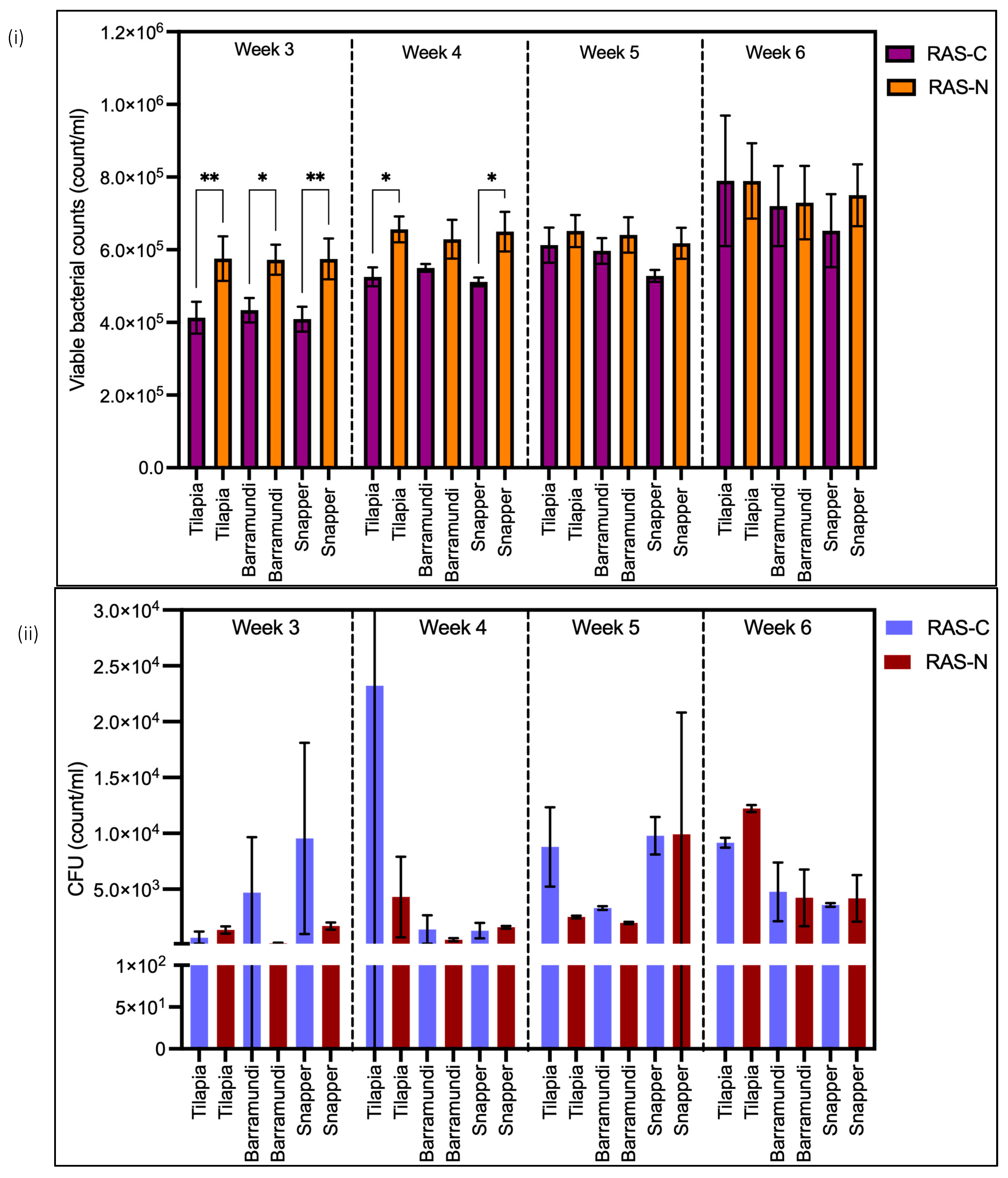
Flow cytometry showed increasing viable bacteria counts in both RAS-N and RAS-C over 4 weeks (Weeks 3–6) that were generally higher in RAS-N with nanobubble injections but were only significantly higher across all three fish species in week 3 and in tilapia and snappers in week 4 (ANOVA) (Figure 2i). There were sporadic spikes in total bacteria plate counts, viz in barramundi and red snapper RAS-N tanks on Day 9, tilapia RAS-C tanks on Day 16, both red snapper RAS-C and RAS-N tanks on Day 23, red snapper RAS-N tanks on Day 25, and both barramundi RAS-C and RAS-N tanks on Day 32. These sporadic spikes detected on total plate bacterial counts (Figure 2ii) were not reflected in viable bacterial counts using flow cytometry. Viable bacteria count based on flow cytometry often exceeded culturable bacteria counts by greater than 30–100-fold.

3.4. LNA and HNA Bacteria Abundance

High Nuclei Acid (HNA) bacteria abundance increased between Weeks 3 and 6 and was significantly higher than Low Nuclei Acid (LNA) bacteria (p < 0.05) in both RAS-N and RAS-C for all fish species, except tilapia tanks in Week 3 (Figure 3). LNA bacteria abundance was consistently higher in RAS-N than in RAS-C.

3.5. Microbiome Profile of Water Samples from RAS with and Without Nanobubbles

The most abundant bacteria amongst the 30 most abundant genus were clustered more closely than the less abundant bacteria genus in both systems. Vibrio, Enterovibrio and Cetobacterium were more abundant in RAS-C. Flavobacteriaceae and Tenacibaculum, and nitrifying bacteria such as Nitrospira and Marinobacter were more abundant in RAS-N, while Fabibacter were significantly more abundant in RAS-C (Figure 4).

4. Discussions

This 6-week study evaluated whether air nanobubble enrichment improves water quality and fish growth performance in marine RAS across three commercially important species: barramundi, red snapper, and tilapia. Despite the significant differences in microbial community composition and bacteria abundance between nanobubble-enriched system and control, air nanobubbles did not significantly improve growth rate, Feed Conversion Ratio or survival percentages in any of the three experimented species.
Flow cytometry-based viable bacterial counts consistently increased from Week 3 to Week 6 and were consistently higher in RAS-N compared to RAS-C throughout the study period. Fish biomass influences organic wastes output which can lead to changes in microbial profiles [35]. Membrane-based ultrafiltration reduced organic matter and significantly improved water quality in RAS, increased microbial diversity, and suppressed bacterial blooms, notably the presence of Mycobacterium [36]. High organic loads fuel the growth of opportunistic bacteria in the water, which may displace beneficial bacteria and impact gut health [37]. Although dissolved oxygen did not show statistical difference between RAS-C and RAS-N, DO was consistently much lower in RAS-N likely due to higher viable bacteria loads. Nanobubbles theoretically enhance oxygen delivery efficiency compared to conventional aeration and can elevate oxygen levels beneficial for high-density aquaculture [38]. Higher oxygen levels promote bacteria growth, particularly the activity of aerobic nitrifying bacteria to break down organic matter [39,40]. However, since DO levels showed no significant difference between RAS-N or RAS-C systems in this study, lower oxygen alone cannot explain the higher bacterial counts in nanobubble systems. An alternative mechanism may involve the negatively charged surface of nanobubbles, which attracts positively charged ions and has been suggested to promote microbial growth [41], though this mechanism requires further investigation in RAS environments.
The 16S rRNA analysis revealed a higher abundance of biofilm-forming bacteria Flavobacteriaceae in RAS-N tanks, since Flavobacteriaceae are dominant in biofilm formation in aquaculture systems [42]. Visual observation showed heavier biofilm formation in control tanks than in nanobubble-enriched tank walls. Dense mats of filamentous bacteria such as Flavobacterium or Tenacibaculum on fish gills and skin are typically triggered by deterioration in water quality, or skin damage due to jellyfish or ectoparasites [43,44]. Similarly, biofilm formation leading to bacterial endocarditis or tooth decay is thought to be triggered by adverse environmental conditions [45,46]. The mechanisms by which nanobubbles reduce biofilm formation on tank surfaces despite elevated Flavobacteriaceae in the water column warrant further investigation.
Nanobubble-enriched systems showed a significantly higher abundance of nitrifying bacteria, particularly Nitrospira and Marinobacter, which corresponded with lower nitrite and higher nitrate concentrations in RAS-N. This pattern indicates more efficient nitrification, a critical process for maintaining water quality in RAS by converting toxic ammonia and nitrite into less harmful nitrate. The enhanced nitrification in RAS-N systems likely contributed to the observed improvements in water quality parameters. Conversely, RAS-N systems exhibited a lower abundance of Vibrio species compared to controls. The use of probiotics containing nitrifying bacteria was demonstrated to reduce total bacteria and total Vibrio counts in artificially made seawater made to mimic wastewater from shrimp culture [47]. Higher abundance of Vibrio, Enterovibrio and Cetobacterium previously reported as gut microbiome suggests the accumulation of fecal wastes in RAS-C [48]. The improved waste clearance in RAS-N may relate to nanobubbles’ ability to attract organic pollutants to their negatively charged surfaces, a property that has been exploited for bioremediation of contaminated groundwater [49]. This mechanism may enhance the removal or processing of fecal matter and other organic waste products in nanobubble-enriched aquaculture systems.
This study demonstrated that flow cytometry (FCM) provides a rapid and effective alternative to traditional culture-based methods for monitoring bacterial populations in RAS. Flow cytometry-based viable bacterial counts consistently exceeded culturable bacteria or total plate counts by 30- to 100-fold throughout the study. This substantial difference reflects the well-established limitation of culture-based methods: slow growing or fastidious bacteria may require special culture media and be missed by conventional bacterial plate counts [50]. Flow cytometry (FCM) proved effective for daily bacterial enumeration of a large number of water samples compared to total bacteria plate counts, which may require several dilutions of water samples to achieve a reasonable count below 300 colonies per plate [51,52]. This study also showed wide variations in total bacteria plate counts in replicate test results, which may be due to a tendency for biofilm-forming bacteria to clump together [53] and differences in water subsamples used to inoculate replicate plates. FCM offers a rapid and effective method for detecting viable bacterial cells, providing results within minutes and correlating well with traditional colony counting [54] and our results observed a small variation in bacterial counts between replicates of water subsamples. This tool is well established for monitoring bacterial communities in various aquatic environments [55,56,57] wastewater treatment plants [58,59,60] and drinking water [61,62]. Recent studies have leveraged flow cytometry to proactively screen for watershed pollutants by quickly assessing metabolic changes in waterborne bacteria [63].
Beyond enumeration, FCM enabled differentiation between High Nuclei Acid (HNA) and Low Nuclei Acid (LNA) bacteria populations, providing additional insights into microbial community dynamics. HNA bacterial numbers increased progressively over 4 weeks with a rise in fish biomass, while LNA bacteria remained relatively stable throughout this study. HNA bacteria are known to dominate in nutrient-rich environments, while LNA bacteria are more prominent in oligotrophic river systems. This may explain the increase in HNA bacteria counts as fish biomass increased in this study. Nuclei acid analysis can be a better indicator of microbial activity in aquatic ecosystems. Spikes in HNA bacteria are linked to increased organic carbon, total nitrogen and biological oxygen demands [29,30,64]. HNA: LNA bacteria ratio may be better indicators for organic loads than total bacteria count [5,65,66].
There were no significant differences in growth, FCR and percentage survival between experimental tanks with and without nanobubble enrichment in marine RAS, for all three fish species. Experimental fish were stocked at densities below 20 kg/m3, which is much lower than in commercial intensive RAS. At lower stocking densities, fish experienced less competition for space and food than under commercial conditions. Water quality parameters were maintained at optimal levels for both control (RAS-C) and experimental systems (RAS-N) with high exchange rates exceeding 100% per hour. A recent study showed no significant difference in FCR in juvenile barramundi kept at three different stocking densities (5.6, 6.7 and 7.7 kg/m3) and fed at three different feed rates [67]. Even though nanobubbles have a long residual time of weeks in water [7,38], it is not known whether nanobubbles dissipated with the high water exchange rates, passage through protein skimmer, degassing tower, UV, or were consumed by the bacteria within the fluidized sand filters in our experimental RAS. Higher fish stocking densities and lower water exchange rates are worth investigating in future studies.
The lack of growth performance improvements in this study contrasts with some previous reports of nanobubble benefits in aquaculture RAS. Ebina et al. (2013) reported that air nanobubble-rich water increased the final biomass of sweetfish (Plecoglossus altivelis) after three weeks and rainbow trout (Oncorhynchus mykiss) after six months. However, Ebina et al. (2013)’s work did not provide details such as stocking density, initial and final fish weights, FCR or any water quality parameters to allow comparison. Other studies reported significant growth improvements in L. vannamei with use of oxygen nanobubbles [10,68]. DO levels in oxygen nanobubble systems with L. vannamei reached a peak of 10.8 mg/L against 4.65 mg/L in control systems with aerators [68]. Therefore, it is unknown whether such reported benefits in L. vannamei were due to higher DO levels or the use of nanobubbles.
The supply of air nanobubbles did not significantly increase DO levels in experimental tanks when compared to the control system in this study, possibly due to the high water exchange rates in the RAS and low fish biomass. DO levels in aquaculture systems are an important factor affecting fish growth by enhancing metabolic rates [69,70]. When dissolved oxygen levels are maintained near their saturation level, feeding is thought to become more efficient, increasing fish growth rates and immune response while reducing FCR [69,71]. Conversely, low levels of DO significantly reduced hatch and survival rates of Northen red snapper (Lutjanus campechanus) larvae [72], and growth, feed utilization and innate immunity of Nile tilapia (Oreochromis niloticus) fingerlings [69].
Future studies investigating nanobubble technology should include control treatments that match oxygen levels through alternative delivery methods (such as oxygen diffusers or Speece cones) to isolate the specific effects of nanobubbles from general oxygenation benefits. Additionally, comparative studies using oxygen-enriched versus air nanobubbles would clarify whether gas composition influences outcomes.

5. Conclusions

This study demonstrated that air nanobubble enrichment in marine RAS significantly altered microbial community structure, with 16S rRNA analysis revealing an increased abundance of nitrifying bacteria (Nitrospira and Marinobacter) that corresponded with enhanced nitrogen cycling and lower nitrite levels. However, these water quality improvements did not translate into enhanced growth performance, Feed Conversion Ratio, or survival in barramundi, red snapper, or tilapia at stocking densities below 20 kg/m3 with high water exchange rates. Flow cytometry proved to be an efficient method for monitoring bacterial abundance in intensive aquaculture systems, with 96-well plate analysis enabling high-throughput, same-day results and revealing 30- to 100-fold higher viable bacterial counts than traditional plate counts. Future research should investigate whether nanobubble benefits become apparent at commercial stocking densities (>30 kg/m3) over full production cycles (12–18 months), comparing air and oxygen nanobubbles with oxygen-matched controls, and utilizing 16S rRNA analysis of replicate tank samples at multiple time points to better characterize the temporal dynamics of microbial community succession.

Author Contributions

A.S.: Conceptualization, data curation, formal analysis, investigation, methodology, writing—original draft, writing—review and editing. T.S.L.: Conceptualization, data curation, formal analysis, investigation, methodology, writing—review and editing. J.A.D.: Conceptualization, methodology, resources, supervision, writing—review and editing. J.A.U.: Methodology, resources, supervision, writing—review and editing. X.S.: Formal analysis, writing—review and editing. S.G.-K.: Conceptualization, funding acquisition, methodology, project administration, resources, supervision, writing—review and editing. All authors have read and agreed to the published version of the manuscript.

Funding

This study was partially funded by Sysmex Asia Pacific Pte Ltd. (funding number: 15092021141421-0001), which provided the CyFlow™ Cube 6 V2m with CyFlow Robby6 Autoload instruments and CyStain reagents for bacterial count analysis.

Institutional Review Board Statement

The animal study protocol was approved by the JCUS Institutional Animal Care and Use Committee (IACUC) (protocol code: 2020-A04, and approval date: 25 March 2021).

Informed Consent Statement

Not applicable.

Data Availability Statement

Raw sequence data were submitted to the National Center for Biotechnology Information Sequence Read Archive under the BioProject accession number PRJNA1284650.

Acknowledgments

We would like to express our gratitude to Hazel Lim (Sysmex Asia Pacific Pte Ltd.), Wendy Phua (Sysmex Asia Pacific Pte Ltd.) and Charlie Chow and James Chow (AquaPro Solutions Pte Ltd.) for their coordination of the administration and provision of the resources for this study. We are also grateful to the following individuals who involved in this study: Nayli Raifana, Justin Yeo, Audrey Lee, Benjamin Pang, Xian Zhe Chew, Celestine Terence, Dorian Baraba, Weiyu Chen, Sarah Nelson, Josiah Poon, and Yu Xun Leow.

Conflicts of Interest

The authors declare no conflicts of interest. The authors declare that this study received funding from Sysmex Asia Pacific Pte Ltd. The funder was not involved in the study design, collection, analysis, interpretation of data, the writing of this article or the decision to submit it for publication.

References

  1. Murray, F.; Bostock, J.; Fletcher, D. Review of Recirculation Aquaculture System Technologies and Their Commercial Application. 2014. Available online: http://www.hie.co.uk/common/handlers/download-document.ashx?id=236008c4-f52a-48d9-9084-54e89e965573 (accessed on 12 May 2025).
  2. Xiao, R.; Wei, Y.; An, D.; Li, D.; Ta, X.; Wu, Y.; Ren, Q. A Review on the Research Status and Development Trend of Equipment in Water Treatment Processes of Recirculating Aquaculture Systems. Rev. Aquac. 2019, 11, 863–895. [Google Scholar] [CrossRef]
  3. Agarwal, A.; Ng, W.J.; Liu, Y. Principle and Applications of Microbubble and Nanobubble Technology for Water Treatment. Chemosphere 2011, 84, 1175–1180. [Google Scholar] [CrossRef]
  4. Phan, K.K.T.; Truong, T.; Wang, Y.; Bhandari, B. Nanobubbles: Fundamental Characteristics and Applications in Food Processing. Trends Food Sci. Technol. 2020, 95, 118–130. [Google Scholar] [CrossRef]
  5. Hu, W.; Zhang, H.; Lin, X.; Liu, R.; Bartlam, M.; Wang, Y. Characteristics, Biodiversity, and Cultivation Strategy of Low Nucleic Acid Content Bacteria. Front. Microbiol. 2022, 13, 900669. [Google Scholar] [CrossRef] [PubMed]
  6. Lyu, T.; Wu, S.; Mortimer, R.J.G.; Pan, G. Nanobubble Technology in Environmental Engineering: Revolutionization Potential and Challenges; ACS Publications: Washington, DC, USA, 2019. [Google Scholar] [CrossRef]
  7. Domingos, J.A.; Huang, Q.; Liu, H.; Dong, H.T.; Khongcharoen, N.; Van, P.T.; Nghia, N.H.; Giang, P.T.; The Viet, P.; St-Hilaire, S. Air-Nanobubbles Ineffective to Reduce Pathogenic Bacteria in Fresh and Brackish Waters. bioRxiv 2021. [Google Scholar] [CrossRef]
  8. Battino, R.; Clever, H.L. The Solubility of Gases in Liquids. Chem. Rev. 1966, 66, 395–463. [Google Scholar] [CrossRef]
  9. Ushikubo, F.Y.; Furukawa, T.; Nakagawa, R.; Enari, M.; Makino, Y.; Kawagoe, Y.; Shiina, T.; Oshita, S. Evidence of the Existence and the Stability of Nano-Bubbles in Water. Colloids Surf. A Physicochem. Eng. Asp. 2010, 361, 31–37. [Google Scholar] [CrossRef]
  10. Rahmawati, A.I.; Saputra, R.N.; Hidayatullah, A.; Dwiarto, A.; Junaedi, H.; Cahyadi, D.; Saputra, H.K.H.; Prabowo, W.T.; Kartamiharja, U.K.A.; Shafira, H. Enhancement of Penaeus vannamei Shrimp Growth Using Nanobubble in Indoor Raceway Pond. Aquac. Fish 2021, 6, 277–282. [Google Scholar] [CrossRef]
  11. Guo, J.; Chen, Y.; Zhang, Y.; Zhang, R.; Inaba, K.; Osato, T.; Zhao, X.; Han, Y.; Ren, T. Oxygen Nanobubble-Induced Hyperoxia: Effects on Growth, Digestive Enzyme Activity, Intestinal Morphology, and Biochemical Parameters in Kuruma Prawn (Penaeus japonicus). Aquac. Rep. 2025, 43, 102882. [Google Scholar] [CrossRef]
  12. Heriyati, E.; Rustadi, R.; Isnansetyo, A.; Triyatmo, B.; Istiqomah, I.; Deendarlianto, D.; Budhijanto, W. Microbubble Aeration in A Recirculating Aquaculture System (RAS) Increased Dissolved Oxygen, Fish Culture Performance, and Stress Resistance of Red Tilapia (Oreochromis Sp.). Trends Sci. 2022, 19, 6251. [Google Scholar] [CrossRef]
  13. Linh, N.V.; Khongcharoen, N.; Nguyen, D.-H.; Dien, L.T.; Rungrueng, N.; Jhunkeaw, C.; Sangpo, P.; Senapin, S.; Uttarotai, T.; Panphut, W. Effects of Hyperoxia during Oxygen Nanobubble Treatment on Innate Immunity, Growth Performance, Gill Histology, and Gut Microbiome in Nile Tilapia, Oreochromis niloticus. Fish Shellfish Immunol. 2023, 143, 109191. [Google Scholar] [CrossRef]
  14. Cardona, E.; Gueguen, Y.; Magré, K.; Lorgeoux, B.; Piquemal, D.; Pierrat, F.; Noguier, F.; Saulnier, D. Bacterial Community Characterization of Water and Intestine of the Shrimp Litopenaeus stylirostris in a Biofloc System. BMC Microbiol. 2016, 16, 1–9. [Google Scholar] [CrossRef]
  15. Khanjani, M.H.; Mohammadi, A.; Emerenciano, M.G.C. Microorganisms in Biofloc Aquaculture System. Aquac. Rep. 2022, 26, 101300. [Google Scholar] [CrossRef]
  16. Del’Duca, A.; Cesar, D.E.; Freato, T.A.; Azevedo, R.d.S.; Rodrigues, E.M.; Abreu, P.C. Variability of the Nitrifying Bacteria in the Biofilm and Water Column of a Recirculating Aquaculture System for Tilapia (Oreochromis niloticus) Production. Aquac. Res. 2019, 50, 2537–2544. [Google Scholar] [CrossRef]
  17. Ward, B.B.; Arp, D.J.; Klotz, M.G. Nitrification; American Society for Microbiology Press: Washington, DC, USA, 2011. [Google Scholar]
  18. Rurangwa, E.; Verdegem, M.C.J. Microorganisms in Recirculating Aquaculture Systems and Their Management. Rev. Aquac. 2015, 7, 117–130. [Google Scholar] [CrossRef]
  19. Chew, X.Z.; Gibson-Kueh, S.; Jerry, D.R.; Shen, X. Comparison of Intestinal Bacterial Communities in Asymptomatic and Diseased Asian Seabass (Lates calcarifer) with Chronic Enteritis and Mixed Bacterial Infections. Aquaculture 2023, 572, 739516. [Google Scholar] [CrossRef]
  20. Kelly, C.; Salinas, I. Under Pressure: Interactions between Commensal Microbiota and the Teleost Immune System. Front. Immunol. 2017, 8, 559. [Google Scholar] [CrossRef] [PubMed]
  21. Xue, S.; Xu, W.; Wei, J.; Sun, J. Impact of Environmental Bacterial Communities on Fish Health in Marine Recirculating Aquaculture Systems. Vet. Microbiol. 2017, 203, 34–39. [Google Scholar] [CrossRef] [PubMed]
  22. You, J.L.; Cao, L.X.; Liu, G.F.; Zhou, S.N.; Tan, H.M.; Lin, Y.C. Isolation and Characterization of Actinomycetes Antagonistic to Pathogenic Vibrio spp. from Nearshore Marine Sediments. World J. Microbiol. Biotechnol. 2005, 21, 679–682. [Google Scholar] [CrossRef]
  23. Bentzon-Tilia, M.; Sonnenschein, E.C.; Gram, L. Monitoring and Managing Microbes in Aquaculture—Towards a Sustainable Industry. Microb. Biotechnol. 2016, 9, 576–584. [Google Scholar] [CrossRef]
  24. Ganesh, E.A.; Das, S.; Chandrasekar, K.; Arun, G.; Balamurugan, S. Monitoring of Total Heterotrophic Bacteria and Vibrio spp. in an Aquaculture Pond. Curr. Res. J. Biol. Sci. 2010, 2, 48–52. Available online: https://www.researchgate.net/publication/267606862 (accessed on 12 May 2025).
  25. Stewart, E.J. Growing Unculturable Bacteria. J. Bacteriol. 2012, 194, 4151–4160. [Google Scholar] [CrossRef]
  26. Zhang, X.-H.; Ahmad, W.; Zhu, X.-Y.; Chen, J.; Austin, B. Viable but Nonculturable Bacteria and Their Resuscitation: Implications for Cultivating Uncultured Marine Microorganisms. Mar. Life Sci. Technol. 2021, 3, 189–203. [Google Scholar] [CrossRef]
  27. Śliwa-Dominiak, J.; Czechowska, K.; Blanco, A.; Sielatycka, K.; Radaczyńska, M.; Skonieczna-Żydecka, K.; Marlicz, W.; Łoniewski, I. Flow Cytometry in Microbiology: A Review of the Current State in Microbiome Research, Probiotics, and Industrial Manufacturing. Cytom. Part A 2025, 107, 145–164. [Google Scholar] [CrossRef]
  28. Khan, M.M.; Pyle, B.H.; Camper, A.K. Specific and Rapid Enumeration of Viable but Nonculturable and Viable-Culturable Gram-Negative Bacteria by Using Flow Cytometry. Appl. Environ. Microbiol. 2010, 76, 5088–5096. [Google Scholar] [CrossRef]
  29. Lebaron, P.; Servais, P.; Agogué, H.; Courties, C.; Joux, F. Does the High Nucleic Acid Content of Individual Bacterial Cells Allow Us To Discriminate between Active Cells and Inactive Cells in Aquatic Systems? Appl. Environ. Microbiol. 2001, 67, 1775–1782. [Google Scholar] [CrossRef] [PubMed]
  30. Lebaron, P.; Servais, P.; Baudoux, A.-C.; Bourrain, M.; Courties, C.; Parthuisot, N. Variations of Bacterial-Specific Activity with Cell Size and Nucleic Acid Content Assessed by Flow Cytometry. Aquatic. Microbial. Ecol. 2002, 28, 131–140. [Google Scholar] [CrossRef]
  31. Marwiyah, U.C.; Mahasri, G.; Ratnasari, R.E.; Wiradana, P.A. Total Plate Count and Identification of Vibrio in Pacific White Shrimp (Litophenaeus vannamei) from Ponds and in Those Exposed to Immunogenic Protein Membrane Zoothamnium penaei. In Proceedings of the IOP Conference Series: Earth and Environmental Science; IOP Publishing: Bristol, UK, 2019; Volume 236, p. 012087. [Google Scholar] [CrossRef]
  32. Majaneva, M.; Diserud, O.H.; Eagle, S.H.C.; Boström, E.; Hajibabaei, M.; Ekrem, T. Environmental DNA Filtration Techniques Affect Recovered Biodiversity. Sci. Rep. 2018, 8, 4682. [Google Scholar] [CrossRef] [PubMed]
  33. Ren, Z.; Qu, X.; Peng, W.; Yu, Y.; Zhang, M. Nutrients Drive the Structures of Bacterial Communities in Sediments and Surface Waters in the River-Lake System of Poyang Lake. Water 2019, 11, 930. [Google Scholar] [CrossRef]
  34. Caporaso, J.G.; Kuczynski, J.; Stombaugh, J.; Bittinger, K.; Bushman, F.D.; Costello, E.K.; Fierer, N.; Peña, A.G.; Goodrich, J.K.; Gordon, J.I. QIIME Allows Analysis of High-Throughput Community Sequencing Data. Nat. Methods 2010, 7, 335–336. [Google Scholar] [CrossRef] [PubMed]
  35. Quero, G.M.; Ape, F.; Manini, E.; Mirto, S.; Luna, G.M. Temporal Changes in Microbial Communities Beneath Fish Farm Sediments Are Related to Organic Enrichment and Fish Biomass Over a Production Cycle. Front. Mar. Sci. 2020, 7, 524. [Google Scholar] [CrossRef]
  36. Fossmark, R.O.; Vadstein, O.; Rosten, T.W.; Bakke, I.; Košeto, D.; Bugten, A.V.; Helberg, G.A.; Nesje, J.; Jørgensen, N.O.G.; Raspati, G.; et al. Effects of Reduced Organic Matter Loading through Membrane Filtration on the Microbial Community Dynamics in Recirculating Aquaculture Systems (RAS) with Atlantic Salmon Parr (Salmo salar). Aquaculture 2020, 524, 735268. [Google Scholar] [CrossRef]
  37. Bugten, A.V.; Attramadal, K.J.K.; Fossmark, R.O.; Rosten, T.W.; Vadstein, O.; Bakke, I. Changes in Rearing Water Microbiomes in RAS Induced by Membrane Filtration Alters the Hindgut Microbiomes of Atlantic Salmon (Salmo salar) Parr. Aquaculture 2022, 548, 737661. [Google Scholar] [CrossRef]
  38. Ebina, K.; Shi, K.; Hirao, M.; Hashimoto, J.; Kawato, Y.; Kaneshiro, S.; Morimoto, T.; Koizumi, K.; Yoshikawa, H. Oxygen and Air Nanobubble Water Solution Promote the Growth of Plants, Fishes, and Mice. PLoS One 2013, 8, e65339. [Google Scholar] [CrossRef]
  39. Van Beijnen, J.; Yan, G. A Breath of Fresh Air: How Nanobubbles Can Make Aquaculture More Sustainable. 2021. Available online: https://thefishsite.com/articles/a-breath-of-fresh-air-how-nanobubbles-can-make-aquaculture-more-sustainable-dissolved-oxygen (accessed on 12 May 2025).
  40. Yao, G.-J.; Ren, J.-Q.; Zhou, F.; Liu, Y.-D.; Li, W. Micro-Nano Aeration Is a Promising Alternative for Achieving High-Rate Partial Nitrification. Sci. Total Environ. 2021, 795, 148899. [Google Scholar] [CrossRef] [PubMed]
  41. Park, J.-S.; Kurata, K. Application of Microbubbles to Hydroponics Solution Promotes Lettuce Growth. Horttechnology 2009, 19, 212–215. [Google Scholar] [CrossRef]
  42. Qu, J.; Yang, H.; Liu, Y.; Qi, H.; Wang, Y.; Zhang, Q. The Study of Natural Biofilm Formation and Microbial Community Structure for Recirculating Aquaculture System. In Proceedings of the IOP Conference Series: Earth and Environmental Science; IOP Publishing: Bristol, UK, 2021; Volume 742, p. 012018. [Google Scholar] [CrossRef]
  43. Avendaño-Herrera, R.; Toranzo, A.E.; Magariños, B. Tenacibaculosis Infection in Marine Fish Caused by Tenacibaculum maritimum: A Review. Dis. Aquat. Organ. 2006, 71, 255–266. [Google Scholar] [CrossRef] [PubMed]
  44. Mabrok, M.; Algammal, A.M.; Sivaramasamy, E.; Hetta, H.F.; Atwah, B.; Alghamdi, S.; Fawzy, A.; Avendaño-Herrera, R.; Rodkhum, C. Tenacibaculosis Caused by Tenacibaculum maritimum: Updated Knowledge of This Marine Bacterial Fish Pathogen. Front. Cell. Infect. Microbiol. 2023, 12, 1068000. [Google Scholar] [CrossRef]
  45. Jefferson, K.K. What Drives Bacteria to Produce a Biofilm? FEMS Microbiol. Lett. 2004, 236, 163–173. [Google Scholar] [CrossRef]
  46. Zhao, A.; Sun, J.; Liu, Y. Understanding Bacterial Biofilms: From Definition to Treatment Strategies. Front. Cell Infect. Microbiol. 2023, 13, 1137947. [Google Scholar] [CrossRef]
  47. Widigdo, B.; Yuhana, M.; Iswantari, A.; Madonsa, C.; Sapitri, I.D.; Wardiatno, Y.; Hakim, A.A.; Nazar, F. The Impact of Nitrifying Probiotic to Population Growth of Pathogenic Bacteria, Vibrio sp., and Toxic Nitrogen Gasses in Marine Shrimp Culture Media under Laboratory Condition. J. Pengelolaan Sumberd. Alam Dan Lingkung. (J. Nat. Resour. Environ. Manag.) 2021, 11, 130–140. [Google Scholar] [CrossRef]
  48. Hassenrück, C.; Reinwald, H.; Kunzmann, A.; Tiedemann, I.; Gärdes, A. Effects of Thermal Stress on the Gut Microbiome of Juvenile Milkfish (Chanos chanos). Microorganisms 2020, 9, 5. [Google Scholar] [CrossRef] [PubMed]
  49. Li, H.; Hu, L.; Xia, Z. Impact of Groundwater Salinity on Bioremediation Enhanced by Micro-Nano Bubbles. Materials 2013, 6, 3676–3687. [Google Scholar] [CrossRef]
  50. Davey, H.; Guyot, S. Estimation of Microbial Viability Using Flow Cytometry. Curr. Protoc. Cytom. 2020, 93, e72. [Google Scholar] [CrossRef] [PubMed]
  51. Koch, A.L. Growth Measurement. In Methods for General and Molecular Microbiology; Wiley Online Library: New York, NY, USA, 2007; pp. 172–199. [Google Scholar] [CrossRef]
  52. Malmberg, C. Evaluation of Flow Cytometry as Replacement for Plating in In Vitro Measurements of Competitive Growth under Antibiotic Stress. 2013, p. 13. Available online: https://uu.diva-portal.org/smash/record.jsf?pid=diva2%3A639728&dswid=4462 (accessed on 12 May 2025).
  53. Wilson, C.; Lukowicz, R.; Merchant, S.; Valquier-Flynn, H.; Caballero, J.; Sandoval, J.; Okuom, M.; Huber, C.; Brooks, T.D.; Wilson, E. Quantitative and Qualitative Assessment Methods for Biofilm Growth: A Mini-Review. Res. Rev. J. Eng. Technol. 2017, 6. [Google Scholar]
  54. Endo, H.; Nakayama, J.; Hayashi, T. Application of Flow Cytometry to Environmental Control in Marine Aquaculture. Mater. Sci. Eng. C 2000, 12, 83–88. [Google Scholar] [CrossRef]
  55. Bernard, L.; Courties, C.; Servais, P.; Troussellier, M.; Petit, M.; Lebaron, P. Relationships among Bacterial Cell Size, Productivity, and Genetic Diversity in Aquatic Environments Using Cell Sorting and Flow Cytometry. Microb. Ecol. 2000, 40, 148–158. [Google Scholar] [CrossRef]
  56. Fiedler, C.J.; Schönher, C.; Proksch, P.; Kerschbaumer, D.J.; Mayr, E.; Zunabovic-Pichler, M.; Domig, K.J.; Perfler, R. Assessment of Microbial Community Dynamics in River Bank Filtrate Using High-Throughput Sequencing and Flow Cytometry. Front. Microbiol. 2018, 9, 2887. [Google Scholar] [CrossRef]
  57. Props, R.; Monsieurs, P.; Mysara, M.; Clement, L.; Boon, N. Measuring the Biodiversity of Microbial Communities by Flow Cytometry. Methods Ecol. Evol. 2016, 7, 1376–1385. [Google Scholar] [CrossRef]
  58. Foladori, P.; Bruni, L.; Tamburini, S.; Ziglio, G. Direct Quantification of Bacterial Biomass in Influent, Effluent and Activated Sludge of Wastewater Treatment Plants by Using Flow Cytometry. Water Res. 2010, 44, 3807–3818. [Google Scholar] [CrossRef]
  59. Ma, L.; Mao, G.; Liu, J.; Yu, H.; Gao, G.; Wang, Y. Rapid Quantification of Bacteria and Viruses in Influent, Settled Water, Activated Sludge and Effluent from a Wastewater Treatment Plant Using Flow Cytometry. Water Sci. Technol. 2013, 68, 1763–1769. [Google Scholar] [CrossRef] [PubMed]
  60. Manti, A.; Boi, P.; Falcioni, T.; Canonico, B.; Ventura, A.; Sisti, D.; Pianetti, A.; Balsamo, M.; Papa, S. Bacterial Cell Monitoring in Wastewater Treatment Plants by Flow Cytometry. Water Environ. Res. 2008, 80, 346–354. [Google Scholar] [CrossRef] [PubMed]
  61. De Roy, K.; Clement, L.; Thas, O.; Wang, Y.; Boon, N. Flow Cytometry for Fast Microbial Community Fingerprinting. Water Res. 2012, 46, 907–919. [Google Scholar] [CrossRef] [PubMed]
  62. Van Nevel, S.; Koetzsch, S.; Proctor, C.R.; Besmer, M.D.; Prest, E.I.; Vrouwenvelder, J.S.; Knezev, A.; Boon, N.; Hammes, F. Flow Cytometric Bacterial Cell Counts Challenge Conventional Heterotrophic Plate Counts for Routine Microbiological Drinking Water Monitoring. Water Res. 2017, 113, 191–206. [Google Scholar] [CrossRef]
  63. Jenkins, J.A.; Mize, S.V.; Johnson, D.; Brown, B.L. Flow Cytometric Detection of Waterborne Bacteria Metabolic Response to Anthropogenic Chemical Inputs to Aquatic Ecosystems. Cells 2025, 14, 352. [Google Scholar] [CrossRef]
  64. Liu, J.; Hao, Z.; Ma, L.; Ji, Y.; Bartlam, M.; Wang, Y. Spatio-Temporal Variations of High and Low Nucleic Acid Content Bacteria in an Exorheic River. PLoS One 2016, 11, e0153678. [Google Scholar] [CrossRef]
  65. Santos, M.; Oliveira, H.; Pereira, J.L.; Pereira, M.J.; Gonçalves, F.J.M.; Vidal, T. Flow Cytometry Analysis of Low/High DNA Content (LNA/HNA) Bacteria as Bioindicator of Water Quality Evaluation. Ecol. Indic. 2019, 103, 774–781. [Google Scholar] [CrossRef]
  66. Wang, Y.; Hammes, F.; Boon, N.; Chami, M.; Egli, T. Isolation and Characterization of Low Nucleic Acid (LNA)-Content Bacteria. ISME J. 2009, 3, 889–902. [Google Scholar] [CrossRef]
  67. Le Boucher, R.; Chung, W.; Ng, J.K.L.; Tan, L.S.E.; Lee, C.S. Balancing Stocking Density and Feed Intake for Barramundi (Lates calcarifer) Raised in Recirculating Aquaculture Systems. Aquac. Res. 2024, 2024, 2264274. [Google Scholar] [CrossRef]
  68. Galang, D.P.; Ashari, A.K.; Sulmatiwi, L.; Mahasri, G.; Prayogo; Sari, L.A. The Oxygen Content and Dissolved Oxygen Consumption Level of White Shrimp Litopenaeus vannamei in the Nanobubble Cultivation System. IOP Conf. Ser. Earth Environ. Sci. 2019, 236, 012014. [Google Scholar] [CrossRef]
  69. Abdel-Tawwab, M.; Hagras, A.E.; Elbaghdady, H.A.M.; Monier, M.N. Effects of Dissolved Oxygen and Fish Size on Nile Tilapia, Oreochromis niloticus (L.): Growth Performance, Whole-Body Composition, and Innate Immunity. Aquac. Int. 2015, 23, 1261–1274. [Google Scholar] [CrossRef]
  70. Jia, Y.; Wang, J.; Gao, Y.; Huang, B. Hypoxia Tolerance, Hematological, and Biochemical Response in Juvenile Turbot (Scophthalmus maximus. L). Aquaculture 2021, 535, 736380. [Google Scholar] [CrossRef]
  71. Mallya, Y.J. The Effects of Dissolved Oxygen on Fish Growth in Aquaculture. The United Nations University Fisheries Training Programme, Final Project 2007. Available online: https://www.grocentre.is/static/gro/publication/58/document/yovita07prf.pdf (accessed on 12 May 2025).
  72. Bardon-Albaret, A.; Saillant, E.A. Effects of Hypoxia and Elevated Ammonia Concentration on the Viability of Red Snapper Embryos and Early Larvae. Aquaculture 2016, 459, 148–155. [Google Scholar] [CrossRef]
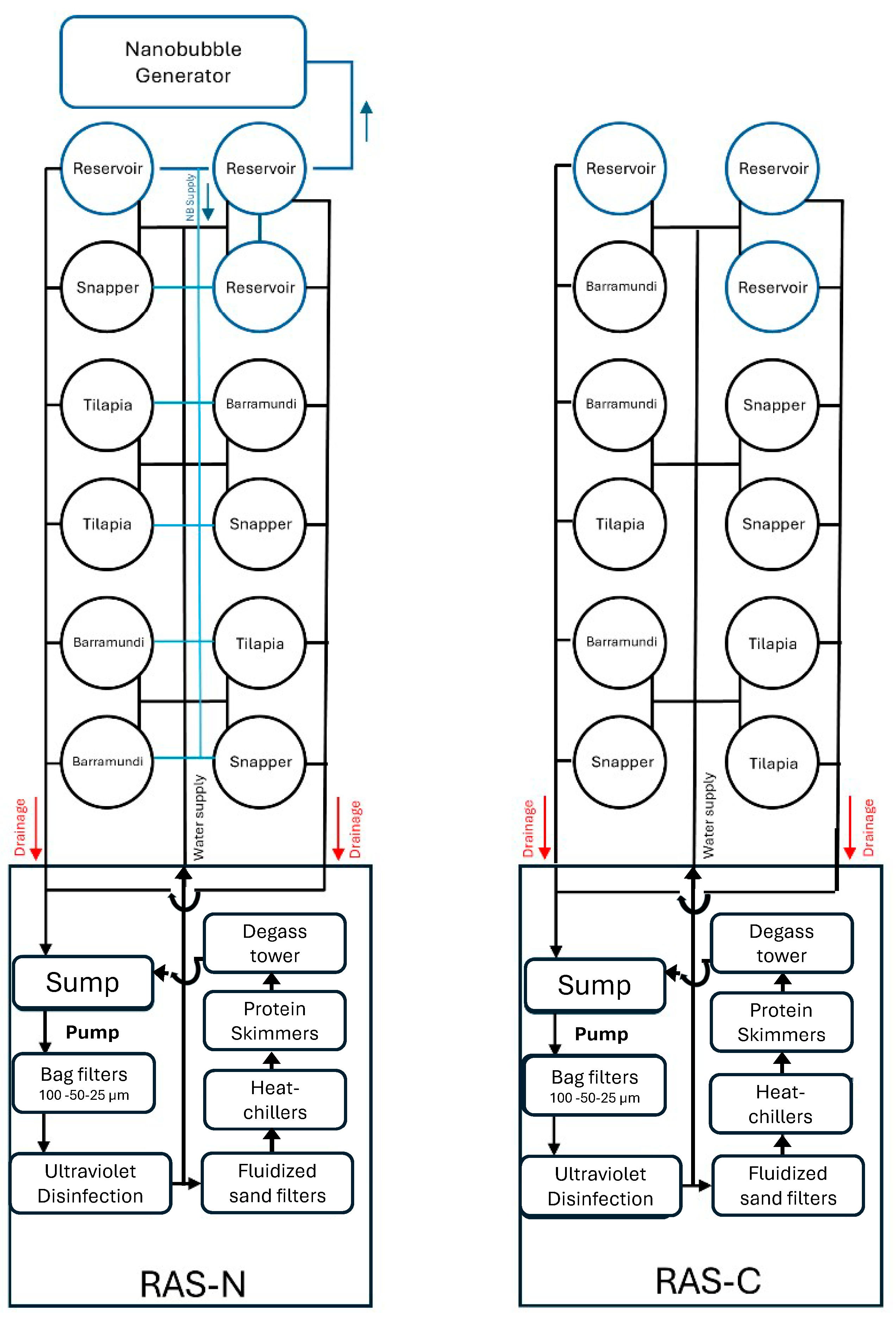
Figure 1. Schematic view of two recirculating aquaculture systems, RAS-N (with air nanobubbles) and RAS-C (control without air nanobubbles) each with triplicate tanks of red snapper (n = 15), barramundi (n = 20) or tilapia (n = 40). The top three tanks in RAS-N served as reservoirs for the nanobubble generator. Water was drawn from the reservoir tanks into the nanobubble generator to produce nanobubble-rich water, which was then distributed to the experimental tanks indicated as blue lines. The flow of water within the RAS is indicated by lines: red lines show water draining to the sump, while black lines represent the overall water flow in the system.
Fishes 10 00550 g001
Figure 2. (i) Flow cytometry: viable bacterial counts increased over Weeks 3 to 6 (4 weeks) in tilapia, barramundi, and red snapper tanks in both RAS-C and RAS-N. RAS-N showed higher total bacterial loads than RAS-C, with statistically significant differences observed during Week 3 in all 3 fish species, and in tilapia and snapper in Week 4. * p < 0.05 and ** p < 0.01. (ii) Conventional total bacteria plate counts on marine agar plates: large standard deviations and apparent sporadic spikes in CFU were observed, with a large variation between replicates. The viable bacterial counts and CFU are means of values taken daily on Monday to Friday each week from triplicate tanks, for each fish species in RAS-C or RAS-N systems.
Fishes 10 00550 g002
Figure 3. Flow cytometry: High Nucleic Acid (HNA) and Low Nucleic Acid (LNA) bacteria total counts increased between Week 3 and 6. HNA bacteria were significantly more abundant than LNA bacteria in all tanks in both RAS-C and RAS-N, irrespective of fish species (tilapia, barramundi, and snapper). **** p < 0.0001, *** p < 0.001, ** p < 0.01 and * p < 0.05.
Fishes 10 00550 g003
Figure 4. (i) A heatmap showing the 30 most abundant genus in water samples. (ii) Top 4 genus with significant differences in relative abundance (%) between (A) control RAS-C and (B) nanobubble RAS-N. * denotes statistical difference at p < 0.01 and ** at p value < 0.05.
Fishes 10 00550 g004
Table 1. Water quality in experimental RAS tanks over 6 weeks.
Water ParameterRAS-CRAS-N
pH8.00 ± 0.098.05 ± 0.07
TAN (mg/L)0.12 ± 0.060.12 ± 0.06
Nitrite (mg/L)1.40 ± 0.25 a0.99 ± 0.16 b
Nitrate (mg/L)165.6 ± 27.7182.2 ± 29.8
Temperature (°C)28.6 ± 0.528.6 ± 0.4
Salinity (ppt)24.8 ± 0.625.2 ± 0.5
DO (mg/L)6.61 ± 0.346.44 ± 0.24
DO (saturation, %)89.4 ± 5.787.7 ± 5.9
Note: Total ammonia nitrogen—TAN, dissolved oxygen—DO. Nitrite levels were significantly lower in RAS-N. Water quality is based on mean ± standard deviation. The different letters denote significant differences between RAS (p < 0.05).
Table 2. Growth performance over 6 weeks, final percentage survival and condition factor.
TreatmentSpeciesInitial Body Weight (g)Final Body Weight (g)Total Feed Intake (g)SGRFCRSurvival (%)Final Total Weight (g)Final Condition Factor
RAS-CMalabar snapper178.8 ± 25.6308.2 ± 45.22250 ± 1371.72 ± 0.120.93 ± 0.0295.6 ± 0.04417 ± 1783.13 ± 0.67
RAS-N177.0 ± 23.6300.8 ± 46.22234 ± 911.74 ± 0.100.95 ± 0.0597.8 ± 0.04411 ± 1683.22 ± 0.28
RAS-CBarramundi132.9 ± 26.9303.8 ± 66.63390 ± 743.19 ± 0.030.76 ± 0.02100.06076 ± 1022.02 ± 0.14
RAS-N132.1 ± 29.9296.8 ± 76.63332 ± 3993.14 ± 0.190.77 ± 0.02100.05937 ± 5522.26 ± 0.28
RAS-CHybrid tilapia9.5 ± 3.085.4 ± 29.72529 ± 1537.98 ± 0.310.80 ± 0.0395.8 ± 0.03272 ± 3003.83 ± 0.54
RAS-N9.2 ± 3.080.7 ± 29.42405 ± 687.92 ± 0.100.81 ± 0.0295.0 ± 0.03968 ± 1603.51 ± 0.52
Note: Values are mean ± standard deviation. Specific Growth Rates (SGR) were calculated as (Ln (Final weight)-Ln (Initial weight)) ∗ 100/Number of days. Feed Conversion Ratio (FCR) is the ratio of total feed intake to overall mean weight gain for each experimental fish group. The Final Condition Factor was calculated as Final body weight (g) * 100/Standard Length3 (cm).
Disclaimer/Publisher’s Note: The statements, opinions and data contained in all publications are solely those of the individual author(s) and contributor(s) and not of MDPI and/or the editor(s). MDPI and/or the editor(s) disclaim responsibility for any injury to people or property resulting from any ideas, methods, instructions or products referred to in the content.