Next Article in Journal
The Postfinasteride Syndrome; An Overview
Previous Article in Journal
Cutis Verticis Gyrata in a Patient with Multiple Basal Cell Carcinomas; Case Presentation and Review of the Literature
 
 
Journal of Mind and Medical Sciences is published by MDPI from Volume 12 Issue 1 (2025). Previous articles were published by another publisher in Open Access under a CC-BY (or CC-BY-NC-ND) licence, and they are hosted by MDPI on mdpi.com as a courtesy and upon agreement with Valparaiso University (ValpoScholar).
Case Report

Perforated Small Intestine in a Patient with T-Cell Lymphoma; A Rare Cause of Peritonitis

by
Petrişor Banu
1,2,*,
Vlad D. Constantin
1,2,
Florian Popa
1,2 and
Mihaela F. Nistor
2
1
Department of Surgery, Carol Davila University, Dionisie Lupu Street No. 37, Sect. 2, 020022 Bucharest, Romania
2
Department of Surgery, St. Pantelimon Hospital, 021659 Bucharest, Romania
*
Author to whom correspondence should be addressed.
J. Mind Med. Sci. 2016, 3(1), 88-98; https://doi.org/10.22543/2392-7674.1029
Submission received: 15 January 2016 / Revised: 25 February 2016 / Accepted: 25 March 2016 / Published: 30 March 2016

Abstract

:
The nontraumatic perforations of the small intestine are pathological entities with particular aspects in respect to diagnosis and treatment. These peculiarities derive from the nonspecific clinical expression of the peritonitis syndrome, and from the multitude of causes that might be the primary sources of the perforation: foreign bodies, inflammatory diseases, tumors, infectious diseases, etc. Accordingly, in most cases intestinal perforation is discovered only by laparotomy and the definitive diagnosis is available only after histopathologic examination. Small bowel malignancies are rare; among them, lymphomas rank third in frequency, being mostly B-cell non Hodgkin lymphomas. Only 10% of non-Hodgkin lymphomas are with T-cell. We report the case of a 57 years’ old woman with intestinal T-cell lymphoma, whose first clinical symptomatology was related to a complication represented by perforation of the small intestine. Laparotomy performed in emergency identified an ulcerative lesion with perforation in the jejunum, which required segmental enterectomy with anastomosis. The nonspecific clinical manifestations of intestinal lymphomas make from diagnosis a difficult procedure. Due to the fact that surgery does not have a definite place in the treatment of the small intestinal lymphomas (for cases complicated with perforation), and beyond the morbidity associated with the surgery performed in emergency conditions, prognosis of these patients is finally given by the possibility to control the systemic disease through adjuvant therapy.

Introduction

The nontraumatic perforation of the small intestine is a rare pathological entity, which has particular aspects in respect to the clinical diagnosis and subsequent therapeutic conduct. These peculiarities derive especially from the difficulty of the preoperative diagnosis. In the majority of cases, the peritonitis syndrome imposes laparotomy which reveals the perforation of the intestine.
In addition, there are multitude of causes that might be the primary sources of the perforation: foreign bodies, inflammatory diseases, tumors, infectious diseases, etc., so that only the histopathologic examination would be conclusive for the establishment of a definitive therapeutic approach.
A variable geographical distribution can be observed when considering the etiology and incidence on age groups of the small intestine’s perforations. The infectious causes, such as typhoid fever or tuberculosis predominate in tropical areas and in the developing countries, being often found at young aged patients, 20-30 years old, as opposed to the western countries where these cases appear much rarer and to older age groups, having as possible causes inflammatory diseases, tumors, radiotherapy, ingestion of foreign bodies or circulatory disorders [1,2,3,4].
The diagnosis becomes a challenging task especially in circumstances in which the patient has no significant medical history, so that the diagnosis can be established only after surgery. Malignant tumors of the small intestine are in general rare (about 3% among digestive neoplasms, and 0.5% within all cancers), having poor clinical expression and a relatively difficult diagnosis with usual investigative methods [5].
Regarding their histology in last decades one can see a reversal of proportions in incidence of carcinoids as against adenocarcinomas while lymphomas ranks third in frequency after these with 17% of all malignant intestinal tumors [6]. Non-Hodgkin's lymphomas are a heterogeneous group of neoplasms that can arise from B or T cells, progenitors or mature, or rarely from natural killer cells. 10 to 30 percent of patients with NHL have extra-nodal disease [7].The digestive tract represents the most frequent location of the non- Hodgkin extra-nodal lymphoma, about 36%, affecting the stomach, small intestine and colon, in order of frequency [8].

Case report

We report the case of a 57 years’ old woman, with no significant medical history, admitted to the hospital for intense and diffuse abdominal pain, which emerged around 3 hour prior to the arrival at the hospital. The patient described the debut of the pain in the upper abdomen, all of a sudden, like a „stubbing” feeling followed soon by the extending to the entire abdominal area.
Physical examination reveals fever 380C, tachycardia and abdominal distention with tenderness to palpation and absence of bowel sounds.
Laboratory tests showed normal values for leucocytes (WBC- 8250 /µL), neutrophil count not elevated (NEU- 6260/ µL), low values for lymphocyte count(LYM- 1170/µL/ 14,22%), red blood cells (RBC) 3,62 /*10^6 µL, Hemoglobin (Hb) 10.2g/dL and HTC31,9%. It was also found decreased levels for sodium (130,5mmol/L) and potassium 3,23mmol/L. No other changes were found in laboratory tests on admission.
Plain X-ray film of the abdomen in upright position showed the presence of free air under the left diaphragm. Chest radiography did not identify changes in lungs or mediastinum. Based on clinical, imaging and laboratory data the presumptive diagnosis was peritonitis from perforated viscera and laparotomy was decided.
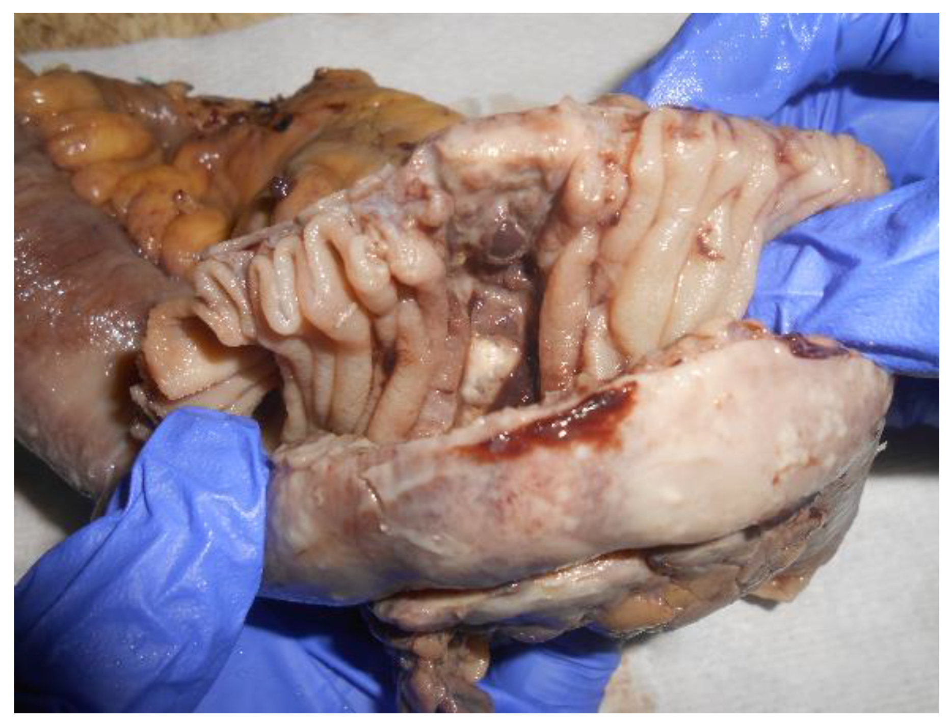
Midline vertical incision was made. The opening of the peritoneal cavity revealed small bowel content in peritoneum (succus entericus) and exploration showed a 3 millimeters perforation situated at the anti-mesenteric border on a jejunum loop about 60 cm from the Treitz angle (Figure 1).
The perforation adjacent area had a pale appearance and the mesentery exhibited multiple enlarged lymph nodes which were conglomerated especially at the root of the mesentery forming large lymphadenopathy masses at this level. The other intraperitoneal organs including liver and spleen had no changes macroscopically.
Small bowel resection and widely excision of adjacent mesentery was performed (Figure 2). The continuity was restored by end to end suturing anastomosis. Postoperative evolution was simple with no complications, and the patient was discharged 7 days after surgery.
Histopathological examination revealed:
Intestinal wall presenting a large ulceration about 1,5cm with perforation and diffuse transmural infiltration with large discoid cells presenting oval and vesicular nuclei and eosinophilic cytoplasm in reduced amount; large number of areas of granulomatous inflammation.
Multiple tumor cells with hyperchromic and hypertrophic nuclei; tumoral cells with multiple nuclei and atypical mitosis. Mesenteric lymphnodes exhibited an altered architecture with sinusal hystiocytosis and irregular germinal centers. This appearance was suggestive of diffuse large cells intestinal lymphoma.
Immunohistochemical examination revealed:
CD3 diffuse positive in tumoral cells, CD2 positive in tumoral cells, CD 25 positive in rare tumoral cells, CD5 negative in tumoral cells, CD30 diffusely positive in tumoral cells cytoplasm and intense positive in some membranes; CD15 positive in numerous granulocytes and in rare tumoral cells with granule appearance near the nuclei; CD20 and CD79a negative in tumoral cells; ALK negative in tumoral cells.
Histopathological aspect and immuno- histochemically tests are compatible with the diagnosis of Malign non Hodgkin lymphoma with T-cell type anaplastic large T-cell lymphoma ALK negative (ALCL, ALK ). Testing for hepatitis B, hepatitis C and HIV serology were negative using the western blotting procedure, and absence of the specific anti-HTLV I/II antibodies was confirmed.

Discussion

The primitive lymphomas of digestive tract remain rare entities, even though in the last decades a slight increase of their incidence (with percentage between 3 and 5) is reported. It is possible that this growth is due to the improvement of the immunohistochemical diagnosis which facilitates their diagnosis [8,9].
The cellular tissue from which these malignancies arise is represented by the MALT (mucosa associated lymphoid tissue), designating a heterogeneous population of cells with immunologic role quartered at the level of digestive tract mucosa, playing an essential role as a protection barrier of body from a large variety of antigens.
The intestine hosts the lymphoid tissue in follicles, in Peyer’s plaques, in lamina propria and intraepithelial and the mesenteric lymph nodes make connection to the peripheral immune system.
The variety of the enteral lymphoid tissue’s cells is very wide, but each phenotype can be individualized with cytogenetic and immunohistochemical techniques, so allowing the anatomo-clinical framing of the lymphomas, the indication of the treatment and the prognostic assessment.
Almost 90% of the gastrointestinal lymphomas derive from B lymphocytes and very few from T lymphocytes [10]. They can be classified into three main categories: immunoproliferative small intestinal disease (IPSID), enteropathy-associated T cell lymphoma (EATL) which arises mostly in gluten-sensitive enteropathy and a third category comprising diffuse large B cell lymphoma, mantle cell lymphoma, Burkitt lymphoma and follicular lymphoma [11].
What causes for lymphomas is still unknown, but a set of conditions seem to favor their development: bacterial infections (e.g. Helicobacter pylori or Helicobacter heilmannii), viruses (HTLV-1 in human T-cell lymphoma virus, Epstein-Barr in Burkitt lymphoma), severe immune deficiencies (AIDS, Waldmann disease) or celiac disease [8,12,13].
Considering frequency order criteria, these locate predominant at stomach level, which sums 50-60% from primary digestive tract lymphomas; 20-50% affect the small intestine, especially the ileum. Very seldom, the primitive lymphomas can be found at the colon and rectum level [14,15,16,17].
The symptoms are uncharacteristic for intestinal lymphomas, for example abdominal bloating and distension, colic abdominal pain, loss of appetite, nausea or may have the clinical expression of an acute complication like perforation, occlusion or bleeding. Other general signs also nonspecific are so called B symptoms (fever, weight loss and night sweats) which occurring in the context of a lymphoma get a prognosis value [18].
Classical methods of imaging diagnosis - barium studies and CT – can reveal tumors as intra- or extraluminal masses, polyps or parietal infiltration areas without offering data regarding the type of the lesion. On the other hand, endoscopy and particularly the “push and pull technique” with double balloon enteroscopy allows both biopsy sampling as well as execution of therapeutic maneuvers [19,20].
The gastrointestinal localization can be secondary in non-Hodgkin lymphomas, appearing most frequently in the context of a widespread nodal disease and much rarely as a primary location.
In order to decide a diagnose to this last form it is necessary for the lesion to be limited to the intestine’s level, the spleen and the liver not to be affected, no mediastinal and peripheral lymph- adenopathy must be present, and the number of white blood cell count must be in normal limits [21,22,23].
The digestive lymphomas evaluation presumes a general balance that compulsory includes the exploration of renal function, hepatic function, serum electrolytes, blood test, lactic dehydrogenase, uric acid, chest x-ray, glycaemia and bone marrow biopsy. The evaluation is completed with the CT exam of the thorax, abdomen and pelvis. The anamnestic data and the complete physical exam can suggest particular etiologies that can be confirmed by specific investigations.
There is still not a consensus regarding the staging system for primary gastrointestinal lymphomas. The classical systems such as type Ann Arbor or Lugano seem to lend less, by omitting the disseminated forms or overlapping some distinctive clinical forms.
The Paris staging resorts to TNM type classification through which one can assess the degree of the tumor extends in the depth of the digestive wall, the contiguous lymph nodes involvement and the specific spread, contributing in this way to offer the therapist an easier and more complex working instrument [24].
WHO (World Health Organization) scheme is the general frame of classification of lymphoid neoplasms unanimously accepted, wherewith different categories of diseases are defined by multidisciplinary approach including clinical, genetic, phenotypic and histological features [25].
The latter two classifications – TNM staging system and WHO scheme–meet the broadest consensus use for primary gastrointestinal lymphomas.
The gastrointestinal lymphomas treatment is multimodal and correlative with the histopathological type and the stage of the disease. Taking into consideration the large variety in lymphomas types, the therapeutically arsenal is complex too, but adaptable concurrently with a better understanding of pathogenesis and the molecular biology’s progress [26].
The Helicobacter pylori infection is considered to be a predisposing factor to the development of the gastrointestinal tract lymphomas, even though it does not associate with a histopathological distinct type of these. The bacteria eradication treatment is part of the gastric lymphomas therapeutic strategies [27,28].
The chemotherapy is administrated in cycles having types of cyclophosphamide-doxorubicin- vincristine combinations, or cyclophosphamide- vincristine to which it can be added prednisone or rituximab [29].
Radiotherapy finds its utility in forms localized at stomach, duodenum or rectum level, but enteral lymphomas may have less benefit from it taking into account that they are often multifocal [30,31].
Only low-grade B-cell lymphoma of the small intestine can benefit from surgical resection as unique therapeutic method. In all the other cases, the role of surgery remains disputable, even though some studies show better survival rates of patients that were both having surgeries and following chemotherapy treatment, as compared to the ones who only had chemotherapy. The small number of patients included in these studies does not allow the establishment of definitive conclusions and the imposition of guidelines [32,33].
Complications of gastrointestinal lymphomas are represented by obstruction, hemorrhage and perforation. For the small bowel as a particular location of lymphomas, perforation occurs in 9% of cases and it is associated with aggressive forms of the disease and chemotherapy. More than half of perforations occur in the first 4-5 weeks of chemotherapy [34,35].
Intraoperative appearance can be often in the form of an ulceration which can be macroscopically indistinguishable from other forms, and in this situation the histopathology examination becomes decisive in the establishment of the lesion’s nature especially when the perforation appears in a case of a patient with no relevant case history.
In order to define the prognosis of these patients, several factors were taken into account but a real clinical significance have proved to have histological B type, symptoms, therapeutic modality, original site, LDH level and complication occurrences.
Worst prognosis is associated with high- grade lymphoma and intestinal location [36].
According to other studies, even though the perforation is a life-threatening complication, it seems to not influence the final prognostic of these subjects, which is ultimately given by the possibility to control the main disease [34].

Conclusions

Primitive gastrointestinal lymphomas represent a rare pathological entity and among these, only 10% are T-cell type. The absence of specific signs and symptoms makes clinical diagnosis to be particularly difficult for these ones.
These malignancies benefit from a multimodal treatment in which surgery has not yet a definitive place. The role of surgery in the treatment of GI lymphomas remains a topic in debate and in literature has conflicting data regarding the therapeutic benefit of the surgical act. This is largely a consequence of the small number of cases included in reports so that clear guidelines cannot be traced yet.
The occurrence of a complication such as perforation compels emergency surgical treatment and lesion’s resolving. Although perforation is a life threatening complication and beyond the morbidity of the surgical acts itself, the prognosis of these patients eventually is given by the ability to control the underlying disease.

References

  1. Eustache, J.M.; Kreis, D.J. Typhoid perforation of the small intestine. Arch Surg 1983, 118, 1269–71. [Google Scholar] [PubMed]
  2. Khanna, A.K.; Misra, M.K. Typhoid perforation of the gut. Postgrad Med J 1984, 60, 523–5. [Google Scholar]
  3. Leijonmarck, C.E.; Fenyo, G.; Raf, L. Nontraumatic perforation of the small intestine. ActaChirScand 1984, 150, 405–411. [Google Scholar]
  4. Kimchi, N.A.; Broide, E.; Shapiro, M.; Scapa, E. Non-traumatic perforation of the small intestine. Report of 13 cases and review of the literature. Hepato-Gastroenterology 2002, 49, 1017–22. [Google Scholar] [PubMed]
  5. Siegel, R.L.; Miller, K.D.; Jemal, A. Cancer statistics, 2015. CA Cancer J Clin. 2015, 65, 5–29. [Google Scholar] [PubMed]
  6. Bilimoria, K.Y.; Bentrem, D.J.; Wayne, J.D.; Ko, C.Y.; Bennett, C.L. ; TalamontiMS Small bowel cancer in the United States: changes in epidemiology, treatment, and survival over the last 20 years. Ann Surg. 2009, 249, 63–71. [Google Scholar]
  7. Anderson, T.; Chabner, B.A.; Young, R.C.; Berard, C.W.; Garvin, A.J.; Simon, R.M.; DeVita, V.T., Jr. Malignant lymphoma. 1. The histology and staging of 473 patients at the National Cancer Institute. Cancer 1982, 50, 2699–707. [Google Scholar]
  8. Abbott, S.; Nikolousis, E.; Badger, I. Intestinal lymphoma--a review of the management of emergency presentations to the general surgeon. Int J Colorectal Dis. 2015, 30, 151–7. [Google Scholar]
  9. Gurney, K.A.; Cartwright, R.A.; Gilman, E.A. Descriptive epidemiology of gastrointestinal non-Hodgkin's lymphoma in a population- based registry. Br J Cancer 1999, 79, 1929–34. [Google Scholar]
  10. Ghimire, P.; Wu, G.Y.; Zhu, L. Primary gastrointestinal lymphoma. World Journal of Gastroenterology 2011, 17, 697–707. [Google Scholar]
  11. Mori, M.; Kobayashi, Y.; Maeshima, A.M.; Gotoda, T.; Oda, I.; Kagami, Y.; Bennett, S.; Nomoto, J.; Azuma, T.; Yokoyama, H.; Maruyama, D.; Kim, S.W.; Watanabe, T.; Matsuno, Y.; Tobinai, K. The indolent course and high incidence of t(14;18) in primary duodenal follicular lymphoma. Ann Oncol. 2010, 21, 1500–5. [Google Scholar] [PubMed]
  12. Ruskoné-Fourmestraux, A.; Rambaud, J.C. Gastrointestinal lymphoma: prevention and treatment of early lesions. Best Pract. Res. Clin. Gastroenterol. 2001, 15, 337–354. [Google Scholar]
  13. Stolte, M.; Bayerdorffer, E.; Morgner, A.; Alpen, B.; Wundisch, T.; Thiede, C.; Neubauer, A. Helicobacter and gastric MALT lymphoma. Gut. 2002, 50, 19–24. [Google Scholar] [CrossRef] [PubMed]
  14. Herrmann, R.; Panahon, A.M.; Barcos, M.P.; Walsh, D.; Stutzman, L. Gastrointestinal involvement in non-Hodgkin's lymphoma. Cancer 1980, 46, 215–22. [Google Scholar] [CrossRef] [PubMed]
  15. Ferreri, A.J.; Montalbán, C. Primary diffuse large B cell lymphoma of the stomach. Crit Rev Oncol Hematol. 2007, 63, 65–71. [Google Scholar]
  16. Koch, P.; del Valle, F.; Berdel, W.E.; Willich, N.A.; Reers, B.; Hiddemann, W.; Grothaus-Pinke, B.; Reinartz, G.; Brockmann, J.; Temmesfeld, A.; Schmitz, R.; Rübe, C.; Probst, A.; Jaenke, G.; Bodenstein, H.; Junker, A.; Pott, C.; Schultze, J.; Heinecke, A.; Parwaresch, R.; Tiemann, M. Primary gastrointestinal non-Hodgkin’s lymphoma: I. anatomic and histologic distribution, clinical features, and survival data of 371 patients registered in the German Multicenter Study GIT NHL 01/92. J Clin Oncol. 2001, 19, 3861–73. [Google Scholar] [CrossRef]
  17. Dionigi, G.; Annoni, M.; Rovera, F.; Boni, L.; Villa, F.; Castano, P.; Bianchi, V.; Dionigi, R. Primary colorectal lymphomas: review of the literature. Surg Oncol. 2007, 16, S169–71. [Google Scholar] [CrossRef]
  18. Anderson, T.; Chabner, B.A.; Young, R.C.; Berard, C.W.; Garvin, A.J.; Simon, R.M.; DeVita, V.T., Jr. Malignant lymphoma. 1. The histology and staging of 473 patients at the National Cancer Institute. Cancer 1982, 50, 2699–707. [Google Scholar]
  19. Pennazio, M. Small-intestinal pathology on capsule endoscopy: spectrum of vascular lesions. Endoscopy 2005, 37, 864–9. [Google Scholar] [CrossRef]
  20. Matsumoto, T.; Nakamura, S.; Esaki, M.; Yada, S.; Moriyama, T.; Yanai, S.; Hirahashi, M.; Yao, T.; Iida, M. Double-Balloon Endoscopy Depicts Diminutive Small Bowel Lesions in Gastrointestinal Lymphom. Dig Dis Sci. 2010, 55, 158–165. [Google Scholar]
  21. Dawson, I.M.; Cornes, J.S.; Morson, B.C. Primary malignant lymphoid tumours of the intestinal tract. Report of 37 cases with a study of factors influencing prognosis. Br J Surg. 1961, 49, 80–89. [Google Scholar]
  22. Cooper, M.J.; Williamson, R.C. Enteric adenoma and adenocarcinoma. World J Surg. 1985, 9, 914–20. [Google Scholar]
  23. Sweetenham, J.W.; Mead, G.M.; Wright, D.H.; McKendrick, J.J.; Jones, D.H.; Williams, C.J.; Whitehouse, J.M. Involvement of the ileocaecal region by non-Hodgkin's lymphoma in adults: clinical features and results of treatment. Br J Cancer. 1989, 60, 366–9. [Google Scholar]
  24. Ruskoné-Fourmestraux, A.; Dragosics, B.; Morgner, A.; Wotherspoon, A.; de Jong, D. Paris staging system for primary gastrointestinal lymphomas. Gut. 2003, 52, 912–913. [Google Scholar] [PubMed]
  25. Campo, E.; Swerdlow, S.H.; Harris, N.L.; Pileri, S.; Stein, H.; Jaffe, E.S. The 2008 WHO classification of lymphoid neoplasms and beyond: evolving concepts and practical applications. Blood. 2011, 117, 5019–5032. [Google Scholar]
  26. Nakamura, S.; Matsumoto, T. Gastrointestinal lymphoma: Recent advances in diagnosis and treatment. Digestion 2013, 87, 182–188. [Google Scholar] [PubMed]
  27. Fischbach, W.; Goebeler-Kolve, M.E.; Dragosics, B.; Greiner, A.; Stolte, M. Long term outcome of patients with gastric marginal zone B cell lymphoma of mucosa associated lymphoid tissue (MALT) following exclusive Helicobacter pylori eradication therapy: experience from a large prospective series. Gut. 2004, 53, 34–7. [Google Scholar]
  28. Hansen, P.B.; Vogt, K.C.; Skov, R.L.; Pedersen- Bjergaard, U.; Jacobsen, M.; Ralfkiaer, E. Primary gastrointestinal non-Hodgkin's lymphoma in adults: a population-based clinical and histopathologic study. J Intern Med. 1998, 244, 71–8. [Google Scholar] [PubMed]
  29. Khosla, D.; Kumar, R.; Kapoor, R.; Kumar, N.; BeraA, *!!! REPLACE !!!*; Sharma, S.C. A retrospective analysis of clinicopathological characteristics, treatment, and outcome of 27 patients of primary intestinal lymphomas. J Gastrointest Canc. 2013, 44, 417–421. [Google Scholar]
  30. Dickson, B.C.; Serra, S.; Chetty, R. Primary gastrointestinal tract lymphoma: diagnosis and management of common neoplasms. Expert Rev Anticancer Ther. 2006, 6, 1609–1628. [Google Scholar]
  31. Ghimire, P.; Wu, G.Y.; Zhu, L. Primary gastrointestinal lymphoma. World Journal of Gastroenterology 2011, 17, 697–707. [Google Scholar]
  32. Chen, J.H.; Ho, C.L.; Chen, Y.C.; Chao, T.Y.; Kao, W.Y. Clinicopathological analysis and prognostic factors of 11 patients with primary non-Hodgkin lymphoma of the small intestine in a single institute. Oncol Lett. 2014, 8, 876–880. [Google Scholar]
  33. Ibrahim, E.M.; Ezzat, A.A.; El-Weshi, A.N.; Martin, J.M.; Khafaga, Y.M.; Al Rabih, A.; AjarimDS, *!!! REPLACE !!!*; Al- Foudeh, M.O.; Zucca, E. Primary intestinal diffuse large B-cell non-Hodgkinslymphoma: clinical features, management, and prognosis of 66 patients. Ann Oncol. 2001, 12, 53–58. [Google Scholar]
  34. Vaidya, R.; Habermann, T.M.; Donohue, J.H.; Ristow, K.M.; Maurer, M.J.; Macon, W.R.; Colgan, J.P.; Inwards, D.J.; Ansell, S.M.; Porrata, L.F.; Micallef, I.N.; Johnston, P.B.; Markovic, S.N.; Thompson, C.A.; Nowakowski, G.S. ; WitzigTE Bowel perforation in intestinal lymphoma: incidence and clinical features. Ann Oncol 2013, 24, 2439–2443. [Google Scholar]
  35. Vaidya, R.; Habermann, T.M.; Donohue, J.H.; Ristow, K.M.; Maurer, M.J.; Macon, W.R.; Colgan, J.P.; Inwards, D.J.; Ansell, S.M.; Porrata, L.F.; Micallef, I.N.; Johnston, P.B.; Markovic, S.N.; Thompson, C.A.; Nowakowski, G.S.; Witzig, T.E. Bowel perforation in intestinal lymphoma: incidence and clinical features. Ann Oncol. 2013, 24, 2439–2443. [Google Scholar]
  36. Li, M.; Zhang, S.; Gu, F.; Xiao, W.; Yao, J.; Chao, K.; Chen, M.; Li, J.; Zhong, B. Clinicopathological characteristics and prognostic factors of primary gastrointestinal lymphoma: a 22-year experience from South China. Int J Clin ExpPathol. 2014, 7, 2718–2728. [Google Scholar]
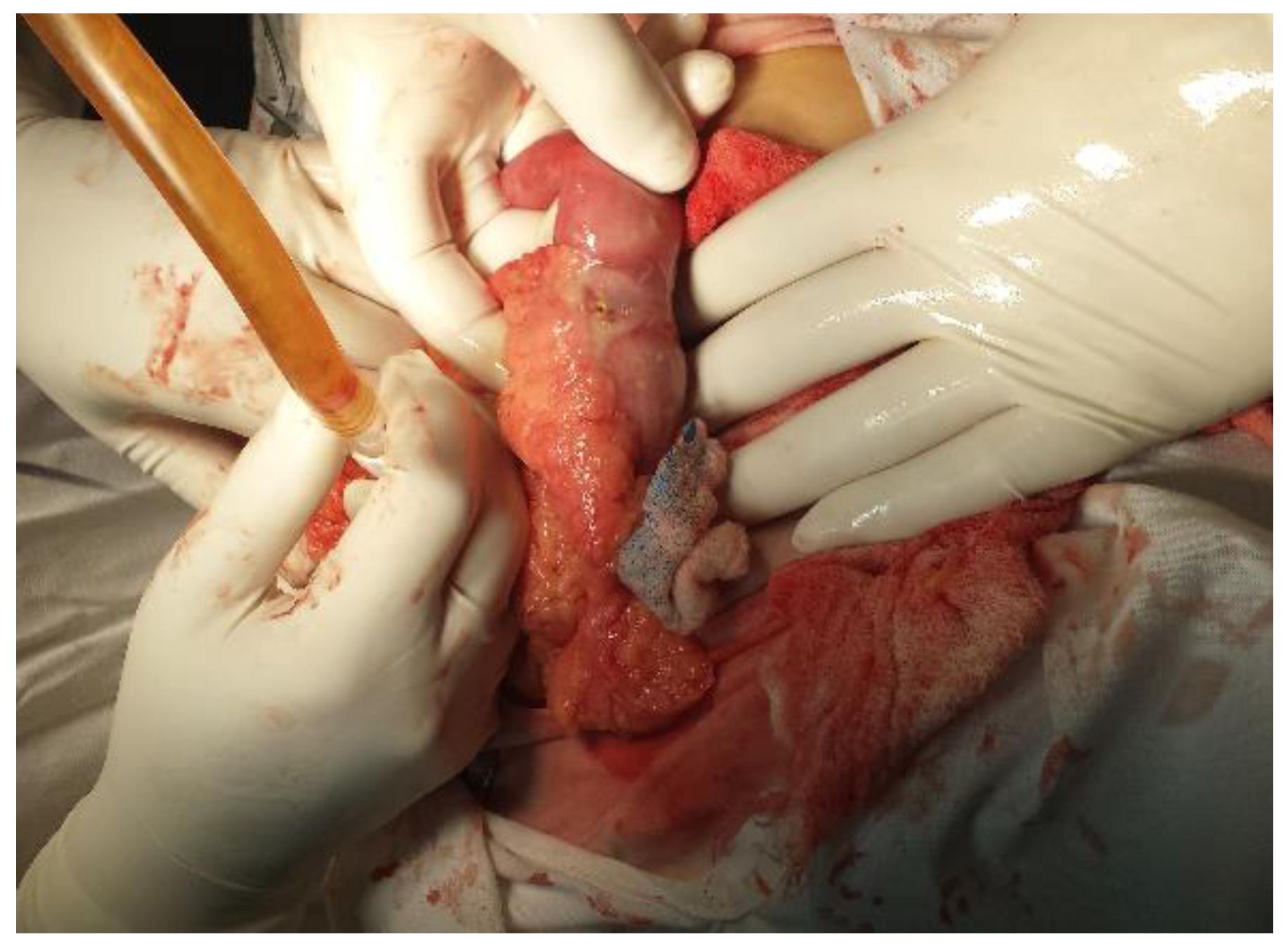
Figure 1.
Figure 1.
Jmms 03 00010 g001
Figure 2.
Figure 2.
Jmms 03 00010 g002