


J. Imaging, Volume 4, Issue 12 (December 2018) – 14 articles
Cover Story (view full-size image):
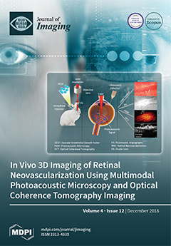
The aim of this study is to investigate a multimodal imaging system for monitoring retinal neovascularization using photoacoustic microscopy (PAM), optical coherence tomography (OCT), fluorescein angiography (FA), and color fundus photography. Retinal neovascularization was generated using intravitreal injection of the vascular endothelial growth factor (VEGF). PAM and OCT demonstrated changes in the retinal morphology and the spatial extent of retinal neovascularization. The multimodal-imaging platform performs imaging with a high lateral spatial resolution of 4.1 µm for PAM and 3.8 µm for OCT, with a high depth of penetration at a safe energy level. This allows for the visualization of retinal neovascularization and microvasculature morphologic changes after VEGF injection. Acquired PAM and OCT imaging provide 3D volumetric images of the vascular network that improve the characterization of the
[...] Read more.
- Issues are regarded as officially published after their release is announced to the table of contents alert mailing list.
- You may sign up for e-mail alerts to receive table of contents of newly released issues.
- PDF is the official format for papers published in both, html and pdf forms. To view the papers in pdf format, click on the "PDF Full-text" link, and use the free Adobe Reader to open them.
Previous Issue
Next Issue