Abstract
The water ligand observed via a gradient spectroscopy type experiment with 19F detection was applied to selectively detect fluorinated compounds with affinity to the target proteins. The 19F signals of bound and unbound compounds were observed as opposite phases, which was advantageous to distinguish the binding state. The proposed NMR method was optimized based on the 19F{1H} saturation transfer difference pulse sequence, and various inversion pulses for the water resonance were evaluated with the aim of high sensitivity.
1. Introduction
NMR spectroscopy can be an effective method for screening compounds with affinity to target proteins. Various NMR-based screening methods to observe 1H ligand signals were proposed, such as NOE-pumping [1], saturation transfer difference (STD) [2], water ligand observed via gradient spectroscopy (WaterLOGSY) [3,4], and reverse NOE-pumping [5] experiments. Some methods were also applied to 19F detection [6,7,8,9]. The characteristic properties of 19F are a 100% natural abundance and high sensitivity for NMR observation. In the 1H→19F STD [9] and 19F{1H} STD experiments [10], on-resonance 1H frequency was set on the protein (e.g., methyl region) and off-resonance 1H frequency was set outside the signal region for reference (e.g., −20 ppm). The 19F signals of the fluorinated compounds bound to proteins were selectively observed in the difference spectra. In the proposed NMR method, the concept of WaterLOGSY was utilized, where 1H magnetization of water, which was transferred to the protein, was again transferred to 19F of the bound ligand via direct or indirect relay processes. The 19F signals of the bound and unbound compounds were observed as opposite phases, which is advantageous to distinguish the binding of each fluorinated compound. Because various fluorinated compounds are used in the pharmaceutical fields, the development of effective screening methods for them is essential. For the purpose of developing more sensitive NMR-based screening methods for fluorinated compounds, a WaterLOGSY type experiment with 19F detection was optimized using the complex of diflunisal (Figure 1) and human serum albumin (HSA). Diflunisal possesses anti-inflammatory, analgesic, and antipyretic activity and is used in the therapy of chronic arthritis. This model system is considered to be suitable for developing the proposed methods.

Figure 1.
The structures of (a) diflunisal and (b) enoxacin.
2. Results and Discussion
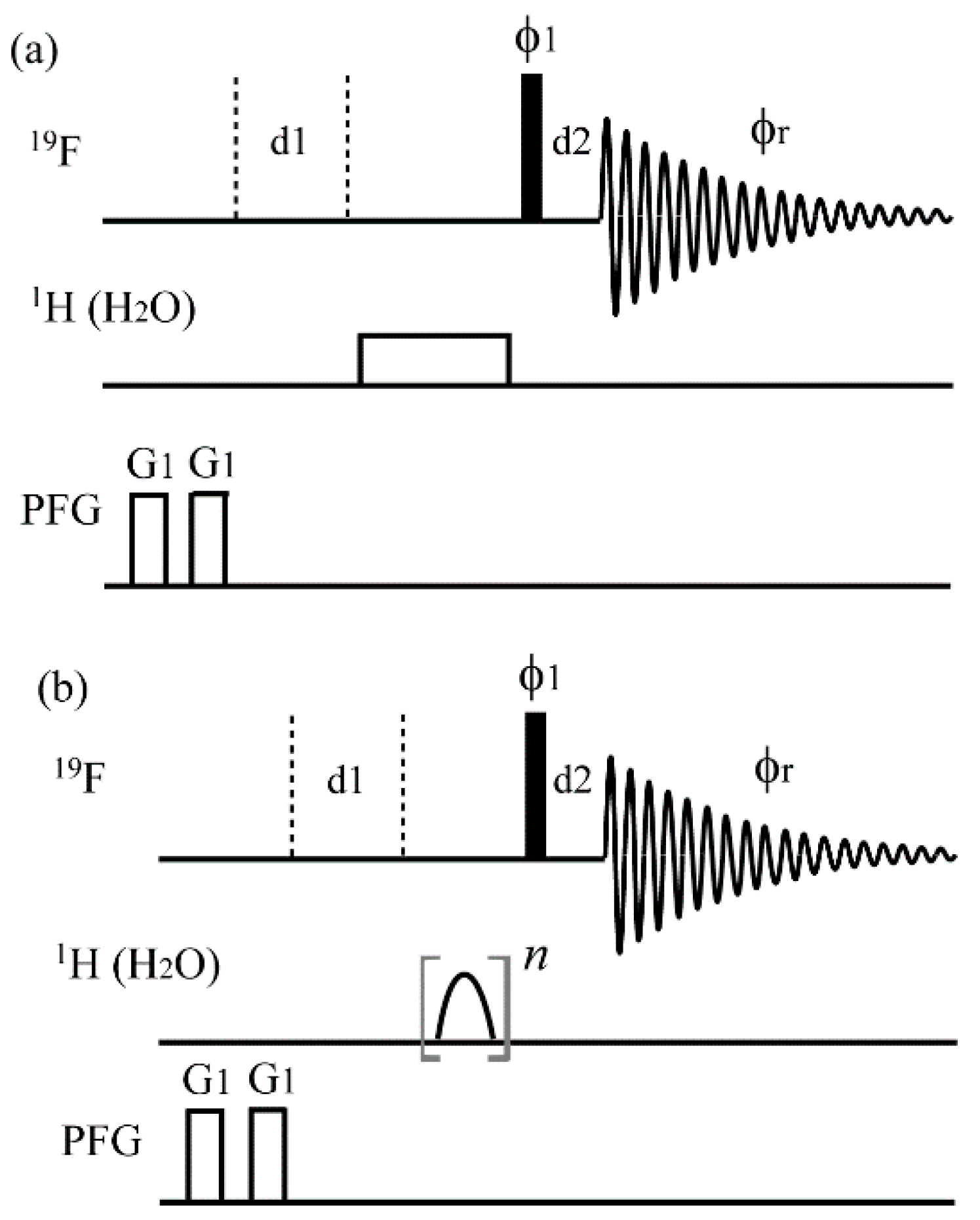
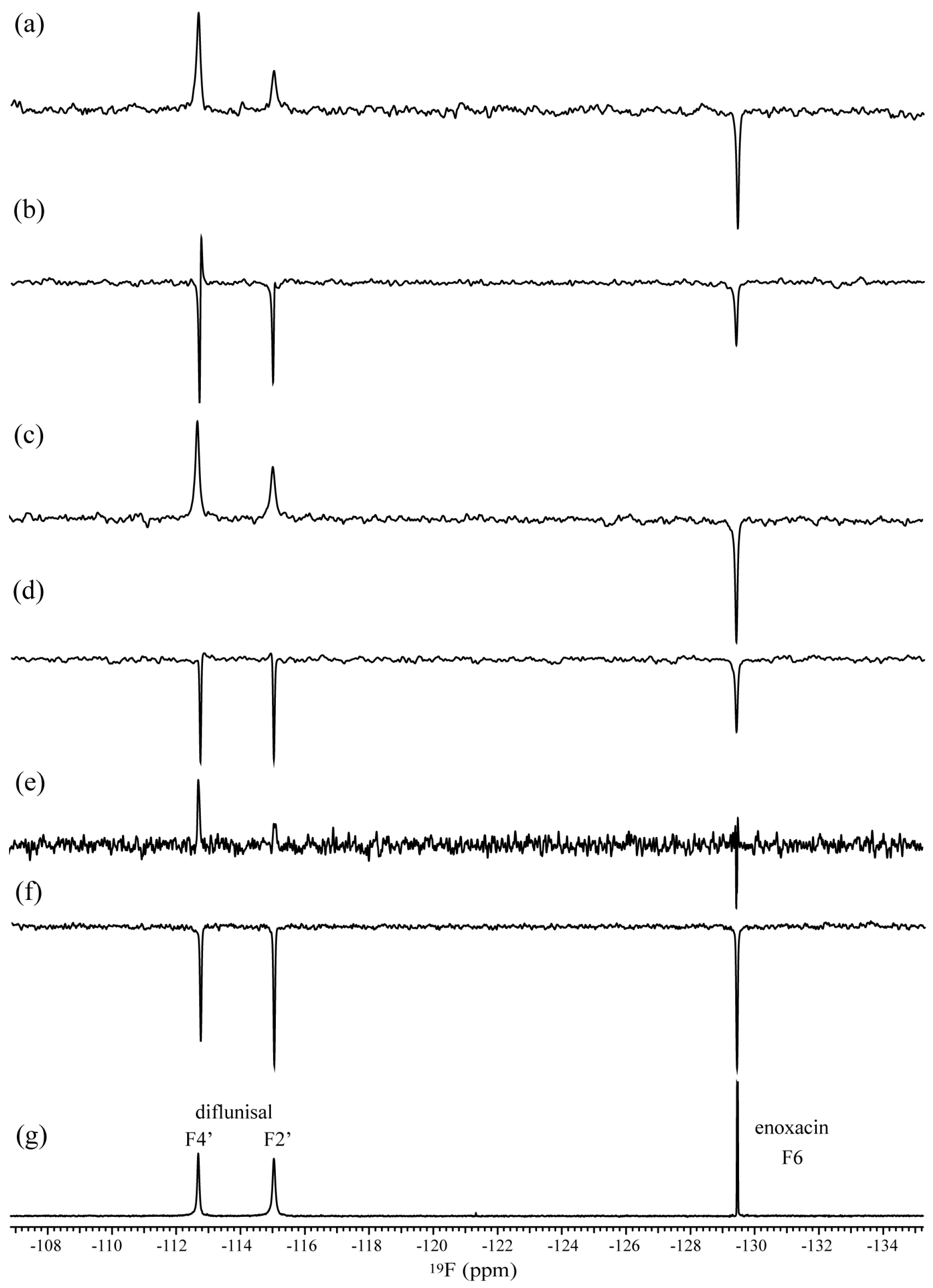
The development of the sensitive 19F-WaterLOGSY-type experiment was carried out based on the 19F{1H} STD pulse sequence (Figure 2) used in our past study [10]. Regarding the inversion pulses of water resonance, several pulse schemes, such as a rectangular pulse and various shaped pulses, were evaluated. The irradiation time was set to 1.0 s in each experiment in consideration of measuring time. A comparison of the 19F-WaterLOGSY-type spectra is shown in Figure 3. In the initial stage of developing this method, a rectangular pulse was used for the irradiation of water resonance [11]. Through the process of implementing several pulse sequences with 19F detection, it was found that the 19F{1H} STD pulse sequence (Figure 2), in which 1H-offset was set to water resonance, was the most sensitive method. Although sensitivity of the spectra acquired using a rectangular pulse was unsatisfactory in this irradiation time (Figure 3e), decent NMR spectra were obtained using Gaussian- and square-shaped pulses (Figure 3a,c). The SN ratios of the spectra using Gaussian- and square-shaped pulses and a rectangular pulse were 32.7, 18.6, and 6.90, respectively, indicating that the Gaussian pulse was more suitable to acquire sensitive spectra. In the other shaped pulses, such as I-BURP-1, Q3, and G3, the SN ratios were 5.40, 5.88, and 8.66, respectively (data not shown), indicating that these shaped pulses were unfeasible for practical use due to low sensitivities. Regarding the selective inversion of water resonance, Gaussian- and square-shaped pulses were appropriate, which was different from the results of irradiating the methyl region [10]. In the spectrum shown in Figure 3b, acquired using a sample solution in the absence of HSA, a phase distortion was observed for the signal of F4′ (δ −112.6). The repetitive square pulses could be the reason for the phase distortion.
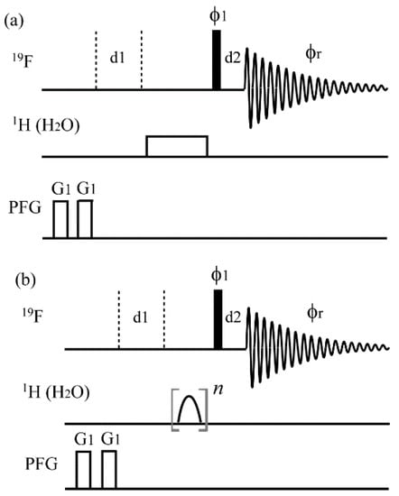
Figure 2.
The pulse sequences of 19F water ligand observed via gradient spectroscopy (WaterLOGSY). The thin bars represent 90° pulses. All pulses were along x unless otherwise shown. In (a), the 1H pulse width (γH1/2π) was 1.0 s (32 Hz). In (b), Gaussian- and square-shaped pulses were used for evaluations. The 1H-offset was set on the water resonance. The repetitive time n was set to adjust the total saturation time. The experimental parameters were; d1 = 1.0 s, d2 = 10 μs, G1 = 7.2 G/cm, gradient pulse width = 2.0 ms. Phase cycling: ϕ1 = x, −x, −x, x, y, −y, −y, y; ϕr = x, x, −x, −x, y, y, −y, −y.
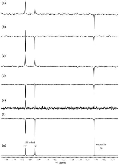
Figure 3.
The 19F-WaterLOGSY spectra acquired using (a,b) square-, (c,d) Gaussian-shaped pulses, and (e,f) a rectangular pulse for the selective water irradiation. In (a,c,e), sample (ii); 10 mM diflunisal in the presence of 0.1 mM human serum albumin (HSA) was used, where enoxacin was prepared in a free state. In (b,d,f), sample (i); 5.0 mM diflunisal and 5.0 mM enoxacin were used. (g) A reference 19F spectrum of sample (i). The Gaussian, square, and rectangular pulse widths (γH1/2π) were 2.2 ms (565 Hz), 3.3 ms (312 Hz), and 1.0 s (32 Hz), respectively.
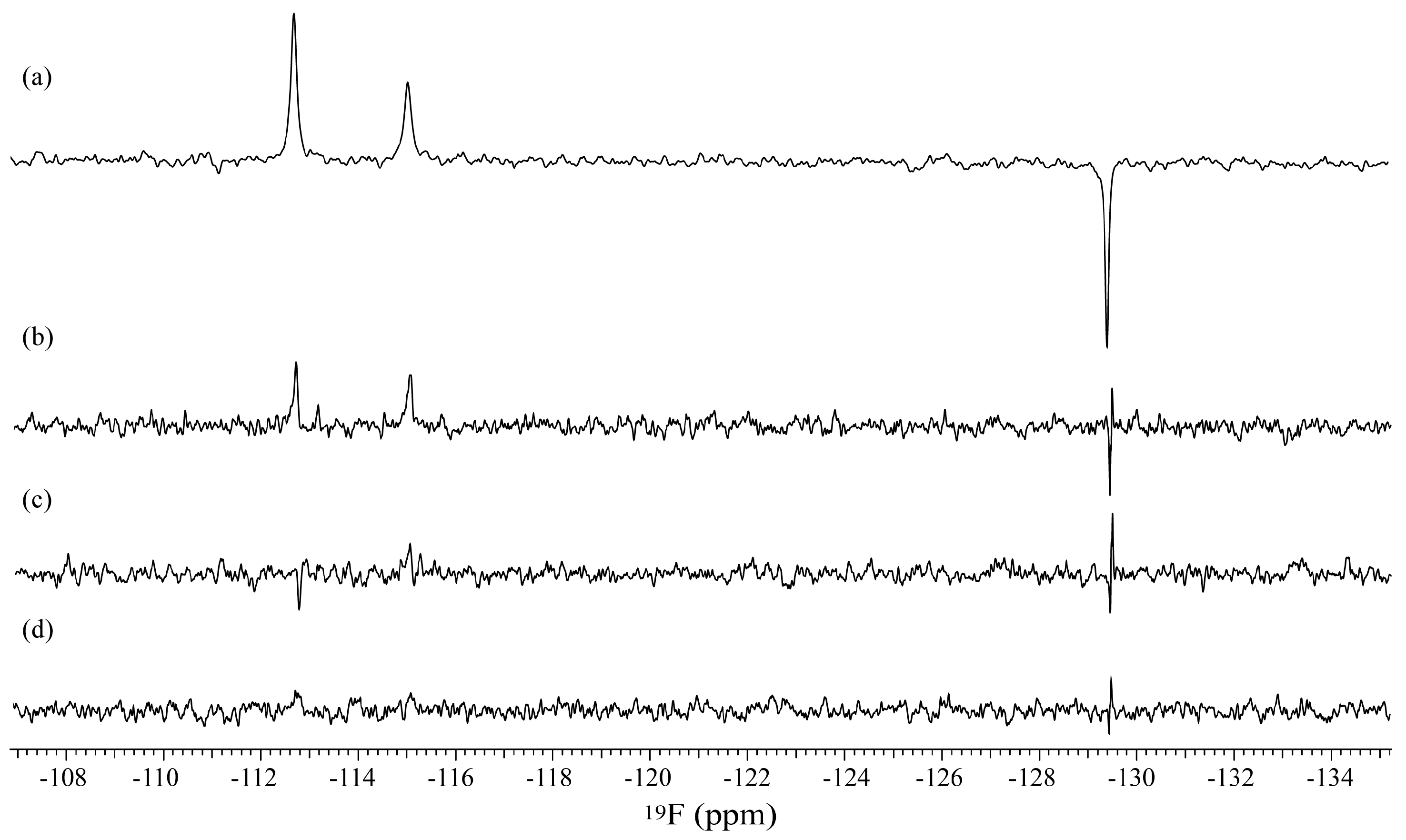
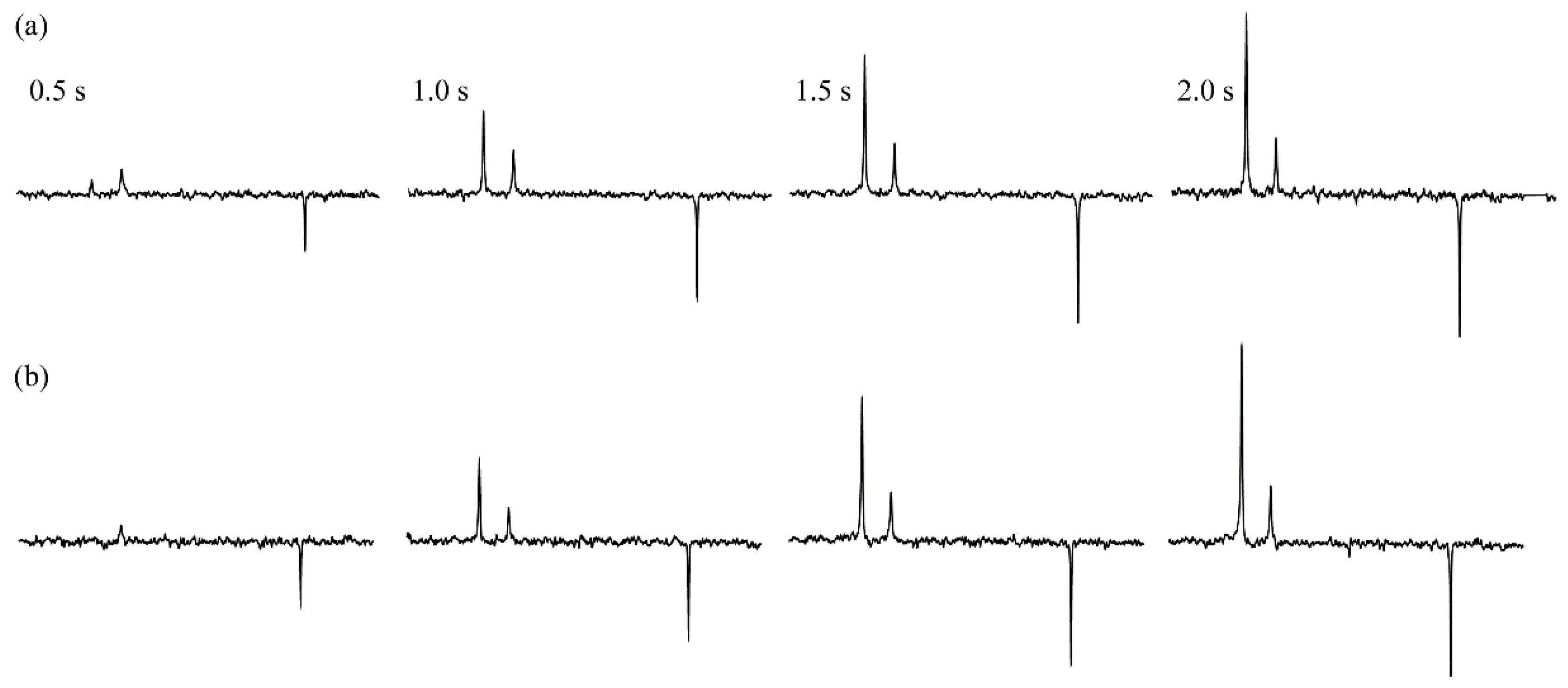
The effect of the Gaussian pulse width was evaluated in terms of sensitivity. The results of four inversion Gaussian pulses, such as 2.2 ms (γH1/2π: 565 Hz), 7.0 ms (179 Hz), 13 ms (100 Hz), and 44 ms (63 Hz), are shown in Figure 4, indicating that the shorter Gaussian pulse width was effective for acquiring the sensitive spectra. Although the relatively wide 1H region near the water was irradiated by the short pulse, the 19F signal of enoxacin (δ −129.5) was not affected in the presence or absence of HSA (Figure 3c,d). To investigate an effect of the irradiation times of water resonance, those using Gaussian- and square-shaped pulses were arrayed in the range of 0.5–2.0 s (Figure 5). The F4′ signal intensity of diflunisal distinctly increased till 2.0 s, while the F2′ signal intensity almost maximized at 1.0 s. The 19F-T1 values of F4′ and F2′ in the free state were 1.98 and 0.87 s, respectively, which could be related to the intensity changes. The heteronuclear cross-relaxation from 1H to 19F is less efficient than the homonuclear case from 1H to 1H, because spectral density for inducing relaxation is significantly lower for hetero- than homonuclear zero-quantum transition frequencies [9]. In consideration of these underlying properties, the proposed experiment could be acceptable for a practical screening method of fluorinated compounds.
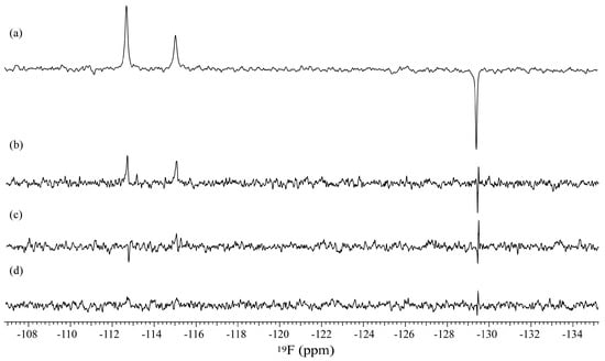
Figure 4.
The 19F-WaterLOGSY spectra acquired using various pulse widths of Gaussian-shaped pulses. The pulse widths (γH1/2π) were (a) 2.2 ms (565 Hz), (b) 7.0 ms (179 Hz), (c) 13 ms (100 Hz), and (d) 44 ms (63 Hz). A sample (ii) was used.
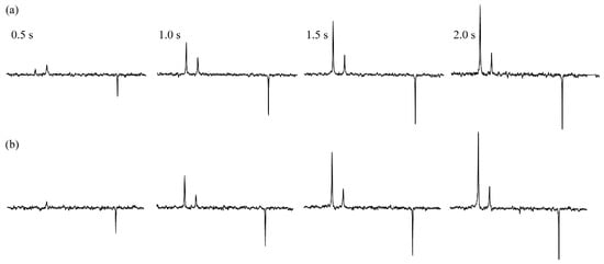
Figure 5.
The 19F-WaterLOGSY spectra acquired with various irradiation times in the range of 0.5–2.0 s, using (a) Gaussian- and (b) square-shaped pulses. The repetitive time n was set to adjust the total irradiation times, and the pulse widths were the same as those used in Figure 3.
3. Materials and Methods
3.1. Chemicals
Diflunisal, enoxacin, and HSA were purchased from Sigma-Aldrich (Tokyo, Japan). Two NMR samples were prepared; sample (i): A 600 μL solution containing 5.0 mM diflunisal and 5.0 mM enoxacin in a 5 mm tube, sample (ii): Double NMR tubes, which comprised a 3 mm tube used as an inner tube containing 10 mM enoxacin and a 5 mm tube used as an outer tube containing 10 mM diflunisal and 0.1 mM HSA. The enoxacin solution, which was prepared in the absence of HSA, was used as a negative control. Each solution contained 90% 1H2O and 10% 2H2O.
3.2. NMR Spectroscopy
All of the NMR spectra were acquired at 20 °C on a Varian 600 MHz NMR system equipped with an HFX probe (Varian, Palo Alto, CA, USA). The experimental parameters of the 19F-WaterLOGSY-type experiment were as follows: Data points = 16,384, 19F spectral width = 120,618 Hz, number of scans = 512 for a sample (i) and 8192 for a sample (ii). The irradiation times for inversion of water resonance were arrayed in the range of 0.5–2.0 s. The repetitive time n (Figure 2) was set to adjust the total irradiation time. The on-resonance frequency of 1H was set to 4.7 ppm corresponding to water, and the off-resonance frequency was set to −30 ppm. The inversion shaped pulses of I-BURP-1, square, Q3, G3, and Gaussian were made using VnmrJ software (Agilent Technologies, Santa Clara, CA, USA).
4. Conclusions
The characteristic difference between 19F{1H} STD and 19F-WaterLOGSY experiments is the 1H on-resonance frequency. The resonances of the methyl region and water are conventionally irradiated in the aforesaid experiments. Because the methyl region cannot always be appropriate as the 1H on-resonance frequency for the compounds possessing methyl groups, the proposed method is expected to be applicable to a wide variety of fluorinated compounds. The sensitivity of the current spectra with 19F detection could be acceptable for practical use of screening, and the proposed method is expected to contribute to the pharmaceutical and biomedical fields.
Author Contributions
Conceptualization, M.T.; methodology, M.T. and K.F.; writing, M.T.; supervision, K.F.; project administration, M.T.; funding acquisition, M.T.
Funding
JSPS KAKENHI Grant Number JP15K05550 and JP18K05185 for M.T.
Acknowledgments
This study was supported by Meisei University and JSPS KAKENHI Grant Number JP15K05550 and JP18K05185 for M.T.
Conflicts of Interest
The authors declare no conflict of interest.
References
- Chen, A.; Shapiro, M.J. NOE Pumping: A novel NMR technique for identification of compounds with binding affinity to macromolecules. J. Am. Chem. Soc. 1998, 120, 10258–10259. [Google Scholar] [CrossRef]
- Maye, M.; Meyer, B. Group epitope mapping by saturation transfer difference NMR to identify segments of a ligand in direct contact with a protein receptor. J. Am. Chem. Soc. 2001, 123, 6108–6117. [Google Scholar]
- Dalvit, C.; Pevarello, P.; Tatò, M.; Veronesi, M.; Vulpetti, A.; Sundström, M. Identification of compounds with binding affinity to proteins via magnetization transfer from bulk water. J. Biomol. NMR 2000, 18, 65–68. [Google Scholar] [CrossRef] [PubMed]
- Dalvit, C.; Fogliatto, G.P.; Stewart, A.; Veronesi, M.; Stockman, B.J. WaterLOGSY as a method for primary NMR screening: Practical aspects and range of applicability. J. Biomol. NMR 2001, 21, 349–359. [Google Scholar] [CrossRef] [PubMed]
- Chen, A.; Shapiro, M.J. NOE Pumping. 2. A high-throughput method to determine compounds with binding affinity to macromolecules by NMR. J. Am. Chem. Soc. 2000, 122, 414–415. [Google Scholar] [CrossRef]
- Dalvit, C.; Fagerness, P.E.; Hadden, S.T.A.; Sarver, R.W.; Stockman, B.J. Fluorine-NMR experiments for high-throughput screening: Theoretical aspects, practical considerations, and range of applicability. J. Am. Chem. Soc. 2003, 125, 7696–7703. [Google Scholar] [CrossRef] [PubMed]
- Dalvit, C.; Flocco, M.; Stockman, B.J.; Veronesi, M. Competition binding experiments for rapidly ranking lead molecules for their binding affinity to human serum albumin. Comb. Chem. High Throughput Screen. 2002, 5, 645–650. [Google Scholar] [CrossRef] [PubMed]
- Sakuma, C.; Kurita, J.; Furihata, K.; Tashiro, M. Achievement of 1H-19F heteronuclear experiments using the conventional spectrometer with a shared single high band amplifier. Magn. Reson. Chem. 2015, 53, 327–329. [Google Scholar] [CrossRef] [PubMed]
- Diercks, T.; Ribeiro, J.P.; Canada, F.J.; Andre, S.; Jimenez-Barbero, J.; Gabius, H. Fluorinated carbohydrates as lectin ligands: Versatile sensors in 19F-Detected saturation transfer difference NMR spectroscopy. Chem. Eur. J. 2009, 15, 5666–5668. [Google Scholar] [CrossRef] [PubMed]
- Furihata, K.; Usui, M.; Tashiro, M. Application of NMR screening methods with 19F detection to fluorinated compounds bound to proteins. Magnetochemistry 2018, 4, 3. [Google Scholar] [CrossRef]
- Furihata, K.; Utsumi, H.; Kato, T.; Sakuma, C.; Tashiro, M. Application of the 19F-Waterlogsy type experiment for NMR-based screening of fluorinated compounds. Pharm. Anal. Chem. Open Access 2016, 2, 111. [Google Scholar] [CrossRef]
© 2019 by the authors. Licensee MDPI, Basel, Switzerland. This article is an open access article distributed under the terms and conditions of the Creative Commons Attribution (CC BY) license (http://creativecommons.org/licenses/by/4.0/).


