Abstract
Jackfruit, primarily cultivated in Nayarit, Mexico, has four notable genotypes: “Agüitada”, “Rumina”, “Licenciada”, and “Karlita”, which require thorough characterization. This study aimed to provide a comprehensive characterization of these genotypes through an integration of morphological, physiological, physicochemical, phytochemical, and DNA fingerprinting analyses. Measurements were taken from physiological maturity to senescence. SSR and SRAP markers were employed for DNA fingerprinting, and a complete randomized design followed by multivariate analysis was used to observe variable relationships. The results revealed that “Rumina” had the largest leaf size, while “Karlita” had the largest fruit size and the highest respiration rate (117.27 mL of CO2·kg−1·h−1). “Licenciada” showed the highest ethylene production (265.45 µL·kg−1·h−1). “Agüitada” and “Licenciada” were associated with orange bulbs, whereas “Rumina” and “Karlita” were associated with yellow ones. Additionally, “Agüitada” demonstrated higher levels of soluble phenols and carotenoids, indicating greater antioxidant capacity. The Jaccard index suggested moderate genetic diversity among the genotypes, and the dendrogram revealed two genetic clusters. “Licenciada” emerged as a promising genotype, combining high genetic diversity with desirable physicochemical traits. This study highlights the need to broaden future genetic analyses to include a wider range of jackfruit genotypes from various regions, offering a more comprehensive understanding of genetic diversity.
1. Introduction
Jackfruit (Artocarpus heterophyllus Lam.) belongs to the Moraceae family, a group of flowering plants that includes economically important species such as figs, mulberries, and breadfruit [1]. Native to India, jackfruit is renowned for its commercial, culinary, and nutritional value. This fruit is rich in vitamins, minerals, carbohydrates, and phytonutrients, making it a highly nutritious option with numerous health benefits [2]. Despite challenges such as a high proportion of non-edible parts and limited processing infrastructure, jackfruit holds significant potential for the development of high-value products, which could enhance its market appeal and benefit various sectors [3]. As a climacteric fruit, its cultivation extends across tropical regions of South and Southeast Asia, with additional genotypes found in Central Africa and the Americas [4,5].
In Mexico, jackfruit production has substantially increased in recent years, with a reported annual production increase of over 218% from 2015 to 2022 [4]. The main jackfruit-producing states in the country are Michoacán, Hidalgo, Colima, Jalisco, Veracruz, and Nayarit, with the latter having the largest cultivated jackfruit area (1632.95 ha) [4]. Jackfruit in Nayarit is in a similar situation, as it is in its native location. Despite existing cultivation management techniques, adequate protocols to reduce genetic variability in the crop are not being considered. It is well known that this species is monoecious, displaying separate male and female inflorescences on the same plant [6]. In this regard, the jackfruit tree’s flowers are pollinated by various vectors such as wind, birds, and insects, resulting in a high rate of cross-pollination [7]. As a consequence of the predominant method of seed propagation, there was notable genetic variability among materials. This often led to the emergence of different genotypes over time [8]. It is important to note that initially, in Nayarit, cultivation propagation was carried out through seeds, resulting in over 15 different jackfruit genotypes in the state [9]. As a result of this propagation, changes in morphology (color, fruit shape, fruit size, and leaves), physicochemical composition (acidity, soluble solids, and pH), physiological processes (respiration and ethylene production), and phytochemical elements (phenolic compounds, volatiles, carotenoids, and vitamin C) were observed [10,11]. To preserve the distinctive and relevant qualities for export, the propagation strategy was moved to cuttings or grafts, which has helped decrease variability among genotypes [9]. It is relevant to highlight the importance of preserving these genotypes, as they hold high commercial value, with over 90% of national production being exported abroad [12].
To date, there is limited information on jackfruit characterization in Mexico, with research primarily focusing on genotype analysis and their morphological, physicochemical, physiological, and phytochemical attributes [13,14,15]. The diversity found in these studies provides a more comprehensive perspective of the phenotypic traits present in jackfruit cultivation. However, it is critical to study these findings using molecular techniques such as molecular markers, as these can help establish linkages between genotypes. Molecular markers offer significant benefits, such as precise genotype selection, detailed genetic information, and assessment of genetic diversity. They are essential for establishing germplasm banks, preserving genetic resources, and developing genetic improvement programs [16]. In the eastern region, research on jackfruit and its genetic diversity, supported by molecular techniques, has been widely studied, being effective in the differentiation of genotypes of this crop. This trend is evident because the crop is native to this region and its economic and cultural importance is different from the rest of the world [17,18,19]. However, in Mexico, being a young crop, there are currently no studies of genetic diversity. Hence, the aim of this study was to conduct a comprehensive characterization encompassing morphological, physiological, physicochemical, phytochemical, and molecular aspects of four commercially significant genotypes in Nayarit, identified as “Agüitada”, “Rumina”, “Licenciada”, and “Karlita”, with the objective of identifying differences and contributing to the conservation efforts for these genotypes.
2. Materials and Methods
2.1. Genotypes and Collection Site
Fruits and leaves of jackfruit of each genotype (“Agüitada”, “Rumina”, “Licenciada”, and “Karlita”) (Figure 1) were collected from an orchard located in Las Varas, Nayarit, Mexico (21°10′28.7″ N and 105°10′27.5″ W), in July 2022. It is important to mention that these materials (fruits and leaves) are from a second crop cycle in the same area evaluated by the authors Morelos-Flores et al. [15]. Propagation in this area mainly relies on cuttings to maintain desired traits. Trees are pruned regularly to prevent heights over 10 m and are spaced 6 to 8 m apart in orchards for optimal growth. Management protocols allow each branch to bear 2 to 3 fruits, with excess fruit removed promptly. The collection area, located at an altitude of 20 m above sea level, is characterized by a warm and humid climate with an average annual rainfall of 58.4 mm, a relative humidity of 56%, and approximately 13 h of daylight, with temperatures reaching up to 32.2 °C [20]. A set of ten 15-year-old trees was selected based on specific genotype criteria, leveraging the growers’ provided morphological data on fruits. Forty-eight fruits at physiological maturity were collected, following the criteria described by Love & Paull [21] for such maturity. Additionally, symmetrical fruits without damage from pests and diseases were selected. Sixty mature leaves were collected, considering those free from pest or disease damage and without discoloration or deformities due to nutrient deficiency; then, they were stored at −10 °C until further conditioning.
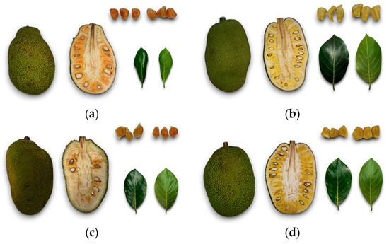
Figure 1.
Jackfruit genotypes of Nayarit (fruits, leaves, and bulbs): “Agüitada” (a), “Rumina” (b), “Licenciada” (c), and “Karlita” (d).
2.2. Sampling of Fruits and Leaves
The reception of fruits and leaves took place at the Laboratorio Integral de Investigacion en Alimentos del Instituto Tecnologico Nacional de Mexico/Instituto Tecnologico de Tepic. The fruits were rinsed with water to remove residues and then immersed in water with thiabendazole (TECTO 60®, Syngenta, Chuo City, Tokyo, Japan) at 800 ppm for 3 min as antifungal treatment. Afterwards, the fruits were allowed to air dry at 25 °C, and the peduncle was sealed with copper oxychloride (Cupravit®, Bayer, México, D.F, México). The fruits were stored in a temperature-controlled chamber at 25 °C with a relative humidity of 90% until further analysis. Once the fruits were evaluated, three bulbs from the bottom, middle, and top parts of each jackfruit were collected. These bulbs were placed in resealable bags and stored at −80 °C for 24 h. Subsequently, the bulbs were lyophilized (Labconco, Freezone 2.5 L, −50 °C, Kansas, MO, USA) and ground (Nutribullet®, NBR-0804B, Los Angeles, CA, USA) to obtain a homogeneous sample. The lyophilized bulbs were stored at −20 °C until further analysis. On the other hand, the leaves were rinsed with water, excess moisture was removed at 20 °C, and they were placed in resealable bags and stored at −80 °C for 24 h. After this, they were lyophilized (Labconco, Freezone 2.5 L, −50 °C, USA), ground (Nutribullet®, NBR-0804B, Los Angeles, CA, USA), and sieved (≤500 µm). The samples were stored at −20 °C until further analysis.
2.3. Morphological Analysis of Fruits and Leaves
The length and width of 50 leaves and 15 fruits of each genotype were measured following the guidelines proposed by the International Plant Genetic Resources Institute [22].
2.4. Physiological Analysis (Respiration Rate, Ethylene Production, and Physiological Weight Loss)
The respiration rate and ethylene production in fruits were obtained following the protocol proposed by Morelos-Flores et al. [13] using a gas chromatograph (GC6890; Hewlett-Packard, Palo Alto, CA, USA) equipped with an HP-PlotQ column (15 m × 0.53 mm and 40 μm film thickness); a thermal conductivity detector and a flame ionization detector were used to detect CO2 and ethylene, respectively. These analyses were conducted every 24 h. A digital balance (L-PCR; Torrey, Monterrey, N.L., Mexico) was used for monitoring physiological weight loss.
2.5. Physicochemical Analysis
The titratable acidity (method 942.15), pH (method 981.12), and total soluble solids (932.14) were measured according to the guidelines established by the AOAC [23]. Firmness and color in the peel and bulbs were also evaluated following the methodology described by Morelos-Flores et al. [13], using a texture analyzer (Stable Micro Systems®, TA.TXplus, Godalming, Surrey, UK) and a colorimeter (M&A Instruments Inc., NH300, Houston, TX, USA). These analyses and the bulb extraction were conducted every 48 h.
2.6. Phytochemical Analysis
2.6.1. Preparation of Phenolic Extract
The aqueous organic extract was obtained following the methodology proposed by Pérez-Jiménez et al. [24]. An aliquot of 5 mL of acidified methanol solution (2% HCl 2 N) was added to 0.5 g of lyophilized sample. The samples were then shaken for 1 h and centrifuged at 13,000 rpm for 10 min at 4 °C (Hermle Larbotechnik, Z36HK, Wehingen, Badén-Württemberg, Germany); the supernatants obtained were recovered in a 10 mL flask. An aliquot of 5 mL of diluted acetone (80:30, v/v) was added to the precipitate, and the process was repeated. The supernatant was mixed with the previous one and adjusted to 10 mL with a combination of acidified methanol and diluted acetone (50:50, v/v).
2.6.2. Quantification of Total Soluble Phenols
Total soluble phenols were determined using the method described by Montreau [25] and modified by Alvarez-Parrilla et al. [26], with the Folin-Ciocalteu reagent. A gallic acid calibration curve was performed to interpolate the absorbances (read at 750 nm) and express the results as milligrams of gallic acid equivalents per gram of dry weight of sample (mg GAE/g DW).
2.6.3. Preparation of Carotenoid Extract
A sample of 2 g of fresh pulp was taken and placed in a beaker, and 0.5 g of magnesium carbonate was added per sample. An aliquot of 10 mL of acetone/ether (80:20 v/v) was added to the beaker and mixed for 1 min using an ultraturrax. Then, the mixture was transferred to a Teflon tube and centrifuged at 15,000 rpm for 30 min at 4 °C. The supernatant was recovered in a dark chamber and transferred to an amber flask at 4 °C. The precipitate was treated with 5 mL of acetone/ether, agitated with a vortex for 1 min, and centrifuged again at 15,000 rpm for 30 min at 4 °C. This procedure was repeated until the pulp turned white. The supernatants were transferred to an amber separation funnel, adding 15 mL of 20% sodium chloride (NaCl) to allow the formation of two distinct layers (organic and aqueous phases). Once the phases were separated, the aqueous phase (lower phase) was drained. Subsequently, washes were carried out with 20 mL of distilled water, again allowing the formation of the two layers and draining the aqueous phase. The acetone/ether extract and the carotenoids (organic phase) were poured into a beaker with a layer of dry sodium sulfate and left to stand for 1 min. After this time, the supernatant was recovered in a 10 mL amber flask, and additional washes were carried out with the acetone/ether solution until the sodium sulfate (Na2SO4) was free of pigments [27].
2.6.4. Quantification of Total Carotenoids
To determine the total carotenoid content, the absorbance of the extract was measured at 440 nm using a spectrophotometer (Visible, 721G/722G, Vernon Hills, IL, USA). A β-carotene calibration curve was used to calculate concentrations, and the results were expressed as micrograms (µg) equivalent to β-carotene per 100 g on a wet weight basis (µg EβC/100 g WW) [27].
2.6.5. Evaluation of the Antioxidant Capacity of Total Soluble Phenols and Carotenoids
The antioxidant capacity (CAOX) by 2,2-Diphenyl-1-picrylhydrazyl (DPPH) method was determined according to the method proposed by Prior [28], with some modifications; 40 μL of extract and 200 μL of DPPH (0.19 mM) were placed in a microplate. The plate was incubated in the dark, shaken for 10 min, and then read at 517 nm on a microplate reader (Biotek®, 800TS, Winooski, VT, USA). The calibration curve was performed with trolox solution, and the results were expressed as trolox equivalents (TEs) in mmol per gram of dry weight (mmol TE/g DW). The ferric reducing antioxidant power (FRAP) method was performed using the methodology of Benzie and Strain (1996) with slight modifications by Álvarez-Parrilla et al. [29]. Readings were taken at 597 nm, and the results were expressed as trolox equivalents in mmol per gram of dry weight (mmol TE/g DW). The 2,2′-azino-bis 3-ethylbenzothiazoline-6-sulfonic acid (ABTS) assay was carried out as described by Re et al. [30] with some modifications by Mercado-Mercado et al. [31]. Readings were taken at 734 nm, and the results were expressed as trolox equivalents in mmol per gram of dry weight (mmol TE/g DW). The same considerations were taken for the evaluation of the antioxidant capacity of soluble phenols and carotenoids, with the only difference being the use of a quartz microplate (Hellma, 730-009-44, Müllheim, Badén-Württemberg, Germany) when evaluating the latter.
2.7. DNA Extraction, Concentration and Quality
DNA extraction was performed following the protocol of Doyle & Doyle [32] with some modifications; 1 mL of 2% cetyltrimethylammonium bromide (CTAB) lysis solution (0.02 M EDTA, pH 7.5; 0.1 M Tris-HCl, pH 8.0; 1.42 M NaCl) was added to a microtube and heated at 65 °C for 5 min in a thermomixer (Benchmark Scientific, H5000-HC, Sayreville, NJ, USA). Then, 3.5 mg of lyophilized leaf was added to the hot microtube and placed on vortex for 30 s. Subsequently, the tube was incubated at 60 °C for 45 min and centrifuged at 10,000 rpm for 20 min at 4 °C (Sigma Laborzentrifugen, Sigma 1-14K, La otra centrifuga es de Lichtenfels, Bavaria, Germany). The supernatant was collected, mixed with phenol–chloroform–isoamyl alcohol (25:24:1 v/v/v), and centrifuged under the same conditions. This step was repeated twice. Then, the supernatant was recovered, it was mixed with chloroform–isoamyl alcohol (24:1 v/v), and centrifugation was repeated under the same conditions. To precipitate the DNA, 100 µL of cold isopropanol was added. The tube was then stored at −20 °C for 30 min and centrifuged to decant the isopropanol. Finally, the precipitated DNA was washed with 75% ethanol, followed by centrifugation and decanting. This step was then repeated with 90% ethanol, followed by another round of centrifugation and decanting. The concentration and purity of the DNA was measured on a micro-volume plate (Agilent, Biotek®, Winooski, VT, USA, Take 3) with the absorbance ratios of A260/A280 nm and A260/A230 nm. The integrity of DNA was verified by electrophoresis in 1% agarose gels. Electrophoresis was carried out at 90 V for 60 min. The gel was visualized in the transilluminator (Bio-Image System 312 nm, Uppsala, Estocolmo, Suecia), and the images were acquired using the Carestream Molecular ImagingSoftware, Version 5.0.
2.7.1. Simple Sequence Repeat Markers (SSR)
Fourteen SSR markers were selected to identify genetic diversity between genotypes (Table 1). These primers were chosen based on the work reported by Anuragi et al. [33]. The PCR mixture consisted of 0.5 µL of forward primer, 0.5 µL of reverse primer, 1 µL of DNA (60 ng/µL), 5 µL of DreamTaq™ (Thermo Scientific, DreamTaq Green PCR master mix 2×, Waltham, MA, USA), and 3 µL of nuclease-free water, with a total volume of 10 µL. The PCR conditions set in the thermocycler (Bio-Rad, T100 thermal cycler, Hércules, CA, USA) were as follows: initial denaturation at 94 °C for 5 min, 35 cycles of denaturation at 94 °C for 40 s, annealing at 55.5 °C for 40 s, and extension at 72 °C for 1 min, followed by a final extension at 72 °C for 10 min.

Table 1.
SSR primers.
2.7.2. Sequence-Related Amplified Polymorphism Markers (SRAPs)
Six pairs of SRAP primers were tested, which were selected based on the research conducted by Talamantes-Sandoval et al. and Lira-Ortiz et al. [34,35] (Table 2). The mixture used for PCR consisted of 0.2 µL of forward primer, 0.2 µL of reverse primer, 1 µL of DNA (60 ng/µL), 5 µL of DreamTaq™ (Thermo Scientific, DreamTaq Green PCR master mix 2×), and 3 µL of nuclease-free water, with a total volume of 10 µL. The thermocycler was set at 94 °C for 4 min (initial denaturation), followed by 5 cycles of denaturation at 94 °C for 1 min, annealing at 35 °C for 1 min, and extension at 75 °C for 1 min (pre-amplification). Subsequently, 35 cycles of denaturation at 94 °C for 1 min, annealing at 50 °C for 1 min, and extension at 72 °C for 2 min were performed. Amplicons were analyzed by electrophoresis on 2.5% agarose gels at 80 V for 2 h and visualized using a PhotoDoc-It Imaging System (Ultra-Violet Products, Ltd., Cambridge, MA, USA).

Table 2.
SRAP primers.
2.7.3. Statistical Analysis
The data were analyzed by a complete randomized design with genotype as the factor. Data were subjected to analysis of variance (ANOVA), followed by a mean comparison test using the Fisher’s least significant difference (LSD) method with a significance level set at p ≤ 0.05. Principal component analysis (PCA) was conducted using morphological, physicochemical, physiological, and phytochemical measurements as variables; while the evaluated genotypes were considered as factors, all data normalization was performed using the minimum–maximum scaling method. ANOVA, data normalization, and PCA were performed using Prism 9 software (GraphPad Software, version 9, USA). In the case of DNA fingerprinting, binary matrices were constructed with the detected bands, reporting their absence (zero) and presence (one). The parameters of the Jaccard index and expected heterozygosity were determined. Genetic distances were calculated using Nei’s genetic distances (D) [36] and represented through graphical clustering analysis (dendrogram) using the Unweighted Pair Group Method with Arithmetic Mean (UPGMA). These analyses were conducted using Ntsys version 2.10e software developed by Rohlf, F.J. [37].
3. Results and Discussion
3.1. Morphological Analysis of Fruits and Leaves
The results from the morphological analysis revealed significant differences (p ≤ 0.05) among the evaluated genotypes, as shown in Figure 2. In leaf measurements, “Rumina” stood out with the largest leaves, averaging 15.55 cm in vertical and 9.05 cm in horizontal length, while “Agüitada” had the smallest values at 11.83 cm and 5.60 cm, respectively. Differences (p ≤ 0.05) between “Licenciada” and “Karlita” were only found in the horizontal length of the leaves. Regarding measurements taken on the fruits, differences (p ≤ 0.05) were observed only in the “Karlita” genotype, indicating that these fruits are larger in size compared to the remaining genotypes. In the study conducted by Biswajit & Kartik [38] in a northeastern region of Assam, the average of six genotypes analyzed recorded a leaf length of 15.19 cm and a width of 8.58 cm, which are smaller values than those observed in the current study. The morphological differences observed in leaf and fruit dimensions among the genotypes result from a combination of genetic variability and environmental conditions [39]. Each genotype possesses unique genetic information that, along with factors such as water availability, nutrient levels, sunlight exposure, and temperature variations, influences its growth and development. This interaction between genetics and environment generates specific phenotypic expressions for each genotype [40]. As Kavya & Gayatri [19] have also noted, morphological differences in jackfruit are linked to genetic clusters identified in molecular analyses. These observations underscore the importance of evaluating both genetic and environmental factors to understand the morphological characteristics and potential of different jackfruit genotypes.
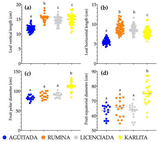
Figure 2.
Scatter plot of the morphological analysis results in four jackfruit genotypes (n = 50 on leaves, n = 15 on fruits): leaf vertical length (a), leaf horizontal length (b), polar diameter of the fruit (c), and equatorial diameter of the fruit (d). A horizontal line is used to indicate the mean value between each data spread. Different letters indicate statistically significant differences (Fisher LSD test; p ≤ 0.05).
3.2. Physiological Analysis (Respiration Rate, Ethylene Production, and Physiological Weight Loss)
The physiological analysis showed statistically significant differences (p ≤ 0.05) among the four analyzed genotypes (Figure 3). “Karlita” recorded the highest respiration rate, with an average of 117.27 mL of CO2·kg−1·h−1, indicating a potentially shorter shelf life due to increased metabolic activity. In contrast, “Rumina” showed the lowest respiration rate, with an average value of 27.73 mL of CO2·kg−1·h−1, suggesting an extended shelf life compared to the other genotypes. Regarding ethylene production, “Licenciada” exhibited the highest production, with values of 265.45 µL·kg−1·h−1, indicating lower ethylene sensitivity. Conversely, “Karlita” produced the least ethylene, 28.08 µL·kg−1·h−1. In terms of physiological weight loss, “Karlita” experienced the greatest loss, with 13.67% of total weight, whereas “Rumina” experienced the least loss, with 9.80%, indicating better fruit preservation capabilities.
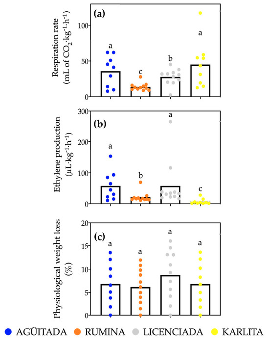
Figure 3.
Physiological analysis of four jackfruit genotypes (n = 11): respiration rate (a), ethylene production rate (b), and physiological weight loss (c). Columns indicate means of each genotype per analysis; different letters denote statistically significant differences (Fisher LSD test; p ≤ 0.05).
Previous results by Mata-Montes de Oca et al. [41] reported a respiration rate of 90.7 mL of CO2·kg−1·h−1 and an ethylene production of 21.4 μL·kg−1·h−1 in an unspecified jackfruit genotype stored at 20 °C. These values differ from those obtained in this study, as “Karlita” had higher peaks of respiration and ethylene, while “Agüitada” had lower peaks of respiration and ethylene compared to those reported by these authors; these differences can be attributed to the different harvest and collection times. Regarding physiological weight loss, the value reported by these authors was 11.96% at the end of the storage period, which is very similar to that reported for the genotypes in this research.
Evaluating these physiological parameters during shelf life is crucial to ensuring fruit quality and proper commercialization. Understanding the respiration rate, ethylene production, and weight loss helps determine the maturity stage and shelf life of the product [42]. Among the genotypes analyzed, “Rumina” demonstrates a clear advantage due to its lower respiration rate, moderate ethylene production, and minimal physiological weight loss. These factors indicate a longer shelf life and better preservation of fruit quality, making “Rumina” the most favorable genotype in terms of these parameters.
The differences observed in the physiological parameters among the genotypes in this study can be attributed to factors such as substrate availability, which affects respiration rate and ethylene production. Different genotypes might vary in their capacity to mobilize and utilize carbohydrate reserves during ripening, impacting these rates [43]. Additionally, substrate availability influences fruit weight loss, as the speed and magnitude of this process depend on the availability of organic compounds for aerobic respiration [44]. Environmental factors and agronomic practices significantly contribute to these disparities, highlighting the importance of specific studies for each variety [45].
3.3. Physicochemical Analysis
The statistical analysis showed significant differences (p ≤ 0.05) in the physicochemical analysis among the different genotypes, as illustrated in Figure 4. Peel color variations were observed during the ripening process. “Agüitada” and “Rumina” maintained similar colors until the end of the shelf-life period, exhibiting a green-brown tone. In contrast, “Licenciada” showed a greenish-yellow color and “Karlita” exhibited an olive-green tone. These findings align with those of Elevitch & Manner [46], who observed a transition from lemon-green (°Hue) to olive-green during maturation. The color of “Agüitada” fruit in this investigation also corroborated the observations made by Mata-Montes de Oca [41]. The variations in peel color are attributed to the accumulation and degradation of chlorophyll, influenced by factors like enzymatic activity and abiotic conditions such as temperature and light exposure [47].
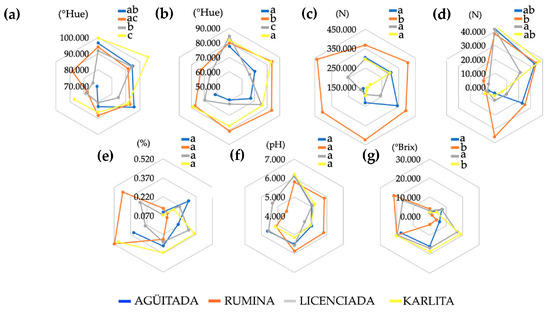
Figure 4.
Radar chart of physicochemical analysis of four jackfruit genotypes (n = 12): peel color (a), bulb color (b), peel firmness (c), bulb firmness (d), titratable acidity (e), pH (f), and total soluble solids (g). Different letters indicate statistically significant differences between genotypes (Fisher LSD test; p ≤ 0.05).
When evaluating the color of the bulbs, “Agüitada” and “Licenciada” shared the same orange color, while “Rumina” showed a yellow tone and “Karlita” a light yellow. Research by Asmady et al. [48] reported saffron-yellow bulbs, within the (°Hue) range (79.32–80.33 °Hue) reported in this study. The bulb color is regulated by the accumulation and degradation of carotenoids, with the concentration and specific combination of these compounds varying due to genetic material and the presence of genes regulating these traits [49,50,51].
The evaluation of peel firmness only differentiated the “Rumina” genotype, which maintained a resistance to penetration of 402.42 N on average during storage. “Karlita” showed the lowest resistance with 224.31 N, while the other genotypes did not show significant differences in this parameter (p ≥ 0.05). Regarding the firmness of the bulbs, “Rumina” stood out for its hard pulp with values of 24.73 N, while “Licenciada” showed the lowest resistance with 15.61 N. In the study conducted by Morelos-Flores et al. [15], which involved four genotypes from the same region, peel firmness ranged from 227.35 to 284.32 N, and bulb firmness ranged from 4.20 to 8.91 N. The findings indicated similarities in peel firmness and dissimilarities in bulb firmness. Peel and bulb firmness are influenced by the cell wall’s composition and structure, which includes cellulose, hemicellulose, pectin, and structural proteins, regulated by enzymes [52,53].
The parameters of titratable acidity and pH did not show significant differences among the genotypes (p ≥ 0.05), suggesting that these parameters are not sufficient to differentiate the genotypes. Despite the above, these results are consistent with those reported by Kamdem et al. [54] (0.1288% TA, pH 5.0). Titratable acidity and pH are regulated by the synthesis and metabolism of organic acids, influenced by enzymatic activity and the transport of protons and ions through cell membranes [55,56].
The analysis of total soluble solids allowed for the distinction of two groups of fruits: sweet and less sweet. “Rumina” and “Karlita” fell into the first group, with values of 21.73 and 20.78 °Brix, respectively, while “Licenciada” and “Agüitada” were in the second group, with values of 18.36 and 17.45, respectively. These results are generally in line with those described by Seleim et al. [57] on three jackfruit cultivars (24.21–26.23 °Brix). The accumulation of simple sugars during ripening is regulated by enzymes involved in the synthesis and degradation of carbohydrates, as well as by the transport and accumulation of these compounds in cellular vacuoles [58].
Based on the above, the “Rumina” genotype offers several attractive physicochemical characteristics for consumers, including good storage stability, appealing yellow bulb color, high firmness ensuring good texture and shelf life, and a moderately sweet taste suitable for those preferring less sweetness.
3.4. Phytochemical Analysis
3.4.1. Quantification of Total Soluble Phenols and Total Carotenoids
The phytochemical analysis revealed significant differences (p ≤ 0.05) in the parameters evaluated among the genotypes (Figure 5). “Agüitada” fruits had the highest total soluble phenols at 1.23 mg GAE/g DW, while “Rumina” had the lowest at 0.54 mg GAE/g DW. For total carotenoids, “Agüitada” again led with 9591.70 µg EβC/100 g WW, followed by “Rumina” and “Licenciada” with 6968.60 and 4656.98 µg EβC/100 g WW, respectively; “Karlita” had the lowest at 3038.40 µg EβC/100 g WW. Different values are reported by other authors, indicating a total soluble phenol content of 9 mg GAE/g [10] and carotenoid concentrations of 820 μg/100 g [59] in unspecified genotypes.
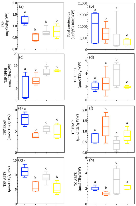
Figure 5.
Boxplot of the phytochemical analysis in four jackfruit genotypes (n = 12): total soluble phenols (a), total carotenoids (b), DPPH total soluble phenols (c), DPPH total carotenoids (d), FRAP total soluble phenols (e), FRAP total carotenoids (f), ABTS total soluble phenols (g), and ABTS total carotenoids (h). The horizontal line denotes the mean value of the assessed variable. The values within the box correspond to the 50% of measurements nearest to the mean. The whiskers depict the measurements furthest from the mean, indicating dispersion. Different letters denote statistically significant differences between genotypes (Fisher LSD test; p ≤ 0.05).
The synthesis of phytochemicals during fruit ripening significantly impacts their final quality and characteristics associated with health benefits. The synthesis of phenolic compounds begins from precursors such as shikimic acid and cinnamic acid, which are metabolized through various biosynthetic pathways [60]. These reactions are regulated by the activity of specific enzymes (lyases, transferases, oxidases) and are influenced by factors such as substrate availability (organic acids), enzymatic activity, and environmental conditions such as temperature and light [61]. Differences between genotypes in these metabolic pathways can result in variations in phenolic content.
Similarly, the synthesis of carotenoids, such as β-carotene, lutein, and zeaxanthin, is derived from the isoprenoid pathway, where isopentenyl pyrophosphate (IPP) and dimethylallyl pyrophosphate (DMAPP) are the main precursors [62]. These molecules are converted into carotenoids through a series of enzymatic reactions, including the condensation of isoprene units, cyclization, and modification of functional groups. Differences between genotypes in carotenoid synthesis can be influenced by environmental factors such as light and temperature, which affect enzymatic activity and metabolic pathways [62]. Plant hormones, such as abscisic acid and auxins, also play a crucial role in modulating carotenoid synthesis in response to environmental and stress signals, contributing to the observed differences between genotypes [63,64].
3.4.2. Evaluation of Antioxidant Capacity
Statistically significant differences (p ≤ 0.05) were observed in the evaluation of CAOX using the DPPH method for both phenolic compounds and total carotenoids. For phenolic compounds, “Karlita” and “Licenciada” exhibited the highest CAOX values, averaging 12.53 and 11.975 µmol TE/g DW, respectively. Regarding the CAOX of total carotenoids using the DPPH method, “Licenciada” showed the highest CAOX with values of 4.01 µmol TE/g DW, followed by “Rumina” with a figure of 2.52 µmol TE/g DW.
The FRAP method provided additional assessment of the mechanism by which phenolic compounds stabilize free radicals. In this regard, “Agüitada” exhibited the highest CAOX by the FRAP method, reporting a value of 7.81 µmol TE/g DW. On the other hand, in the evaluation of CAOX by the FRAP method for total carotenoids, “Rumina” emerged as the genotype with the highest value, reaching a CAOX of 1.20 µmol TE/g DW.
For the ABTS method, significant differences were also observed among the analyzed genotypes (p ≤ 0.05). “Licenciada” exhibited the best radical stabilization capacity for total carotenoids with a value of 3.04 µmol TE/g DW, followed by “Karlita” and “Agüitada” with values of 2.18 and 1.94 µmol TE/g DW, respectively (ABTS).
In general, values presented in these assays fell within the range of similarity with the genotypes reported by Morelos-Flores et al. [14], from 3.06 to 7.82 mmol TE/g (DPPH), 5.3 to 10.21 mmol TE/g (FRAP), and 4.41 to 10.43 mmol TE/g (ABTS).
The quantified phytochemicals exhibit antioxidant capacity due to their intrinsic chemical properties. Phenols, such as flavonoids, phenolic acids, and tannins, possess hydroxyl groups that can donate electrons to neutralize reactive oxygen species (ROS) and free radicals, acting as reducing agents [65]. Additionally, they have the ability to chelate pro-oxidant metals, thus inhibiting ROS formation.
Furthermore, carotenoids, such as β-carotene and lutein, are capable of dissipating absorbed light energy, thereby reducing the formation of reactive oxygen species in the photosystem [66]. They also act as antioxidants by donating electrons to free radicals, stabilizing them, and preventing oxidative damage to biomolecules [66]. The variation in antioxidant capacity among genotypes of the same fruit implies differences in the quantity and activity of antioxidant compounds present in each variety [67]. This may be due to variations in the concentration of antioxidant metabolites, whose disparities can influence the fruit’s resistance to oxidative stress, its ability to combat cellular damage, and its longevity during storage, which in turn can have implications for the quality, health, and commercialization of agricultural products [65].
All of the above suggests that “Licenciada” has a greater amount of antioxidant phytochemicals than the rest of the genotypes evaluated, giving it greater potential for quality and commercialization.
3.5. Principal Component Analysis
The principal component analysis (PCA) reveals that the first two principal components (PC1 and PC2) together explain 78.31% of the total variability in the data, indicating that these components suffice to condense essential information about the differences between genotypes (Figure 6). The application of Kaiser–Guttman’s criterion reveals that both PC1 and PC2 have eigenvalues greater than 1 (9.34 and 7.87, respectively), thus validating the significance of both components and confirming their ability to explain the observed variability in the data.
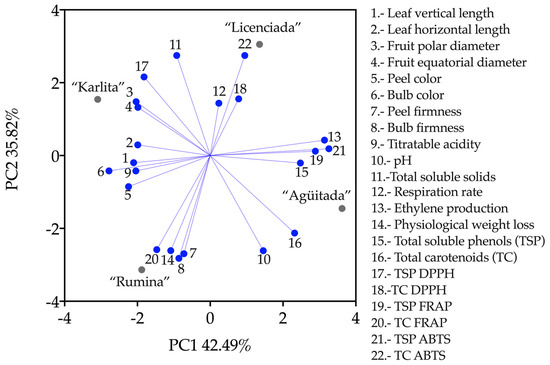
Figure 6.
Principal component analysis of morphological, physiological, physicochemical, and phytochemical analysis in jackfruit genotypes.
PC1 is positively correlated with variables related to the analyzed phytochemical compounds and their antioxidant capacities, as well as ethylene production (13, 15, 16, 19, and 20). This correlation is congruent, as ethylene may regulate the activation of metabolic pathways for the production of bioactive compounds (phenols and carotenoids). Consequently, the accumulation of these compounds will impact antioxidant capacity. This positive correlation implies that these characteristics contribute most to the differentiation between genotypes in PC1—in other words, genotypes distinguished by higher production of phenolic compounds, carotenoids, and ethylene production.
On the other hand, inverse correlations include parameters related to morphology and physicochemical composition of the jackfruits. The four morphological parameters are correlated along with peel color, bulbs, and titratable acidity (1, 2, 3, 4, 5, 6, and 9). The correlation of these variables suggests that genotypes with smaller fruit/leaf sizes and lower content of bioactive compounds and organic acids will exhibit lower antioxidant capacity and reduced rates of ethylene, phenols, and carotenoids production. The remaining components contribute little to explaining the behavior of the genotypes; however, they are indispensable for the distribution of variables and cases in this analysis (7, 8, 10, 11, 12, 14, 17, 18, 20, and 22).
PC2 exhibits positive correlations with variables associated with the production of total soluble solids and the antioxidant capacity TSP DPPH and TC ABTS (11, 17, and 22).
While soluble solids include sugars that do not possess direct antioxidant properties, their presence and relationship with antioxidant capacities may be associated with the presence of other compounds such as organic acids, vitamins, and phenolic compounds. In contrast, PC2 shows negative correlations with variables reflecting fruit quality parameters such as firmness, physiological weight loss, total carotenoids, and their antioxidant capacity by FRAP (7, 8, 10, 14, 16, and 20). There were also parameters that did not contribute to explaining PC2; however, they had a greater contribution to PC1 (1, 2, 3, 4, 5, 6, 9, 12, 13, 15, 18, 19, and 21).
The objective of conducting the PCA was twofold: first, to identify and elucidate the underlying correlations among the diverse set of variables, which range from morphological traits such as leaf and fruit sizes to physicochemical properties and bioactive compounds, and second, to group the genotypes based on these correlated variables to better understand the differentiation and similarities among them.
In terms of genotype distribution, the PCA reveals distinctive grouping patterns. “Agüitada” primarily follows PC1, indicating a strong correlation with variables associated with bioactive compounds and antioxidant capacity. “Licenciada” shows some affinity with PC1 but shares more similarities with “Agüitada” on PC2, particularly in traits linked to total soluble solids and antioxidant capacities. Conversely, “Karlita” follows the direction established by PC1 in reverse, suggesting it has opposing traits in the variables contributing to PC1. “Rumina” shows a lower affinity with PC1 but is similar to “Karlita” on PC2, indicating comparable characteristics in fruit quality parameters like firmness and weight loss. The arrangement on PC2 highlights “Licenciada” in the positive zone, associating it with high total soluble solids and antioxidant capacities, while “Rumina” in the negative zone shares similarities with “Agüitada” in traits like fruit firmness and carotenoid content.
3.6. Molecular Markers
The SSR markers selected based on the PCR band pattern were SSR LMCH 144, 122, 114, and 96, and from the SRAP markers, the combinations me1+em15, me3+em15, and me4+em15 were chosen. The results of the genetic distance dendrogram show two clusters of jackfruit genotypes (Figure 7). In the first cluster, the genotype “Rumina” is observed with a genetic distance of 0.56, which is separated from the rest of the genotypes. In the second cluster, “Karlita” is separated from “Agüitada” and “Licenciada”, with a genetic distance of 0.45; the genotypes “Agüitada” and “Licenciada” were found to be closer to each other in terms of genetic similarity, with a genetic distance of 0.22. Heterozygosity analysis on jackfruit genotypes, as presented in Table 3, used a threshold of 0.5 to differentiate between low and high heterozygosity. According to Table 4, the genotype “Agüitada” showed a low value of 0.375, while “Licenciada” showed the highest genetic diversity (0.625). The Jaccard index matrix shows the similarity between jackfruit genotypes; index values indicate that “Agüitada” has moderate similarity with “Rumina” (0.4815) and “Karlita” (0.4483) and relatively high similarity with “Licenciada” (0.6667). “Rumina” shows moderate similarity with Licenciada (0.3571) and “Karlita” (0.3793). “Licenciada” and “Karlita” also exhibit moderate similarity with each other (0.4815). These results reveal different levels of genetic similarity among the studied jackfruit genotypes.
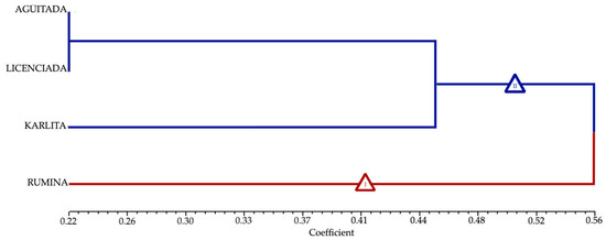
Figure 7.
Dendrogram illustrating the genetic distances among the analyzed genotypes of jackfruit.

Table 3.
Heterozygosity in analyzed genotypes of jackfruit.

Table 4.
Jaccard indices from analyzed genotypes of jackfruit.
The observed genetic divergence among jackfruit genotypes can be attributed to various genetic, environmental, and evolutionary factors. According to Palupi et al. [18], two main clusters were also found among 11 jackfruit genotypes, with lower genetic distances when they originated from the same region. This supports our findings, as geographical distribution and cross-pollination in the orchard can limit gene flow, resulting in greater genetic differentiation [18].
The link between the genetic distance dendrogram and the morphological, physiological, physicochemical, and phytochemical investigations is clear. “Rumina” consistently stands out with larger leaves, a lower respiration rate, less physiological weight loss, and firm yellow bulbs, supported by low levels of soluble phenols and carotenoids. This phenotype matches its genetic distinction in the dendrogram. “Agüitada” and “Licenciada” display genetic proximity, reflected in their similar orange bulb color, the high levels of phenols and carotenoids in “Agüitada”, and the high ethylene production in “Licenciada”. These biochemical and physiological similarities align with their close genetic relationship. “Karlita”, which is distinctly separated in the dendrogram, shows larger fruits, a higher respiration rate, and lower firmness, significantly differentiating it from the other genotypes in both phenotypic and genetic terms.
Heterozygosity values provide insights into genetic diversity within each genotype. High heterozygosity, as seen in “Licenciada”, suggests greater genetic variability, beneficial for adaptability and survival against environmental changes. Conversely, low heterozygosity in “Agüitada” indicates reduced genetic diversity and potentially lower adaptability [68].
The Jaccard index matrix aids in identifying phylogenetic relationships among genotypes, revealing greater diversity in our samples compared to those evaluated by Palupi et al. [18]. Despite the greater genetic diversity observed in the “Licenciada” genotype (Table 3), it is noteworthy that it shows genetic similarity with “Agüitada”, as indicated by the Jaccard index, which also aligns with the genetic distance dendrogram (Figure 7). Additionally, the dissimilarity of “Rumina” with the other genotypes is highlighted, which was also observed in the dendrogram. This comprehensive understanding of genetic diversity and phylogenetic relationships is essential for the management and conservation of jackfruit genetic resources.
4. Conclusions
The purpose of this study was to comprehensively analyze jackfruit genotypes through morphological, physiological, physicochemical, phytochemical, and molecular marker evaluations to understand their genetic diversity and potential for agricultural improvement. The findings highlighted “Licenciada” as a particularly promising genotype, combining high genetic diversity, strong antioxidant potential, and desirable physicochemical traits. Significant genetic diversity was observed among the genotypes, with clear differentiation supported by principal component analysis and dendrogram analysis. These findings will guide breeding programs to enhance yield, quality, and commercial viability. Additionally, the molecular tools employed will support the establishment of a germplasm bank, facilitating the selection of superior varieties for export and the development of genetic improvement initiatives. Future research should expand genetic analysis to include a wider range of jackfruit genotypes from various regions, providing a more comprehensive understanding of genetic diversity.
Author Contributions
Conceptualization, M.d.L.G.-M., E.M.-G., A.S.-V., V.M.Z.-G., M.A.C.-L., and G.B.-V.; Formal analysis, D.A.M.-F., V.M.Z.-G., G.B.-V., and M.d.L.G.-M.; Investigation, D.A.M.-F.; Methodology, D.A.M.-F., G.B.-V., E.M.-G., and M.d.L.G.-M.; Project administration, M.d.L.G.-M.; Supervision, G.B.-V., E.M.-G., and M.d.L.G.-M.; Writing—review and editing, G.B.-V. and M.d.L.G.-M. All authors have read and agreed to the published version of the manuscript.
Funding
This research received no external funding.
Data Availability Statement
The original contributions presented in the study are included in the article, further inquiries can be directed to the corresponding author.
Acknowledgments
The authors thank CONACYT (México) for the scholarship awarded to David Antonio Morelos-Flores (9348) as well as the Tecnologico Nacional de México/Instituto Tecnologico de Tepic and the Universidad Autonoma de Nayarit for their support in the development of this project.
Conflicts of Interest
The authors declare no conflicts of interest.
References
- Somashekhar, M.; Nayeem, N.; Sonnad, B. A Review on Family Moraceae (Mulberry) with a Focus on Artocarpus Species. 2013. Available online: www.wjpps.com (accessed on 25 August 2024).
- Khan, A.U.; Ema, I.J.; Faruk, R.; Tarapder, S.A.; Khan, A.U.; Noreen, S.; Adnan, M. Review on Importance of Artocarpus heterophyllus L. (Jackfruit). J. Multidiscip. Appl. Nat. Sci. 2021, 1, 106–116. [Google Scholar] [CrossRef]
- Anaya-Esparza, L.M.; González-Aguilar, G.A.; Domínguez-Ávila, J.A.; Olmos-Cornejo, J.E.; Pérez-Larios, A.; Montalvo-González, E. Effects of Minimal Processing Technologies on Jackfruit (Artocarpus heterophyllus Lam.) Quality Parameters. Food Bioproc. Tech. 2018, 11, 1761–1774. [Google Scholar] [CrossRef]
- SIAP. Anuario Estadístico de la Producción Agrícola, Cierre de la Producción Agrícola. 2024. Available online: https://nube.siap.gob.mx/cierreagricola/ (accessed on 10 March 2024).
- De Oliveira, F.F.; Souto, A.G.L.; Cavalcante, L.; De Oliveira, F.F.; Nascimento, J.A.M.D.; Bezerra, F.T.C. Salt tolerance of Soft and Hard jackfruit varieties in the seedling stage. Emir. J. Food Agric. 2022, 34, 703–710. [Google Scholar] [CrossRef]
- Mijin, S.; Ding, P.; Saari, N.; Ramlee, S. Effects of pollination techniques and harvesting stage on the physico-chemical characteristics of jackfruit. Sci. Hortic. 2021, 285, 110199. [Google Scholar] [CrossRef]
- Luna, E.G.; Alejo, S.G.; Ramírez, G.; Arévalo, Y.G. La Yaca un Fruto de Exportación. Agro Product. 2013, 6, 65–70. [Google Scholar]
- Morelos-Flores, D.A.; Montalvo-González, E.; Chacón-López, M.A.; Santacruz-Varela, A.; Zamora-Gasga, V.M.; Torres-García, G.; de Lourdes García-Magaña, M. Comparative Study of Four Jackfruit Genotypes: Morphology, Physiology and Physicochemical Characterization. Horticulturae 2022, 8, 1010. [Google Scholar] [CrossRef]
- Villalobos, M.C. Jaca (Artocarpus heterophyllus Lam.) Centenario de Nayarit. In Enciclopedia Centenario de Nayarit, 1st ed.; Ladrón, L.C.P., Ed.; Consejo Estatal para la Cultura y Las Artes de Nayarit: Tepic, Mexico, 2017; Volume 91, pp. 25–30. [Google Scholar]
- Vargas-Torres, A.; Becerra-Loza, A.S.; Sayago-Ayerdi, S.G.; Palma-Rodríguez, H.M.; García-Magaña, M.d.L.; Montalvo-González, E. Combined effect of the application of 1-MCP and different edible coatings on the fruit quality of jackfruit bulbs (Artocarpus heterophyllus Lam) during cold storage. Sci. Hortic. 2017, 214, 221–227. [Google Scholar] [CrossRef]
- Barros-Castillo, J.C.; Calderón-Santoyo, M.; Cuevas-Glory, L.F.; Pino, J.A.; Ragazzo-Sánchez, J.A. Volatile profiles of five jackfruit (Artocarpus heterophyllus Lam.) cultivars grown in the Mexican Pacific area. Food Res. Int. 2021, 139, 109961. [Google Scholar] [CrossRef] [PubMed]
- SIAP. Boletín De Export. Jackfruit . 2017. Available online: https://www.gob.mx/cms/uploads/attachment/file/229919/Boletin_de_exportaciones_jackfruit_2017_06.pdf (accessed on 25 August 2024).
- Morelos-Flores, D.A.; Nolasco-González, Y.; Gutiérrez-Martínez, P.; Hernández-Fuentes, L.M.; Montalvo-González, E.; García-Magaña, M.L. Study of marketing simulation in jackfruit (Artocarpus heterophyllus Lam) treated with 1–methylcyclopropene. Rev. Int. Investig. Innovación Tecnológica 2021, 9, 90–111. [Google Scholar]
- Morelos-Flores, D.A.; Anzaldo-Mendiola, R.L.; Montalvo-González, E.; Zamora-Gasga, V.M.; Chacón-López, M.A.; Santacruz-Varela, A.; García-Magaña, M.D.L. Characterization and antioxidant capacity of phenolic compounds of jackfruit genotypes from Nayarit, Mexico. Food Chem. Adv. 2023, 3, 100470. [Google Scholar] [CrossRef]
- Morelos-Flores, D.A.; Montalvo-González, E.; Chacón-López, M.A.; Santacruz-Varela, A.; Zamora-Gasga, V.M.; Torres-Garcia, G.; García-Magaña, d.L.M. Jackfruit in Mexico: Characterization of four genotypes from the south of Nayarit. Fruits 2024, 79, 1–9. [Google Scholar] [CrossRef]
- Salgotra, R.K.; Chauhan, B.S. Genetic Diversity, Conservation, and Utilization of Plant Genetic Resources. Genes 2023, 14, 174. [Google Scholar] [CrossRef] [PubMed]
- Singh, D.K.; Pandey, A.; Choudhary, S.B.; Kumar, S.; Tribhuvan, K.U.; Mishra, D.C.; Bhati, J.; Kumar, M.; Tomar, J.; Bishnoi, S.; et al. Development of genic-SSR markers and their application in revealing genetic diversity and population structure in an Eastern and North-Eastern Indian collection of Jack (Artocarpus heterophyllus Lam.). Ecol. Indic. 2021, 131, 108143. [Google Scholar] [CrossRef]
- Palupi, D.; Rahayu, S.S.B.; Daryono, B.S. Genetic diversity in jackfruit (Artocarpus heterophyllus Lam.) based on molecular characters in Indonesia. SABRAO J. Breed Genet. 2019, 51, 57–67. [Google Scholar]
- Kavya, K.; Shyamalamma, S.; Gayatri, S. Morphological and molecular genetic diversity analysis using SSR markers in Jackfruit (Artocarpus heterophyllus Lam.) genotypes for pulp colour. Indian J. Agric. Res. 2019, 53, 8–16. [Google Scholar] [CrossRef]
- CONAGUA. Servicio Meteorologico Nacional, Normales Climatológica por Estado. 2023. Available online: https://smn.conagua.gob.mx/es/informacion-climatologica-por–estado?estado=nay (accessed on 10 July 2023).
- Love, K.; Paull, R.E. Jackfruit; College of Tropical Agriculture and Human Resources: Honolulu, HI, USA, 2011; pp. 1–7. [Google Scholar]
- IPGRI Artocarpus heterophyllus. In Powdered Crude Drug Microscopy of Leaves and Barks; International Plant Genetic Resources Institute: Rome, Italy, 2000; p. 64. [CrossRef]
- AOAC. Official Methods of Analysis of AOAC International; Association of Official Analytical Chemists: Rockville, MA, USA, 2005. [Google Scholar]
- Pérez-Jiménez, J.; Arranz, S.; Tabernero, M.; Díaz-Rubio, M.E.; Serrano, J.; Goñi, I.; Saura-Calixto, F. Updated methodology to determine antioxidant capacity in plant foods, oils and beverages: Extraction, measurement and expression of results. Food Res. Int. 2008, 41, 274–285. [Google Scholar] [CrossRef]
- Montreau, F.R. Sur le dosage des composés phénoliques totaux dans les vins par la méthode Folin-Ciocalteu. OENO One 1972, 24, 397–404. [Google Scholar] [CrossRef]
- García, J.R.; De la Rosa, L.A.; González-Barrios, A.G.; Herrera-Duenez, B.; López-Díaz, J.A.; González-Aguilar, G.A.; Ruíz-Cruz, S.; Álvarez-Parrilla, E. Cuantificación de polifenoles y capacidad antioxidante en duraznos comercializados en ciudad Juárez, México. Tecnociencia Chihuah. 2011, 5, 67–75. [Google Scholar]
- Philip, T.; Chen, T. Development of a Method for the Quantitative Estimation of Provitamin A Carotenoids in Some Fruits. J. Food Sci. 1988, 53, 1703–1706. [Google Scholar] [CrossRef]
- Prior, R.L.; Cao, G. Antioxidant Phytochemicals in Fruits and Vegetables: Diet and Health Implications. HortScience 2000, 35, 588–592. [Google Scholar] [CrossRef]
- Alvarez-Parrilla, E.; De La Rosa, L.A.; Legarreta, P.; Saenz, L.; Rodrigo-García, J.; González-Aguilar, G.A. Daily consumption of apple, pear and orange juice differently affects plasma lipids and antioxidant capacity of smoking and non-smoking adults. Int. J. Food Sci. Nutr. 2010, 61, 369–380. [Google Scholar] [CrossRef] [PubMed]
- Re, R.; Pellegrini, N.; Proteggente, A.; Pannala, A.; Yang, M.; Rice-Evans, C. Antioxidant activity applying an improved abts radical cation decolorization assay. Free Radic. Biol. Med. 1999, 26, 1231–1237. [Google Scholar] [CrossRef] [PubMed]
- Mercado Mercado, G.; López Teros, V.; Montalvo González, E.; González Aguilar, G.A.; Álvarez Parrilla, E.; Sáyago Ayerdi, S.G. Efecto de la extracción asistida por ultrasonido en la liberación y bioaccesibilidad in vitro de carotenoides, en bebidas elaboradas con mango (Mangifera indica L.) ‘Ataulfo’. Nova Sci. 2018, 10, 100–132. [Google Scholar] [CrossRef]
- Doyle, J.J.; Doyle, J.L. A rapid DNA isolation for small quantities of fresh leaf tissue. Phytochem. Bull. 1987, 19, 11–15. [Google Scholar]
- Anuragi, H.; Dhaduk, H.L.; Kumar, S.; Dhruve, J.J.; Parekh, M.J.; Sakure, A.A. Molecular diversity of Annona species and proximate fruit composition of selected genotypes. 3 Biotech 2016, 6, 204. [Google Scholar] [CrossRef]
- Talamantes-Sandoval, C.A.; Cortés-Cruz, M.; Balois-Morales, R.; López-Guzmán, G.G.; Palomino-Hermosillo, Y.A. Molecular analysis of genetic diversity in soursop (Annona muricata L.) using SRAP markers. Rev. Fitotec. Mex. 2019, 42, 209–214. [Google Scholar] [CrossRef]
- Lira-Ortiz, R.; Cortés-Cruz, M.A.; López-Guzmán, G.G.; Palomino-Hermosillo, Y.A.; Sandoval-Padilla, I.; Ochoa-Jiménez, V.A.; Sánchez-Herrera, L.M.; Balois-Morales, R.; Berumen-Varela, G. Genetic diversity of soursop populations (Annona muricata L.) in Nayarit, Mexico using SSR and SRAP markers. Acta Biol. Colomb. 2022, 27, 104–112. [Google Scholar] [CrossRef]
- Saitou, N.; Nei, M. The neighbor-joining method: A new method for reconstructing phylogenetic trees. Mol. Biol. Evol. 1987, 4, 406–425. [Google Scholar]
- Rohlf, F.J. NTSYS-pc, Version 1.80; Distribution by Exeter Software: New York, NY, USA, 1993. [Google Scholar]
- Dey, B.; Baruah, K. Morphological Characterization of Jackfruit (Artocarpus heterophyllus Lam.) of Assam. Int. J. Curr. Microbiol. Appl. Sci. 2019, 8, 1005–1016. [Google Scholar] [CrossRef]
- Mahla, J.S.; Soni, N.V.; Patel, P.C.; Patel, A.V.; Dasalania, J.P.; Roul, S. Variability, character association and path analysis for Annona yield and quality attributes. Emergent Life Sci. Res. 2022, 8, 229–239. [Google Scholar] [CrossRef]
- Marais, D.L.D.; Lasky, J.R.; Verslues, P.E.; Chang, T.Z.; Juenger, T.E. Interactive effects of water limitation and elevated temperature on the physiology, development and fitness of diverse accessions of Brachypodium distachyon. New Phytol. 2017, 214, 132–144. [Google Scholar] [CrossRef] [PubMed]
- De Tepic, I.T.; de Oca, M.M.-M.; Osuna-García, J.; Hernández-Estrada, A.; Ochoa-Villarreal, M.; Tovar-Gómez, B. Efecto del 1-Metilciclopropeno (1-MCP) Sobre la Fisiología y Calidad de Frutos de Jaca (Artocarpus heterophyllus Lam.). Rev. Chapingo. Ser. Hortic. 2007, 13, 165–170. [Google Scholar] [CrossRef]
- Salehi, F. Recent Advances in the Modeling and Predicting Quality Parameters of Fruits and Vegetables during Postharvest Storage: A Review. Int. J. Fruit Sci. 2020, 20, 506–520. [Google Scholar] [CrossRef]
- Ray, B.J.; Chandra, K.; Viswavidyalaya, A.; Choudhury, S.G.; Kalindi, D. Physiology and Biochemistry of Fruit ripening: A Review. Scientist 2023, 2, 197–204. [Google Scholar] [CrossRef]
- Xanthopoulos, G.T.; Templalexis, C.G.; Aleiferis, N.P.; Lentzou, D.I. The contribution of transpiration and respiration in water loss of perishable agricultural products: The case of pears. Biosyst. Eng. 2017, 158, 76–85. [Google Scholar] [CrossRef]
- Lufu, R.; Ambaw, A.; Opara, U.L. Mechanisms and modelling approaches to weight loss in fresh fruit: A review. Technol. Hortic. 2024, 4, e006. [Google Scholar] [CrossRef]
- Elevitch, C.R.; Manner, H.I. Artocarpus heterophyllus (jackfruit). Species Profiles Pac. Isl. Agrofor. 2006, 1, 16. [Google Scholar]
- Ebrahimi, P.; Shokramraji, Z.; Tavakkoli, S.; Mihaylova, D.; Lante, A. Chlorophylls as Natural Bioactive Compounds Existing in Food By-Products: A Critical Review. Plants 2023, 12, 1533. [Google Scholar] [CrossRef]
- Asmady, N.H.; Abidin, M.Z. Effect of Vacuum Packaging on Sensory and Texture Properties of Fresh Cut Jackfruit. Sci. Technol. 2023, 3, 452–459. [Google Scholar] [CrossRef]
- Tatarowska, B.; Milczarek, D.; Wszelaczyńska, E.; Pobereżny, J.; Keutgen, N.; Keutgen, A.J.; Flis, B. Carotenoids Variability of Potato Tubers in Relation to Genotype, Growing Location and Year. Am. J. Potato Res. 2019, 96, 493–504. [Google Scholar] [CrossRef]
- Zafar, J.; Aqeel, A.; Shah, F.I.; Ehsan, N.; Gohar, U.F.; Moga, M.A.; Festila, D.; Ciurea, C.; Irimie, M.; Chicea, R. Biochemical and immunological implications of lutein and zeaxanthin. Int. J. Mol. Sci. 2021, 22, 10910. [Google Scholar] [CrossRef] [PubMed]
- González-Peña, M.A.; Ortega-Regules, A.E.; de Parrodi, C.A.; Lozada-Ramírez, J.D. Occurrence, Properties, Applications, and Encapsulation of Carotenoids—A Review. Plants 2023, 12, 313. [Google Scholar] [CrossRef]
- Li, X.; Xu, C.; Korban, S.S.; Chen, K. Regulatory mechanisms of textural changes in ripening fruits. CRC Crit. Rev. Plant. Sci. 2010, 29, 222–243. [Google Scholar] [CrossRef]
- Su, Q.; Li, X.; Wang, L.; Wang, B.; Feng, Y.; Yang, H.; Zhao, Z. Variation in Cell Wall Metabolism and Flesh Firmness of Four Apple Cultivars during Fruit Development. Foods 2022, 11, 3518. [Google Scholar] [CrossRef]
- Bemmo, U.L.K.; Bindzi, J.M.; Kamseu, P.R.T.; Ndomou, S.C.H.; Tambo, S.T.; Zambou, F.N. Physicochemical properties, nutritional value, and antioxidant potential of jackfruit (Artocarpus heterophyllus) pulp and seeds from Cameroon eastern forests. Food Sci. Nutr. 2023, 11, 4722–4734. [Google Scholar] [CrossRef]
- Famiani, F.; Farinelli, D.; Frioni, T.; Palliotti, A.; Battistelli, A.; Moscatello, S.; Walker, R.P. Malate as substrate for catabolism and gluconeogenesis during ripening in the pericarp of different grape cultivars. Biol. Plant 2016, 60, 155–162. [Google Scholar] [CrossRef]
- Mirza, A.; Tripathi, K.; Kumar, P.; Kumar, R.; Saxena, R.; Kumar, A.; Badoni, H.; Goyal, B. Efficacy of jackfruit components in prevention and control of human disease: A scoping review. J. Educ. Health Promot. 2023, 12, 361. [Google Scholar] [CrossRef]
- Seleim, M.A.A.; Hassan, M.A. Physicochemical Properties and Nutritional Evaluation of Jackfruits (Artocarpus heterophyllus L.). Int. Adv. Res. J. Sci. Eng. Technol. 2019, 6, 75–84. [Google Scholar] [CrossRef]
- Saxena, A.; Bawa, A.S.; Raju, P.S. Jackfruit (Artocarpus heterophyllus Lam.). In Postharvest Biology and Technology od Tropical and Subtropical Fruits; Woodhead Publishing Limited: Sawston, UK, 2011. [Google Scholar] [CrossRef]
- Saxena, A.; Bawa, A.; Raju, P. Phytochemical changes in fresh-cut jackfruit (Artocarpus heterophyllus L.) bulbs during modified atmosphere storage. Food Chem. 2009, 115, 1443–1449. [Google Scholar] [CrossRef]
- Santos-Sánchez, N.F.; Salas-Coronado, R.; Hernández-Carlos, B.; Villanueva-Cañongo, C. Shikimic Acid Pathway in Biosynthesis of Phenolic Compounds. In Plant Physiological Aspects of Phenolic Compounds; IntechOpen: London, UK, 2019. [Google Scholar] [CrossRef]
- Nieto Ramírez, M.I.; García Trejo, J.F.; Caltzontzin Rabell, V.; Chávez Jaime, R.; Estrada Sánchez, M.D.L.L. Efecto de las condiciones de cultivo en la producción de fenoles, flavonoides totales y su capacidad antioxidante en el árnica (Heterotheca inuloides). Rev. Mex. Cienc. Agric. 2018, 21, 4296–4305. [Google Scholar] [CrossRef][Green Version]
- Zhao, X.; Li, Y.; Zhang, M.M.; He, X.; Ahmad, S.; Lan, S.; Liu, Z.J. Research advances on the gene regulation of floral development and color in orchids. Gene 2023, 888, 147751. [Google Scholar] [CrossRef] [PubMed]
- Stanley, L.; Yuan, Y.W. Transcriptional Regulation of Carotenoid Biosynthesis in Plants: So Many Regulators, So Little Consensus. Front. Plant Sci. 2019, 10, 1017. [Google Scholar] [CrossRef] [PubMed]
- Yuan, H.; Zhang, J.; Nageswaran, D.; Li, L. Carotenoid metabolism and regulation in horticultural crops. Hortic. Res. 2015, 2, 15036. [Google Scholar] [CrossRef] [PubMed]
- Francenia Santos Sánchez, N.; Salas-Coronado, R.; Villanueva-Cañongo, C.; Hernández-Carlos, B. Antioxidant Compounds and Their Antioxidant Mechanism. In Antioxidants; Shalaby, E., Ed.; Intechopen: London, UK, 2019. [Google Scholar]
- Kesawat, M.S.; Satheesh, N.; Kherawat, B.S.; Kumar, A.; Kim, H.U.; Chung, S.M.; Kumar, M. Regulation of Reactive Oxygen Species during Salt Stress in Plants and Their Crosstalk with Other Signaling Molecules—Current Perspectives and Future Directions. Plants 2023, 12, 864. [Google Scholar] [CrossRef]
- Nurzyńska-Wierdak, R. Phenolic Compounds from New Natural Sources—Plant Genotype and Ontogenetic Variation. Molecules 2023, 28, 1731. [Google Scholar] [CrossRef]
- Amevoin, K.; Agboyi, L.K.; Gomina, M.; Kounoutchi, K.; Bassimbako, K.H.; Djatoite, M.; Dawonou, A.V.; Tagba, A. Fruit fly surveillance in Togo (West Africa): State of diversity and prevalence of species. Int. J. Trop. Insect Sci. 2021, 41, 3105–3119. [Google Scholar] [CrossRef]
Disclaimer/Publisher’s Note: The statements, opinions and data contained in all publications are solely those of the individual author(s) and contributor(s) and not of MDPI and/or the editor(s). MDPI and/or the editor(s) disclaim responsibility for any injury to people or property resulting from any ideas, methods, instructions or products referred to in the content. |
© 2024 by the authors. Licensee MDPI, Basel, Switzerland. This article is an open access article distributed under the terms and conditions of the Creative Commons Attribution (CC BY) license (https://creativecommons.org/licenses/by/4.0/).