Article

Species Identification and Fungicide Sensitivity of Fusarium spp. Causing Peanut Root Rot in Henan, China

1
College of Plant Protection, Henan Agricultural University, No. 218, Ping’an Avenue, Zhengzhou 450046, China
2
Henan Key Laboratory of Creation and Application of New Pesticide, Henan Agricultural University, Zhengzhou 450046, China
3
Henan Research Center of Green Pesticide Engineering and Technology, Henan Agricultural University, Zhengzhou 450046, China
4
Institute of Plant Protection Research, Henan Academy of Agricultural Sciences, Zhengzhou 450002, China
*
Authors to whom correspondence should be addressed.
J. Fungi 2025, 11(6), 433; https://doi.org/10.3390/jof11060433
Submission received: 4 March 2025 / Revised: 30 May 2025 / Accepted: 4 June 2025 / Published: 6 June 2025

Abstract

:
Peanut Root Rot (PRR) is a devastating disease that significantly limits peanut production worldwide. Although PRR has been frequently reported in Henan Province of China, the predominant Fusarium species and their sensitivity to different fungicides remain unclear. Between 2021 and 2023, we surveyed 81 peanut fields across 17 cities in Henan Province, China, to assess PRR prevalence and Fusarium species distribution. A total of 1131 Fusarium isolates were identified based on the morphological characters and phylogenetic analyses and classified into 11 recognized Fusarium species: F. solani (56.06%), F. oxysporum (20.87%), F. neocosmosporiellum (13.62%), F. proliferatum (4.69%), F. acuminatum (1.33%), F. commune (1.15%), F. graminearum (1.06%), F. pseudograminearum (0.35%), F. ipomoeae (0.35%), F. lacertarum (0.26%), and F. armeniacum (0.26%). Pathogenicity assessments showed that all 11 Fusarium species were capable of causing PRR, with F. solani exhibiting the highest isolation frequency and widespread distribution in all areas. Furthermore, the four Fusarium species (F. solani, F. oxysporum, F. neocosmosporiellum, and F. proliferatum) were highly sensitive to the six fungicides, including prochloraz (EC50 values of 0.02 ± 0.00~0.06 ± 0.01 mg/L), pydiflumetofen (EC50 values of 0.31 ± 0.07~0.67 ± 0.06 mg/L), tetramycin (EC50 values of 0.11 ± 0.02~0.58 ± 0.08 mg/L), tebuconazole (EC50 values of 0.26 ± 0.07~0.65 ± 0.10 mg/L), prothioconazole (EC50 values of 1.14 ± 0.16~3.15 ± 0.81 mg/L), and difenoconazole (EC50 values of 0.62 ± 0.12~3.58 ± 0.76 mg/L). This comprehensive study is the first systematic documentation on the prevalence, virulence, and fungicide sensitivity of PRR pathogens in Henan Province. The findings of the current study will provide a theoretical basis for the effective management of peanut root rot in Henan, China.

1. Introduction

Peanut (Arachis hypogaea L.) is an important oil and economic crop widely cultivated in tropical and subtropical agro-climatic regions [1]. China is the largest producer of peanuts in the world, with a peanut planting area of 4,850,656 ha in 2023, producing 19.27 MT peanuts, accounting for approximately 35.51% of the global peanut yield (54.27 MT) [2]. China’s peanut growing areas are mainly distributed in the Henan Province, which contributes significantly to the national peanut yield and planting area at 33.22% and 27.24%, respectively (http://www.stats.gov.cn, accessed on 10 February 2025). In recent years, due to the adjustment of the cropping system and changes in climate conditions, peanut root rot (PRR) has been frequently and widely reported in Henan Province, posing a serious threat to the peanut yield and quality [3].
PRR is a devastating disease affecting peanut crops, which has been found in peanut fields in Australia [4], Vietnam [5], Pakistan [6], Argentina [7], Egypt [8], India [9], Ethiopia [10] and China [11,12,13,14]. The early symptoms of the disease are brown spots on the root and wilting on the lower leaves [12,13,15]. As the disease progresses, infected peanut plants exhibit stunted, yellowing leaves, diminished plant vigor or sudden wilting, and the presence of brown/black discoloration and rot on the infected roots [4,11,12,13]. Eventually, the affected plants collapse and die [11,12,13,15]. The disease usually causes from 20% to 40% of disease incidence in peanut fields [4,13,14]. In serious cases, the incidence of this disease was found in up to 75% in peanut fields [10]. Especially during seasons with continuous drought stress, this disease could cause a 95% disease incidence in some peanut fields [16].
PRR is caused by multiple pathogenic fungal species [6,7,8,9,11,12,17,18]. Among them, various Fusarium species have been implicated in PRR worldwide, with F. solani and F. oxysporum being the most frequently reported pathogens across multiple regions, including Argentina [7], Egypt [8,19], Ethiopia [10], and so on. However, the Fusarium species of PRR are remarkably varied with different geographical regions. In Egypt, F. solani, F. moniliforme, F. equesti, F. semitectum, and F. oxysporum have been reported to cause root rot in peanut fields [8,19]. In Argentina, F. oxysporum and F. solani have been implicated as responsible for PRR [7]. In India, F. neocosmosporiellum and F. solani are isolated from diseased peanut plants, which caused PRR [20]. In Australia and Vietnam, PRR disease is caused by F. neocosmosporiellum [4,5]. In eastern Ethiopia, F. oxysporum and F. solani are isolated from peanut roots, and identified as PRR pathogens [10]. In China, F. equiseti, F. incarnatum, F. oxysporum, F. proliferatum, F. solani, F. fujikuroi, and F. acuminatum have been identified as PRR pathogens [13,14,21,22]. However, it remains uncertain which specific Fusarium species are most prevalent and play a significant role in causing PRR in Henan Province.
A thorough comprehension of pathogens involved in PRR is essential for effective disease management. Previous studies have identified Fusarium species as causative agents of PRR in various provinces of China, including Jiangxi [21], Shandong [14], and Henan [13,22]. However, comprehensive surveys of PRR in these provinces, especially in Henan Province, are lacking. In present, only two species (F. fujikuroi and F. proliferatum) have been reported in Henan Province [13]. In a recent investigation, 41 fungal isolates were collected from the diseased peanut roots in six cities within Henan Province (Anyang, Jiaozuo, Pingdingshan, Xinxiang, Xuchang, and Zhoukou). These isolates were identified to F. equiseti, F. incarnatum, F. oxysporum, F. proliferatum, and F. solani, respectively, all of which were confirmed to be the pathogens of PRR [22]. However, due to the limited sampling areas and a small number of isolates, these data may not accurately reflect the prevalence and distribution of pathogens associated with PRR in Henan Province. Therefore, a comprehensive understanding of biology, population dynamics, and epidemiological factors contributing to disease exacerbation, is necessary for effective disease control strategies.
At present, various management measures have been reported to be effective approaches for control of PRR, such as chemical control [23,24], biological control [15,16,17], and agricultural practices like crop rotation or intercropping [21,25]. However, the application of fungicides remains the more effective approach for disease control. Although different fungicides have been widely used to control PRR, there is limited information on the sensitivity of PRR-causing pathogens to fungicides, particularly in China. To effectively manage PRR, it is crucial to understand the sensitivity of local pathogens to various fungicides utilized to manage this disease.
Therefore, the main objectives of this research were to (i) identify the Fusarium species causing root rot on peanut in the major peanut-cultivation regions of Henan; (ii) determine the virulence of each Fusarium species obtained from peanut roots; and (ⅲ) assess the inhibitory effect of registered and other alternative potential fungicides against mycelial growth of the four Fusarium species (F. solani, F. oxysporum, F. neocosmosporiellum, and F. proliferatum), the most frequent pathogens of PRR in the current study.

2. Materials and Methods

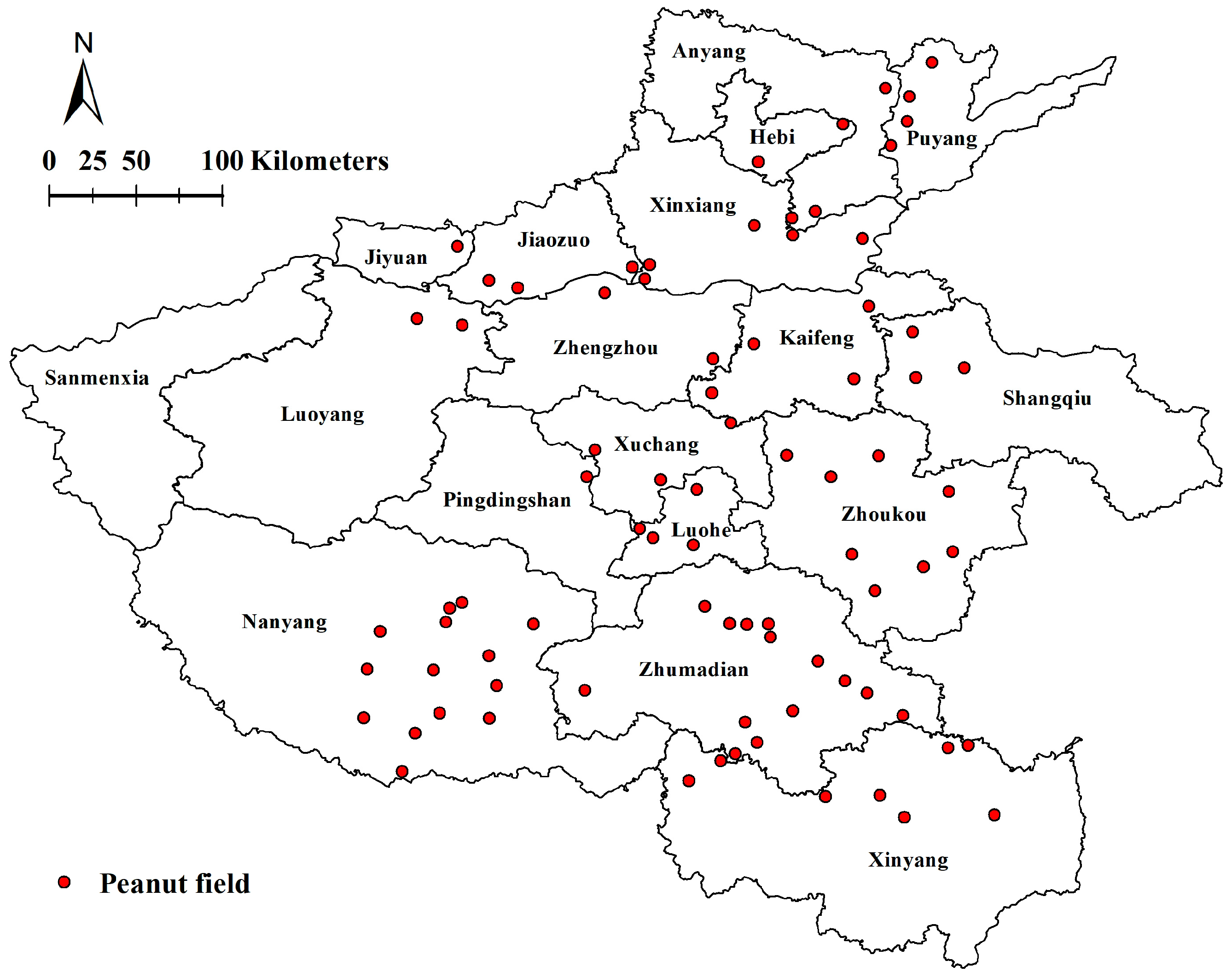
2.1. Sample Collection

During 2021 to 2023, peanut samples with typical root rot symptoms were collected from the seedling stage to flowering stage in Henan Province, China. A total of 81 peanut fields in 17 cities (including Puyang, Anyang, Hebi, Xinxiang, Jiaozuo, Jiyuan, Luoyang, Zhengzhou, Kaifeng, Shangqiu, Zhoukou, Xuchang, Luohe, Pingdingshan, Nanyang, Zhumadian and Xinyang) were investigated, representing the main peanut-producing regions in Henan Province (Figure 1). The peanut fields were arbitrarily selected, with a minimum distance of 5 km between each other. For each peanut field, five sampling points were made in a zig zag pattern [26], with each point being 20 m apart. At each of the five points, approximately 20 peanut plants were assessed, and on return to the laboratory 3–4 plants with typical symptoms of root rot were randomly selected from each transect point, for a total of 15–20 plants per field. All samples obtained were stored at 4 °C before fungal isolation.

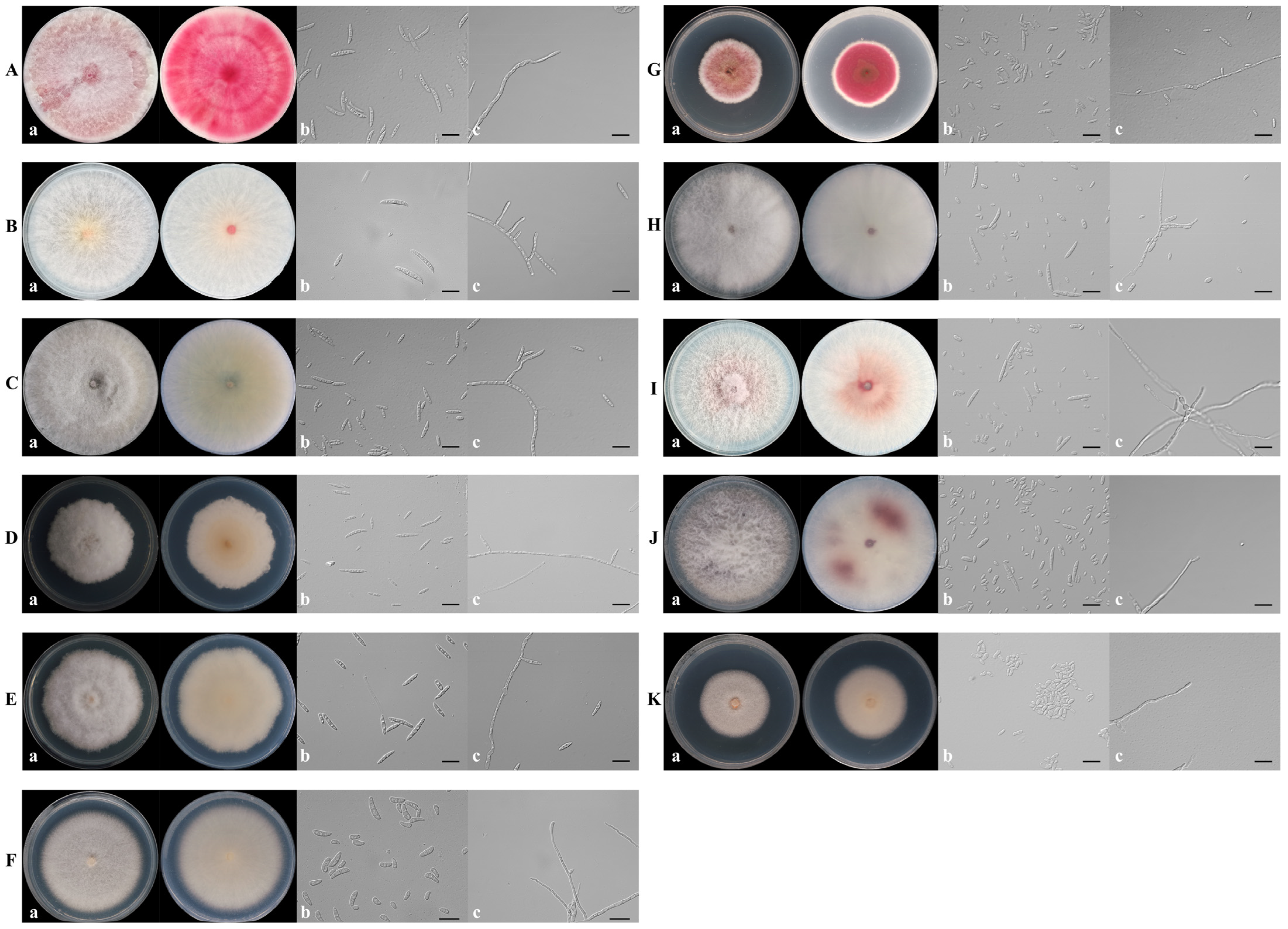
2.2. Isolation of Fungi and Morphological Observation

The infected peanut roots were washed with running water, dried on filter paper, and then 5 mm length pieces were taken from diseased to healthy tissue. The pieces were surface-disinfected for 1 min with 3% NaClO solution, rinsed three times in sterile distilled water, and dried on sterilized filter paper. Five pieces of each root were placed on potato dextrose agar (PDA) plates containing ampicillin (100 mg/L). These plates were incubated at 25 °C in the dark for 2 days. When fungal colonies appeared, they were chopped using sterile toothpicks and transferred to the new PDA plates. Each isolate obtained were further purified by using the single spore isolation method [27]. All pure fungal isolates obtained in the present study were stored in PDA tubes at 4 °C.
To confirm whether these fungal isolates were Fusarium species, they were preliminarily identified based on morphological characteristics as described in the literature [28,29]. Briefly, all pure fungal isolates were cultured on PDA plates and colonies morphology were observed after 7 days of incubation in the dark at 25 °C. Furthermore, all pure fungal isolates were cultured in carboxymethyl cellulose medium (CMC) to induce sporulation in darkness at 25 °C, 175 rpm and micromorphological characteristics (microconidia, macroconidia, and conidiophores) were observed using a Zeiss Imager M2 compound microscope with differential interference contrast (Carl Zeiss AG, Oberkochen, Germany) with the Zeiss software ZEN 3.4 after 3 days.

2.3. DNA Extraction, PCR Amplification and Sequencing

Genomic DNA of all isolates were extracted using a modified CTAB method as described by Özer et al. [30]. Five gene fragments, including ITS, TEF-1α, RPB2, TUB2, and CAM of 49 representative isolates (Table S1), were amplified with the primer pairs ITS1/ITS4 [31], EF1/EF2 [32], 5f2/7cr [33,34], T1/T2 [35], and CL1/CL2A [36], respectively. For the other isolates, only the TEF-1α gene region was sequenced. The primer pairs used in the study are shown in Table 1. DNA amplification was conducted in a total volume of 25 μL reaction solution that contained 22 μL 1.1 × T3 Super PCR Mix (Tsingke Biotech Co. Ltd., Beijing, China), 1 μL of each primer (10 mM), and 1 μL template DNA (50 ng/μL). The PCR cycling conditions were initiated with 98 °C for 2 min, followed by 35 cycles of denaturation at 98 °C for 10 s, annealing at a suitable temperature for 10 s for different loci: 56 °C for ITS, 55 °C for TEF-1α, 58 °C for RPB2, 54 °C for TUB2, and 55 °C for CAM, and extension at 72 °C for 10 s, and a final elongation step at 72 °C for 2 min. The PCR products were separated by agarose gel electrophoresis (1%, wt/vol) and sequenced by Tsingke Biotech Co. Ltd., Beijing. The obtained sequences were examined and aligned using BioEdit version 7.0.5.2 [37]. For the TEF-1α gene region, a sequence match of more than 99% similarity was regarded as the indicative of a fungal species in the present study [38].

2.4. Phylogenetic Analysis

Based on morphology and TEF-1α sequence data, 49 isolates were selected to perform the phylogenetic analysis, including 5 F. solani isolates, 5 F. oxysporum isolates, 5 F. neocosmosporiellum isolates, 5 F. proliferatum isolates, 5 F. acuminatum isolates, 5 F. commune isolates, 5 F. graminearum isolates, 4 F. pseudograminearum isolates, 4 F. ipomoeae isolates, 3 F. lacertarum isolates and 3 F. armeniacum isolates (Table S1). Multiple sequence alignments for each of five gene sequence fragments (ITS, TEF-1α, RPB2, TUB2, and CAM), including sequences generated from the present study and sequences from related taxa (Tables S1 and S2), were initially implemented using the MAFFT version 7.110 [39] with default parameters setting before being manually adjusted using BioEdit version 7.0.5.2 [37]. The phylogenetic analysis was performed by two independent algorithms, including Maximum likelihood (ML) and Bayesian inference (BI). The ML analysis was executed in the IQ-TREE version 1.6.8 [40] utilizing the GTR model with 1000 ultrafast bootstrap (BS) replications. Bayesian inference (BI) was utilized to construct phylogenetic relationship via MrBayes version 3.2.6 [41]. The best nucleotide substitution model of each partition was created by MrModeltest version 2.3 [42]. Markov Chain Monte Carlo (MCMC) analysis with four chains was conducted twice from a random tree topology for 3,000,000 generations. Trees were sampled every 1000 generations, which lasted until the average standard deviation of split frequencies was <0.01. After discarding the first 25% of saved trees (burn-in), the remaining trees were used to calculate the 50% majority rule consensus trees and posterior probability (PP) values. Clades with PP ≥ 0.95 and bootstrap values (BS) ≥ 70% were regarded as well supported [43]. Phylogenetic trees were estimated and adjusted using FIGTREE version 1.4.2 (http://tree.bio.ed.ac.uk/software/figtree/, accessed on 25 May 2025). Fusarium ventricosum CBS 748.79 served as the outgroup taxon in the phylogenetic analysis of Fusarium spp. The GenBank numbers of all isolates chosen for the phylogenetic analyses are shown in Tables S1 and S2.

2.5. Prevalence

The prevalence of fungal species in the main peanut-producing regions of Henan province was estimated as previously described [44]. The Isolation Frequency (FI) of individual species was calculated by the formula, FI% = (NS/NI) × 100, where NS was the number of isolates per fungal species, and NI was the total number of isolates from all fungal species obtained.

2.6. Pathogenicity Tests

Forty-nine isolates from 11 Fusarium species (Table S1) were selected to evaluate their pathogenicity on peanut seedlings by a modified inoculum layer method [45]. Briefly, all isolates selected were grown on PDA at 25 °C for 14 d, and the spores were suspended in 5 mL of sterile deionized water by scraping the colony with a sterile inoculating loop, filtered, then adjusted to the concentrations of 106 conidia per mL. The triangular flasks (250 mL) containing 50 g autoclaved millet seeds were inoculated with 2 mL spore suspension (106 conidia per mL), which were incubated at 25 °C for 7 days and were shaken daily for 30 s to ensure uniform colonization. Seeds of peanut (cv. Yuhua 22, a widely cultivated variety in Henan Province) were soaked for 1 min in 75% alcohol, rinsed in sterile water, and then germinated on sterile paper towels saturated with sterile water for 2 days at 25 °C. For planting, a 2 g layer of millet seed inoculums was placed in a plastic pot (7 cm × 7 cm × 8 cm) containing 15 g of sterilized premium grade coarse dry vermiculite and then covered with 8 g of vermiculite. Subsequently, two germinated peanut seeds with healthy and consistent growth were selected and sown on the top of the vermiculite, and 8 g layer of vermiculite was added to the top of the seeds. Finally, the sterilized water (80 mL) was added slowly to the pots to saturate the vermiculite. The sterile millet seed without mycelia was regarded as the negative control. Each four pots were used as a replicate unit, with three replications of each isolate tested. The experiments were repeated in triplicate. All pots were provided with natural daylight and water regularly, in a greenhouse with a temperature of 25–30 °C. At 3 weeks after of planting, the peanut seedlings were carefully taken out from the vermiculite and roots washed under tap water. The severity of peanut root rot symptoms was recorded using a 0 to 4 rating scale, as in previous studies, with a slight modification [16,46], where 0 = no symptoms, 1 = mild symptoms (discoloration but no visible lesions), 2 = obvious lesions (severe discoloration with lateral root reduction), 3 = severe lesions on the taproot and lateral root and diminished plant vigor, and 4 = hypocotyl rotten, plant dead. Disease severity index (DI) was calculated using the following equations [46]: DI = [Σ (number of diseased peanut plant in each scale × disease scale)/(the total number of observed peanut plants × highest scale)] × 100. The pathogenic degree of the different fungal species was assessed according to Bertoldo et al. [47]’s descriptions, namely non virulence (N), DI = 0 to 10: low virulence (L), DI = 11 to 30; moderate virulence (M). DI = 31 to 60; and high virulence (H), DI = 61 to 100.
For each isolate, three inoculated roots with diseased symptoms were selected to confirm Koch’s postulates. The pathogens were re-isolated from diseased root tissues as described above. To determine whether the recovered isolates were the same species as those used for inoculations, morphological observations and TEF-1α sequencing of the recovered isolates were conducted and compared against the inoculated isolates.

2.7. Fungicide Sensitivity Assays

Tebuconazole (97.30% a.i., Guangxi Tianyuan Biochemical Co., Ltd., Nanning, China), difenoconazole (98.40% a.i., Qingdao Taisheng Biological Technology Co., Ltd., Qingdao, China), prothioconazole (96.00% a.i., Guangxi Tianyuan Biochemical Co., Ltd.), prochloraz (96.50% a.i., Qingdao Taisheng Biological Technology Co., Ltd.), tetramycin standard (15.00% a.i., Liaoning Wkioc Bioengineering Co., Ltd., Chaoyang, China), pyraclostrobin (98.60% a.i., Qingdao Taisheng Biological Technology Co., Ltd.), and pydiflumetofen (99.66% a.i., Beijing Qincheng Yixin Technology Development Co., Ltd., Beijing, China) were dissolved in dimethyl sulfoxide (DMSO) to prepare stock solutions of 10,000 mg/L.
The antifungal activity of seven fungicide against four fungal species (F solani, F. oxysporum, F. neocosmosporiellum and F. proliferatum) was determined using a mycelium growth rate method in vitro. Mycelial plugs (5 mm in diameter) cut from the edge of 7-day-old colonies were transferred to PDA plates amended with different concentrations for each of the fungicides (Table S3). PDA plates containing the same volume of DMSO were used as controls. For pyraclostrobin, salicylhydroxamic acid (SHAM) was added to the fungicide-amended/fungicide-free PDA plates at a final concentration of 100 μg/mL to block the alternative oxidase pathway [48]. The above PDA plates were incubated at 25 °C in the dark. When colonies in the control treatment covered the entire plate area, colony diameter under each treatment was measured by the cross method, and the mycelial growth inhibition rate was calculated according to the formula: inhibition rate = [(the diameter of control-5 mm) − (the diameter of treatment-5 mm)]/(the diameter of control-5 mm) × 100. The EC50 (the effective concentration for 50% inhibition) values of the fungicides was calculated by linear regression analysis of the percentage of growth inhibition plotted against the log10 fungicide concentrations. Five representative isolates from each fungal species were designated for the assay (Table S1). Three replicate plates were used for each isolate/fungicide/combination, and all the experiments were performed in triplicate.

2.8. Statistical and Analysis

All data were analyzed using SPSS 18.0 software (SPSS Inc., Chicago, IL, USA). Data were checked for normality (Shapiro–Wilk test for normality, p > 0.05) and equality of variances (Levene’s Test, p > 0.05) prior to statistical analysis. Differences among different treatments were determined by analysis of variance (ANOVA), and mean values were compared by Tukey’s HSD test at the 5% significance level. Data are presented here as means ± SDs (standard deviation).

3. Results

3.1. Collection of Fungal Isolates

In the surveyed peanut field, PRR showed the symptoms including chlorotic leaves, stunting and wilting on the above-ground parts of the diseased peanut plants (Figure 2a–c), and light brown to black lesions on the infected root tissues (Figure 2d–g). From these peanut fields, 1555 PRR samples were collected and 1131 Fusarium isolates were obtained. Other fungal species were also isolated, including Rhizoctonia solani (n = 19 isolates), Aspergillus niger (n = 14 isolates), Rhizopus arrhizus (n = 3 isolates), and Lasiodiplodia theobromae (n = 2 isolates). Nevertheless, due to the predominance and diversity of Fusarium spp. that are associated with root rot of peanut, other fungal species were not included in this work.

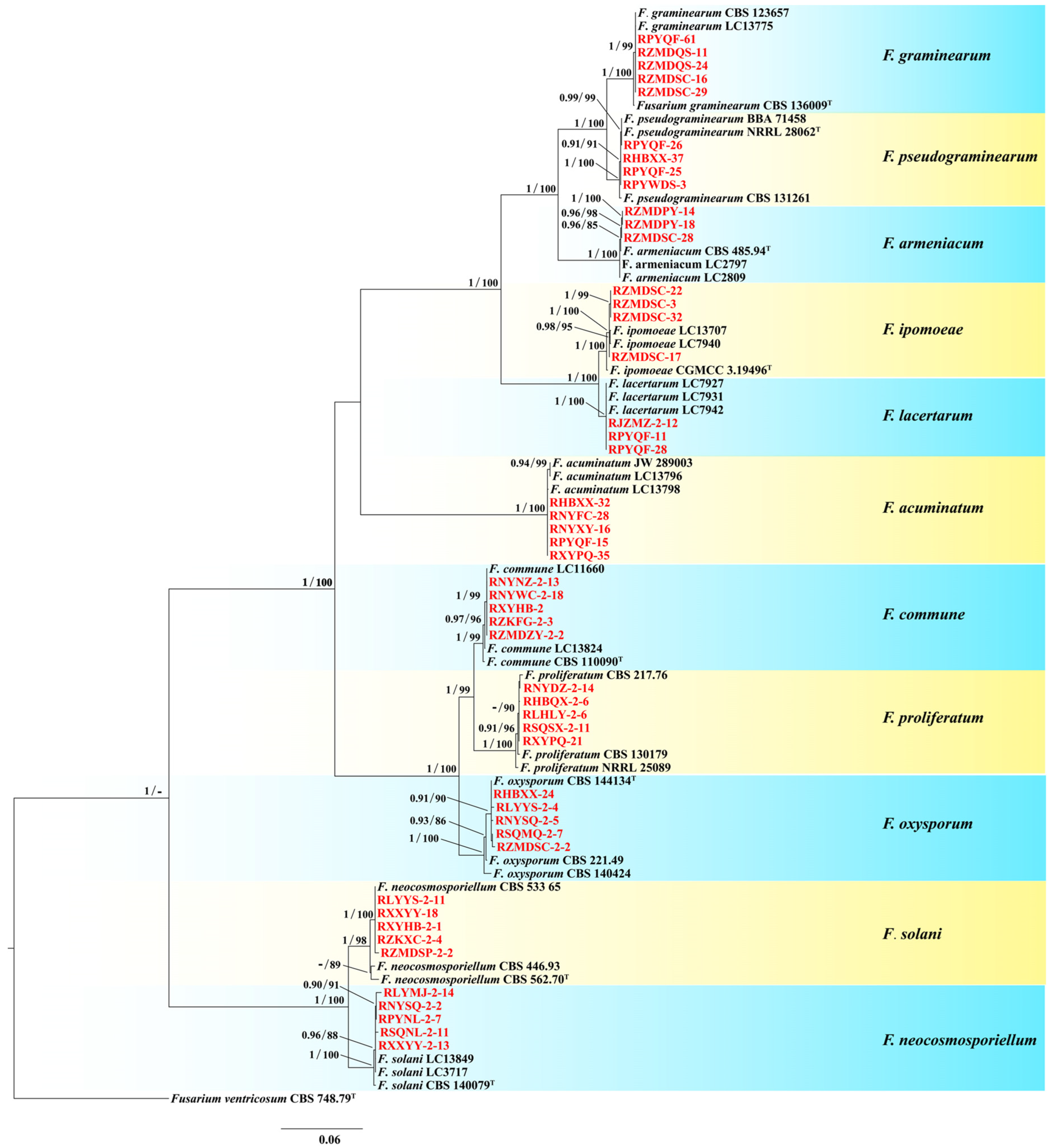
3.2. Species Identification and Phylogenetic Analysis

Based on the colony morphology, these 1131 Fusarium isolates were preliminarily classified into 11 Fusarium species (Figure 3). For further molecular verification, the phylogenetic tree based on the combined sequences of ITS–TEF-1αRPB2TUB2CAM gene partition was constructed. The concatenated ITS, TEF-1α, RPB2, TUB2, and CAM sequences dataset involved 83 isolates, including 49 Fusarium isolates from this study, 33 reference Fusarium isolates and the outgroup Fusarium ventricosum (CBS 748.79) (Tables S1 and S2). The concatenated dataset contained 2980 characters including gaps (467 for ITS, 670 for TEF-1α, 722 for RPB2, 442 for TUB2, and 679 for CAM). In BI analyses, the GTR + I + G model of evolution was selected for ITS and TEF-1a, the GTR + G model for RPB2, the HKY + G model for TUB2, and the SYM + I + G model for CAM. The phylogenetic trees from BI and ML analyses showed congruent topological structure. The phylogenetic trees revealed that these Fusarium isolates also clustered together with 11 species, which were consistent with the morphological result (Figure 4). Thus, based on morphological and phylogenetic trees, these isolates were identified as F. armeniacum, F. pseudograminearum, F. graminearum, F. acuminatum, F. commune, F. ipomoeae, F. lacertarum, F. neocosmosporiellum, F. solani, F. oxysporum, and F. proliferatum (Figure 3 and Figure 4).

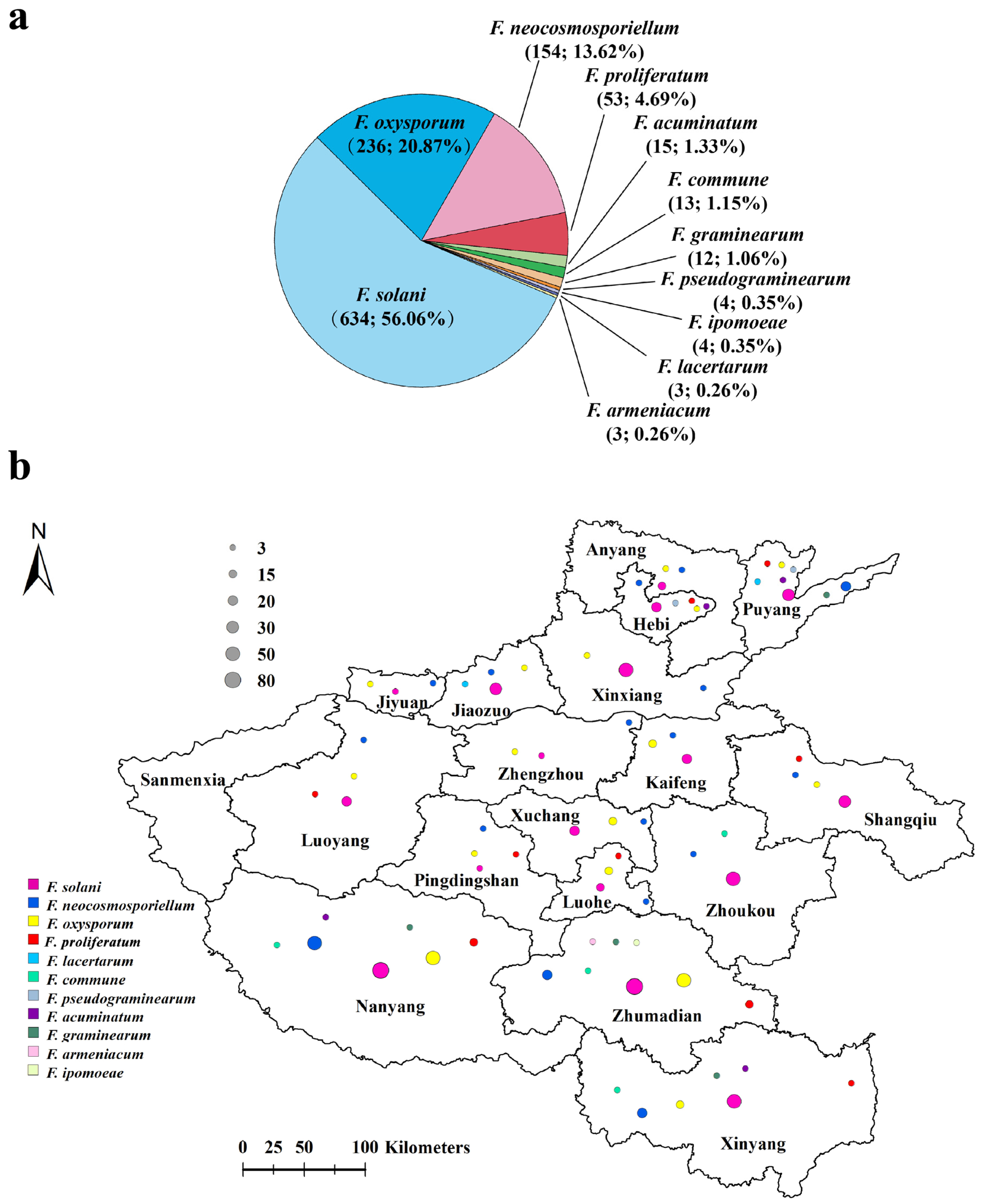
3.3. Prevalence of Fungal Species

Prevalence analyses revealed that F. solani (634 isolates, 56.06% of the total isolates, obtained from all 17 sampled areas), F. oxysporum (236 isolates, 20.87%, isolated from 16 sampled areas except for Zhoukou), and F. neocosmosporiellum (154 isolates, 13.62%, isolated from all 17 sampled areas) were the most frequent species associated with PRR in Henan Province, followed by F. proliferatum (53 isolates, 4.69%, isolated from Xinyang, Nanyang, Shangqiu, Pingdingshan, Luoyang, Luohe, Puyang, Zhumadian and Hebi), F. acuminatum (15 isolates, 1.33%, isolated from Xinyang, Nanyang, Puyang, and Hebi), F. commune (13 isolates, 1.15%, isolated from Xinyang, Nanyang, Zhoukou, and Zhumadian), and F. graminearum (12 isolates, 1.06%, isolated from Xinyang, Nanyang, Puyang, and Zhumadian) (Figure 5a,b). The remaining four Fusarium species, including F. pseudograminearum (four isolates, 0.35%), F. ipomoeae (four isolates, 0.35%), F. lacertarum (three isolates, 0.26%) and F. armeniacum (three isolates, 0.26%), were detected in a less than 1% prevalence and only found in one or two areas (Figure 5a,b).

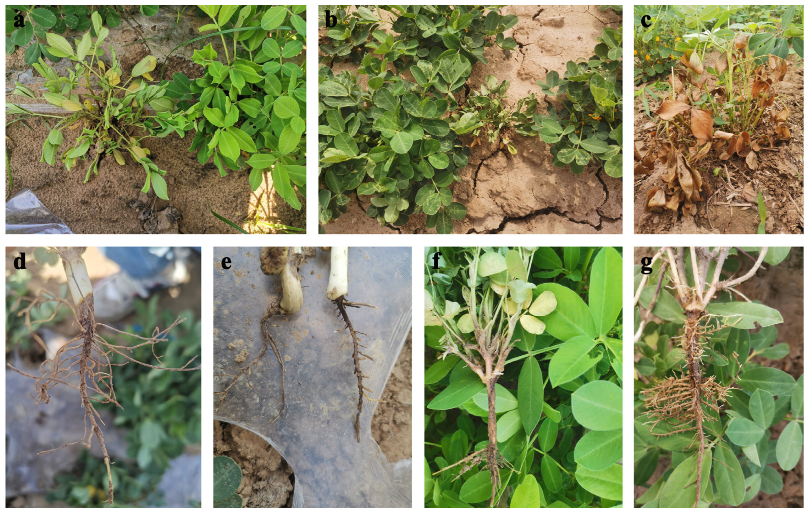
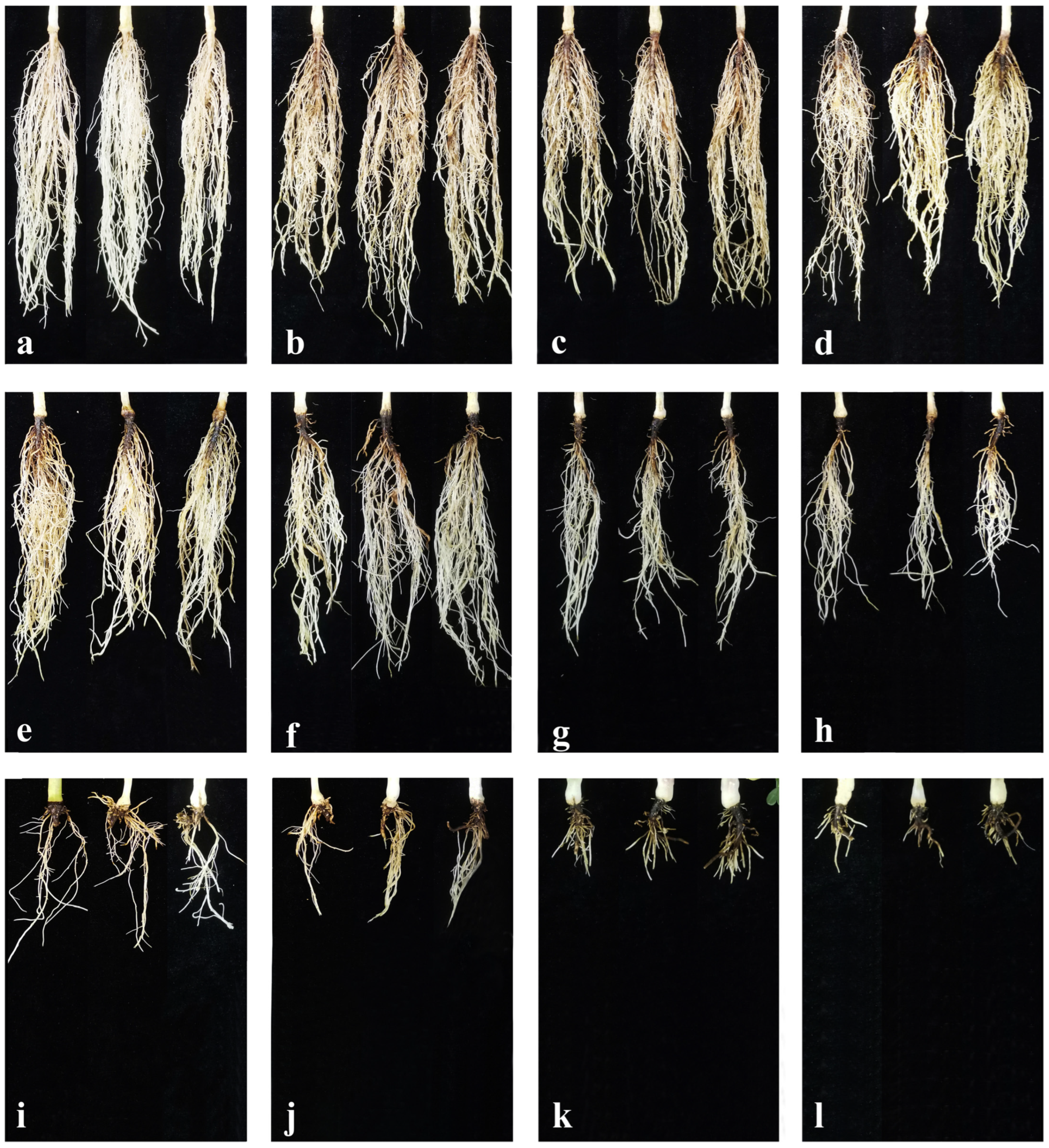
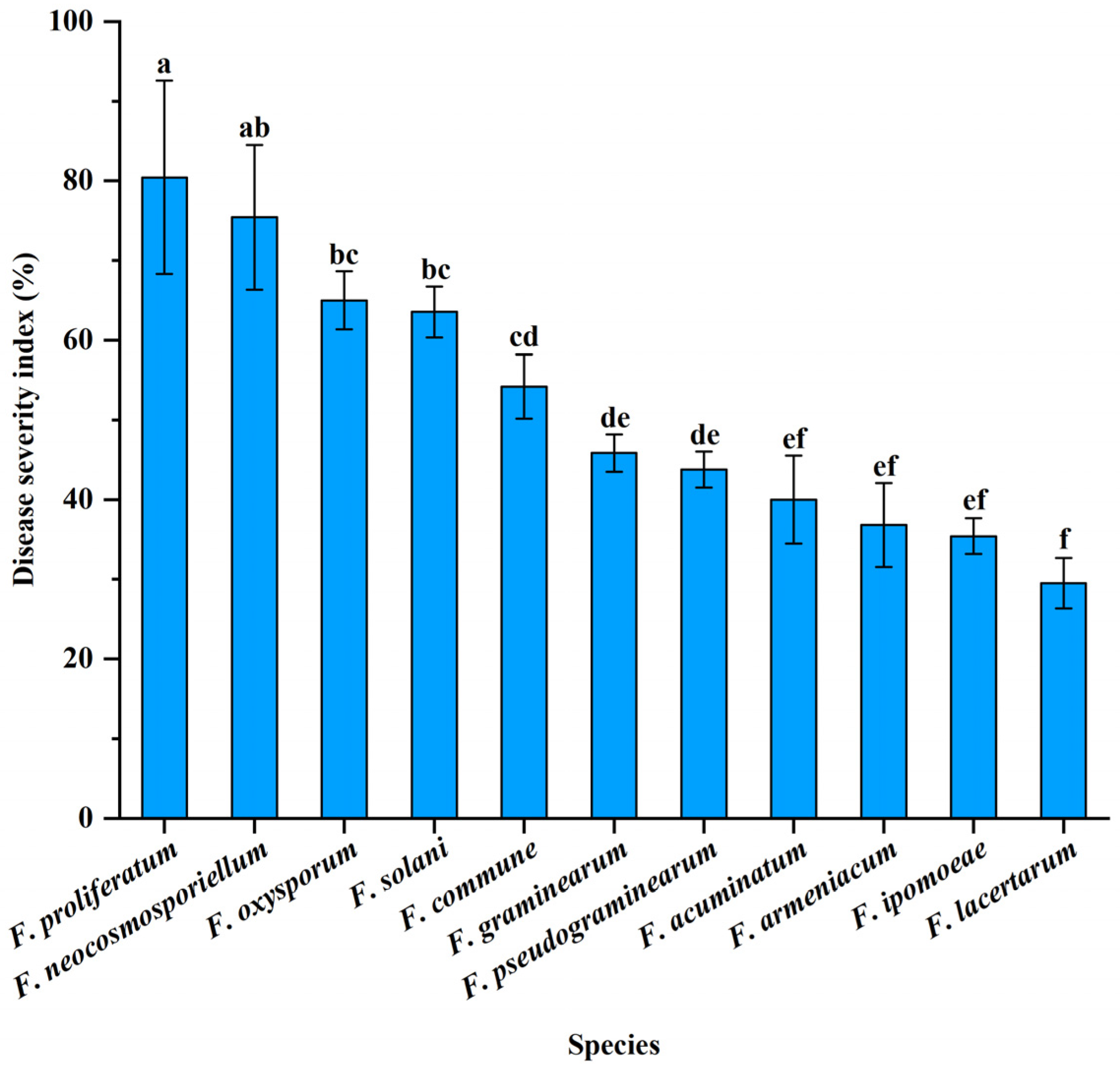
3.4. Pathogenicity Tests

The fungus of 11 Fusarium species were used to conduct the pathogenicity tests. After 21 days, peanuts inoculated with each of the 11 Fusarium species exhibited typical symptoms of PRR, similar to the symptoms initially observed on naturally infected peanut roots, while no symptoms appeared on the negative controls (Figure 6a–l). All Fusarium isolates of aforementioned test were successfully re-isolated from the infected peanut roots to satisfy Koch’s postulates. Although these Fusarium species can infect peanut, they exhibited the different symptoms on infected roots of peanut (Figure 6b–l). The symptoms of F. lacertarum and F. ipomoeae showed as light brown to dark brown discoloration on the infected root tissues (Figure 6b,c); F. armeniacum and F. acuminatum were able to induce brown discoloration with conspicuous black necrotic spots (Figure 6d,e). F. pseudograminearum, F. graminearum and F. commune could cause brown/black lesions with secondary root reduction (Figure 6f–h). F. solani, F. oxysporum, F. neocosmosporiellum and F. proliferatum produced brown/black lesions with main and lateral root reduction (Figure 6i–l).
In addition, based on the calculated DI, these pathogenic fungal species showed different levels of virulence (Figure 7). F. proliferatum, F. neocosmosporiellum, F. oxysporum, and F. solani were rated as the highly virulent pathogens, but the DI of F. proliferatum was significantly greater than those of F. oxysporum and F. solani (p < 0.05). F. commune, F. graminearum, F. pseudograminearum, F. acuminatum, F. armeniacum and F. ipomoeae were considered to be moderately virulent, whereas F. commune exhibited a significantly higher DI compared with F. acuminatum, F. armeniacum and F. ipomoeae (p < 0.05). The remaining species F. lacertarum was determined to have low virulence.

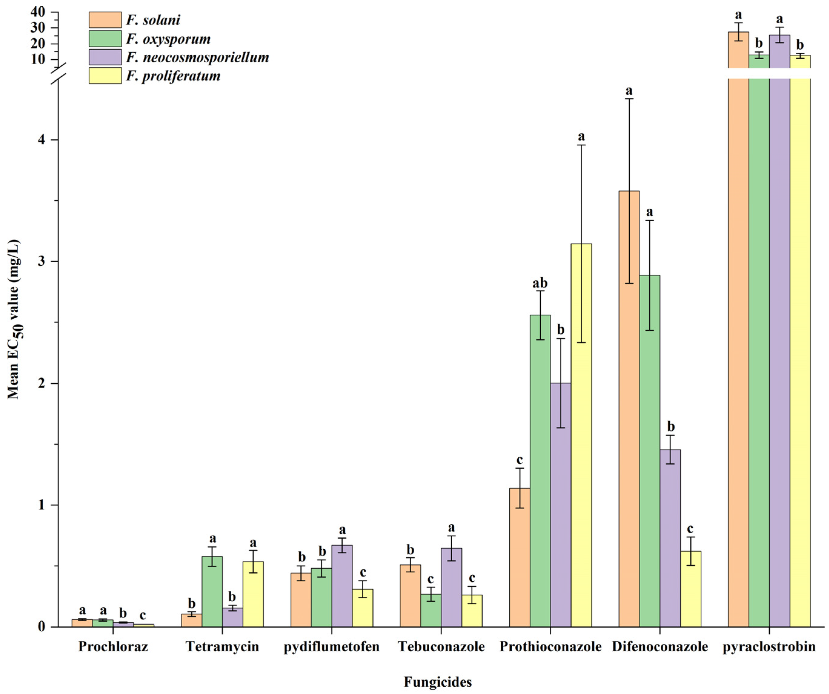
3.5. Fungicide Sensitivity Assays

To identify effective fungicides for controlling PRR, seven fungicides were classified into five classes (imidazoles, antibiotics, demethylation inhibitors [DMIs], quinone outside inhibitors [QoIs], and succinate dehydrogenase inhibitors [SDHIs]) and evaluated in this study. First, four Fusarium species, which exhibited high isolation frequency, wide geographical distribution and high virulence, were selected to perform the sensitivity assay. All seven fungicides had different degrees of inhibitory effect on the mycelial growth of the four Fusarium species, and the average EC50 values ranged from 0.02~27.49 mg/L (Figure 8; Figures S1–S7). Based on the EC50 values of the different fungicides, the four Fusarium species were sensitive to prochloraz (0.02 ± 0.00~0.06 ± 0.01 mg/L), pydiflumetofen (0.31 ± 0.07~0.67 ± 0.06 mg/L), tetramycin (0.11 ± 0.02~0.58 ± 0.08 mg/L), tebuconazole (0.26 ± 0.07~0.65 ± 0.10 mg/L), prothioconazole (1.14 ± 0.16~3.15 ± 0.81 mg/L) and difenoconazole (0.62 ± 0.12~3.58 ± 0.76 mg/L) (Figure 7). Based on the different Fusarium species, the sensitivities of these seven fungicides significantly varied by species (p < 0.05) (Figure 8). F. proliferatum exhibited a higher sensitivity to prochloraz and difenoconazole compared to F. solani, F. oxysporum and F. neocosmosporiellum (p < 0.05), and F. proliferatum had less sensitivity to tetramycin and prothioconazole compared with F. solani and F. neocosmosporiellum except for F. oxysporum (p < 0.05). The EC50 values of F. neocosmosporiellum to tebuconazole and pydiflumetofen was separately higher than those of the other three species, respectively (p < 0.05), and the EC50 values of F. oxysporum and F. proliferatum to pyraclostrobin were lower than those of the other two species (p < 0.05).

4. Discussion

Previous studies have shown that the pathogens of PRR are mainly associated with Fusarium species [8,10,19,22]. In 2021 and 2023, a total of 1131 Fusarium isolates were collected from 17 cities in Henan, an area known for its high incidence of PRR. Based on colony morphology and phylogenetic analyses, the identified Fusarium isolates were classified into 11 distinct species, including F. armeniacum, F. pseudograminearum, F. graminearum, F. acuminatum, F. commune, F. ipomoeae, F. lacertarum, F. neocosmosporiellum, F. solani, F. oxysporum, and F. proliferatum (Figure 3 and Figure 4), and Koch’s postulates confirm that the isolates are pathogenic. Interestingly, the frequency and composition of these Fusarium species differed from previous reports [10,20,22], with F. solani being the most prevalent species, accounting for 54.23% of PRR pathogens in the region, followed by F. oxysporum. Conversely, Debele et al. [10] found that F. oxysporum is the most frequent species associated with PRR in eastern Ethiopia, followed by F. solani. The reasons for the difference may be due to the climatic conditions, the geographical locations, tillage patterns and peanut variety. Notably, F. neocosmosporiellum was identified across major peanut-growing areas in Henan, with a 13.17% isolation frequency, aligning with previous results of its association with diseased peanut roots [20,49]. This study is also the first record of F. neocosmosporiellum as a PRR pathogen in Henan. F. proliferatum was another common pathogen among these Fusarium species, with 53 isolates collected from nine cities, further confirming its presence in the root tissues of peanuts in Henan [13,22]. The remaining seven species, including F. pseudograminearum, F. graminearum, F. commune, F. ipomoeae, F. lacertarum, F. armeniacum and F. acuminatum, were detected with a low occurrence in Henan. Notably, F. acuminatum had been reported as a PRR pathogen in Shandong, China [14], while the other six Fusarium species were first recorded as PRR pathogens globally. Moreover, F. equiseti and F. incarnatum had also been reported as PRR pathogens in Henan [22]. However, they were not detected during this survey. This may be related to the fact that the investigations of Pan et al. [22] were conducted in the pudding period to the full fruit maturity period, while our survey was carried out during the seedling stage to the flowering stage.
This study showed that different Fusarium species displayed different aggressiveness on the peanut roots. Among all tested Fusarium species, F. proliferatum and F. neocosmosporiellum were determined to be highly virulent pathogens, which complemented the previous studies by other researchers [4,5,13,22], who only described that they were pathogenic and responsible for PRR by the Koch’s postulates. Additionally, F. oxysporum and F. solani were also identified as highly pathogenic to peanut roots. The results corroborate the previous studies [10,19], which found that F. oxysporum and F. solani could lead to severe root rot of peanut. Furthermore, although F. commune, F. graminearum, F. pseudograminearum, F. acuminatum, F. armeniacum and F. ipomoeae had the lowest frequency of occurrences, these species exhibited moderate virulence on peanut roots. This indicated that these species may be an emerging threat to peanut production in Henan and need be closely monitored in the future. The remaining Fusarium species, F. lacertarum, exhibited low virulence on the peanut roots. However, it was infrequently isolated in the present study and was considered to be of secondary importance as a pathogen of PRR.
No crop completely immune to the various isolates of Fusarium spp. has been reported so far. At present, chemical control is still considered as the most effective and important management strategy for controlling the root rot disease. To date, several fungicides have been registered against PRR in China, including prochloraz, carbendazim, azoxystrobin, pyraclostrobin, difenoconazole, tetramycin, thifluzamide, and so on (http://www.icama.org.cn, accessed on 10 February 2025). However, carbendazim-resistant F. graminearum populations have increased in the field in recent years [50]. Succinate dehydrogenase inhibitors (SDHIs) have been applied to manage various plant fungal diseases, but most show poor activity against Fusarium spp. [51]. Therefore, to identify suitable fungicides for controlling the most frequent pathogens of PRR in the current research, including F. oxysporum, F. neocosmosporiellum, F. proliferatum and F. solani, seven fungicides with different modes of action were evaluated in this study. Our results showed that all species were highly sensitive to prochloraz, tetramycin, and difenoconazole. This result indicated that prochloraz, tetramycin, and difenoconazole can continue to be applied to the management of PRR caused by the four Fusarium species. Considering that there are few registered fungicides for controlling PRR, we also determined the sensitivity of the four Fusarium species to three unregistered fungicides (tebuconazole, prothioconazole, and pydiflumetofen). The results showed that these three fungicides have great application prospects in the control of F. oxysporum, F. neocosmosporiellum, F. proliferatum and F. solani. However, the effects of different fungicides on Fusarium spp. under field conditions remain unknown and necessitate further evaluation through pot or field experiments.

5. Conclusions

In conclusion, we firstly demonstrated that F. solani is the most prevalent species associated with PRR in Henan province, China. In addition, it is the first report that F. armeniacum, F. pseudograminearum, F. graminearum, F. commune, F. ipomoeae, and F. lacertarum occur as pathogens in peanut roots globally. Moreover, it is also the first documentation of F. neocosmosporiellum and F. acuminatum as a pathogen of PRR in Henan. Fungicide sensitivity assays showed that F. oxysporum, F. neocosmosporiellum, F. proliferatum and F. solani exhibited higher sensitivity to prochloraz, tetramycin, tebuconazole, prothioconazole, difenoconazole, and pydiflumetofen compared to pyraclostrobin. Overall, the results of this study provide crucial information for the effective management of PRR in Henan Province. Further work is needed to monitor the development of resistance to these fungicides for successful control of PRR in Henan Province.

Supplementary Materials

The following supporting information can be downloaded at: https://www.mdpi.com/article/10.3390/jof11060433/s1, Table S1. Isolates from peanut root rot included in this study; Table S2. Origin, culture and sequence GenBank accession numbers of strains used for phylogenetic analyses; Table S3. Concentrations of the seven fungicides (prochloraz, tetramycin, tebuconazole, prothioconazole, difenoconazole, pyraclostrobin and pydiflumetofen) used in the current study; Figure S1. Effects of prochloraz on the mycelial growth of the four Fusarium species (F. solani, F. oxysporum, F. neocosmosporiellum, and F. proliferatum); Figure S2. Effects of tebuconazole on the mycelial growth of the four Fusarium species (F. solani, F. oxysporum, F. neocosmosporiellum, and F. proliferatum); Figure S3. Effects of difenoconazole on the mycelial growth of the four Fusarium species (F. solani, F. oxysporum, F. neocosmosporiellum, and F. proliferatum); Figure S4. Effects of prothioconazole on the mycelial growth of the four Fusarium species (F. solani, F. oxysporum, F. neocosmosporiellum, and F. proliferatum); Figure S5. Effects of tetramycin on the mycelial growth of the four Fusarium species (F. solani, F. oxysporum, F. neocosmosporiellum, and F. proliferatum); Figure S6. Effects of pyraclostrobin on the mycelial growth of the four Fusarium species (F. solani, F. oxysporum, F. neocosmosporiellum, and F. proliferatum); Figure S7. Effects of pydiflumetofen on the mycelial growth of the four Fusarium species (F. solani, F. oxysporum, F. neocosmosporiellum, and F. proliferatum).

Author Contributions

Conceptualization, M.L., X.M. and L.Z.; methodology, M.L. and Y.D.; software, M.L. and L.C.; validation, M.L., L.H., X.M. and L.Z.; formal analysis, L.C.; investigation, M.L. and L.C.; resources, M.L. and Q.W.; data curation, M.L. and Q.W.; writing—original draft preparation, M.L.; writing—review and editing, M.L. and X.M.; visualization, M.L. and X.M.; supervision, L.Z.; project administration, L.Z.; funding acquisition, L.Z. All authors have read and agreed to the published version of the manuscript.

Funding

This study was financially supported by the National Key Research and Development Program (2024YFD1400084), the Henan Provincial Science and Technology Major Project (221100110100), the Construction Project of the Modern Agricultural Industrial Technology System of Henan Province (HARS-22-05-G3) and the Science and technology Innovation Fund of Henan Agricultural University (2023CXZX013).

Institutional Review Board Statement

Not applicable.

Informed Consent Statement

Not applicable.

Data Availability Statement

The sequences from the present study were submitted to the NCBI database (https://www.ncbi.nlm.nih.gov/, accessed on 25 May 2025). The accession numbers are listed in Table S1.

Conflicts of Interest

The authors declare no conflicts of interest.

Abbreviations

The following abbreviations are used in this manuscript:
PRRPeanut Root Rot
PDAPotato Dextrose Agar
TEF1-αThe translation elongation factor 1-α
MLMaximum Likelihood
BIBayesian Inference
MCMCMarkov Chain Monte Carlo
PPPosterior Probability
BSBootstrap Values
FIIsolation Frequency
DIDisease Severity Index
DMSODimethyl Sulfoxide
SHAMSalicylhydroxamic Acid
ANOVAAnalysis of Variance
SD Standard Deviation

References

  1. Akram, N.A.; Shafiq, F.; Ashraf, M. Peanut (Arachis hypogaea L.): A prospective legume crop to offer multiple health benefits under changing climate. Compr. Rev. Food Sci. Food Saf. 2018, 17, 1325–1338. [Google Scholar] [CrossRef] [PubMed]
  2. FAOSTAT. Food and Agriculture Organization Statistical Database. Available online: http://www.fao.org/faostat/en/#data/QC (accessed on 10 February 2025).
  3. Min, H.; Li, H.H.; Zhou, L.; Mao, Q.; Hao, R. Recent Occurrence of Peanut Diseases in Henan Province and Integrated Application of Disease Management Strategies. China Plant Prot. 2024, 44, 74–77. [Google Scholar]
  4. Fuhlbohm, M.F.; Tatnell, J.R.; Ryley, M.J. Neocosmospora vasinfecta is pathogenic on peanut in Queensland. Australas. Plant Dis. Notes 2007, 2, 3–4. [Google Scholar] [CrossRef]
  5. Dau, V.T.; Pham, L.T.; Luong, T.M.; Huynh, L.M.T.; Tran, N.T.; Ho, T.D.; Hoang, H.M.T.; Phan, H.T.; Burgess, L.W. First report of Neocosmospora vasinfecta associated with the root rot complex of peanuts in Vietnam. Australas. Plant Dis. Notes 2010, 5, 79–81. [Google Scholar] [CrossRef]
  6. Zaman, N.; Ahmed, S. Survey of root rot of groundnut in rainfed areas of Punjab, Pakistan. Afr. J. Biotechnol. 2012, 11, 4791–4794. [Google Scholar] [CrossRef]
  7. Casasnovas, F.; Fantini, E.N.; Palazzini, J.M.; Giaj-Merlera, G.; Chulze, S.N.; Reynoso, M.M.; Torres, A.M. Development of amplified fragment length polymorphism (AFLP)-derived specific primer for the detection of Fusarium solani aetiological agent of peanut brown root rot. J. Appl. Microbiol. 2013, 114, 1782–1792. [Google Scholar] [CrossRef]
  8. Abd-Elmagid, W.M.; Aly, M.M.E.S.; El-Sharkawy, R.M. Control of peanut root and pod rots diseases using certain bioagents. J. Phytopathol. Pest. Manag. 2020, 7, 79–90. [Google Scholar]
  9. Sridhar, P.; Venkateshbabu, G.; Hemalakshmi, D.; Kirthika, V.M.; Palani, P. New disease and first report of marasmioid fungus, Marasmius palmivorus (Sharples), causing white root rot in Arachis hypogaea L. Lett. Appl. Microbiol. 2022, 75, 368–377. [Google Scholar] [CrossRef]
  10. Debele, S.; Fininsa, C.; Dejene, M.; Tana, T. Distribution of groundnut (Arachis hypogaea L.) root rot complex and associated pathogens in eastern Ethiopia. Afr. J. Plant Sci. 2023, 17, 18–29. [Google Scholar] [CrossRef]
  11. Xu, M.L.; Yang, J.G.; Wang, F.L.; Wu, J.X.; Chi, Y.C. First report of Rhizopus arrhizus (syn. R. oryzae) causing root rot of peanut in China. Plant Dis. 2015, 99, 1448. [Google Scholar] [CrossRef]
  12. Xu, M.L.; Yang, J.G.; Wu, J.X.; Chi, Y.C.; Xie, L.H. First Report of Aspergillus niger Causing Root Rot of Peanut in China. Plant Dis. 2015, 99, 284. [Google Scholar] [CrossRef] [PubMed]
  13. Sun, W.; Lei, T.; Yuan, H.; Chen, S. Occurrence of root rot caused by Fusarium fujikuroi and Fusarium proliferatum on peanut in China. Plant Dis. 2023, 107, 940. [Google Scholar] [CrossRef] [PubMed]
  14. Li, Y.; Yu, J.; Guo, Z.; Song, X.; Xu, M.; He, K.; Zhang, X.; Chi, Y. First report of peanut root rot caused by Fusarium acuminatum in Shandong Province, China. Plant Dis. 2023, 107, 2882. [Google Scholar] [CrossRef]
  15. Erazo, J.G.; Palacios, S.A.; Pastor, N.; Giordano, F.D.; Rovera, M.; Reynoso, M.M.; Venisse, J.S.; Torres, A.M. Biocontrol mechanisms of Trichoderma harzianum ITEM 3636 against peanut brown root rot caused by Fusarium solani RC 386. Biol. Control 2021, 164, 104774. [Google Scholar] [CrossRef]
  16. Rojo, F.G.; Reynoso, M.M.; Ferez, M.; Chulze, S.N.; Torres, A.M. Biological control by Trichoderma species of Fusarium solani causing peanut brown root rot under field conditions. Crop Prot. 2007, 26, 549–555. [Google Scholar] [CrossRef]
  17. Shanmugam, V.; Senthil, N.; Raguchander, T.; Ramanathan, A.; Samiyappan, R. Interaction of Pseudomonas fluorescens with Rhizobium for their effect on the management of peanut root rot. Phytoparasitica 2002, 30, 169–176. [Google Scholar] [CrossRef]
  18. Raja Mohan, K.; Balabaskar, P. Survey on the incidence of groundnut root rot disease in cuddalore district of Tamil Nadu and assessing the cultural characters and pathogenicity of Macrophomina phaseolina (Tassi.) Goid. Asian J. Sci. Technol. 2012, 3, 90–94. [Google Scholar]
  19. Ahmed, M.S.; Sallam, N.M.; Mohamed, A.A.; Hassan, M.H. Effect of mycorrhiza and biofertilisers on reducing the incidence of Fusarium root and pod rot diseases of peanut. Arch. Phytopathol. Plant Prot. 2013, 46, 868–881. [Google Scholar] [CrossRef]
  20. Sakhuja, P.K.; Sethi, C.L. Frequency of occurrence of various plants parasitic nematodes and root-rot fungi on groundnut in Punjab. Indian J. Nematol. 1985, 15, 191–194. [Google Scholar]
  21. Li, X.G.; De Boer, W.D.; Zhang, Y.N.; Ding, C.F.; Zhang, T.L.; Wang, X.X. Suppression of soil-borne Fusarium pathogens of peanut by intercropping with the medicinal herb Atractylodes lancea. Soil Biol. Biochem. 2018, 116, 120–130. [Google Scholar] [CrossRef]
  22. Pan, X.; Yan, S.W.; Hu, X.Y.; Du, P.Q.; Zhang, X.T.; Zhou, L.; Gao, F. Isolation and Identification of Fusarium from Roots, Stems and Pods of Peanut in Henan Province. J. Peanut Sci. 2023, 52, 25–32. [Google Scholar] [CrossRef]
  23. Guan, L.; Guo, B.B.; Wang, X.K.; Zhang, D.X.; Wang, K.; Liu, F. Control Efficacies of Two Preparations of Difenoconazole and Fluazinam by Seed-Coating Against Peanut Soil-Borne Fungal Diseases. Sci. Agric. Sin. 2015, 48, 2176–2186. [Google Scholar] [CrossRef]
  24. Guan, L.; Guo, B.B.; Wang, X.K.; Li, B.X.; Zhang, D.X.; Liu, F. Seed-coating treatment of four fungicides against peanut crown rot and root rot diseases. Acta Phytophylacica Sin. 2016, 43, 842–849. [Google Scholar] [CrossRef]
  25. Oddino, C.M.; Marinelli, A.D.; Zuza, M.; March, G.J. Influence of crop rotation and tillage on incidence of brown root rot of peanut caused by Fusarium solani in Argentina. Can. J. Plant Pathol. 2008, 30, 575–580. [Google Scholar] [CrossRef]
  26. Tunali, B.; Nicol, J.M.; Hodson, D.; Uçkun, Z.; Büyük, O.; Erdurmuş, D.; Hekimhan, H.; Aktaş, H.; Akbudak, M.A.; Bağci, S.A. Root and Crown Rot Fungi Associated with Spring, Facultative, and Winter Wheat in Turkey. Plant Dis. 2008, 92, 1299–1306. [Google Scholar] [CrossRef] [PubMed]
  27. Maryani, N.; Lombard, L.; Poerba, Y.S.; Subandiyah, S.; Crous, P.W.; Kema, G.H.J. Phylogeny and genetic diversity of the banana Fusarium wilt pathogen Fusarium oxysporum f. sp. cubense in the Indonesian centre of origin. Stud. Mycol. 2019, 92, 155–194. [Google Scholar] [CrossRef]
  28. Leslie, J.F.; Summerell, B.A. (Eds.) The Fusarium Laboratory Manual; Blackwell: Ames, IO, USA, 2006. [Google Scholar]
  29. Chang, X.; Dai, H.; Wang, D.; Zhou, H.; He, W.; Fu, Y.; Ibrahim, F.; Zhou, Y.; Gong, G.; Shang, J.; et al. Identification of Fusarium species associated with soybean root rot in Sichuan Province, China. Eur. J. Plant Pathol. 2018, 151, 563–577. [Google Scholar] [CrossRef]
  30. Özer, G.; Paulitz, T.C.; Imren, M.; Alkan, M.; Muminjanov, H.; Dababat, A.A. Identity and Pathogenicity of Fungi Associated with Crown and Root Rot of Dryland Winter Wheat in Azerbaijan. Plant Dis. 2020, 104, 2149–2157. [Google Scholar] [CrossRef]
  31. White, T.J.; Bruns, T.; Lee, S.; Taylor, J. Amplification and direct sequencing of fungal ribosomal RNA genes for phylogenetics. In PCR Protocols: A Guide to Methods and Applications; Innis, M.A., Gelfand, D.H., Sninsky, J.J., White, T.J., Eds.; Academic Press: SanDiego, CA, USA, 1990; pp. 315–322. [Google Scholar]
  32. O’Donnell, K.; Kistler, H.C.; Cigelnik, E.; Ploetz, R.C. Multiple evolutionary origins of the fungus causing Panama disease of banana: Concordant evidence from nuclear and mitochondrial gene genealogies. Proc. Natl. Acad. Sci. USA 1998, 95, 2044–2049. [Google Scholar] [CrossRef]
  33. Reeb, V.; Lutzoni, F.; Roux, C. Contribution of RPB2 to multilocus phylogenetic studies of the euascomycetes (Pezizomycotina, Fungi) with special emphasis on the lichen-forming Acarosporaceae and evolution of polyspory. Mol. Phylogenet. Evol. 2004, 32, 1036–1060. [Google Scholar] [CrossRef]
  34. Liu, Y.J.; Whelen, S.; Hall, B.D. Phylogenetic relationships among ascomycetes: Evidence from an RNA polymerse II subunit. Mol. Biol. Evol. 1999, 16, 1799–1808. [Google Scholar] [CrossRef]
  35. O’Donnell, K.; Cigelnik, E. Two divergent intragenomic rDNA ITS2 types within a monophyletic lineage of the fungus Fusarium are nonorthologous. Mol. Phylogenet. Evol. 1997, 7, 103–116. [Google Scholar] [CrossRef]
  36. O’Donnell, K.; Nirenberg, H.I.; Aoki, T.; Cigelnik, E. A multigene phylogeny of the Gibberella fujikuroi species complex: Detection of additional phylogenetically distinct species. Mycoscience 2000, 41, 61–78. [Google Scholar] [CrossRef]
  37. Hall, T.; Biosciences, I.; Carlsbad, C. BioEdit: An important software for molecular biology. GERF Bull. Biosci. 2011, 2, 60–61. [Google Scholar]
  38. Rivedal, H.M.; Tabima, J.F.; Stone, A.G.; Johnson, K.B. Identity and Pathogenicity of Fungi Associated with Root, Crown, and Vascular Symptoms Related to Winter Squash Yield Decline. Plant Dis. 2022, 106, 1660–1668. [Google Scholar] [CrossRef]
  39. Katoh, K.; Rozewicki, J.; Yamada, K.D. MAFFT online service: Multiple sequence alignment, interactive sequence choice and visualization. Brief. Bioinform. 2019, 20, 1160–1166. [Google Scholar] [CrossRef]
  40. Nguyen, L.T.; Schmidt, H.A.; von Haeseler, A.; Minh, B.Q. IQ-TREE: A fast and effective stochastic algorithm for estimating maximum-likelihood phylogenies. Mol. Biol. Evol. 2015, 32, 268–274. [Google Scholar] [CrossRef]
  41. Ronquist, F.; Teslenko, M.; van der Mark, P.; Ayres, D.L.; Darling, A.; Höhna, S.; Larget, B.; Liu, L.; Suchard, M.A.; Huelsenbeck, J.P. MrBayes 3.2: Efficient Bayesian phylogenetic inference and model choice across a large model space. Syst. Biol. 2012, 61, 539–542. [Google Scholar] [CrossRef]
  42. Nylander, J.A.A. MrModeltest V2. Program Distributed by the Author Evolutionary Biology Centre; Uppsala University: Uppsala, Sweden, 2004. [Google Scholar]
  43. Huelsenbeck, J.; Rannala, B. Frequentist properties of Bayesian posterior probabilities of phylogenetic trees under simple and complex substitution models. Syst. Biol. 2004, 53, 904–913. [Google Scholar] [CrossRef]
  44. Bozoğlu, T.; Derviş, S.; Imren, M.; Amer, M.; Özdemir, F.; Paulitz, T.C.; Morgounov, A.; Dababat, A.A.; Özer, G. Fungal Pathogens Associated with Crown and Root Rot of Wheat in Central, Eastern, and Southeastern Kazakhstan. J. Fungi 2022, 8, 417. [Google Scholar] [CrossRef]
  45. Bilgi, V.N.; Bradley, C.A.; Khot, S.D.; Grafton, K.F.; Rasmussen, J.B. Response of Dry Bean Genotypes to Fusarium Root Rot, Caused by Fusarium solani f. sp. phaseoli, Under Field and Controlled Conditions. Plant Dis. 2008, 92, 1197–1200. [Google Scholar] [CrossRef] [PubMed]
  46. Chang, X.; Yan, L.; Naeem, M.; Khaskheli, M.I.; Zhang, H.; Gong, G.; Zhang, M.; Song, C.; Yang, W.; Liu, T.; et al. Maize/Soybean Relay Strip Intercropping Reduces the Occurrence of Fusarium Root Rot and Changes the Diversity of the Pathogenic Fusarium Species. Pathogens 2020, 9, 211. [Google Scholar] [CrossRef] [PubMed]
  47. Bertoldo, C.; Gilardi, G.; Spadaro, D.; Gullino, M.L.; Garibaldi, A. Genetic diversity and virulence of italian strains of Fusarium oxysporum isolated from Eustoma grandiflorum. Eur. J. Plant Pathol. 2015, 141, 83–97. [Google Scholar] [CrossRef]
  48. Cruz Jimenez, D.R.; Ellis, M.L.; Munkvold, G.P.; Leandro, L.F.S. Isolate–cultivar interactions, in vitro growth, and fungicide sensitivity of Fusarium oxysporum isolates causing seedling disease on soybean. Plant Dis. 2018, 102, 1928–1937. [Google Scholar] [CrossRef]
  49. Wenham, K. Investigation into the Emerging Soil Borne Disease of Peanut–Neocosmospora Root Rot. Ph.D. Thesis, The University of Queensland, Brisbane, Australia, 2018. [Google Scholar]
  50. Hou, Y.P.; Mao, X.W.; Wang, J.X.; Zhan, S.W.; Zhou, M.G. Sensitivity of Fusarium asiaticum to a novel succinate dehydrogenase inhibitor fungicide pydiflumetofen. Crop Prot. 2017, 96, 237–244. [Google Scholar] [CrossRef]
  51. Avenot, H.F.; Michailides, T.J. Progress in understanding molecular mechanisms and evolution of resistance to succinate dehydrogenase inhibiting (SDHI) fungicides in phytopathogenic fungi. Crop Prot. 2010, 29, 643–651. [Google Scholar] [CrossRef]
Figure 1. The sample distribution map of peanut fields in the different peanut-producing regions of Henan Province in 2021 to 2023.
Jof 11 00433 g001
Figure 2. Representative symptoms of peanut root rot in the field. (ac) the above-ground parts of diseased peanut plants; (dg) the infected root tissues.
Jof 11 00433 g002
Figure 3. Typical colonies of 11 Fusarium species observed after culture on PDA medium at 25 °C for 7 d and conidia and conidiophores in carboxymethyl cellulose (CMC) medium after incubation for 3 d at 25 °C, 175 rpm. (A) F. pseudograminearum. (B) F. graminearum. (C) F. armeniacum. (D) F. ipomoeae. (E) F. lacertarum. (F) F. solani. (G) F. acuminatum. (H) F. commune. (I) F. oxysporum. (J) F. proliferatum. (K) F. neocosmosporiellum. The images from a to c were colony morphology, macro- or microconidia, and conidiophores. Scale bar = 20 μm.
Jof 11 00433 g003
Figure 4. Phylogenetic tree based on Bayesian inference (BI) using MrBayes with the combined sequences of ITS–TEF-1αRPB2TUB2CAM gene partition of Fusarium isolates. Bayesian posterior probability (PP ≥ 0.90) and IQtree bootstrap support values (BS ≥ 70%) are shown at the nodes (PP/BS). Ex-type isolates were indicated with superscript ‘‘T”. Isolates obtained from the present study are indicated in red font. Scale bar indicates the expected number of changes per site.
Jof 11 00433 g004
Figure 5. The prevalence of Fusarium species isolated from diseased peanut roots. (a) The number and overall isolation rate (%) of each Fusarium species; (b) distribution of fungal species in Henan Province, China, each color represents one fungal species, and the size of the circle represents the number of isolates.
Jof 11 00433 g005
Figure 6. Root rot symptoms on peanuts were observed after 21 days of inoculation with 11 Fusarium species through millet seeds. (a) Control without inoculation; (b) F. lacertarum; (c) F. ipomoeae; (d) F. armeniacum; (e) F. acuminatum; (f) F. pseudograminearum; (g) F. graminearum; (h) F. commune; (i) F. solani; (j) F. oxysporum; (k) F. neocosmosporiellum; (l) F. proliferatum.
Jof 11 00433 g006
Figure 7. Variation in virulence of the different Fusarium species. Means followed by the same letter are not significantly different at p = 0.05 based on analysis of variance (ANOVA) and Tukey’s HSD test. Bars and error bars represent means ± SDs.
Jof 11 00433 g007
Figure 8. The EC50 values of four Fusarium species (F. solani, F. oxysporum, F. neocosmosporiellum and F. proliferatum) from peanut root to prochloraz, tetramycin pydiflumetofen, tebuconazole, prothioconazole, difenoconazole and pyraclostrobin. Fusarium species followed by the same letter are not significantly different at p = 0.05 based on analysis of variance (ANOVA) with Tukey’s HSD test. Bars and error bars represent means + SDs (n = 5).
Jof 11 00433 g008
Table 1. Primers used for PCR and sequencing in the present study.
LocusPrimerDirectionSequence (5′ to 3′)
ITSITS1ForwardTCCGTAGGTGAACCTGCGG
ITS4ReverseTCCTCCGCTTATTGATATGC
TEF-1aEF1ForwardATGGGTAAGGARGACAAGAC
EF2ReverseGGARGTACCAGTSATCATG
RPB25f2ForwardGGGGWGAYCAGAAGAAGGC
7crReverseCCCATRGCTTGYTTRCCCAT
TUB2T1ForwardAACATGCGTGAGATTGTAAGT
T2ReverseTAGTGACCCTTGGCCCAGTTG
CAMCL1ForwardGARTWCAAGGAGGCCTTCTC
CL2AReverseTTTTTGCATCATGAGTTGGAC
Disclaimer/Publisher’s Note: The statements, opinions and data contained in all publications are solely those of the individual author(s) and contributor(s) and not of MDPI and/or the editor(s). MDPI and/or the editor(s) disclaim responsibility for any injury to people or property resulting from any ideas, methods, instructions or products referred to in the content.

