Article

Variation in Growth, Morphology, and Fungicide Sensitivity Among Monilinia Species from South Tyrol’s Alpine Orchards

1
Institute for Plant Health, Laimburg Research Centre, Laimburg 6, 39040 Auer (Ora), Italy
2
Faculty of Agricultural, Environmental and Food Sciences, Free University of Bozen-Bolzano, Piazza Università 5, 39100 Bolzano, Italy
*
Author to whom correspondence should be addressed.
J. Fungi 2025, 11(10), 690; https://doi.org/10.3390/jof11100690
Submission received: 22 August 2025 / Revised: 16 September 2025 / Accepted: 19 September 2025 / Published: 23 September 2025
(This article belongs to the Section Fungal Pathogenesis and Disease Control)

Abstract

Brown rot, caused by Monilinia species, is a major disease affecting stone and pome fruits. The most relevant species are M. fructigena, M. laxa, M. polystroma, and M. fructicola. These four species exhibit morphological differences, but comparative data on these traits remain limited. In European integrated fruit production, pre-harvest control of brown rot mainly relies on the fungicides cyprodinil, boscalid, and tebuconazole. Given the coexistence of multiple Monilinia species, understanding differences in fungicide sensitivity is crucial for optimizing control strategies. In this study, mycelial growth, colony and conidial morphology, and sporulation capacity on potato dextrose agar (PDA), tomato sauce agar (TSA), and apple fruit were investigated. Fungicide sensitivity was assessed by measuring mycelial growth on apples following treatment with the three active ingredients. Tebuconazole was the most effective fungicide, particularly against M. laxa and M. polystroma, while cyprodinil and boscalid were less effective. These results highlight the need for species-specific fungicide strategies, Monilinia populations, and the effectiveness of disease control under various environmental conditions. All Monilinia species sporulated successfully on TSA, underscoring its suitability for sporulation studies. Conidial size varied significantly among species; nevertheless, overlapping sizes prevent reliable species or strain discrimination based on this trait alone.

Graphical Abstract

1. Introduction

Brown rot, caused by Monilinia species, is one of the most important diseases in stone fruit orchards, both pre- and postharvest. Monilinia species are a significant threat to stone and pome fruit crops, particularly in temperate climates. The most important pathogens include Monilinia fructigena Honey, Monilinia laxa Honey, Monilinia polystroma L.M. Kohn, and Monilinia fructicola Honey [1]. All Monilinia species affect the flowers and twigs of stone fruit, causing blossom blight and cankers [2,3,4]. Additionally, the symptoms of brown rot appear on the stone fruit. Initially, small, superficial, brown circular spots are observed, which expand in diameter until they cover the entire fruit [5]. The fungus overwinters primarily in mummified fruits, which either remain on the trees or fall to the ground [6,7]. In pome fruit cultivation, Monilinia is mainly recognized as an important post-harvest pathogen [4,6]. However, brownish dieback has also been observed on the leaf petioles, laminas, and on small fruits and fruit pedicels of apple [8]. Since brown rot symptoms are caused by different closely related species, morphological characteristics of individual strains are crucial for distinguishing Monilinia species and understanding the consistency between strains.
Management of brown rot relies on cultural practices, chemical treatments, biological control, and, finally, physical treatments to reduce or eliminate harvest losses. The highest efficacy is achieved with chemical control, particularly through preventive applications [9]. In Europe, chemical post-harvest treatments against Monilinia are not authorized [10]. As a result, brown rot infection after harvest ranges up to 30%, with peaks up to 50% [11,12].
Three active ingredients are commonly applied in the orchards: cyprodinil, boscalid, and tebuconazole. Cyprodinil operates through a systemic mode of action (MoA) as a single-site methionine inhibitor, targeting the enzyme cysteine β-lyase. Based on its MoA, the Fungicide Resistance Action Committee (FRAC) classifies it in Group 9 as an anilinopyrimidine (AP) [13]. Boscalid acts as a contact fungicide with a likely multi-site MoA. It inhibits the ubiquinone binding site QP, disrupting the electron flow to Complex III and coenzyme Q, causing oxidative stress, blocking respiration, and resulting in ATP depletion [13,14,15]. Boscalid belongs to FRAC Group 7, succinate dehydrogenase inhibitors (SDHI) [13]. Tebuconazole is a systemic fungicide with a single-site MoA that inhibits demethylation and other processes in sterol biosynthesis. Its effects include mycelial growth inhibition and suppression of sporulation [16,17]. Tebuconazole is classified in FRAC Group 3 as a demethylation inhibitor (DMI) [13]. Demethylation refers to the chemical process of removing a methyl group (CH3) from a molecule. To assess the damage potential of different species, it is essential to know the species-specific sensitivity to fungicides.
Recent studies on Monilinia species in stone and pome fruit have identified all four species in South Tyrol (Italy), where the present study was conducted [18,19,20]. Research on Monilinia strains from this region is particularly valuable because South Tyrol is an alpine area where stone fruit cultivation occurs mainly in cooler mountain regions. This environment may favor brown rot caused by M. laxa rather than M. fructicola, as the optimal growing temperature for M. fructicola is 26 °C, which is significantly higher than the 20 °C and 23 °C optimal for M. fructigena and M. laxa, respectively [1,21].
Despite several studies on pathogenicity, limited information exists regarding the morphological, phylogenetic, and fungicide-resistance profiles of various Monilinia species. In particular, the species M. polystroma lacks detailed information [1,22,23,24]. Therefore, additional information on this species would be valuable for distinguishing M. polystroma from other species.
Hence, the present study aims to investigate in detail the morphological and phylogenetic characteristics of M. fructigena, M. laxa, and M. polystroma, as well as a reference isolate of M. fructicola. Furthermore, the sensitivity of these species to the active substances cyprodinil, boscalid, and tebuconazole was assessed to determine if species-specific tolerance is present. Particular attention was given to M. polystroma due to the limited available information on this species.

2. Materials and Methods

2.1. Fungi Isolates and Experimental Conditions

The used strains were isolated from fruit mummies and infected fruit sampled in 2020 and 2021 in South Tyrol [18,19,20]. To obtain single Monilinia strains, single-spore isolates were prepared from symptomatic fruit. Spores were picked with a needle under sterile conditions using a stereo microscope and transferred to Petri dishes (90 mm diameter) containing potato dextrose agar (PDA; 4 g L−1 potato starch, 20 g L−1 dextrose, 15 g L−1 agar, pH 5.2; Difco™, Bordeaux, France). Details on single strains are available in Table 1. Incubation and all experiments were performed in a climatic chamber at 20 ± 1 °C, 40% RH, and under a 12:12 h L:D (light:dark) photoperiod. Light was generated by an LED Osram Fluora L 36W/77 (Fluora Leuchten Lichtmanufaktur AG, Flawil, Switzerland), providing white light.

2.2. Phylogeny

Total DNA was extracted from monoconidial culture grown on PDA using the DNeasy® Plant Mini Kit (Qiagen GmbH, Hilden, Germany) according to the manufacturer’s instructions with minor modifications: the fungal material was disrupted by adding 400 μL of AP1 lysis buffer and three 3 mm tungsten carbide beads (Qiagen) in a mixer mill (MM 400; Retsch GmbH, Haan, Germany) for 3 min at 30 beats per s. No RNase was added. The DNA was eluted two times with 50 μL nuclease-free water. The DNA concentration of the samples was quantified using a NanoDrop 1000 spectrophotometer (Thermo Fisher Scientific, Wilmington, NC, USA) and stored at −20 °C until use [19].
The internal transcribed spacer (ITS) region was amplified using the universal primers ITS5 and ITS4 [25]. For the PCR reactions, 30 µL reaction mixture was used: 1 × Phusion HF Buffer, 0.2 mM dNTPs, 0.4 µM of each primer, 0.02 U µL−1 PhusionTM High-Fidelity DNA polymerase (Thermo Fisher Scientific Inc., Waltham, MA, USA), and 3 µL DNA template. The cycling conditions consisted of an initial denaturation at 95 °C for 2 min, followed by 35 cycles of denaturation at 95 °C for 30 s, annealing at 56 °C for 30 s, and extension at 72 °C for 45 s, with a final extension at 72 °C for 5 min.
PCR products were run on a 1.5% agarose gel, stained with GelRed® Nucleic Acid Stain (Merck KGaA, Darmstadt, Germany), and viewed on a ChemiDocTM Imaging System with Image LabTM Software version 5.2.1 (Universal Hood III, Bio-Rad Laboratories Inc., Hercules, CA, USA). Successfully amplified products were sent for Sanger sequencing to LGC Genomics GmbH (Berlin, Germany). Raw sequence data were checked manually and trimmed using Geneious v.11.1.5 (Biomatters Ltd., Auckland, New Zealand). Multiple sequence alignment was performed using the ClustalW algorithm to prepare the sequences for phylogenetic analysis. Subsequent phylogenetic analysis was performed, including one with MEGA v. 12.0.11 [26]. A model test to identify the lowest Bayesian information criterion (BIC) score was performed, and the Kimura 2-parameter (K2) model was applied for maximum likelihood (ML) tree reconstruction. For bootstrap analysis, 1000 replicates above 85% were performed to estimate node support.

2.3. Growth Rate and Colony Morphology on PDA

To characterize the Monilinia strains, in vitro tests were conducted on PDA (39 g L−1). Petri dishes (90 mm diameter) were inoculated with mycelial plugs from a PDA plate, mycelial side down, and sealed with parafilm. Radial growth (mm) and colony morphology were assessed at 6, 10, 15, 20, and 27 days post-inoculation (dpi). Measurements were taken on the back of the plate at the edge of the inoculated area, and the mean value per strain was calculated from four radial measurements. Five replicates were performed (n = 5).

2.4. Mycelial Growth Rate and Morphology on Apple

Differences in the characterization of Monilinia species strains were evaluated through in planta tests on apples of the Golden Delicious cultivar from organic production after five months of storage. Apples were surface sterilized using cotton swabs soaked in 75% ethanol. A 3 mm deep hole was punched into each fruit with a 7.8 mm diameter cork borer and a mycelial plug, matching the hole’s diameter and taken from the colony periphery, was placed inside with the mycelial side facing the pulp. Sterile PDA plugs were used as controls. Apples were placed on the halves of 6 cm diameter Petri dishes and incubated in sterile, transparent plastic boxes (22 × 12 × 33 cm; Gió Style, Rome, Italy) containing 100 mL of sterile water-soaked bibulous paper. The box lids were left slightly ajar to allow air circulation. Five apples per strain were placed in each box. The relative humidity inside the boxes was nearly 100%. Radial fungal growth on the apples was monitored until the fruit was fully colonized. Measurements were taken at 7, 9, 14, 16, 21, and 23 dpi. Fungal coverage was measured at four positions from the edge of the inoculated hole to the fungal margin using a ruler, and the growth rate was calculated. The mean of these four measurements was used for statistical analysis, with each apple serving as a replicate (n = 5).

2.5. Conidia Production and Characterization

Sporulation and conidial size were assessed using the same apples described above. Conidia from inoculated apples were collected and placed on Fuchs–Rosenthal chambers for evaluation. Incubation period of the apples ranged from 9 to 21 dpi. If no sporulation occurred on apple, it was induced on tomato sauce agar (TSA; 227 g tomato sauce (Casar Srl, Serramanna, Italy), 20 g agar (Merck, Darmstadt, Germany), 750 mL deionized water) following the method of [27]. Dishes were inoculated with mycelial plugs taken from the periphery of PDA-grown cultures, with two technical replicates.
Sporulating samples were scraped using sterile inoculating loops, and conidia were suspended in tap water on Fuchs–Rosenthal chambers. Microscopic analysis was conducted at 200× and 400× total magnification (20× or 40× objective plus 10× ocular). The length and width of 50 conidia per sample were measured (n = 50).

2.6. Fungicide Screening on Apples

Three plant protection products, each authorized for use in integrated pest management (IPM) in Italy (Table 2), were tested for their efficacy. Fungicide sensitivity was evaluated under in planta conditions, following the methodology described by Chen et al. (2013) [27]. Apples were washed twice with tap water, sterilized using cotton swabs soaked in 75% ethanol, and then left to air dry. Application rates for the active substances were based on the manufacturers’ recommended field doses for controlling Monilinia species (http://www.fitosanitari.salute.gov.it, accessed on 15 March 2025). Approximately 1.5 mL of each fungicide was sprayed onto the apples using a hand mister, ensuring coverage to the point of runoff. Sterile deionized water was used as a control treatment. Each treatment included five apples (n = 5). Inoculation and evaluation procedures were conducted as described above. Growth inhibition was calculated as the coverage in the fungicide treatment relative to the mean coverage in the control. A 100% inhibitory effect indicates complete growth suppression, whereas 0% inhibition corresponds to growth equal to or greater than that of the control.

2.7. Statistical Analyses

All statistical analyses were performed using R version 4.3.2 (R Foundation for Statistical Computing). Mycelial growth on PDA and apples, as well as fungicide efficacy, was evaluated using linear mixed-effects models (LMMs) fitted via restricted maximum likelihood (REML) with the lme4 package [28]. Species or strain were included as fixed effects, while dpi was treated as a random effect. Data from different species were analyzed separately to assess the effect of individual strains. Model selection was based on Akaike Information Criterion (AIC) values, and residuals were examined to verify error distribution assumptions. Pairwise comparisons of treatments were conducted using Tukey’s test. The lmerTest and multcomp packages were used for statistical inference, and figures were generated using ggplot2 [29,30,31].

3. Results

3.1. Phylogeny

The identity of the four Monilinia species was confirmed through BLASTN analysis of the obtained ITS sequences against reference sequences in the NCBI GenBank database. The isolates clustered with the reference sequences of M. fructicola CBS 127259 (MH864497), M. fructigena CBS 101500 (MH862738), M. laxa CBS 298.31 (MH855219), and M. polystroma CBS 122306 (MH863200) [32]. No genetic differences were observed between strains originating from different geographic locations or host fruits within the same Monilinia species (Figure 1).

3.2. Growth Rate and Colony Morphology on PDA

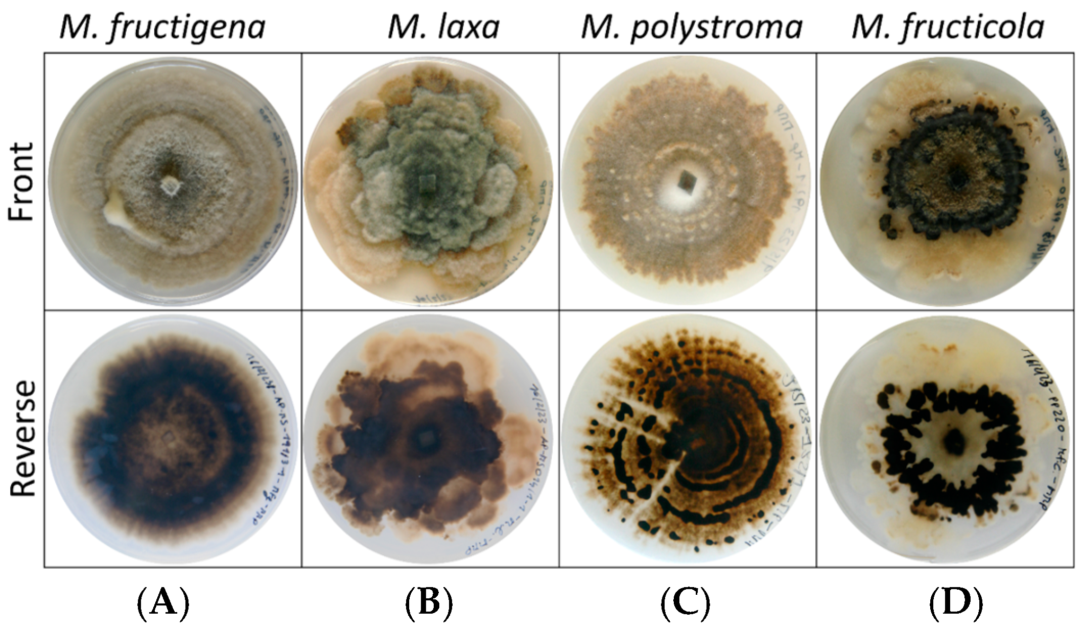
The four Monilinia species exhibited distinct morphological characteristics when cultured on PDA. Colonies of M. fructigena were characterized by entire or slightly undulate margins and a whitish surface. The reverse side of the colonies appeared dark and frequently displayed concentric rings. M. polystroma formed colonies with entire to slightly undulate margins and a grayish coloration. On the reverse side, black stromatal plates arranged in concentric rings were clearly visible, and in some cases, these structures were also observable from the front of the colony. In contrast, colonies of M. laxa showed strongly undulated margins and a grayish to brownish coloration. No concentric rings were visible on the reverse side. M. fructicola produced colonies with undulated margins and a whitish appearance. Black stromata were evident on the front side of the colony, while the reverse side displayed irregular, undulated stromatal plates that did not originate from the colony center (Figure 2).
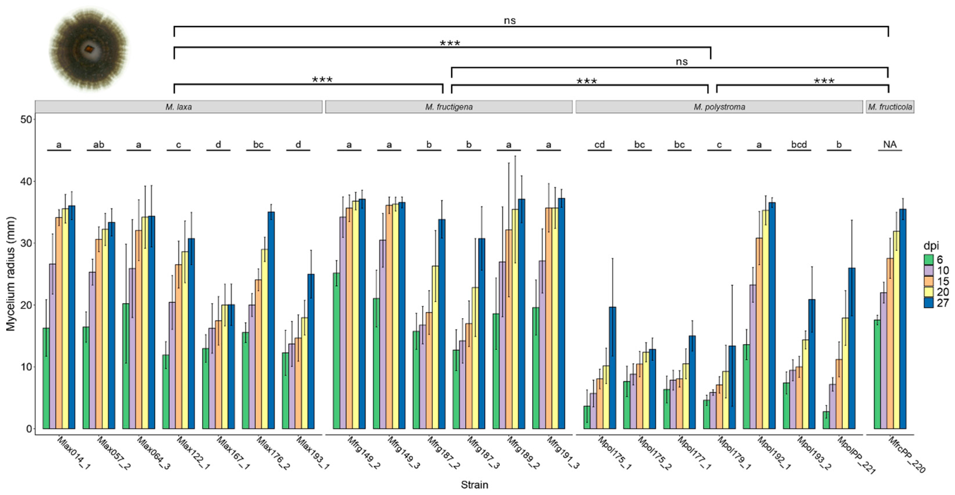
The mycelial growth rate on PDA varied significantly among the Monilinia species (F3,520 = 145.57, p < 0.001). Over the 27-day observation period, M. laxa exhibited a growth rate ranging from 0.64 to 1.45 mm per day. M. fructigena grew at a rate of 0.89 to 1.56 mm per day, while M. fructicola showed a growth rate between 1.23 and 1.38 mm per day. M. polystroma demonstrated a growth rate ranging from 0.22 to 1.39 mm per day (Figure 3).
Significant differences in growth rate were also observed between strains within each species. In M. laxa, the effect of strain was significant (F6,170 = 41.351, p < 0.001). Similarly, growth rate differences among strains of M. fructigena (F5,145 = 34.987, p < 0.001) and M. polystroma (F6,170 = 67.478, p < 0.001) were statistically significant (Figure 3).

3.3. Mycelial Growth Rate and Colony Morphology on Apples

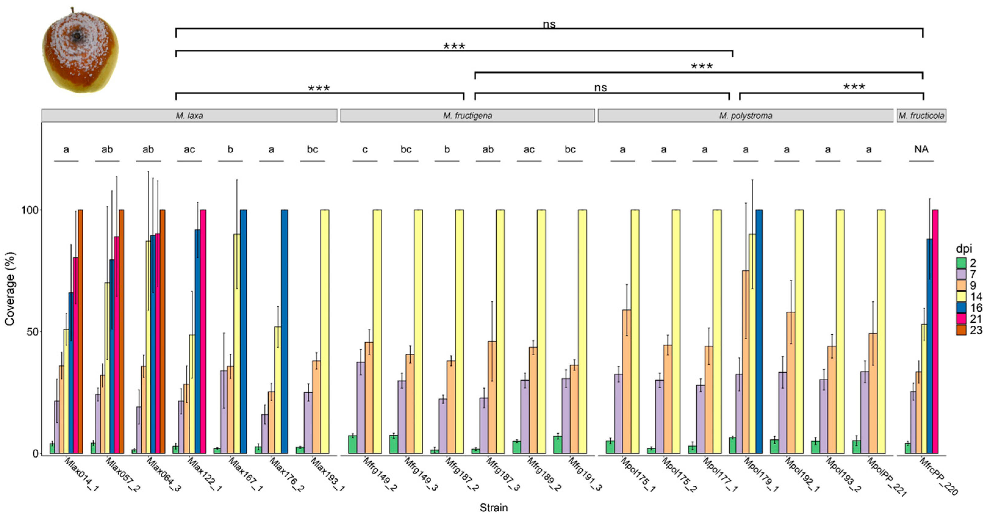
The mycelial growth rate on apples varied considerably among Monilinia species and strains (Figure 4). All strains of M. fructigena reached full surface coverage within 14 dpi, whereas certain strains of M. laxa required up to 23 dpi to achieve full coverage. Statistically, the overall growth rate differed significantly between the Monilinia species (F3,493 = 33.651, p < 0.001). Among them, M. laxa displayed the slowest growth, with full coverage reached between 14 and 23 dpi, confirming its significantly reduced colonization rate over the experimental period compared to the other species.
In contrast, both M. fructigena and M. polystroma exhibited significantly higher growth rates than the tested strain of M. fructicola. Most M. polystroma strains reached 100% apple surface coverage within 14 dpi. However, one exception was observed for strain Mpol179_1, in which one replicate required 16 dpi to reach complete coverage.
Within species, strain-level differences in growth rate were also significant: In M. laxa, growth rate varied significantly between strains (F6,136 = 5.892, p < 0.001). Similarly, significant variation was found among strains of M. fructigena (F5,116 = 5.381, p < 0.001) and M. polystroma (F6,136 = 1.918, p < 0.001), indicating intraspecific variability in colonization ability on apple fruit (Figure 4).
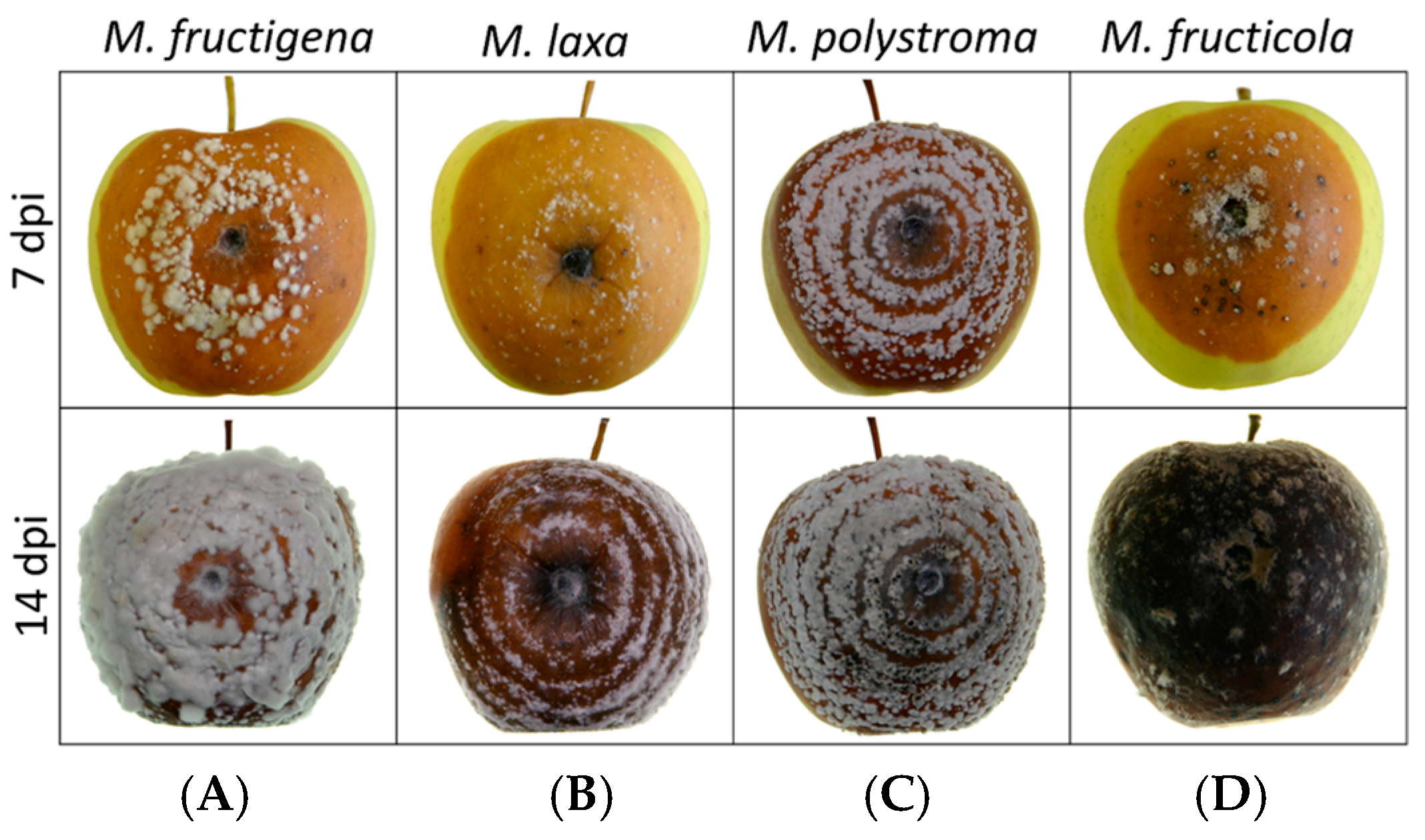
The colony morphology of the four species on apples was largely similar (Figure 5). In M. fructigena, the lesion initially appears light brown, accompanied by dense white mycelial growth forming concentric rings around the inoculation site. As the infection progresses, the center of the lesion darkens, while the mycelium continues to develop a distinct ring-like pattern. In M. laxa, the lesion also exhibits a brown discoloration that spreads outward in well-defined concentric rings. However, these rings are more sparsely and less distinctly distributed compared to M. fructigena, and white mycelial structures are less prominent. In M. polystroma, the lesion is brown with clearly developed concentric rings and abundant white sporulation across the surface. The rings are more closely spaced than in M. laxa, and the strong contrast between them gives the colony a distinct patterned appearance. M. fructicola also causes a brown lesion, nearly black, 14 dpi. The colony displayed dense, dark sporulation with scattered white stromata, giving it a patchy and irregular appearance. The colony margin is less distinct, and the overall pattern is more diffuse compared to the other species.

3.4. Conidia Production and Characterization from PDA, Apple, and TSA

Of the 21 strains tested, only one strain of M. laxa sporulated on PDA. In contrast, all strains of M. fructigena and M. fructicola sporulated on apple between 9 and 21 dpi, whereas M. laxa and M. polystroma did not sporulate on PDA. On TSA, all Monilinia species were capable of sporulation, except for three strains of M. polystroma, which showed no sporulation (Table 3).
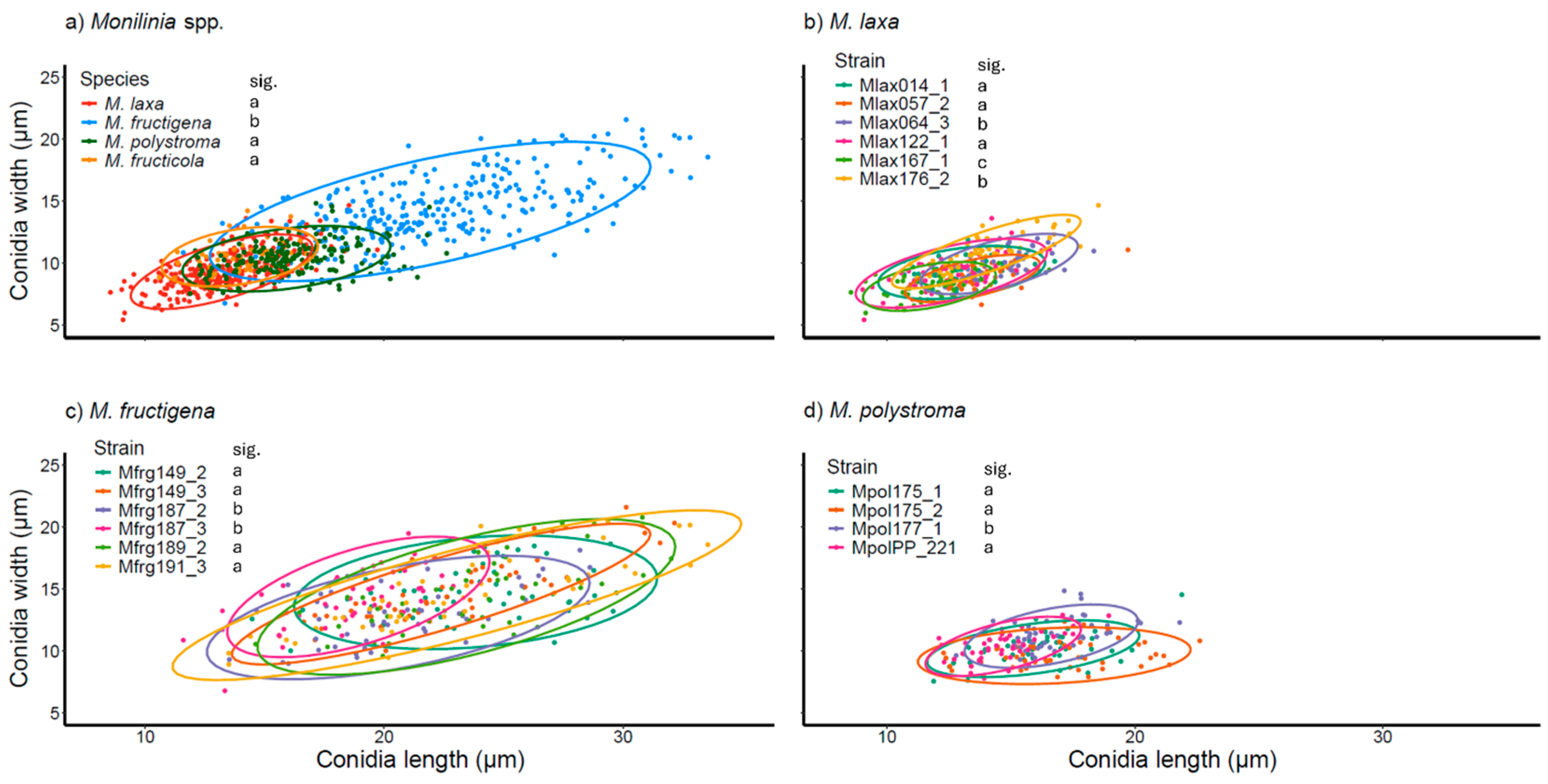
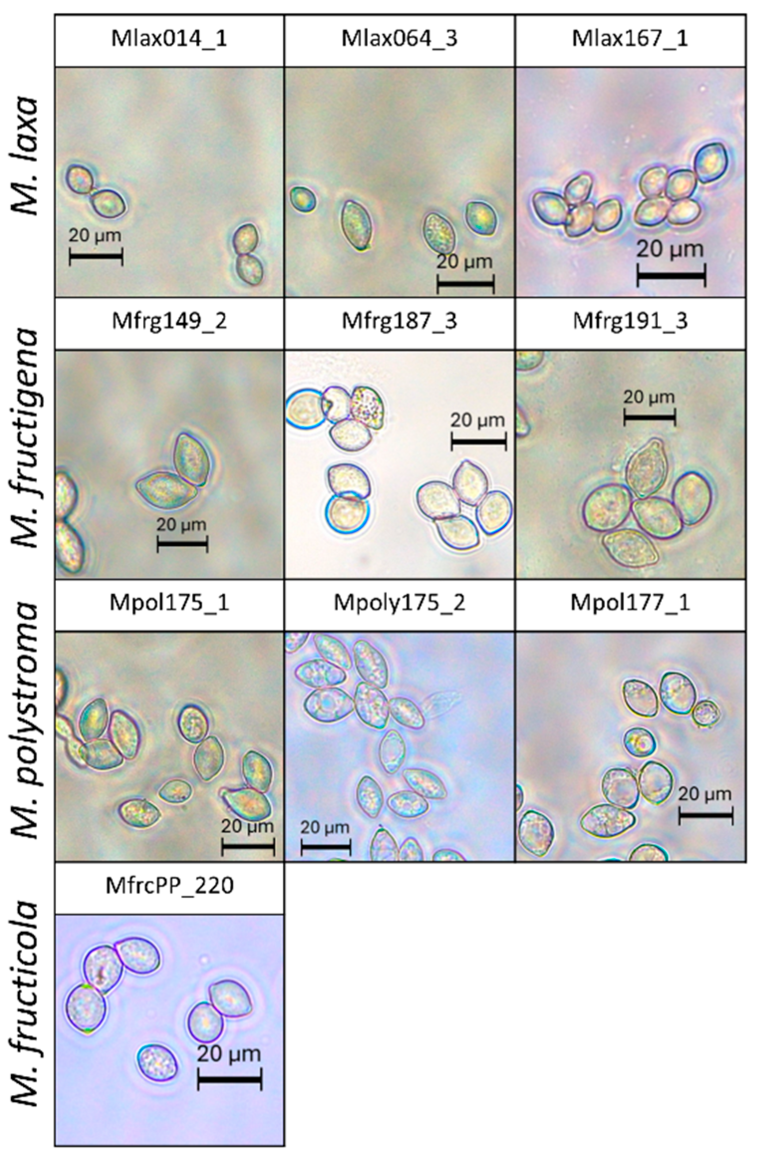
Conidial size differed significantly among Monilinia species (F3,17 = 59.229, p < 0.001). M. fructigena produced significantly larger conidia (6.78–21.59 µm × 11.60–33.55 µm) compared to the other species. No significant differences were observed between M. laxa (5.44–14.69 µm × 8.55–19.72 µm), M. fructicola (8.81–14.24 µm × 10.60–16.75 µm), and M. polystroma (7.55–14.85 µm × 11.89–22.62 µm) (Figure 6a). Within species, strain identity also had a significant effect on conidial size (M. laxa: F5 = 33.73, p < 0.001; M. fructigena: F5 = 8.269, p < 0.001; M. polystroma: F3 = 12.174, p < 0.001) (Figure 6b,c). However, due to overlapping 95% confidence intervals, conidial size alone does not allow for reliable discrimination between species or strains (Figure 7).

3.5. Fungicide Screening on Apples

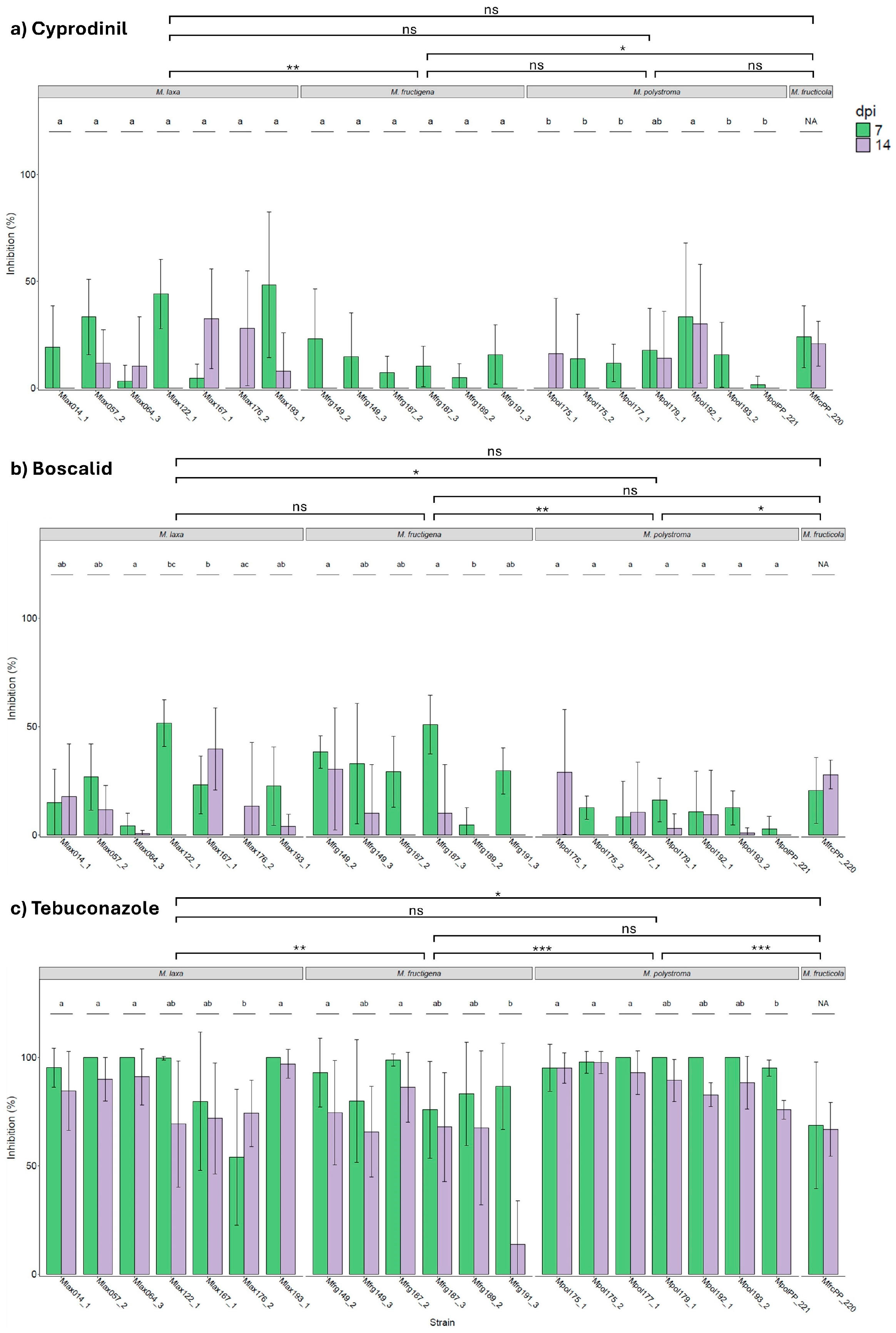
The minimum and maximum inhibition of mycelial growth by cyprodinil on apples ranged from 0 to 89.30% at 7 dpi and from 0 to 60% at 14 dpi. Inhibition was significantly affected by Monilinia species (F3,208 = 5.142, p = 0.002). The highest inhibition rates were observed for M. laxa (16.67% at 7 dpi; 12.90% at 14 dpi) and M. fructicola (24.03% at 7 dpi; 20.82% at 14 dpi). M. fructigena showed the lowest inhibition (12.58% at 7 dpi; 0% at 14 dpi), followed by M. polystroma (13.39% at 7 dpi; 8.59% at 14 dpi) (Figure 8a). Among the four species, significant strain-level variation was observed only for M. polystroma (F6,70 = 3.751, p = 0.003).
The minimum and maximum inhibition by boscalid ranged from 0 to 70.97% at 7 dpi and 0 to 66% at 14 dpi. A significant effect of species was detected (F3,208 = 5.448, p = 0.001), with M. polystroma exhibiting significantly lower inhibition (8.98% at 7 dpi; 1.52% at 14 dpi) compared to M. laxa (20.42% at 7 dpi; 12.41% at 14 dpi), M. fructigena (30.85% at 7 dpi; 8.39% at 14 dpi), and M. fructicola (20.57% at 7 dpi; 27.85% at 14 dpi) (Figure 8b). Significant strain effects were found for M. laxa (F6,68 = 3.431, p = 0.005) and M. fructigena (F5,58 = 5.145, p < 0.001).
Tebuconazole exhibited the strongest inhibitory effect, with inhibition ranging from 0 to 100% at both 7 and 14 dpi. Inhibition differed significantly among species (F3,208 = 12.381, p < 0.001). The highest inhibition was recorded for M. polystroma (98.28% at 7 dpi; 88.84% at 14 dpi) and M. laxa (89.78% at 7 dpi; 82.53% at 14 dpi). In contrast, M. fructigena (86.19% at 7 dpi; 99.33% at 14 dpi) and M. fructicola (68.66% at 7 dpi; 66.78% at 14 dpi) showed significantly lower inhibition (Figure 8c). Significant strain effects were detected for M. laxa (F6,68 = 4.975, p < 0.001), M. fructigena (F5,58 = 3.683, p = 0.006), and M. polystroma (F6,68 = 3.373, p = 0.006).
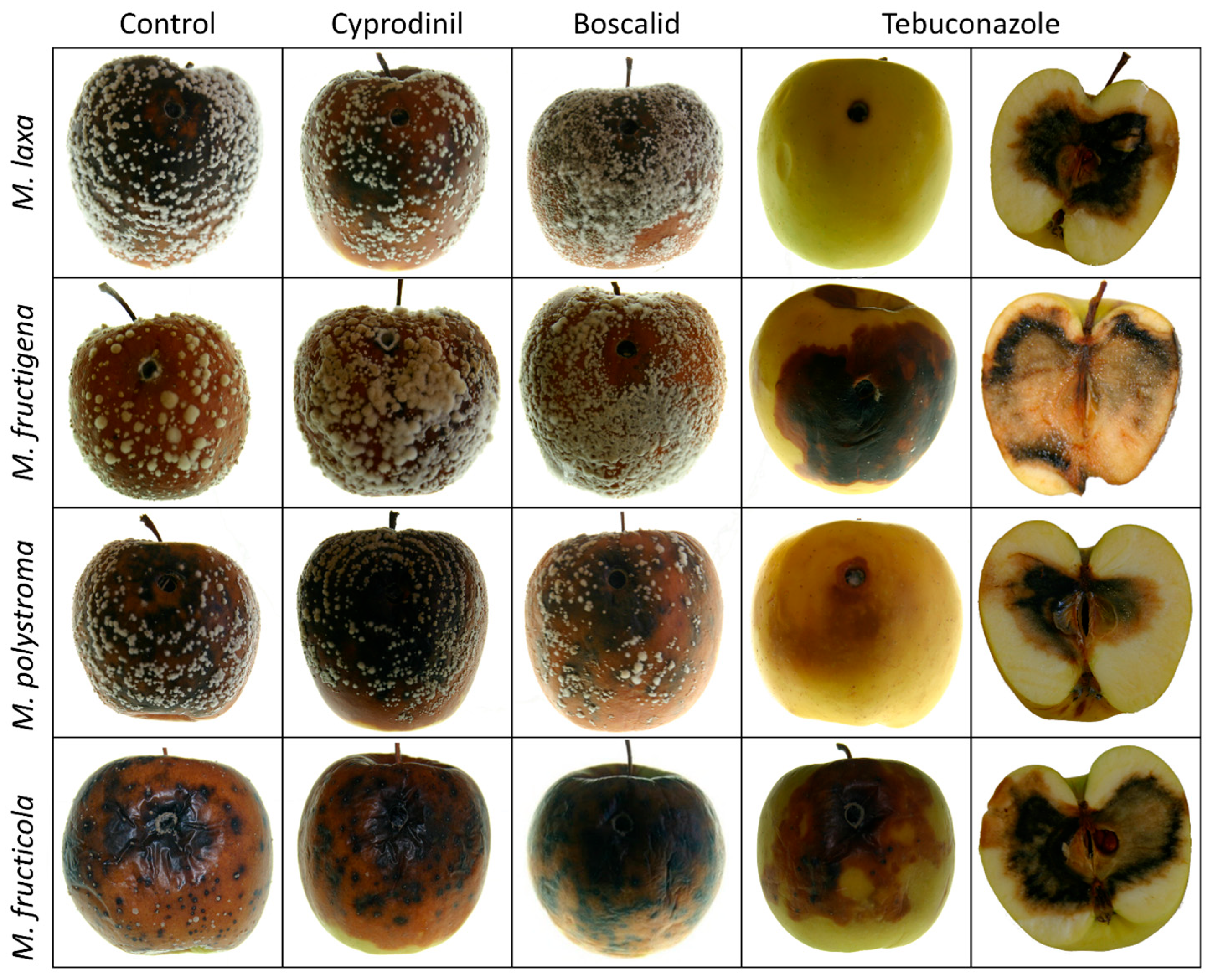
While apples treated with cyprodinil and boscalid showed mycelial growth and brown rot symptoms on the fruit surface, treatment with tebuconazole provided protection on the fruit surface, resulting in fungal spread and symptom development being concentrated in the fruit interior (Figure 9).

4. Discussion

This study aimed to characterize four Monilinia species (M. fructigena, M. laxa, M. polystroma, and M. fructicola) based on morphological traits, phylogenetic identity, growth dynamics, and fungicide sensitivity. In particular, the need for information on M. polystroma, a relatively recently described species [22], to address existing knowledge gaps and assess its potential impact on disease management strategies.
Molecular identification using universal primers ITS5 and ITS4 successfully differentiated among all four Monilinia species, supporting the reliability of ITS-based diagnostics for this fungal genus [25]. Sequence homogeneity within species indicated stable ITS regions, with no intraspecific variation observed. Nevertheless, the lack of intraspecific resolution in the ITS region shows that additional molecular markers (e.g., β-tubulin, GAPDH) or whole-genome sequencing is necessary to distinguish different strains based on fungicide sensitivity [33]. The ITS region exhibits generally low variability within Monilinia species, yet it remains a useful marker for distinguishing the four described species from others that have not been detected so far in South Tyrol’s alpine orchards [24,34,35]. Morphologically, not all species exhibited distinguishable colony characteristics on PDA; especially, M. fructigena and M. polystroma displayed entire to slightly undulate margins, highlighting the need for complementary molecular identification.
Colony growth at 20 °C and under short-day conditions revealed significantly lower growth rates compared to the literature values obtained at optimal temperatures [1], reflecting the adaptation to the cooler mountain climates of South Tyrol. Despite these constraints, in vitro growth rate differences among species were consistent with known thermal preferences. In planta growth on apples confirmed significant interspecific differences, underscoring the importance of intraspecies diversity in disease progression. Colony morphology and conidial characteristics generally aligned with descriptions from earlier studies [36,37]. Conidia of M. fructigena were significantly larger than those of M. laxa, M. fructicola, and M. polystroma, although their 95% confidence intervals overlapped. Therefore, it is not possible to assign Monilinia spores to a specific species based on their size.
Sporulation success was highly dependent on the substrate, with TSA outperforming apples in inducing conidial production. However, some M. polystroma isolates failed to sporulate on TSA, indicating strain-specific responses to induction media. The results explain the missing sporulation on PDA, which was reported for several experiments to fulfill Koch’s postulates, where mycelial plugs were used [20,38].
The fungicide trials revealed differential sensitivities among the species. The reduced performance of cyprodinil for single strains may indicate the early stages of resistance development, which is consistent with its classification as a high-risk, single-site inhibitor of methionine biosynthesis [13,39,40]. For M. laxa isolates, the half maximal effective concentration (EC50) values for cyprodinil ranged from 1.11 to 0.03 mg L−1, and for M. fructicola, from 0.29 to 0.06 mg L−1 [39]. The observed cyprodinil inhabitation at a dose of 250 mg L−1 ranged from 0 to 89.30%, which indicates a high strain and species-specific variation.
Boscalid, belonging to the SDHI class (FRAC Group 7) and acting by the inhibition of Complex II of mitochondrial respiration [13,14,15], showed moderate efficacy. Resistance arises from mutations in the Sdh gene complex (SDHA–D subunits) and overexpression of efflux transporters, reducing intracellular fungicide concentrations [15,27]. For M. fructicola isolates, the EC50 values for boscalid range from 0.42 to 11.94 mg L−1 [27]. At a boscalid concentration of 200 mg L−1, inhibition across Monilinia species ranged from 0 to 70.97%, again highlighting a substantial variation between strains and species.
Tebuconazole, a DMI fungicide, showed the highest efficacy across species, overall, close to the skin of treated apples. This aligns with its systemic activity and broad spectrum of effectiveness [16,17]. For M. laxa isolates, the EC50 values ranged from 0.42 to 0.02 mg L−1, and for M. fructicola, from 0.37 to 0.13 mg L−1 [41]. The observed tebuconazole inhibition at a dose of 125 mg L−1 ranged from 0 to 100%, which indicates a high strain and species-specific variation, as it was observed for the other fungicides. Overall high tolerances in M. laxa and M. polystroma indicate the presence of resistant strains.
The findings suggest that fungicide application strategies should account for species-specific sensitivities. Reduced efficacy of cyprodinil and boscalid could stem from repeated applications and selection pressure [1]. Indeed, despite operating through a distinct mode of action, the observed partial tolerance to both cyprodinil and boscalid in some strains could hint at emerging cross-resistance mechanisms, potentially linked to multidrug efflux transporters or stress response pathways [5]. Rotational and integrated fungicide programs, as recommended by FRAC, are necessary to delay resistance onset [13]. Post-harvest brown rot management is particularly challenging in Europe due to the absence of authorized chemical treatments [10]. This makes pre-harvest fungicide efficacy critical for disease suppression.
In the alpine region of South Tyrol, the climate and cultivation practices may favor M. laxa dominance, but the presence of M. polystroma and M. fructicola, species with distinct thermal preferences, could complicate disease dynamics, especially under climate change scenarios. Studies from central and southern Europe have shown varying dominance of Monilinia species and differences in fungicide sensitivity patterns [41,42,43]. Comparing our findings with those studies highlights the importance of region-specific monitoring programs and suggests that climatic conditions and orchard practices significantly shape pathogen populations.

5. Conclusions

In conclusion, ITS sequencing with primers ITS5 and ITS4 successfully differentiated all species, with no intraspecific variation observed. Colony morphology and conidial size showed species-specific trends, but overlapping ranges prevent reliable identification based on morphology alone. All species sporulated on TSA, while PDA was less suitable. Some M. polystroma isolates failed to sporulate on TSA, indicating strain-specific differences. Further studies should focus on identifying compounds that are responsible for the induction of sporulation. Fungicide trials at field concentration revealed significant variation in sensitivity. The variability across and within species emphasizes that the occurrence of species and strains can substantially affect fungicide efficacy. This underlines the importance of studies addressing strain-specific differences in fungicide sensitivity and resistance, especially in fruit growing regions such as South Tyrol’s alpine orchards.

Author Contributions

Conceptualization, M.M.P., S.O., Y.P., and U.S.; methodology, M.M.P. and U.S.; validation, M.M.P.; resources, S.O., E.D., and U.S.; data curation, M.M.P. and U.S.; writing—original draft preparation, M.M.P. and U.S.; writing—review and editing, M.M.P., S.O., E.D., Y.P., and U.S.; supervision, Y.P. and U.S.; project administration S.O., Y.P., and U.S. All authors have read and agreed to the published version of the manuscript.

Funding

The Laimburg Research Centre is funded by the Autonomous Province of Bozen/Bolzano.

Institutional Review Board Statement

Not applicable.

Informed Consent Statement

Not applicable.

Data Availability Statement

The ITS sequences are identical to those already available in the NCBI GenBank database (accession numbers: MH864497, MH862738, MH855219, MH863200). The corresponding raw data and raw sequencing files have been deposited in Zenodo and are available at: https://doi.org/10.5281/zenodo.17167654.

Acknowledgments

The authors would like to thank Aldo Matteazzi from the Laimburg Research Centre for providing Melanie M. Pagano with the opportunity to work at the Institute for Plant Health. We also acknowledge the Consortium of South Tyrolean Fruit Growers Cooperatives for supplying the apples used in the trials and the Department of Innovation, Research, University and Museums of the Autonomous Province of Bozen/Bolzano for covering the publication costs.

Conflicts of Interest

The authors declare no conflicts of interest.

References

  1. Abate, D.; Pastore, C.; Gerin, D.; de Miccolis Angelini, R.M.; Rotolo, C.; Pollastro, S.; Faretra, F. Characterization of Monilinia spp. populations on stone fruit in South Italy. Plant Dis. 2018, 102, 1708–1717. [Google Scholar] [CrossRef]
  2. Holb, I.J. Brown rot blossom blight of pome and stone fruits: Symptom, disease cycle, host resistance, and biological control. Int. J. Hortic. Sci. 2008, 14, 15–21. [Google Scholar] [CrossRef]
  3. Obi, V.I.; Barriuso, J.J.; Gogorcena, Y. Peach brown rot: Still in search of an ideal management option. Agriculture 2018, 8, 125. [Google Scholar] [CrossRef]
  4. Wenneker, M.; Thomma, B.P.H.J. Latent postharvest pathogens of pome fruit and their management: From single measures to a systems intervention approach. Eur. J. Plant Pathol. 2020, 156, 663–681. [Google Scholar] [CrossRef]
  5. Hu, M.-J.; Cox, K.D.; Schnabel, G.; Luo, C.-X. Monilinia species causing brown rot of peach in China. PLoS ONE 2011, 6, e24990. [Google Scholar] [CrossRef]
  6. Byrde, R.; Willetts, H.J. The brown rot fungi of fruit: Their biology and control. Mycologia 1977, 69, 1240. [Google Scholar] [CrossRef]
  7. Rungjindamai, N.; Jeffries, P.; Xu, X.-M. Epidemiology and management of brown rot on stone fruit caused by Monilinia laxa. Eur. J. Plant Pathol. 2014, 140, 1–17. [Google Scholar] [CrossRef]
  8. Petróczy, M.; Palkovics, L. First report of Monilia polystroma on apple in Hungary. Eur. J. Plant Pathol. 2009, 125, 343–347. [Google Scholar] [CrossRef]
  9. Balardin, R.; Madalosso, M.; Debortoli, M.; Lenz, G. Factors Affecting Fungicide Efficacy in the Tropics; Federal University of Santa Maria: Santa Maria, Brazil, 2010. [Google Scholar]
  10. Casals, C.; Teixidó, N.; Viñas, I.; Llauradó, S.; Usall, J. Control of Monilinia spp. on stone fruit by curing treatments. Postharvest Biol. Technol. 2010, 56, 19–25. [Google Scholar] [CrossRef]
  11. Garcia-Benitez, C.; Casals, C.; Usall, J.; Sánchez-Ramos, I.; Melgarejo, P.; de Cal, A. Impact of postharvest handling on preharvest latent infections caused by Monilinia spp. in nectarines. J. Fungi 2020, 6, 266. [Google Scholar] [CrossRef]
  12. Akhoon, B.A.; Gupta, S.K.; Dhar, M.K. Dissecting the genome, secretome, and effectome repertoires of Monilinia spp.: The causal agent of brown rot disease: A comparative analysis. Postharvest Biol. Technol. 2023, 195, 112120. [Google Scholar] [CrossRef]
  13. Adaskaveg, J.; Gubler, D.; Michailides, T.; Holtz, B. Efficacy and Timing of Fungicides, Bacteriocides, and Biologicals for Deciduous Tree Fruit, Nut, Strawberry and Vine Crops; Department of Plant Pathology, UC Davis: Davis, CA, USA, 2011. [Google Scholar]
  14. Rustin, P.; Munnich, A.; Rötig, A. Succinate dehydrogenase and human diseases: New insights into a well-known enzyme. Eur. J. Hum. Genet. 2002, 10, 289–291. [Google Scholar] [CrossRef] [PubMed]
  15. Sang, H.; Lee, H.B. Molecular mechanisms of succinate dehydrogenase inhibitor resistance in phytopathogenic fungi. Res. Plant Dis. 2020, 26, 1–7. [Google Scholar] [CrossRef]
  16. Adaskaveg, J.E.; Förster, H.; Thompson, D.F. Identification and etiology of visible quiescent infections of Monilinia fructicola and Botrytis cinerea in sweet cherry fruit. Plant Dis. 2000, 84, 328–333. [Google Scholar] [CrossRef]
  17. Zeng, F.; Arnao, E.; Zhang, G.; Olaya, G.; Wullschleger, J.; Sierotzki, H.; Ming, R.; Bluhm, B.H.; Bond, J.P.; Fakhoury, A.M.; et al. Characterization of quinone outside inhibitor fungicide resistance in Cercospora sojina and development of diagnostic tools for its identification. Plant Dis. 2015, 99, 544–550. [Google Scholar] [CrossRef]
  18. Spitaler, U.; Oettl, S.; Deltedesco, E. First report of brown rot caused by Monilinia polystroma on quince in Italy. Plant Dis. 2023, 107, 229. [Google Scholar] [CrossRef]
  19. Spitaler, U.; Pfeifer, A.; Deltedesco, E.; Hauptkorn, S.; Oettl, S. Detection of Monilinia spp. by a multiplex real-time PCR assay and first report of Monilinia fructicola in South Tyrol (northern Italy). J. Plant Dis. Prot. 2022, 129, 1013–1020. [Google Scholar] [CrossRef]
  20. Deltedesco, E.; Oettl, S.; Spitaler, U. First report of brown rot caused by Monilinia polystroma on sweet cherry and almond in Italy. Plant Dis. 2023, 107, 2252. [Google Scholar] [CrossRef]
  21. Angeli, S.S.; de Mio, L.L.M.; Amorim, L. Comparative analysis of Monilinia fructicola and M. laxa isolates from Brazil: Monocyclic components of peach brown rot. Cienc. Rural 2017, 47, e20160300. [Google Scholar] [CrossRef]
  22. van Leeuwen, G.C.; Baa Yen, R.P.; Holb, I.J.; Jeger, M.J. Distinction of the Asiatic brown rot fungus Monilia polystroma sp. nov. from M. fructigena. Mycol. Res. 2002, 106, 444–451. [Google Scholar] [CrossRef]
  23. Petróczy, M.; Szigethy, A.; Palkovics, L. Monilinia species in Hungary: Morphology, culture characteristics, and molecular analysis. Trees 2012, 26, 153–164. [Google Scholar] [CrossRef]
  24. Vasić, M.; Vico, I.; Jurick, W.M.; Duduk, N. Distribution and characterization of Monilinia spp. causing apple fruit decay in Serbia. Plant Dis. 2018, 102, 359–369. [Google Scholar] [CrossRef]
  25. White, T.J.; Bruns, T.; Lee, S.; Taylor, J. Amplification and direct sequencing of fungal ribosomal RNA genes for phylogenetics. In PCR Protocols; Elsevier: Amsterdam, The Netherlands, 1990; pp. 315–322. ISBN 9780123721808. [Google Scholar]
  26. Kumar, S.; Stecher, G.; Suleski, M.; Sanderford, M.; Sharma, S.; Tamura, K. MEGA12: Molecular evolutionary genetic analysis version 12 for adaptive and green computing. Mol. Biol. Evol. 2024, 41, msae263. [Google Scholar] [CrossRef] [PubMed]
  27. Chen, F.; Liu, X.; Chen, S.; Schnabel, E.; Schnabel, G. Characterization of Monilinia fructicola strains resistant to both propiconazole and boscalid. Plant Dis. 2013, 97, 645–651. [Google Scholar] [CrossRef] [PubMed]
  28. Bates, D.; Mächler, M.; Bolker, B.; Walker, S. Fitting linear mixed-effects models using lme4. J. Stat. Soft. 2015, 67, 48. [Google Scholar] [CrossRef]
  29. Hothorn, T.; Bretz, F.; Westfall, P. Simultaneous inference in general parametric models. Biom. J. 2008, 50, 346–363. [Google Scholar] [CrossRef]
  30. Wickham, H. Getting Started with ggplot2. In ggplot2; Wickham, H., Ed.; Springer International Publishing: Cham, Switzerland, 2016; pp. 11–31. ISBN 978-3-319-24275-0. [Google Scholar]
  31. Kuznetsova, A.; Brockhoff, P.B.; Christensen, R.H.B. lmerTest package: Tests in linear mixed effects models. J. Stat. Soft. 2017, 82, 1–26. [Google Scholar] [CrossRef]
  32. Vu, D.; Groenewald, M.; de Vries, M.; Gehrmann, T.; Stielow, B.; Eberhardt, U.; Al-Hatmi, A.; Groenewald, J.Z.; Cardinali, G.; Houbraken, J.; et al. Large-scale generation and analysis of filamentous fungal DNA barcodes boosts coverage for kingdom fungi and reveals thresholds for fungal species and higher taxon delimitation. Stud. Mycol. 2019, 92, 135–154. [Google Scholar] [CrossRef]
  33. Bradshaw, M.J.; Guan, G.-X.; Nokes, L.; Braun, U.; Liu, S.-Y.; Pfister, D.H. Secondary DNA barcodes (CAM, GAPDH, GS, and RpB2) to characterize species complexes and strengthen the powdery mildew phylogeny. Front. Ecol. Evol. 2022, 10, 918908. [Google Scholar] [CrossRef]
  34. Poniatowska, A.; Michalecka, M.; Puławska, J. Phylogenetic relationships and genetic diversity of Monilinia spp. isolated in Poland based on housekeeping- and pathogenicity-related gene sequence analysis. Plant Pathol. 2021, 70, 1640–1650. [Google Scholar] [CrossRef]
  35. Zhu, X.-Q.; Niu, C.-W.; Chen, X.-Y.; Guo, L.-Y. Monilinia Species Associated with Brown Rot of Cultivated Apple and Pear Fruit in China. Plant Dis. 2016, 100, 2240–2250. [Google Scholar] [CrossRef]
  36. Martini, C.; Mari, M. Monilinia fructicola, Monilinia laxa (Monilinia rot, brown rot). In Postharvest Decay; Academic Press: Cambridge, MA, USA, 2014; pp. 233–265. [Google Scholar] [CrossRef]
  37. Verde-Yáñez, L.; Usall, J.; Teixidó, N.; Vall-Llaura, N.; Torres, R. Deciphering the effect of light wavelengths in Monilinia spp. DHN-Melanin production and their interplay with ROS metabolism in M. fructicola. J. Fungi 2023, 9, 653. [Google Scholar] [CrossRef]
  38. Munda, A. First report of brown rot on peach caused by Monilia polystroma in Slovenia. Plant Dis. 2015, 99, 1281. [Google Scholar] [CrossRef]
  39. Avenot, H.F.; Quattrini, J.; Puckett, R.; Michailides, T.J. Different levels of resistance to cyprodinil and iprodione and lack of fludioxonil resistance in Botrytis cinerea isolates collected from pistachio, grape, and pomegranate fields in California. Crop Prot. 2018, 112, 274–281. [Google Scholar] [CrossRef]
  40. Scott, J.; Sueiro-Olivares, M.; Thornton, B.P.; Owens, R.A.; Muhamadali, H.; Fortune-Grant, R.; Thomson, D.; Thomas, R.; Hollywood, K.; Doyle, S.; et al. Targeting methionine synthase in a fungal pathogen causes a metabolic imbalance that impacts cell energetics, growth, and virulence. mBio 2020, 11, e01985-20. [Google Scholar] [CrossRef]
  41. Dardani, G.; Guarnaccia, V.; Nari, L.; Testempasis, S.I.; Karaoglanidis, G.S.; Gullino, M.L. Identification of pathogens causing brown rot of stone fruit in Cuneo province (Italy) and assessment of sensitivity to azoxystrobin, cyprodinil, fenhexamid, fludioxonil, and tebuconazole. Phytopathol. Mediterr. 2023, 62, 455–465. [Google Scholar] [CrossRef]
  42. Egüen, B.; Melgarejo, P.; de Cal, A. The effect of fungicide resistance on the structure of Monilinia laxa populations in Spanish peach and nectarine orchards. Eur. J. Plant Pathol. 2016, 145, 815–827. [Google Scholar] [CrossRef]
  43. Hrustić, J.; Mihajlović, M.; Grahovac, M.; Delibašić, G.; Tanović, B. Fungicide sensitivity, growth rate, aggressiveness and frost hardiness of Monilinia fructicola and Monilinia laxa isolates. Eur. J. Plant Pathol. 2018, 151, 389–400. [Google Scholar] [CrossRef]
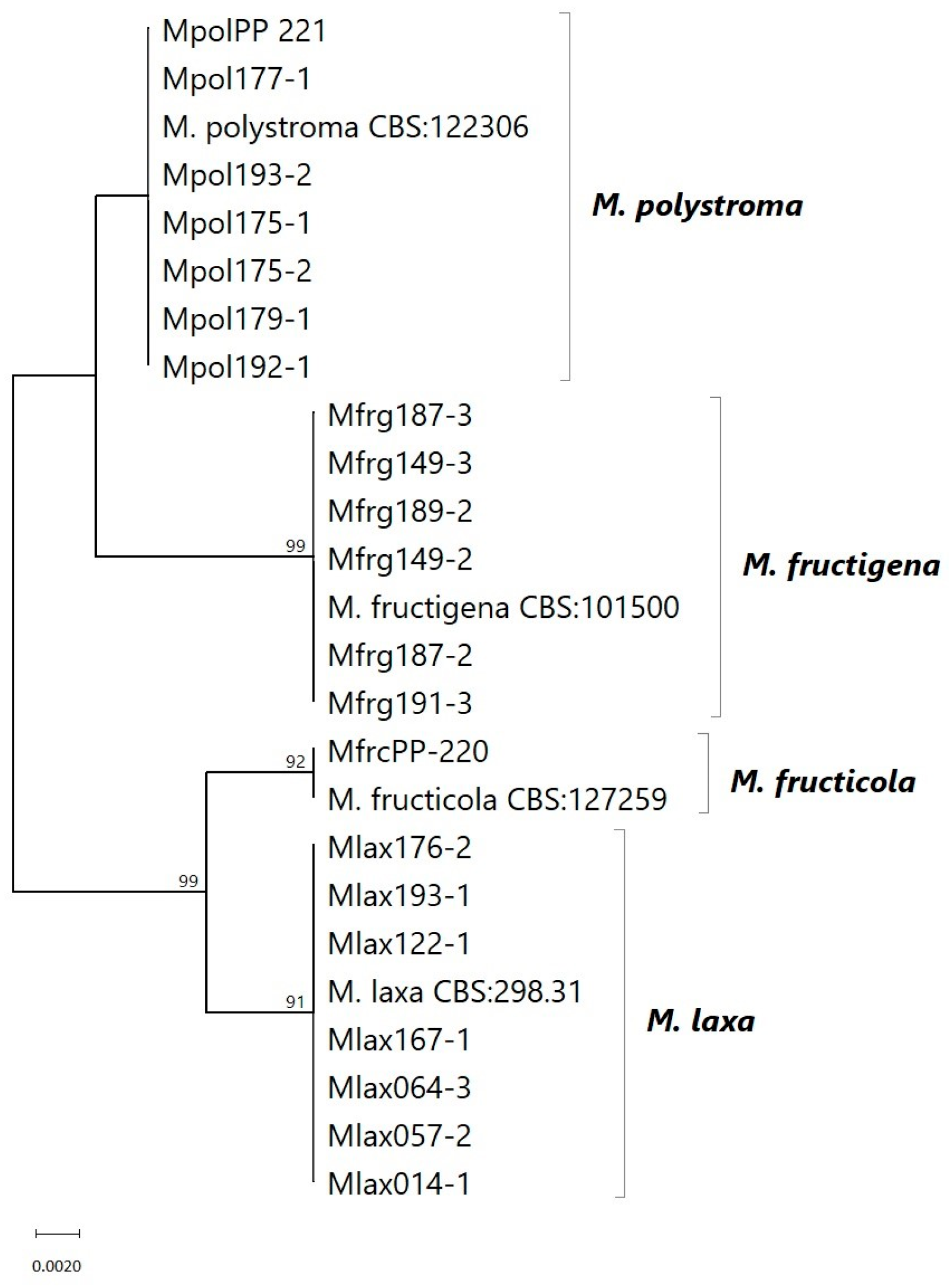
Figure 1. Maximum likelihood (ML) tree based on ITS sequences from Monilinia strains collected in different locations in South Tyrol, Italy. Sequences from NCBI GenBank were used as references. Bootstrap support values (1000 replicates) above 85% are shown at the branches.
Jof 11 00690 g001
Figure 2. Samples assessed at 27 dpi of (A) M. fructigena strain 191_3, (B) M. laxa strain 14_1, (C) M. polystroma strain 192_1, and (D) M. fructicola strain PP_220 grown on PDA at 20 °C under a 12:12 h L:D photoperiod. Front and back of colony.
Jof 11 00690 g002
Figure 3. Mean mycelium radius (±SD; n = 5) of strains of M. fructigena, M. laxa, M. polystroma, and M. fructicola grown on PDA at 20 °C under a 12:12 h L:D photoperiod over 27 days. Different lowercase letters denote significant differences in growth between strains within the same Monilinia spp. (p < 0.05, n = 5). NA means no statistically different values. Asterisks denote significant differences between treatments (ns p > 0.05, *** p ≤ 0.001).
Jof 11 00690 g003
Figure 4. Mean coverage (±SD; n = 5) of strains of M. fructigena, M. laxa, M. polystroma, and M. fructicola on apples at 20 °C under a 12:12 h L:D photoperiod. Different lowercase letters denote significant differences in growth between strains within the same Monilinia spp. (p < 0.05, n = 5). NA means no statistically significant values. Asterisks denote significant differences between treatments (ns p > 0.05, *** p ≤ 0.001).
Jof 11 00690 g004
Figure 5. Samples assessed at 7 and 14 dpi of (A) M. fructigena strain 191_3, (B) M. laxa strain 14_1, (C) M. polystroma strain 192_1, and (D) M. fructicola strain PP_220 grown on apples at 20 °C under a 12:12 h L:D photoperiod.
Jof 11 00690 g005
Figure 6. Conidia length and width (n = 50) of (a) Monilinia spp., (b) M. laxa strains, (c) M. fructigena strains, and (d) M. polystroma strains. Ellipses show 95% confidence level. Different lowercase letters indicate significant differences between species or strains (p < 0.05).
Jof 11 00690 g006
Figure 7. Conidia morphology of selected Monilinia strains.
Jof 11 00690 g007
Figure 8. Mean inhibition (±SD; n = 5) of Monilinia spp. by (a) cyprodinil, (b) boscalid, and (c) tebuconazole on apples at 7 and 14 dpi. Means not sharing any letter are significantly different by the Tukey test (p < 0.05). Different lowercase letters denote significant differences in growth between strains within the same Monilinia species (p < 0.05, n = 5). NA means no statistical values. Asterisks denote significant differences between treatments (ns p > 0.05, * p ≤ 0.05, ** p ≤ 0.01, *** p ≤ 0.001).
Jof 11 00690 g008
Figure 9. Morphology of Monilinia spp. on apples 14 dpi from the control, cyprodinil, boscalid, and tebuconazole treatments. Furthermore, the pulp of apples treated with tebuconazole is shown.
Jof 11 00690 g009
Table 1. Descriptive table of 21 Monilinia strains: assigned species, strain ID, host fruit from which they were collected, and the respective sampling site in German and Italian.
SpeciesStrain IDHost FruitSampling Sites *
M. laxaMlax014_1PlumJenesien/San Genesio
Mlax057_2PeachPartschins/Parcines
Mlax064_3CherryAllitz/Lasa
Mlax122_1CherryRitten/Renon
Mlax167_1ApricotKollmann/Colma
Mlax176_2PeachPiglon/Piccolungo
Mlax193_1QuinceKaltern/Caldaro
M. fructigenaMfrg149_2PlumKollmann/Colma
Mfrg149_3PlumKollmann/Colma
Mfrg187_2AppleTerlan/Terlano
Mfrg187_3AppleTerlan/Terlano
Mfrg189_2AlmondPiglon/Piccolungo
Mfrg191_3AlmondPiglon/Piccolungo
M. polystromaMpol175_1CherryLaimburg
Mpol175_2CherryLaimburg
Mpol177_1AlmondPiglon/Piccolungo
Mpol179_1AlmondPiglon/Piccolungo
Mpol192_1QuinceKaltern/Caldaro
Mpol193_2QuinceKaltern/Caldaro
MpolPP_221AppleJapan (CBS 102686)
M. fructicolaMfrcPP_220CherryBari, Italy (CBS 144849)
* Sampling site names are provided in German and Italian.
Table 2. Fungicides used in laboratory bioassays, including active ingredient (AI), trade name, manufacturer/distributor, formulation, and applied dose.
Active IngredientTrade NameManufacturer/DistributorComposition (AI)Applied Dose (AI)
BoscalidCantus®BASF Italia S.p.A. (Cesano Maderno, Italy)500 g kg−1200 mg L−1
CyprodinilChorus®Syngenta Italia S.p.A. (Milan, Italy)500 g kg−1250 mg L−1
TebuconazoleFolicur® WGBayer CropScience S.r.l. (Milan, Italy)250 g kg−1125 mg L−1
Table 3. Sporulation capability of Monilinia species on PDA, apple, and TSA of the 21 strains and the respective days required for sporulation to happen (“X” = sporulation occurred, “-” = not sporulating).
SpeciesIsolateSporulation
PDAdpiAppledpiTSAdpi
M. laxaMlax014_1----X6
Mlax057_2----X12
Mlax064_3----X6
Mlax122_1----X6
Mlax167_1X12--X12
Mlax176_2----X6
Mlax193_1----X12
M. fructigenaMfrg149_2--X9X6
Mfrg149_3--X9X7
Mfrg187_2--X14X7
Mfrg187_3--X9X6
Mfrg189_2--X14X7
Mfrg191_3--X21X6
M. polystromaMpol175_1----X6
Mpol175_2----X12
Mpol177_1----X12
Mpol179_1------
Mpol192_1------
Mpol193_2------
MpolPP_221----X6
M. fructicolaMfrcPP_220--X16X6
Disclaimer/Publisher’s Note: The statements, opinions and data contained in all publications are solely those of the individual author(s) and contributor(s) and not of MDPI and/or the editor(s). MDPI and/or the editor(s) disclaim responsibility for any injury to people or property resulting from any ideas, methods, instructions or products referred to in the content.