
A Preliminary Evaluation of the Comparative Efficacy of Gel-Based and Oil-Based CBD on Hematologic and Biochemical Responses in Dogs


 , , and
Simple Summary
Abstract
1. Introduction
2. Materials and Methods
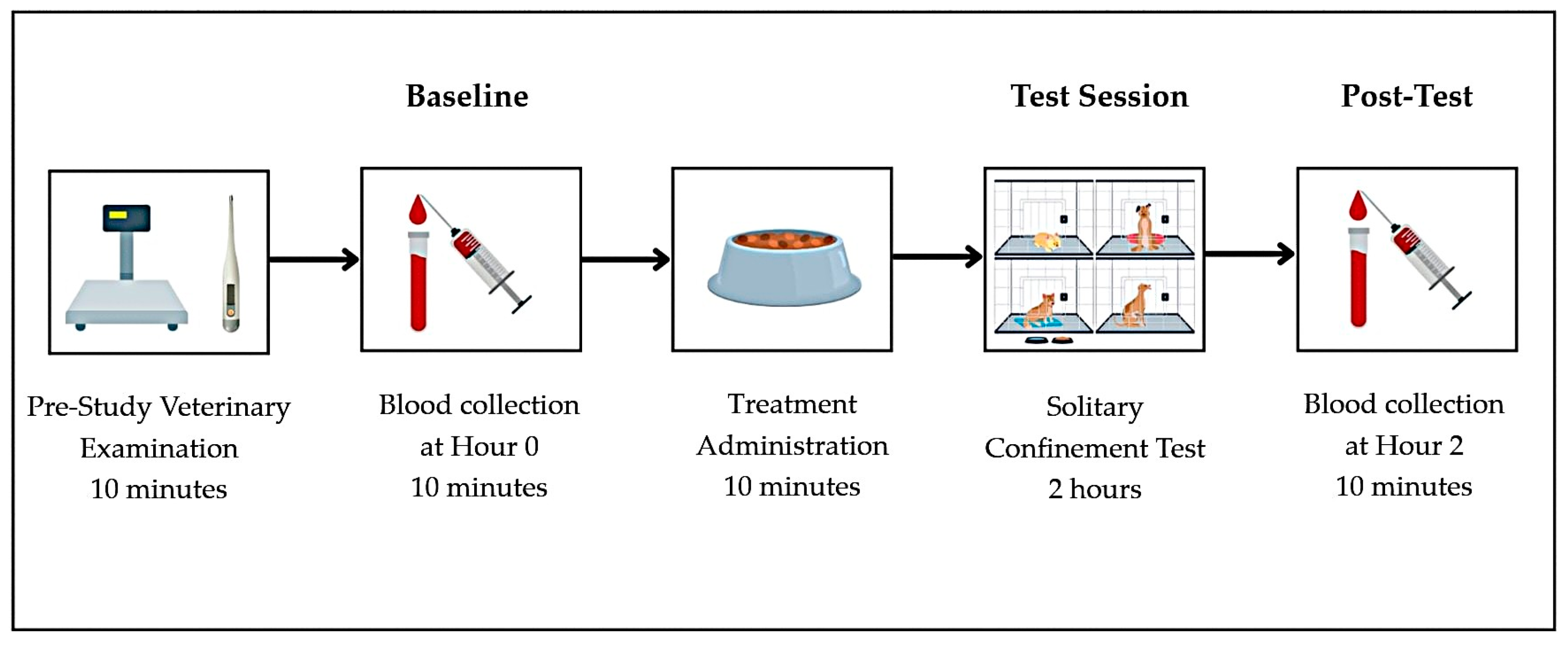
2.1. Animals and Housing
2.2. Study Design and Treatment
2.3. Stress Induction
2.4. Blood Collections and Analyses
2.5. Statistical Analysis
3. Results
3.1. Hematological and Biochemical Parameters
3.2. Serum Cortisol and Stress Response
3.3. Immunological Responses
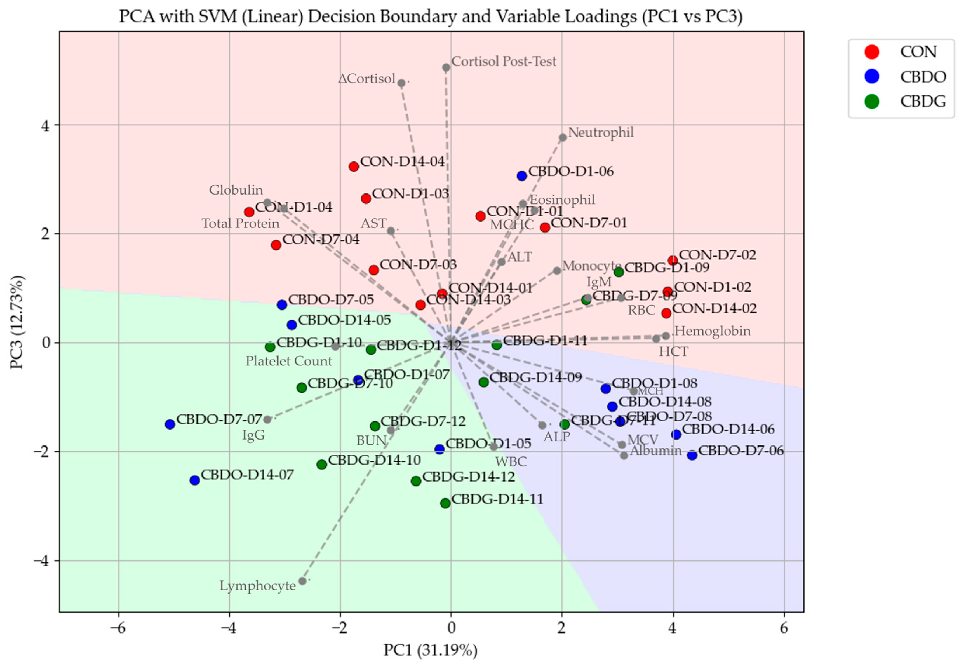
3.4. Principal Component Analysis and Blood Parameter Associations
4. Discussion
5. Conclusions
Author Contributions
Funding
Institutional Review Board Statement
Informed Consent Statement
Data Availability Statement
Acknowledgments
Conflicts of Interest
References
- Peyravian, N.; Deo, S.; Daunert, S.; Jimenez, J.J. Cannabidiol as a Novel Therapeutic for Immune Modulation. ImmunoTargets Ther. 2020, 9, 131–140. [Google Scholar] [CrossRef] [PubMed]
- Beerda, B.; Schilder, M.B.H.; van Hooff, J.A.; de Vries, H.W. Manifestations of chronic and acute stress in dogs. Appl. Anim. Behav. Sci. 1997, 52, 307–319. [Google Scholar] [CrossRef]
- Demirbas, Y.S.; Isparta, S.; Saral, B.; Yılmaz, N.K.; Adıay, D.; Matsui, H.; Töre-Yargın, G.; Musa, S.A.; Atilgan, D.; Öztürk, H.; et al. Acute and chronic stress alter behavioral laterality in dogs. Sci. Rep. 2023, 13, 4092. [Google Scholar] [CrossRef]
- van der Laan, J.E.; Vinke, C.M.; Arndt, S.S. Evaluation of hair cortisol as an indicator of long-term stress responses in dogs in an animal shelter and after subsequent adoption. Sci. Rep. 2022, 12, 5117. [Google Scholar] [CrossRef]
- Gamble, L.-J.; Boesch, J.M.; Frye, C.W.; Schwark, W.S.; Mann, S.; Wolfe, L.; Brown, H.; Berthelsen, E.S.; Wakshlag, J.J. Pharmacokinetics, Safety, and Clinical Efficacy of Cannabidiol Treatment in Osteoarthritic Dogs. Front. Vet. Sci. 2018, 5, 165. [Google Scholar] [CrossRef]
- Deabold, K.A.; Schwark, W.S.; Wolf, L.; Wakshlag, J.J. Single-Dose Pharmacokinetics and Preliminary Safety Assessment with Use of CBD-Rich Hemp Nutraceutical in Healthy Dogs and Cats. Animals 2019, 9, 832. [Google Scholar] [CrossRef]
- Gugliandolo, E.; Licata, P.; Peritore, A.F.; Siracusa, R.; D’amico, R.; Cordaro, M.; Fusco, R.; Impellizzeri, D.; Di Paola, R.; Cuzzocrea, S.; et al. Effect of Cannabidiol (CBD) on Canine Inflammatory Response: An Ex Vivo Study on LPS Stimulated Whole Blood. Vet. Sci. 2021, 8, 185. [Google Scholar] [CrossRef]
- Mejia, S.; Duerr, F.M.; Griffenhagen, G.; McGrath, S. Evaluation of the Effect of Cannabidiol on Naturally Occurring Osteoarthritis-Associated Pain: A Pilot Study in Dogs. J. Am. Anim. Hosp. Assoc. 2021, 57, 81–90. [Google Scholar] [CrossRef]
- Di Salvo, A.; Conti, M.B.; della Rocca, G. Pharmacokinetics, efficacy, and safety of cannabidiol in dogs: An update of current knowledge. Front. Vet. Sci. 2023, 10, 1204526. [Google Scholar] [CrossRef]
- Millar, S.A.; Maguire, R.F.; Yates, A.S.; O’Sullivan, S.E. Towards Better Delivery of Cannabidiol (CBD). Pharmaceuticals 2020, 13, 219. [Google Scholar] [CrossRef]
- Limsuwan, S.; Phonsatta, N.; Panya, A.; Asasutjarit, R.; Tansakul, N. Pharmacokinetics behavior of four cannabidiol preparations following single oral administration in dogs. Front. Vet. Sci. 2024, 11, 1389810. [Google Scholar] [CrossRef]
- Williams, N.N.B.; Ewell, T.R.; Abbotts, K.S.S.; Harms, K.J.; Woelfel, K.A.; Dooley, G.P.; Weir, T.L.; Bell, C. Comparison of Five Oral Cannabidiol Preparations in Adult Humans: Pharmacokinetics, Body Composition, and Heart Rate Variability. Pharmaceuticals 2021, 14, 35. [Google Scholar] [CrossRef] [PubMed]
- O’sullivan, S.E.; Jensen, S.S.; Kolli, A.R.; Nikolajsen, G.N.; Bruun, H.Z.; Hoeng, J. Strategies to Improve Cannabidiol Bioavailability and Drug Delivery. Pharmaceuticals 2024, 17, 244. [Google Scholar] [CrossRef] [PubMed]
- Stella, B.; Baratta, F.; Della Pepa, C.; Arpicco, S.; Gastaldi, D.; Dosio, F. Cannabinoid Formulations and Delivery Systems: Current and Future Options to Treat Pain. Drugs 2021, 81, 1513–1557. [Google Scholar] [CrossRef] [PubMed]
- Tittle, D.J.; Wakshlag, J.J.; Schwark, W.S.; Lyubimov, A.; Zakharov, A.; Gomez, B. Twenty-Four Hour and One-Week Steady State Pharmacokinetics of Cannabinoids in Two Formulations of Cannabidiol and Cannabidiolic Acid Rich Hemp in Dogs. Med. Res. Arch. 2022, 10, 1–10. [Google Scholar] [CrossRef]
- Pinto, T.C.; Martins, A.J.; Pastrana, L.; Pereira, M.C.; Cerqueira, M.A. Oleogel-Based Systems for the Delivery of Bioactive Compounds in Foods. Gels 2021, 7, 86. [Google Scholar] [CrossRef]
- Iwanaga, K.; Sumizawa, T.; Miyazaki, M.; Kakemi, M. Characterization of organogel as a novel oral controlled release formulation for lipophilic compounds. Int. J. Pharm. 2010, 388, 123–128. [Google Scholar] [CrossRef]
- O’Sullivan, C.M.; Davidovich-Pinhas, M.; Wright, J.A.; Barbut, S.; Marangoni, G.A. Ethylcellulose oleogels for lipophilic bioactive delivery—Effect of oleogelation on in vitro bioaccessibility and stability of beta-carotene. Food Funct. 2017, 8, 1438–1451. [Google Scholar] [CrossRef]
- Marliani, G.; Vaccari, L.; Cavallini, D.; Montesano, C.S.; Buonaiuto, G.; Accorsi, P.A. Assessing the effectiveness of cannabidiol additive supplementation on canine behavior and cortisol levels. Heliyon 2024, 10, e31345. [Google Scholar] [CrossRef]
- Freeman, L.; Becvarova, I.; Cave, N.; MacKay, C.; Nguyen, P.; Rama, B.; Takashima, G.; Tiffin, R.; van Beukelen, P.; Yathiraj, S.; et al. WSAVA Nutritional Assessment Guidelines. Small Anim. Pract. Feline Med. Surg. 2011, 52, 385–396. [Google Scholar] [CrossRef]
- Rooney, N.J.; Gaines, S.A.; Bradshaw, J.W. Behavioural and glucocorticoid responses of dogs (Canis familiaris) to kennelling: Investigating mitigation of stress by prior habituation. Physiol. Behav. 2008, 92, 847–854. [Google Scholar] [CrossRef]
- Wróblewska, M.; Szymańska, E.; Szekalska, M.; Winnicka, K. Different Types of Gel Carriers as Metronidazole Delivery Systems to the Oral Mucosa. Polymers 2020, 12, 680. [Google Scholar] [CrossRef]
- Klaassen, J.K. Reference Values in Veterinary Medicine. Lab. Med. 1999, 30, 194–197. [Google Scholar] [CrossRef]
- Protopopova, A. Effects of sheltering on physiology, immune function, behavior, and the welfare of dogs. Physiol. Behav. 2016, 159, 95–103. [Google Scholar] [PubMed]
- Hunt, A.B.G.; Flint, H.E.; Logan, D.W.; King, T. A single dose of cannabidiol (CBD) positively influences measures of stress in dogs during separation and car travel. Front. Vet. Sci. 2023, 10, 1112604. [Google Scholar] [CrossRef]
- Morris, E.M.; Kitts-Morgan, S.E.; Spangler, D.M.; Gebert, J.; Vanzant, E.S.; McLeod, K.R.; Harmon, D.L. Feeding Cannabidiol (CBD)-Containing Treats Did Not Affect Canine Daily Voluntary Activity. Front. Vet. Sci. 2021, 8, 645667. [Google Scholar] [CrossRef]
- Vaughn, D.; Kulpa, J.; Paulionis, L. Preliminary Investigation of the Safety of Escalating Cannabinoid Doses in Healthy Dogs. Front. Vet. Sci. 2020, 7, 51. [Google Scholar] [CrossRef]
- Vaughn, D.M.; Paulionis, L.J.; Kulpa, J.E. Randomized, placebo-controlled, 28-day safety and pharmacokinetics evaluation of repeated oral cannabidiol administration in healthy dogs. Am. J. Vet. Res. 2021, 82, 405–416. [Google Scholar] [CrossRef]
- Raudies, C.; Waiblinger, S.; Arhant, C. Characteristics and Welfare of Long-Term Shelter Dogs. Animals 2021, 11, 194. [Google Scholar] [CrossRef]
- Chen, Y.-H.; Chen, C.-I.; Lin, C.-Y.; Teng, K.T.-Y. Prevalent and Severe Conditions That Compromise the Welfare of Shelter Dogs: Opinions from the Taiwanese Experts. Animals 2025, 15, 592. [Google Scholar] [CrossRef]
- Klein, T.W. Cannabinoid-based drugs as anti-inflammatory therapeutics. Nat. Rev. Immunol. 2005, 5, 400–411. [Google Scholar] [CrossRef] [PubMed]
- Nagarkatti, P.; Pandey, R.; Rieder, S.A.; Hegde, V.L.; Nagarkatti, M. Cannabinoids as Novel Anti-Inflammatory Drugs. Future Med. Chem. 2009, 1, 1333–1349. [Google Scholar] [CrossRef]
- Zuardi, A.W. Cannabidiol: From an inactive cannabinoid to a drug with wide spectrum of action. Braz. J. Psychiatry. 2008, 30, 271–280. [Google Scholar] [CrossRef]
- Calheiros, L.; Pedro, G.; da Silva, T.O.; Amorim, R.M.; Alves, C.E.F.; Laufer-Amorim, R. In Vitro Antitumor Effect of Oils Rich in CBD and THC Cannabis Extract in Canine Prostate Carcinoma Cell Lines. Vet. Sci. 2024, 11, 501. [Google Scholar] [CrossRef]
- Gross, C.; Ramirez, D.A.; McGrath, S.; Gustafson, D.L. Cannabidiol Induces Apoptosis and Perturbs Mitochondrial Function in Human and Canine Glioma Cells. Front. Pharmacol. 2021, 12, 725136. [Google Scholar] [CrossRef]
- Blessing, E.M.; Steenkamp, M.M.; Manzanares, J.; Marmar, C.R. Cannabidiol as a Potential Treatment for Anxiety Disorders. Neurotherapeutics 2015, 12, 825–836. [Google Scholar] [CrossRef] [PubMed]
- Henson, J.D.; Vitetta, L.; Quezada, M.; Hall, S. Enhancing Endocannabinoid Control of Stress with Cannabidiol. J. Clin. Med. 2021, 10, 5852. [Google Scholar] [CrossRef]
- Corsetti, S.; Borruso, S.; Malandrucco, L.; Spallucci, V.; Maragliano, L.; Perino, R.; D’Agostino, P.; Natoli, E. Cannabis sativa L. may reduce aggressive behaviour towards humans in shelter dogs. Sci. Rep. 2021, 11, 2773. [Google Scholar] [CrossRef] [PubMed]
- Beerda, B.; Schilder, M.B.; Bernadina, W.; van Hooff, J.A.; de Vries, H.W.; Mol, J.A. Chronic stress in dogs subjected to social and spatial restriction. II. Hormonal and immunological responses. Physiol. Behav. 1999, 66, 243–254. [Google Scholar] [CrossRef]
- Yun, S.; Yun, T.; Cha, S.; Oh, J.; Lee, D.; Koo, Y.; Chae, Y.; Yang, M.P.; Kang, B.T.; Kim, H. Can neutrophil-to-lymphocyte and platelet-to-lymphocyte ratios be used as markers for hypercortisolism in dogs? Top. Companion Anim. Med. 2024, 61, 100890. [Google Scholar] [CrossRef]
- McGrath, S.; Bartner, L.R.; Rao, S.; Kogan, L.R.; Hellyer, P.W. A Report of Adverse Effects Associated with the Administration of Cannabidiol in Healthy Dogs. J. Am. Holist. Vet. Med. Assoc. 2018, 52, 34–38. [Google Scholar]
- Yu, C.H.J.; Rupasinghe, H.P.V. Cannabidiol-based natural health products for companion animals: Recent advances in the management of anxiety, pain, and inflammation. Res. Vet. Sci. 2021, 140, 38–46. [Google Scholar] [CrossRef] [PubMed]
- Mills, T.; Myers, S.; Hughes, D.; Wakshlag, J. Tolerability of 2 and 4 mg/kg Dosing Every 12 Hour of a Cannabidiol- and Cannabidiolic Acid-Rich Hemp Extract on Mixed-Breed Dogs Utilized for Teaching in a Closed Colony. Animals 2024, 14, 1863. [Google Scholar] [CrossRef] [PubMed]
- Pawar, V.U.; Dessai, A.D.; Nayak, U.Y. Oleogels: Versatile Novel Semi-Solid System for Pharmaceuticals. AAPS PharmSciTech 2024, 25, 146. [Google Scholar] [CrossRef]
- Kogan, L.R.; Hellyer, P.W.; Robinson, N.G. Consumers’ perceptions of hemp products for animals. J. Am. Holist. Vet. Med. Assoc. 2016, 42, 40–48. [Google Scholar]
- Kirtane, A.R.; Karavasili, C.; Wahane, A.; Freitas, D.; Booz, K.; Le, D.T.H.; Hua, T.; Scala, S.; Lopes, A.; Hess, K.; et al. Development of oil-based gels as versatile drug delivery systems for pediatric applications. Sci. Adv. 2022, 8, eabm8478. [Google Scholar] [CrossRef]
- Liu, L.; Gao, Z.; Chen, G.; Yao, J.; Zhang, X.; Qiu, X.; Liu, L. A comprehensive review: Impact of oleogel application on food texture and sensory properties. Food Sci. Nutr. 2024, 12, 3849–3862. [Google Scholar]

| Parameter (Ref. Range [23]) | CON | CBDO | CBDG | p (G) | p (T) | p (Int.) | ||||||
|---|---|---|---|---|---|---|---|---|---|---|---|---|
| Day 1 | Day 7 | Day 14 | Day 1 | Day 7 | Day 14 | Day 1 | Day 7 | Day 14 | ||||
| WBC (4.0–15.5 × 10³/mm3) | 9.50 ± 3.21 | 10.78 ± 3.37 | 12.40 ± 5.96 | 13.83 ± 4.94 | 16.25 ± 6.16 | 17.33 ± 6.40 | 12.73 ± 4.97 | 16.00 ± 9.75 | 12.20 ± 6.81 | 0.502 | 0.093 | 0.251 |
| RBC (4.8–9.3 ×10⁶/mm3) | 4.50 ± 0.28 | 4.98 ± 0.53 | 5.10 ± 0.18 | 4.40 ± 0.63 | 4.88 ± 1.09 | 4.68 ± 0.95 | 4.70 ± 1.07 | 4.90 ± 1.27 | 4.78 ± 0.56 | 0.922 | 0.076 | 0.786 |
| Hemoglobin (12.1–20.3 g/dL) | 9.45 ± 1.56 | 10.18 ± 1.49 | 10.43 ± 1.16 | 9.50 ± 1.98 | 10.30 ± 3.19 | 10.00 ± 2.86 | 9.40 ± 2.13 | 9.95 ± 2.70 | 9.58 ± 0.76 | 0.961 | 0.207 | 0.929 |
| Hct (36–60%) | 25.00 ± 4.08 | 28.25 ± 3.77 | 29.50 ± 2.52 | 25.00 ± 5.35 | 28.75 ± 9.00 | 28.25 ± 8.26 | 26.50 ± 5.51 | 27.25 ± 6.99 | 26.75 ± 2.87 | 0.980 | 0.050 | 0.590 |
| Platelets count (170–400 × 103/mm3) | 129.75 ± 47.83 | 75.00 ± 19.11 | 141.00 ± 90.06 | 111.00 ± 66.17 | 132.75 ± 76.14 | 105.50 ± 38.92 | 95.25 ± 43.25 | 96.00 ± 48.03 | 67.00 ± 35.67 | 0.538 | 0.833 | 0.205 |
| MCV (58–79 fl) | 55.00 ± 6.38 | 56.25 ± 6.34 | 55.50 ± 6.03 | 56.50 ± 4.65 | 58.25 ± 5.50 | 57.75 ± 5.56 | 55.75 ± 1.71 | 56.00 ± 1.63 | 56.50 ± 1.29 | 0.844 | 0.053 | 0.124 |
| MCH (19–28 pg) | 21.25 ± 2.63 | 20.50 ± 2.52 | 20.25 ± 2.63 | 21.50 ± 1.73 | 20.75 ± 2.06 | 21.00 ± 1.83 | 20.00 ± 0.00 | 20.25 ± 0.50 | 20.25 ± 0.96 | 0.787 | 0.058 | 0.056 |
| MCHC (30–38 g/dL) | 38.00 ± 0.00 | 36.50 ± 0.58 | 39.75 ± 0.50 | 38.00 ± 0.00 | 35.75 ± 0.50 | 36.00 ± 0.00 | 36.00 ± 1.15 | 36.75 ± 0.96 | 35.75 ± 0.96 | 0.114 | 0.051 | 0.101 |
| Neutrophils (58–85%) | 62.75 ± 9.18 | 62.25 ± 14.22 | 63.00 ± 5.72 | 49.50 ± 11.90 | 46.00 ± 5.89 | 49.50 ± 6.81 | 49.00 ± 16.27 | 54.50 ± 9.11 | 46.75 ± 3.59 | 0.085 | 0.900 | 0.443 |
| Lymphocytes (8–21%) | 29.75 ± 10.90 | 28.00 ± 12.52 | 28.50 ± 5.80 a | 32.25 ± 6.65 A | 37.25 ± 10.05 AB | 43.75 ± 5.54 bB | 31.50 ± 9.95 A | 36.75 ± 9.91 B | 46.75 ± 6.55 bB | 0.016 | 0.011 | 0.027 |
| Monocytes (2–10%) | 6.00 ± 1.63 | 5.75 ± 1.71 | 5.50 ± 1.29 | 6.00 ± 2.45 | 6.25 ± 3.30 | 4.50 ± 2.08 | 5.75 ± 0.50 | 6.50 ± 2.38 | 4.75 ± 2.87 | 0.987 | 0.285 | 0.927 |
| Eosinophils (0–9%) | 1.50 ± 1.00 | 3.75 ± 1.71 | 3.00 ± 0.82 | 2.25 ± 2.22 | 0.50 ± 0.58 | 2.25 ± 2.06 | 3.75 ± 5.56 | 2.25 ± 1.50 | 1.75 ± 0.50 | 0.602 | 0.922 | 0.212 |
| Parameter (Ref. Range [23]) | CON | CBDO | CBDG | p (G) | p (T) | p (Int.) | ||||||
|---|---|---|---|---|---|---|---|---|---|---|---|---|
| Day 1 | Day 7 | Day 14 | Day 1 | Day 7 | Day 14 | Day 1 | Day 7 | Day 14 | ||||
| Serum Chemistry | ||||||||||||
| BUN (6–25 mg/dL) | 12.75 ± 4.27 | 9.25 ± 2.06 | 13.25 ± 4.35 | 14.75 ± 7.85 | 17.50 ± 4.80 | 26.50 ± 13.67 | 16.25 ± 3.86 | 10.75 ± 3.86 | 13.00 ± 1.63 | 0.053 | 0.148 | 0.198 |
| Total protein (5.0–7.4 g/dL) | 11.48 ± 1.72 | 11.08 ± 1.26 | 11.43 ± 1.49 | 10.45 ± 1.01 | 10.90 ± 1.14 | 11.15 ± 0.98 | 10.50 ± 1.57 | 10.48 ± 1.02 | 10.50 ± 1.59 | 0.632 | 0.601 | 0.648 |
| Albumin (2.7–4.4 g/dL) | 2.18 ± 0.25 | 2.25 ± 0.26 | 2.25 ± 0.25 | 2.55 ± 0.58 | 2.43 ± 0.75 | 2.45 ± 0.77 | 2.30 ± 0.24 | 2.20 ± 0.20 | 2.15 ± 0.24 | 0.336 | 0.836 | 0.929 |
| Globulin (1.6–3.6 g/dL) | 9.30 ± 1.94 | 9.18 ± 1.73 | 8.83 ± 1.45 | 7.90 ± 1.56 | 8.73 ± 1.72 | 8.45 ± 1.84 | 8.20 ± 1.79 | 8.30 ± 1.68 | 8.33 ± 1.06 | 0.663 | 0.701 | 0.772 |
| Liver Function | ||||||||||||
| AST (15–66 U/L) | 29.00 ± 5.60 | 38.00 ± 6.48 | 34.50 ± 12.12 | 29.75 ± 7.80 | 27.00 ± 4.55 | 28.00 ± 7.79 | 36.50 ± 9.98 | 39.75 ± 11.93 | 35.00 ± 13.19 | 0.331 | 0.402 | 0.353 |
| ALT (12–118 U/L) | 33.25 ± 14.73 | 44.00 ± 22.70 | 43.50 ± 22.84 | 40.75 ± 24.54 | 31.00 ± 7.79 | 33.00 ± 5.03 | 54.75 ± 24.66 | 38.25 ± 17.46 | 32.75 ± 13.72 | 0.817 | 0.308 | 0.064 |
| ALP (5–131 U/L) | 58.00 ± 31.27 | 63.50 ± 52.13 | 64.00 ± 41.67 | 70.00 ± 47.34 | 47.00 ± 7.96 | 54.75 ± 15.00 | 58.25 ± 17.71 | 71.50 ± 20.92 | 83.00 ± 22.80 | 0.766 | 0.631 | 0.376 |
| Immunoglobulin | ||||||||||||
| IgM (100–400 mg/dL) | 128.58 ± 43.00 | 112.68 ± 49.74 | 112.33 ± 50.26 | 133.20 ± 91.77 | 149.75 ± 83.73 | 154.75 ± 80.26 | 74.18 ± 11.97 | 68.78 ± 13.48 | 66.53 ± 8.27 | 0.200 | 0.897 | 0.233 |
| IgG (670–1650 mg/dL) | 1598.00 ± 44.99 | 1665.00 ± 99.02 | 1636.50 ± 101.09 | 1643.25 ± 117.00 | 1705.75 ± 118.65 | 1660.25 ± 120.25 | 1629.00 ± 70.82 | 1652.50 ± 64.69 | 1664.50 ± 48.84 | 0.848 | 0.002 | 0.218 |
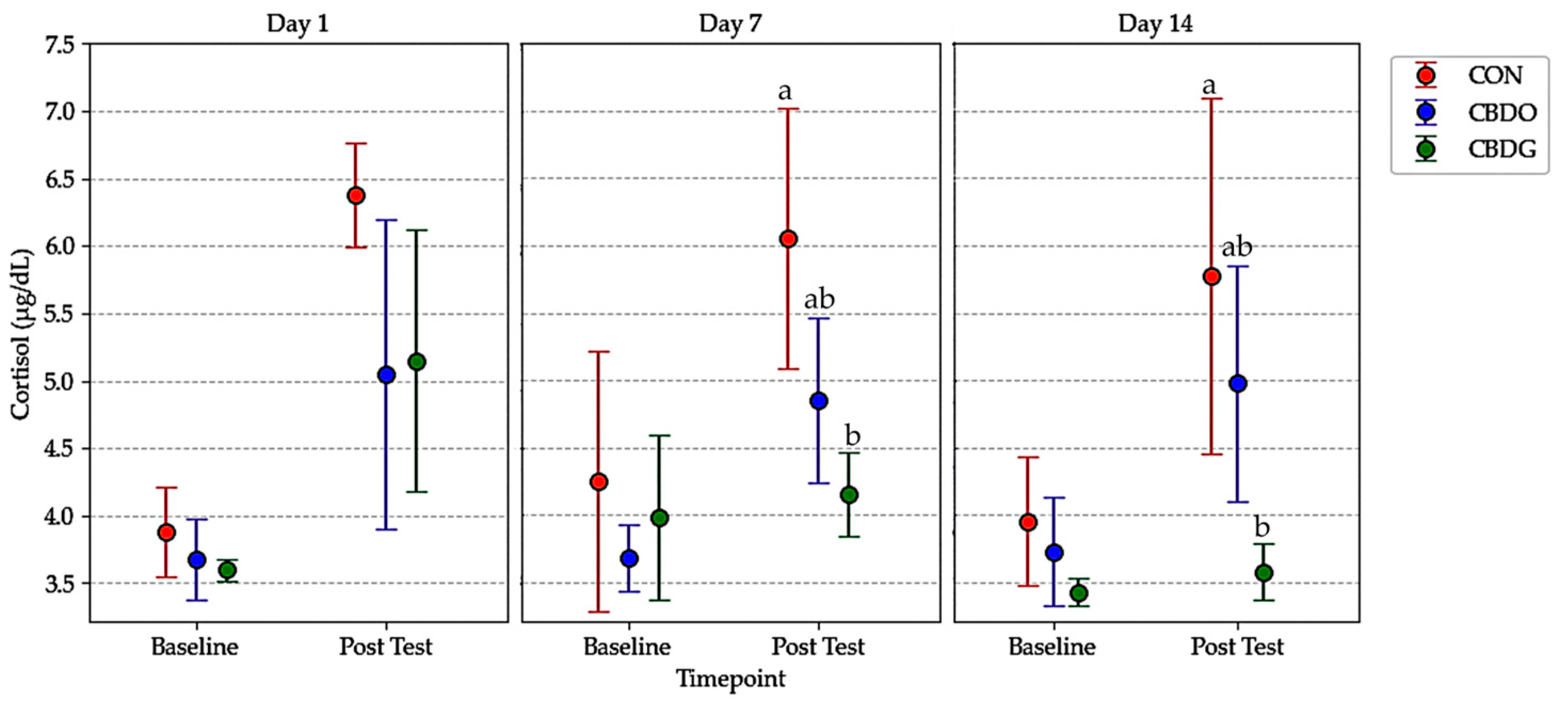
| Serum Cortisol | ||||||||||||
| Cortisol baseline (0.5–5.5 µg/dL) | 3.88 ± 0.33 | 4.25 ± 0.97 | 3.95 ± 0.48 | 3.68 ± 0.30 | 3.68 ± 0.25 | 3.73 ± 0.40 | 3.60 ± 0.08 | 3.98 ± 0.61 | 3.43 ± 0.05 | 0.220 | 0.280 | 0.713 |
| Cortisol Post-Test (5.0–17.0 µg/dL) | 6.38 ± 0.39 | 6.05 ± 0.97 a | 5.78 ± 1.33 a | 5.05 ± 1.15 | 4.85 ± 0.61 ab | 4.98 ± 0.88 ab | 5.15 ± 0.97 A | 4.15 ± 0.31 bAB | 3.58 ± 0.21 bB | 0.001 | 0.042 | 0.615 |
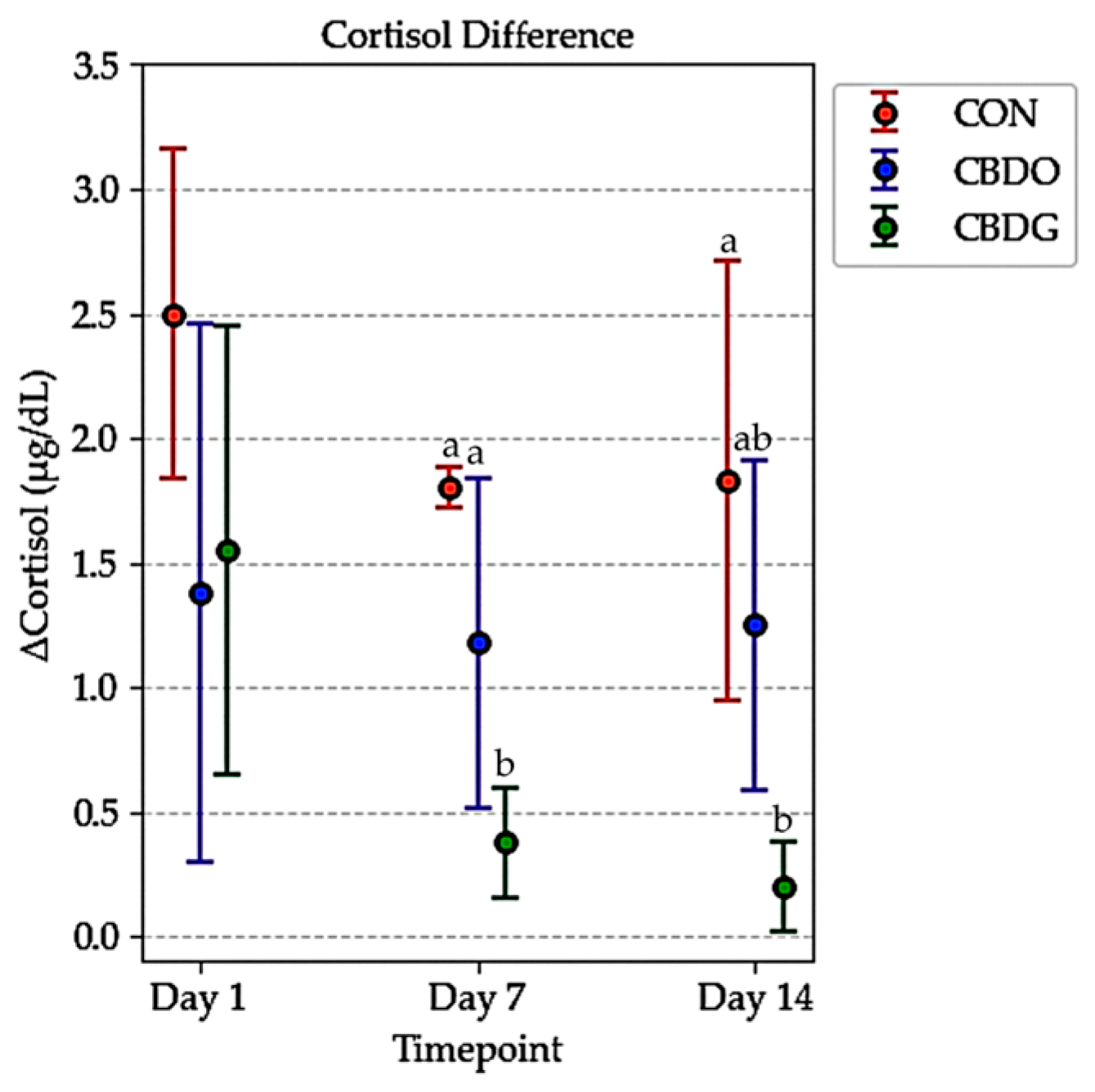
| ∆Cortisol µg/dL | 2.50 ± 0.66 | 1.80 ± 0.08 a | 1.83 ± 0.88 a | 1.38 ± 1.08 | 1.18 ± 0.66 a | 1.25 ± 0.66 ab | 1.55 ± 0.90 A | 0.38 ± 0.22 bB | 0.20 ± 0.18 bB | 0.001 | 0.047 | 0.556 |
Disclaimer/Publisher’s Note: The statements, opinions and data contained in all publications are solely those of the individual author(s) and contributor(s) and not of MDPI and/or the editor(s). MDPI and/or the editor(s) disclaim responsibility for any injury to people or property resulting from any ideas, methods, instructions or products referred to in the content. |
© 2025 by the authors. Licensee MDPI, Basel, Switzerland. This article is an open access article distributed under the terms and conditions of the Creative Commons Attribution (CC BY) license (https://creativecommons.org/licenses/by/4.0/).
Share and Cite
Puttharaksa, W.; Charoensook, R.; Tungtrakanpoung, R.; Hoidokhom, N.; Rungchang, S.; Brenig, B.; Numthuam, S. A Preliminary Evaluation of the Comparative Efficacy of Gel-Based and Oil-Based CBD on Hematologic and Biochemical Responses in Dogs. Vet. Sci. 2025, 12, 342. https://doi.org/10.3390/vetsci12040342
Puttharaksa W, Charoensook R, Tungtrakanpoung R, Hoidokhom N, Rungchang S, Brenig B, Numthuam S. A Preliminary Evaluation of the Comparative Efficacy of Gel-Based and Oil-Based CBD on Hematologic and Biochemical Responses in Dogs. Veterinary Sciences. 2025; 12(4):342. https://doi.org/10.3390/vetsci12040342
Chicago/Turabian StylePuttharaksa, Wassana, Rangsun Charoensook, Rongdej Tungtrakanpoung, Niramon Hoidokhom, Saowaluk Rungchang, Bertram Brenig, and Sonthaya Numthuam. 2025. "A Preliminary Evaluation of the Comparative Efficacy of Gel-Based and Oil-Based CBD on Hematologic and Biochemical Responses in Dogs" Veterinary Sciences 12, no. 4: 342. https://doi.org/10.3390/vetsci12040342
APA StylePuttharaksa, W., Charoensook, R., Tungtrakanpoung, R., Hoidokhom, N., Rungchang, S., Brenig, B., & Numthuam, S. (2025). A Preliminary Evaluation of the Comparative Efficacy of Gel-Based and Oil-Based CBD on Hematologic and Biochemical Responses in Dogs. Veterinary Sciences, 12(4), 342. https://doi.org/10.3390/vetsci12040342