Article

Profiling the Differential Distribution of Ginsenosides Across Ginseng Tissues Using High-Resolution Mass Spectrometry

Institute of Special Animal and Plant Sciences, Chinese Academy of Agricultural Sciences, Changchun 130112, China
*
Author to whom correspondence should be addressed.
Separations 2025, 12(7), 170; https://doi.org/10.3390/separations12070170
Submission received: 27 May 2025 / Revised: 16 June 2025 / Accepted: 23 June 2025 / Published: 26 June 2025

Abstract

This study investigates the compositional characteristics and quantitative differences of ginsenosides across various ginseng tissues, with a particular focus on the specific accumulation patterns of malonyl ginsenosides. Five tissue samples—ginseng fruit (F), leaf (L), taproot (TR), lateral root (LR), and fibrous root (FR)—were analyzed using Orbitrap Fusion high-resolution liquid chromatography–mass spectrometry. A total of 413 ginsenosides, including 33 standards, were identified, encompassing 172 protopanaxadiol (PPD)-type, 188 protopanaxatriol (PPT)-type, 14 oleanolic acid (OA)-type, and 12 ocotillol (OT)-type ginsenosides, of which 160 were malonyl ginsenosides. Statistical analysis revealed significant variations in the relative content per unit mass of malonyl ginsenosides across tissues, with the highest levels in fibrous roots, followed by fruits, lateral roots, leaves, and taproots. Distinct tissue-specific differences in malonyl ginsenoside types and quantities were observed: fruits exhibited 51 malonyl ginsenosides with significantly higher levels, compared to 8, 14, and 17 in lateral roots, fibrous roots, and leaves, whereas TR showed no significant enrichment. This study elucidates the diversity and unique distribution of malonyl ginsenosides in ginseng roots, leaves, and fruits, providing a valuable basis for the targeted selection of tissues with high malonyl ginsenoside content and the development of functional food and medicinal products.

1. Introduction

Ginseng has been revered globally as a “panacea” due to its extensive health benefits [1]. It is widely utilized in traditional Chinese medicine, health supplements, and functional foods [2]. Ginsenosides, the principal secondary metabolites of ginseng, are the key bioactive compounds responsible for its pharmacological effects. They exhibit diverse pharmacological activities, including applications in cancer treatment, anti-inflammatory effects, and cardiovascular and neuroprotective benefits. For example, ginsenoside Rg5 inhibits osteosarcoma cell proliferation by suppressing the PI3K/Akt/mTORC1 pathway and activating the LC3 autophagy pathway to induce apoptosis [3]. Ginsenoside Rg3, in combination with cyclophosphamide, shows significant protective effects against ovarian cancer [4]. In neurodegenerative disease research, ginsenosides have demonstrated the ability to block the extracellular deposition of β-amyloid (Aβ) fibrils and reduce the density of neurofibrillary tangles (NFTs), thereby delaying the pathological progression of Alzheimer’s disease [5].
Malonyl ginsenosides account for 50–60% of the total ginsenoside content in ginseng and are widely distributed across its roots, stems, leaves, flowers, and fruits [6]. These highly polar acidic ginsenosides are characterized by malonyl groups ester-linked to the terminal hydroxyl groups of glycosides, with one or more substituents. Advances in separation and analytical technologies, such as macroporous resin chromatography, silica gel column chromatography, and high-performance liquid chromatography (HPLC), have enabled the isolation of various malonyl ginsenoside monomers, including malonyl Notoginsenoside A and malonyl Ginsenoside Rb1, as well as novel compounds such as double malonyl ginsenosides (e.g., DMR1) [7].
Studies have shown that malonyl ginsenosides possess antidiabetic properties and improve insulin resistance. Their mechanisms involve enhancing insulin sensitivity via the RS1/PI3K/Akt and GLUT4 signaling pathways, resulting in significant hypoglycemic effects, and activating the hepatic AMPK/ACC pathway to regulate lipid metabolism [8]. Wang Dongsheng [9] reported that malonyl ginsenosides from ginseng stems and leaves protect against APAP-induced acute liver injury in mice by significantly reducing serum levels of AST, ALT, IL-1β, and TNF-α, increasing hepatic glutathione (GSH) levels, and inhibiting MDA production. Histopathological analysis revealed that malonyl ginsenosides effectively mitigated hepatocyte necrosis, and Western blotting indicated that this protective effect is closely related to the PI3K/AKT-mediated anti-apoptotic mechanism. These findings systematically elucidate the molecular pharmacological mechanisms of malonyl ginsenosides in metabolic diseases and organ protection.
The accumulation patterns of malonyl ginsenosides vary significantly across different ginseng organs. PARK et al. [10] compared malonyl ginsenoside content in the peeled and unpeeled main roots, lateral roots, and fibrous roots of Korean white ginseng. They found that fibrous roots contained the highest levels of malonyl ginsenosides compared to main and lateral roots. The malonyl ginsenoside content in unpeeled main roots was 20.08 mg/g, while peeled main roots contained only 2.58 mg/g. Furthermore, malonyl ginsenosides are recognized as characteristic components for evaluating diverse ginseng products [11], and tissue-specifically accumulated malonyl ginsenosides can serve as potential chemical quality markers for the detection of ginseng and its processed products, providing a significant chemical basis for the authenticity identification of medicinal materials [12].
Liquid chromatography–mass spectrometry (LC-MS) has significant advantages in the quality control of traditional Chinese medicines, particularly in detecting trace chemical components essential for in-depth analyses [13,14]. LC-MS provides critical data on molecular weights, formulas, and fragmentations, aiding the structural elucidation of compounds. The Orbitrap Fusion system, based on next-generation ultra-high-field Orbitrap technology, offers enhanced resolution due to its reduced trap volume and increased voltage [15,16]. This technique has not only been successfully applied to the discovery of novel malonyl ginsenosides (e.g., double malonyginsenoside Rc, double malonyginsenoside Rb2) [17], but also demonstrates irreplaceable technical superiority in the rapid screening and structural characterization of malonyl ginsenosides within complex matrices [18].
Despite its significance, a systematic investigation of the specific distribution patterns of malonyl ginsenosides across ginseng tissues remains lacking. In this study, ultra-performance liquid chromatography–mass spectrometry (UPLC-MS) was employed to conduct a comprehensive metabolomic analysis of ginseng’s main root, lateral root, fibrous root, leaves, and fruits. This study aims to investigate the compositional characteristics and content variations of ginsenosides in different plant parts, with a particular focus on elucidating the specific accumulation patterns of malonyl ginsenosides.

2. Materials and Methods

2.1. Materials and Reagents

2.1.1. Raw Materials

The ginseng plants used in this study were collected from the Medicinal Plant Resource Garden of the Institute of Special Animal and Plant Sciences, Chinese Academy of Agricultural Sciences (44.05° N, 126.10° E; altitude 266 m). Five parts of 4-year-old ginseng plants—fruits (F), leaf (L), taproot (TR), lateral root (LR), and fibrous root (FR)—were harvested in October 2023, with three biological replicates per part. After being rinsed thoroughly with distilled water, the samples were immediately frozen in liquid nitrogen and stored at −80 °C for subsequent ginsenoside analysis.

2.1.2. Reagents and Consumables

Chromatography-grade methanol, acetonitrile, and formic acid were purchased from Thermo Fisher Scientific Co., Ltd. (Waltham, MA, USA). Ginsenoside standards were obtained from Beijing Solarbio Science & Technology Co., Ltd. (Beijing, China), with a purity >99%.

2.2. Experimental Methods

2.2.1. Sample Preparation

The collected samples were freeze-dried and ground into fine powder using a mortar and pestle. A 20 mg portion of the powder was weighed and mixed with 1 mL of methanol in a 1.5 mL centrifuge tube. The mixture was subjected to ultrasonic treatment at room temperature for 30 min, followed by standing at 4 °C overnight. After centrifugation at 12,000 rpm for 20 min, the supernatant was collected and filtered through a 0.22 μm membrane. The filtrate was transferred into sample vials for further analysis.

2.2.2. LC and MS Conditions

Ginsenoside separation was conducted using a Thermo Scientific™ Dionex™ Vanquish™ 3000 UHPLC system with a Hypersil GOLD™ aQ C18 column (2.1 mm ×  100 mm, 1.9 μm) at a flow rate of 300 µL/min and a column temperature of 35 °C. Binary mobile phases consisted of (A) 1% formic acid and (B) acetonitrile. A linear gradient was applied, starting with 15% B and linearly increasing to 55% B at 34 min, followed by a further linear increase to 98% B at 35 min. Subsequently, the column was washed for the next 1 min, and equilibration was maintained until 40 min. A 1 μL aliquot of the extracted sample was injected for detection using the Thermo Scientific ™ Orbitrap Fusion™ hybrid quadrupole–Orbitrap mass spectrometer equipped with a heated H-ESI source. Untargeted profiling involved a full scan (300–1500 m/z) at 60,000 resolutions, followed by Top 10 data-dependent MS/MS at 15,000 resolutions under negative ionization mode. Key MS/MS parameters included the following: RF lens—50%, AGC target—400,000, maximum IT—50 ms, intensity filter—intensity threshold 5.0 × 10⁴–1.0 × 10²⁰, mass tolerance—±10 ppm, isolation window—1.4 m/z, stepped collision energy—20%, 40%, 60%, dynamic exclusion—6 s. Ionization conditions were optimized and operated at a final spray voltage of −2.7 kV, with heater and capillary temperatures set at 320 °C each. Sheath gas and sweep gas were configured at 40 Arb and 5 Arb, respectively [19].

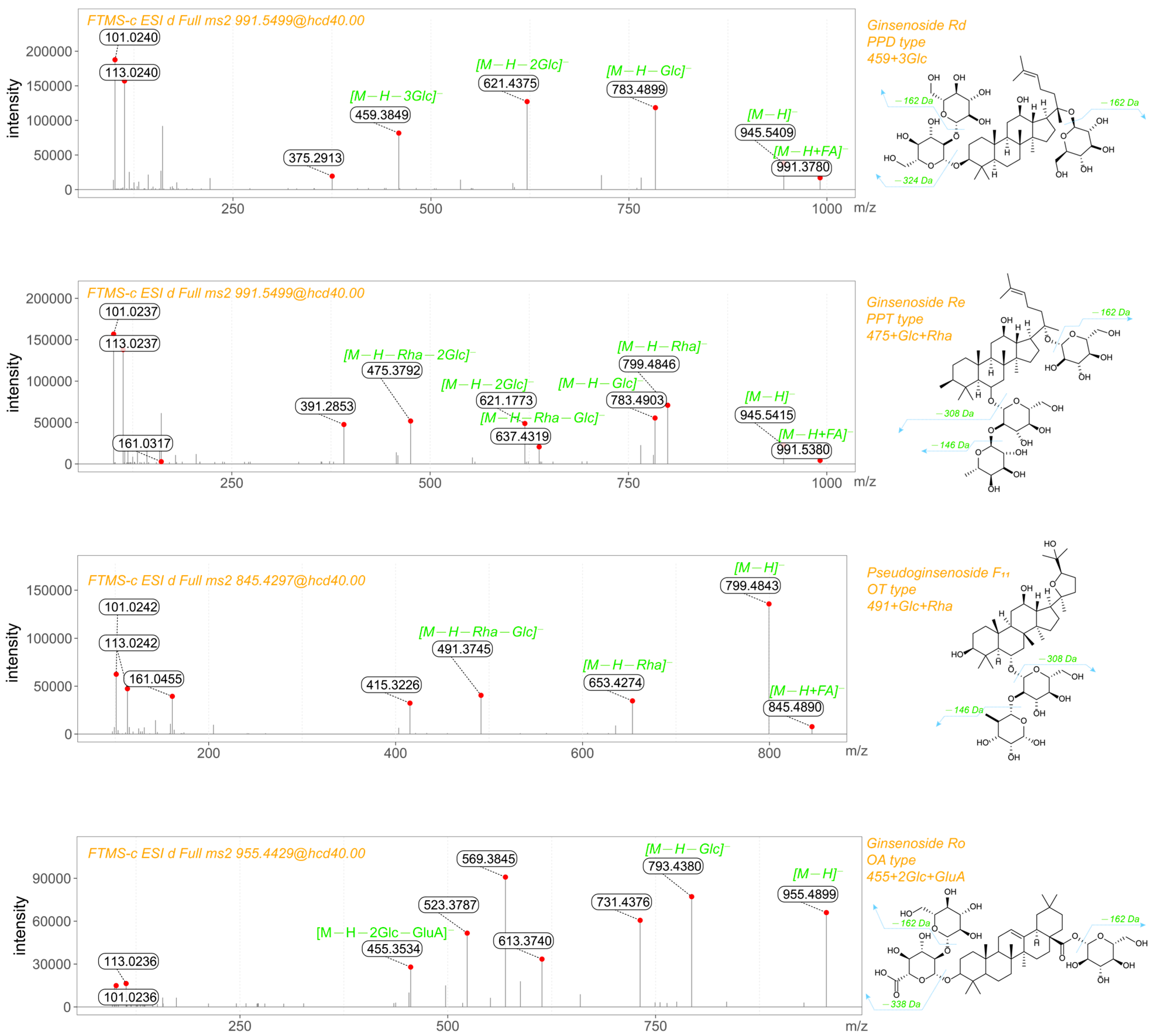
2.2.3. Ginsenoside Identification

The characteristic structure of ginsenosides comprises three components: an aglycone core, glycosyl substituents, and acyl substituents. The aglycone types include protopanaxadiol (PPD), protopanaxatriol (PPT), ocotillol-type (OT), oleanolic acid (OA), and specific-type variants. Specific-type aglycones typically originate from PPD or PPT derivatives with modified C17-side chains or altered aglycone skeletons. Due to structural isomerism among these aglycones, their precise configurations cannot be differentiated through mass spectrometry. Therefore, this study employed molecular weight characterization for identification. A few specific aglycones share identical molecular weights with conventional types and were temporarily classified as standard ginsenosides [20].
The glycosyl moieties consist of glucuronic acid (GluA), glucose (Glc), rhamnose (Rha), arabinose (Ara), and xylose (Xyl). However, Ara and Xyl residues cannot be distinguished via mass spectrometry. For analytical purposes, Xyl was used to represent both arabinose and xylose substituents in this study.
Acyl substituents were categorized into two groups: polar types (e.g., malonyl [Mal]) and nonpolar types (e.g., acetyl [Ace], butenoyl [But], and heptenoyl [Hep]) [21] (Figure 1).
Ginsenoside standards used in this study are listed in Table 1. The detected ginsenosides were named based on their aglycone mass-to-charge ratio (m/z) and substituents (e.g., sugars, malonyl groups, or acetyl groups) in conjunction with their retention times. For instance, aglycones with an m/z of 475, 459, 455, and 491 correspond to PPT-type, PPD-type, OA-type, and OT-type ginsenosides, respectively. Sugar moieties were denoted as follows: Glc (glucose), Xyl (xylose), GluA (glucuronic acid), and Rha (rhamnose). Malonyl (M), dimalonyl (Dim), acetyl (Ace), and butenoyl (But) groups were also included in the naming conventions.
The quality control criteria for ginsenoside identification were established as follows. The relative standard deviation (RSD) of peak areas for the same metabolite in QC samples was ≤30%; the parts-per-million (ppm) difference between the measured and theoretical m/z values was within ±5; the retention time drift was less than 0.2 min.

2.3. Data Processing

A local database was constructed using mzVault (v2.3), with peak area extraction and retrieval performed through Compound Discoverer (v3.3). Experimental data were statistically organized using Excel 2016; differential metabolite analysis was performed with MetaboAnalystR 1.0.1 in R version 3.5.1, where differential metabolites were screened based on Variable Importance in Projection (VIP) scores from the OPLS-DA model, fold-change (FC) values, and FDR correction; volcano plots and PCA plots were generated using the online cloud tool platform [https://cloud.metware.cn/#/home, accessed on 15 December 2024]; bar charts were created with Origin 2021; clustering heatmaps were constructed with TBtools-II v2.098 using MAX-MIN normalized data processed by hierarchical clustering.
The quality control criteria for ginsenoside identification were established as follows: 1. The relative standard deviation (RSD) of peak areas for the same metabolite in QC samples was ≤30%; 2. The parts-per-million (ppm) difference between the measured and theoretical m/z values was within ±5; 3. The retention time drift was less than 0.2 min.

3. Results

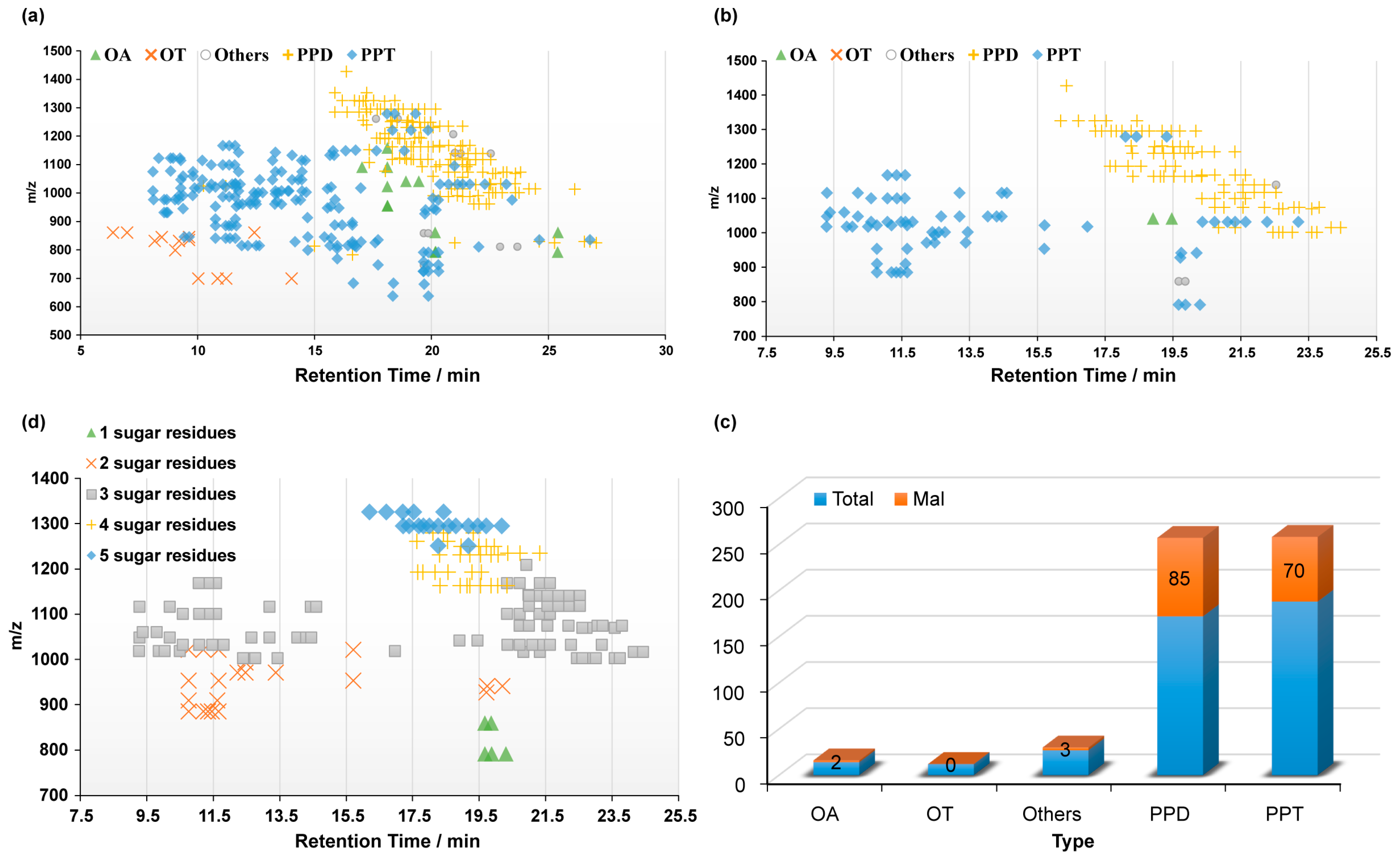
3.1. Composition and Retention Analysis of Ginsenosides

A total of 413 ginsenosides, including 33 standards, were identified across ginseng fruit (F), leaf (L), taproot (TR), lateral root (LR), and fibrous root (FR) samples. These comprised 172 protopanaxadiol (PPD)-type, 188 protopanaxatriol (PPT)-type, 14 oleanolic acid (OA)-type, 12 ocotillol (OT)-type, and 27 other ginsenosides. OT-type ginsenosides exhibited the highest polarity, with retention times between 5 and 15 min, followed by PPT-type (7–20 min). PPD-type and OA-type ginsenosides showed similar polarity values, with retention times concentrated within 15–25 min (Figure 2a).
Ginsenosides with malonyl modifications were predominantly of the PPD and PPT types, accounting for 85 and 70 ginsenosides, respectively, with only 2 OA-type and 3 other-type malonyl ginsenosides identified. No malonyl-modified OT-type ginsenosides were detected. The number of glycosyl groups carried by malonyl ginsenosides followed a specific pattern, primarily consisting of three glycosyl groups: 6 with one glycosyl group, 22 with two glycosyl groups, 85 with three, 30 with four, and 20 with five glycosyl groups.

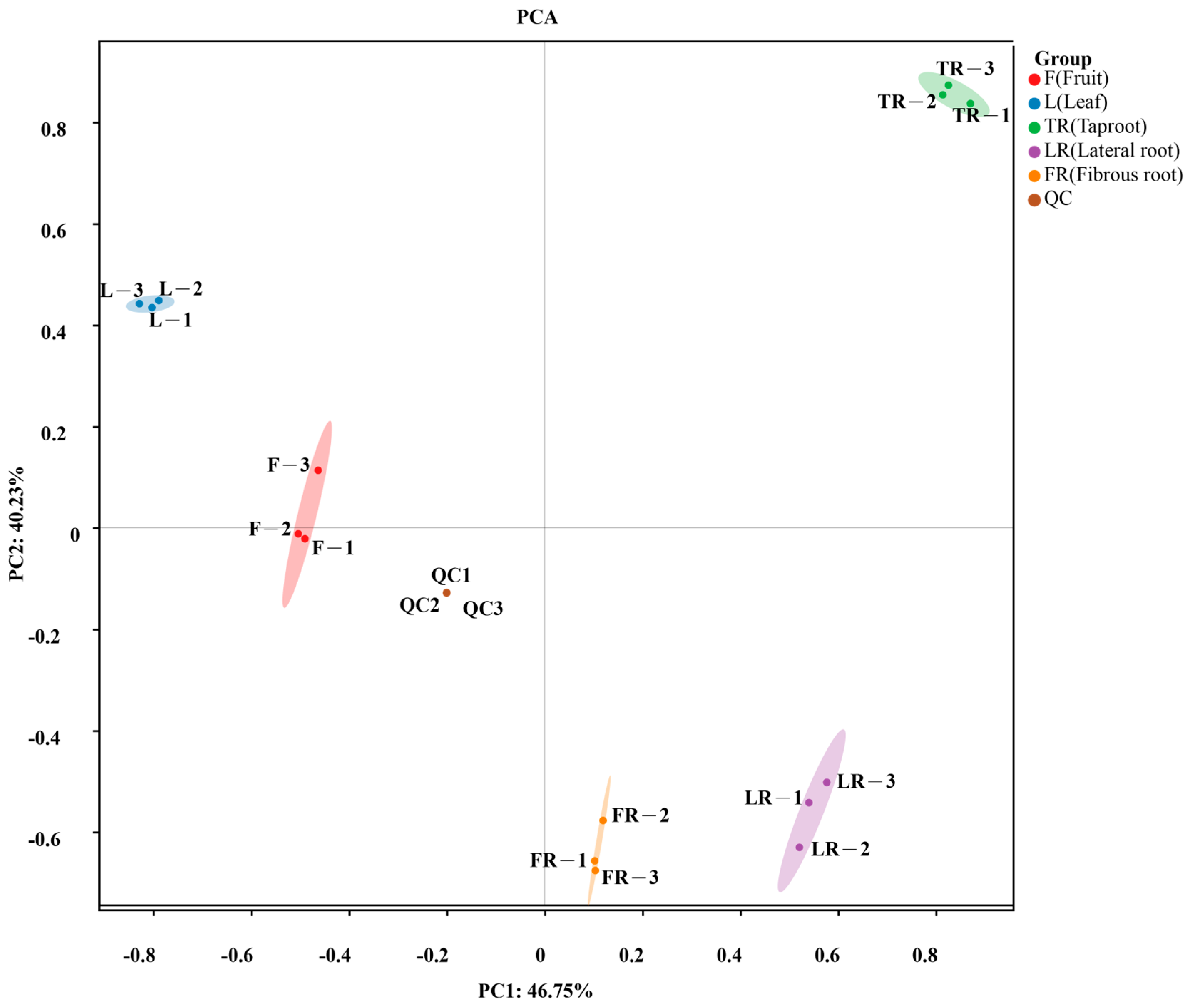
3.2. Principal Component Analysis (PCA) of Ginsenosides Across Tissues

Principal component analysis (PCA) was performed to evaluate differences in ginsenoside distribution among ginseng tissues. The first two principal components cumulatively explained 86.98% of the variance, with PC1 and PC2 contributing 46.75% and 40.23%, respectively (Figure 3). Distinct clustering patterns were observed, indicating significant differences in ginsenoside composition across tissues. Samples within the same group exhibited high intra-group similarity, as evidenced by compact clustering. Quality control (QC) samples showed tight clustering, further supporting the stability of the UHPLC-MS methodology. Quality control (QC) samples showed tight clustering, further supporting the stability of the UHPLC-MS methodology. Fibrous roots (FR) and lateral roots (LR) were closely positioned, suggesting comparable ginsenoside profiles.

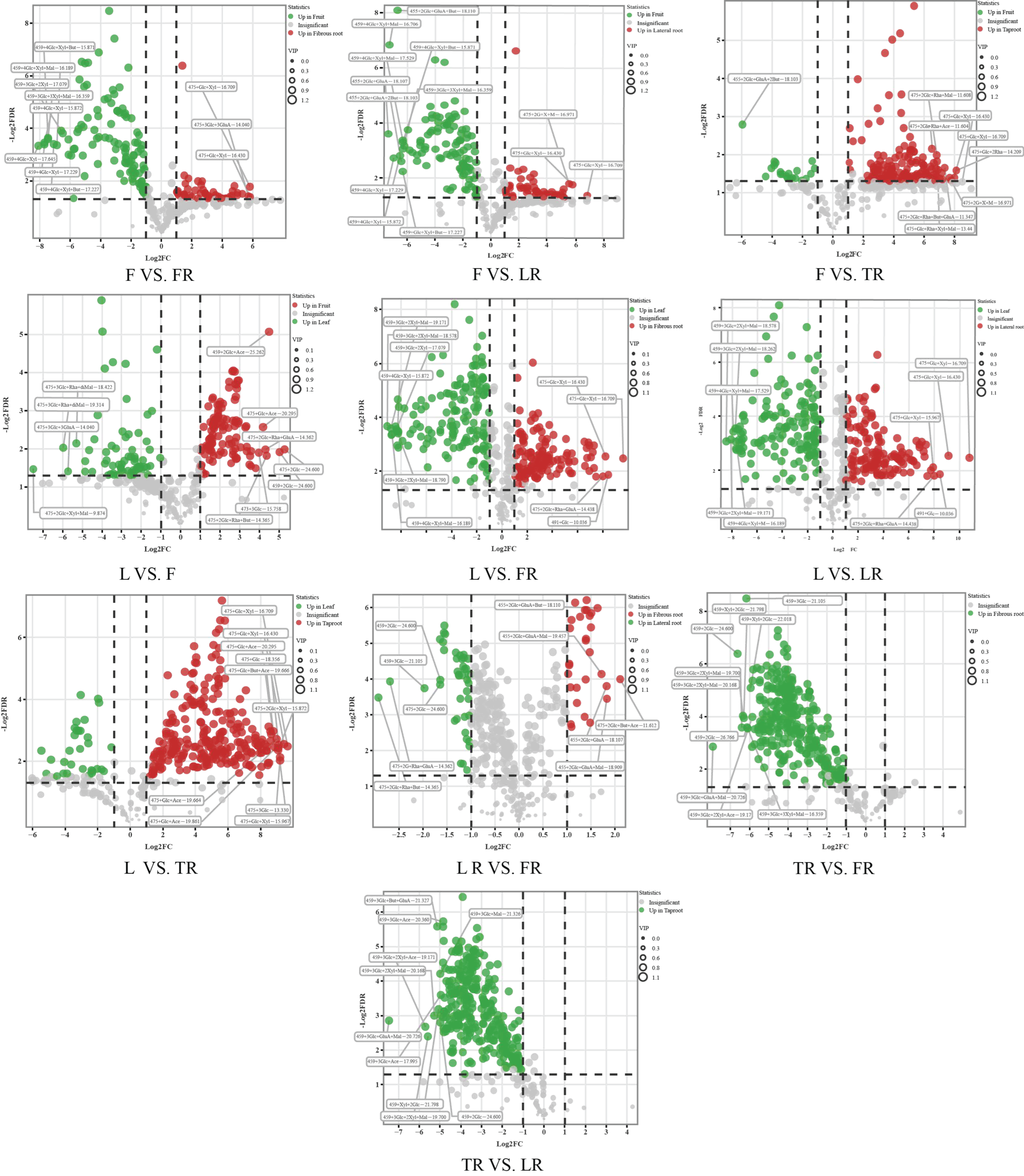
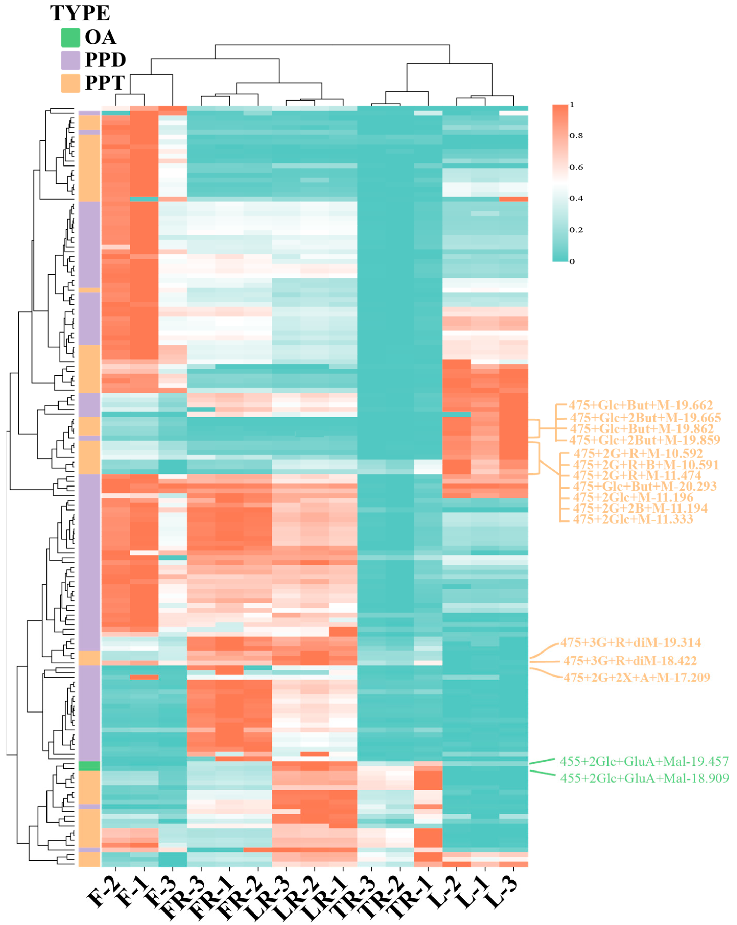
3.3. Analysis of Ginsenoside Content Differences in Different Ginseng Parts

Based on the screening criteria of VIP > 1, fold change > 2, and FDR < 0.05, differential ginsenosides were identified among the sample groups, as shown in Table 2. The LR vs. FR group exhibited the fewest differential ginsenosides, with only 72, whereas the other pairwise comparisons involved over 165 differential ginsenosides. The TR vs. FR and TR vs. LR groups, both representing underground parts, showed the highest numbers of differential ginsenosides, with 301 and 306, respectively, all of which were less abundant in the main root. Among the aboveground parts, the F vs. L group displayed 187 differential ginsenosides. Additionally, the L vs. LR, L vs. FR, and L vs. TR groups each exhibited 294 differential ginsenosides. The F vs. FR, F vs. LR, and F vs. TR groups presented 165, 170, and 172 differential ginsenosides, respectively.
Among the top 10 significantly different ginsenosides in each comparison group (Figure 4), certain patterns emerged; in comparisons between leaves and the three root parts, the differential ginsenoside 475+Glc+Xyl-16.709 was shared. Similarly, comparisons between fruits and the three root parts revealed the common differential ginsenosides 475+Glc+Xyl-16.430 and 475+Glc+Xyl-16.709. Additionally, the ginsenosides 459+3Glc+2Xyl+Mal-19.700, 459+3Glc+2Xyl+Mal-20.168, 459+3Glc+2Xyl+Ace-19.171, 459+2Glc-24.600, and 459+Xyl+2Glc-21.798 were significantly different in the TR vs. FR and TR vs. LR groups.

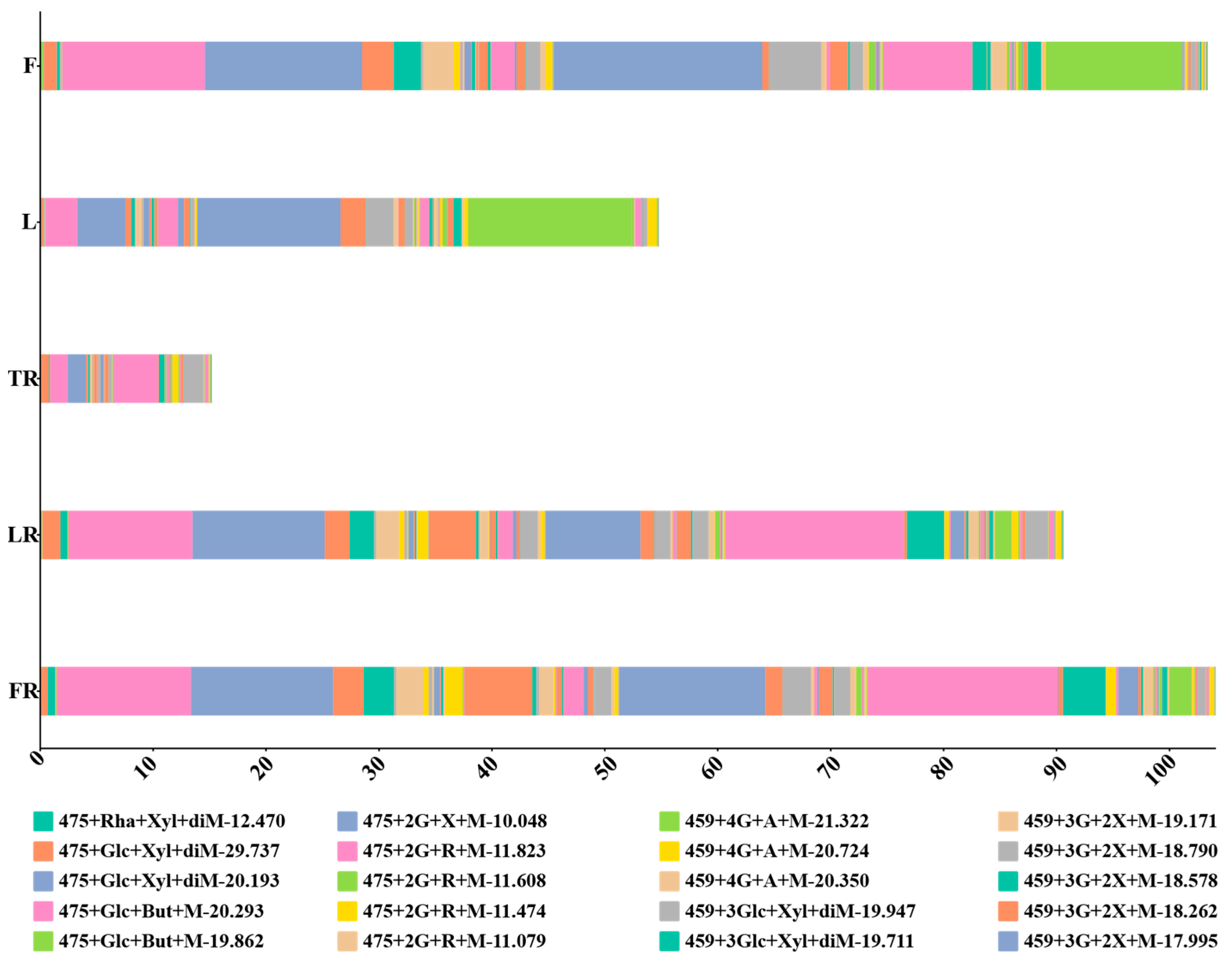
3.4. Analysis of Malonylated Ginsenoside Content Differences in Different Ginseng Parts

In total, 160 malonyl ginsenosides were identified from 15 samples, and detailed information is presented in Supplementary Table S1. As shown in Figure 5, the relative contents of malonylated ginsenosides per unit mass significantly varied among the five ginseng parts, ranked from highest to lowest as FR > F > LR > L > TR. The composition and distribution of malonylated ginsenosides also displayed noticeable differences, reflecting the distinct accumulation and metabolic characteristics in each part.
To systematically explore the accumulation patterns of malonylated ginsenosides in ginseng, the identified ginsenosides were normalized, and a heatmap was generated (Figure 6). The heatmap revealed that most malonylated ginsenosides exhibited high accumulation levels in fruits, except for two OA-type ginsenosides (455+2Glc+GluA+Mal-18.909 and 455+2Glc+GluA+Mal-19.457), which showed low accumulation levels. In the fibrous roots, 32 PPD-type malonylated ginsenosides exhibited high contents. The lateral roots contained both high levels of PPD-type malonylated ginsenosides similar to the fibrous roots and a unique accumulation of PPT-type malonylated ginsenosides, forming a distinct distribution pattern. Although leaves showed generally low malonylated ginsenoside contents, specific PPT-type ginsenosides, such as 475+Glc+But+Mal-20.293, 475+2Glc+Rha+But+Mal-10.591, 475+2Glc+2But+Mal-11.194, and 475+2Glc+Rha+Mal-11.474, displayed notably high levels, highlighting the unique accumulation characteristics in leaves.
Regarding the number of differential malonylated ginsenosides, fruits exhibited the highest number, with 53 significantly different compounds compared to other parts. The lateral roots had 9, the fibrous roots had 17, and the leaves had 23 (Table 3). Seven PPT-type malonylated ginsenosides, including 475+2Glc+Rha+Mal-11.823, 475+3Glc+Mal-14.037, 475+3Glc+But+Mal-14.601, 475+2Glc+Rha+Mal-11.608, 475+2Glc+Rha+2But+Mal-11.349, 475+2Glc+Rha+But+Mal-11.352, and 475+2Glc+Rha+2But+Mal-11.596, were significantly more abundant in fruits and leaves than in the three root parts. Conversely, the ginsenosides 459+4Glc+Mal-18.555, 459+2Glc+2Xyl+Ace+diMal-18.550, 459+4Glc+Mal-17.645, 475+3Glc+Rha+diMal-19.314, 475+3Glc+Rha+diMal-18.422, and 475+2Glc+2Xyl+Ace+Mal-17.209 were significantly more abundant in the fibrous and lateral roots than in the other three parts.

4. Discussion

In recent years, the compositional analysis and functional characterization of ginsenosides across different ginseng organs have emerged as pivotal research topics in traditional Chinese medicine. Extensive studies have demonstrated significant organ-specific variations in the distribution and bioactivity profiles of ginsenosides across roots, stems, and leaves. As pharmacologically valuable secondary metabolites, investigations into organ-specific accumulation patterns of malonyl ginsenosides have primarily centered on stems, leaves, and roots, with preliminary insights elucidating their organ-specific mechanisms [22]. Using metabolomic relative quantification, this study identified 160 malonyl ginsenosides from 15 samples, revealing distinct differences in their relative unit mass contents across tissues. Fibrous roots exhibited the highest abundance, followed by fruits, lateral roots, leaves, and main roots. This organ-specific distribution pattern aligns closely with the findings by Zhang Furui et al. [23], who employed HPLC quantification and chemometric analyses to confirm fibrous roots as the principal accumulation site for malonyl ginsenosides. While leaves exhibited lower overall accumulation of malonyl ginsenosides, certain characteristic malonyl ginsenosides were uniquely enriched, underscoring the metabolic modification capabilities distinctive to leaf tissues.
These pronounced distribution differences are unlikely to be coincidental, instead reflecting organ-specific metabolic regulation. For instance, specific malonyl ginsenosides, such as 459+3Glc+2Xyl+Mal-18.578 and 459+3Glc+2Xyl+Mal-19.171, exhibited significantly higher relative abundances in fibrous roots compared to other organs, alongside their neutral saponin precursors (e.g., 459+3Glc+2Xyl-17.079), which were also abundant in fibrous roots. This suggests that fibrous roots harbor highly active biosynthetic pathways for precursor saponins, facilitating the preferential accumulation of malonylated derivatives. Furthermore, the biosynthesis of leaf-specific modified ginsenosides may be linked to the unique enzymatic microenvironment of leaves, such as light-induced cofactors. These organ-preferential metabolic pathways constitute a fundamental basis for the chemical diversity and functional specialization of ginsenosides.
The biosynthesis and accumulation of ginsenosides exhibit clear organ differentiation. Previous studies have demonstrated that aerial organs (e.g., leaves, stems, flowers) predominantly accumulate protopanaxatriol (PPT)-type ginsenosides, likely due to their involvement in photosynthetic product allocation, light stress response, or aboveground defense mechanisms. In contrast, underground organs (e.g., roots, rhizomes) primarily enrich protopanaxadiol (PPD)-type ginsenosides, with rhizomes showing the highest levels of Ro (OA-type saponins) [24]. As critical derivatives of PPD- and PPT-type ginsenosides, the high accumulation of malonyl ginsenosides in fibrous roots further refines the metabolic compartmentalization of saponins within underground organs. Fibrous roots, as absorptive structures, are metabolically active and possess specific cellular microenvironments (e.g., pH, cofactor concentrations) that may favor malonylation reactions and ensure the stable storage of malonylated ginsenosides, potentially within vacuoles. Malonylation not only enhances the water solubility of saponins, but may also influence their bioactivity, stability, and transport efficiency within the plant. Consequently, the high abundance of malonyl ginsenosides in fibrous roots likely represents a specific metabolic strategy evolved by ginseng to adapt to underground environments (e.g., interactions with soil microbiota, nutrient uptake regulation) or to optimize the storage and transport of bioactive compounds.

5. Conclusions

Through the comprehensive analysis of ginsenosides across different ginseng parts, a total of 413 ginsenosides (including isomers and 33 standards) were identified, comprising 172 PPD-type, 188 PPT-type, 14 OA-type, 12 OT-type, and 27 other types, including 160 malonylated ginsenosides. The concentrations of these compounds varied significantly among tissues, with the highest levels found in fibrous roots, followed by fruits, lateral roots, leaves, and taproots. Distinct differences in the types and quantities of malonylated ginsenosides were observed across parts.
In total, 53 malonylated ginsenosides were predominantly abundant in fruits, while lateral roots, fibrous roots, and leaves contained 8, 14, and 23 characteristic malonylated ginsenosides, respectively. Seven malonylated ginsenosides, including 475+2Glc+Rha+Mal-11.823, were significantly more abundant in fruits and leaves than in root tissues, whereas six compounds, such as 459+4Glc+Mal-18.555, were more concentrated in fibrous and lateral roots. These findings highlight the diversity and specificity of malonylated ginsenoside distribution in ginseng roots, leaves, and fruits, providing an important foundation for the targeted selection of tissues with high malonylated ginsenoside content and the development of related food and medicinal products.

Supplementary Materials

The following supporting information can be downloaded at: https://www.mdpi.com/article/10.3390/separations12070170/s1, Figure S1: The stacked histograms of malonyl ginsenosides content of each sample; Figure S2: Row-normalized Heatmap of Malonyl Ginsenoside Content of Each Sample; Figure S3: Chromatograms of ginsenosides detected in five tissues of ginseng; Table S1: Malonyl ginsenosides detectd in 15 samples.

Author Contributions

Conceptualization, C.L.; Methodology, Z.L.; Software, Z.L.; Validation, C.L.; Formal analysis, Y.W.; Investigation, Y.W.; Resources, S.Q.; Data curation, H.X.; Writing—original draft, H.X.; Writing—review & editing, S.Q.; Visualization, H.X.; Supervision, H.Z.; Project administration, H.Z.; Funding acquisition, H.Z. All authors have read and agreed to the published version of the manuscript.

Funding

This study was supported by the National Key R&D Program of China (2021YFD1600901), the Jilin Provincial Major Science and Technology Special Program (20230304001YY), and the Innovation Program of the Chinese Academy of Agricultural Sciences (CAAS-CSIAF-202303).

Data Availability Statement

The original contributions presented in the study are included in the article, further inquiries can be directed to the corresponding author.

Conflicts of Interest

The authors declare that they have no conflicts of interest.

References

  1. Loo, S.; Kam, A.; Dutta, B.; Zhang, X.; Feng, N.; Sze, S.K.; Liu, C.F.; Wang, X.; Tam, J.P. Broad-Spectrum Ginsentides Are Principal Bioactives in Unraveling the Cure-All Effects of Ginseng. Acta Pharm. Sin. B 2024, 14, 653–666. [Google Scholar] [CrossRef] [PubMed]
  2. Riaz, M.; Rahman, N.U.; Zia-Ul-Haq, M.; Jaffar, H.Z.E.; Manea, R. Ginseng: A Dietary Supplement as Immune-Modulator in Various Diseases. Trends Food Sci. Technol. 2019, 83, 12–30. [Google Scholar] [CrossRef]
  3. Liu, M.Y.; Liu, F.; Li, Y.J.; Yin, J.N.; Gao, Y.L.; Wang, X.Y.; Yang, C.; Liu, J.G.; Li, H.J. Ginsenoside Rg5 Inhibits Human Osteosarcoma Cell Proliferation and Induces Cell Apoptosis through PI3K/Akt/mTORC1-Related LC3 Autophagy Pathway. Oxid. Med. Cell. Longev. 2021, 2021, 5040326. [Google Scholar] [CrossRef] [PubMed]
  4. Xu, T.; Xin, Y.; Cui, M.; Jiang, X.; Gu, L. Inhibitory Effect of Ginsenoside Rg3 Combined with Cyclophosphamide on Growth and Angiogenesis of Ovarian Cancer. Chin. Med. J. 2007, 120, 584–588. [Google Scholar] [CrossRef]
  5. Shi, Z.; Chen, H.; Zhou, X.; Yang, W.; Lin, Y. Pharmacological Effects of Natural Medicine Ginsenosides against Alzheimer’s Disease. Front. Pharmacol. 2022, 13, 952332. [Google Scholar] [CrossRef]
  6. Liu, Z.; Li, Y.; Li, X.; Ruan, C.C.; Wang, L.J.; Sun, G.Z. The Effects of Dynamic Changes of Malonyl Ginsenosides on Evaluation and Quality Control of Panax ginseng C.A. Meyer. J. Pharm. Biomed. Anal. 2012, 64, 56–63. [Google Scholar] [CrossRef] [PubMed]
  7. Cui, Y.; Wu, J.; Qu, C.; Li, J.; Li, Z.; Liu, Z. Isolation and identification of two new malonyl ginsenosides from Panax quinquefolius. Chin. Tradit. Herb. Drugs 2023, 54, 6940–6945. [Google Scholar]
  8. Wang, D.S.; Wang, J.M.; Zhang, F.R.; Lei, F.J.; Wen, X.; Song, J.; Sun, G.Z.; Liu, Z. Ameliorative Effects of Malonyl Ginsenoside from Panax ginseng on Glucose-Lipid Metabolism and Insulin Resistance via IRS1/PI3K/Akt and AMPK Signaling Pathways in Type 2 Diabetic Mice. Am. J. Chin. Med. 2022, 50, 863–882. [Google Scholar] [CrossRef]
  9. Wang, D. Chemical Constituents of Malonyl Ginsenosides from Stems and Leaves of Panax ginseng C. A. Meyer and Their Protective Effects on Acetaminophen-Induced Liver Injury in Mice. Master’s Thesis, Jilin Agricultural University, Changchun, China, 2024. [Google Scholar]
  10. Park, Y.S.; Oh, M.H.; Lee, H.; Jung, J.T.; Jo, Y.H.; Pyo, M.K. Comparison of Malonyl Ginsenoside Contents in Parts of Korean Ginseng. Korean J. Pharmacogn. 2017, 48, 82–87. [Google Scholar]
  11. Wu, W.; Sun, L.; Zhang, Z.; Guo, Y.; Liu, S. Profiling and Multivariate Statistical Analysis of Panax ginseng Based on Ultra-High-Performance Liquid Chromatography Coupled with Quadrupole-Time-of-Flight Mass Spectrometry. J. Pharm. Biomed. Anal. 2015, 107, 141–150. [Google Scholar] [CrossRef]
  12. Yao, C.; Wang, J.; Li, Z.; Qu, H.; Pan, H.; Li, J.; Wei, W.; Zhang, J.; Bi, Q.; Guo, D. Characteristic Malonyl Ginsenosides from the Leaves of Panax Notoginseng as Potential Quality Markers for Adulteration Detection. J. Agric. Food Chem. 2021, 69, 4849–4857. [Google Scholar] [CrossRef]
  13. Jiao, X.; Li, R.; Ocholi, S.S.; Wang, H.; Cui, T.; Chen, B.; Wang, L.; Fu, Z.; Liu, E.; Wang, F.; et al. A Multi-Level Strategy Based on Comprehensive Two-Dimensional Liquid Chromatography-Q-Orbitrap-Mass Spectrometry Combined with PLS Regression Model and RT-Ensemble Pred Model to Intelligently Distinguish Different Geographical Locations of Huanglian Sample. J. Pharm. Biomed. Anal. 2025, 262, 116864. [Google Scholar] [CrossRef]
  14. Yang, J.; Wu, Y.; Wei, W.; Guo, W.; Li, M.; Jia, J.; Xu, Y.; Wang, Y. Study on Cold and Hot Properties of Chinese Materia Medica Using Liquid Chromatography-Mass Spectrometry-Based Metabolomics Combined with Network Pharmacology Analysis. Biomed. Chromatogr. 2025, 39, e70070. [Google Scholar] [CrossRef] [PubMed]
  15. Zhang, P.; Li, S.; Li, J.; Wei, F.; Cheng, X.; Zhang, G.; Ma, S.; Liu, B. Identification of Ophiocordyceps Sinensis and Its Artificially Cultured Ophiocordyceps Mycelia by Ultra-Performance Liquid Chromatography/Orbitrap Fusion Mass Spectrometry and Chemometrics. Molecules 2018, 23, 1013. [Google Scholar] [CrossRef]
  16. Tu, B.; Wang, Y.; Wu, Z.; Zhou, W.; Tang, X.; Zhang, C.; Gao, Y. DIA-Based Serum Proteomics Revealed the Protective Effect of Modified Siwu Decoction against Hypobaric Hypoxia. J. Ethnopharmacol. 2024, 319, 117303. [Google Scholar] [CrossRef] [PubMed]
  17. Qu, Z.; Zheng, P.; Li, Y.; Hou, W.; Wang, Y. Rapid Identification of Saponins in Panax Notoginseng Fruits by Ultra High-Performance Liquid Chromatography-Orbitrap High-Resolution Mass Spectrometry. Chin. J. Anal. Chem. 2022, 50, 225–259. [Google Scholar] [CrossRef]
  18. Yang, W.; Ye, M.; Qiao, X.; Liu, C.; Miao, W.; Bo, T.; Tao, H.; Guo, D. A Strategy for Efficient Discovery of New Natural Compounds by Integrating Orthogonal Column Chromatography and Liquid Chromatography/Mass Spectrometry Analysis: Its Application in Panax ginseng, Panax quinquefolium and Panax notoginseng to Characterize 437 Potential New Ginsenosides. Anal. Chim. Acta 2012, 739, 56–66. [Google Scholar] [CrossRef] [PubMed]
  19. Pang, S.; Piao, X.; Zhang, X.; Chen, X.; Zhang, H.; Jin, Y.; Li, Z.; Wang, Y. Discrimination for Geographical Origin of Panax quinquefolius L. Using UPLC Q-Orbitrap MS-Based Metabolomics Approach. Food Sci. Nutr. 2023, 11, 4843–4852. [Google Scholar] [CrossRef]
  20. Jin, Y.; Hao, Y.; Zhang, H.; Qu, Z.; Wang, Y.; Piao, X. Dynamic Changes of Ginsenosides in Panax quinquefolium Fruit at Different Development Stages Measured Using UHPLC-Orbitrap MS. Rapid Commun. Mass Spectrom. 2022, 36, e9270. [Google Scholar] [CrossRef]
  21. Yang, W.; Zhang, Y.; Wu, W.; Huang, L.; Guo, D.; Liu, C. Approaches to Establish Q-Markers for the Quality Standards of Traditional Chinese Medicines. Acta Pharm. Sin. B 2017, 7, 439–446. [Google Scholar] [CrossRef]
  22. Wan, J.; Li, S.; Chen, J.; Wang, Y. Chemical Characteristics of Three Medicinal Plants of the Panax Genus Determined by HPLC-ELSD. J. Sep. Sci. 2007, 30, 825–832. [Google Scholar] [CrossRef] [PubMed]
  23. Zhang, F.; Han, X.; Li, D.; Liu, Z. Differences of malonyl ginsenosides and neutral ginsenosides in different parts of Panax ginseng and Panax notoginseng by high performance liquid chromatography and chemometric analysis. China Food Addit. 2024, 35, 238–245. [Google Scholar] [CrossRef]
  24. Piao, X.; Zhang, H.; Kang, J.P.; Yang, D.U.; Li, Y.; Pang, S.; Jin, Y.; Yang, D.C.; Wang, Y. Advances in Saponin Diversity of Panax ginseng. Mol. Basel Switz. 2020, 25, 3452. [Google Scholar] [CrossRef] [PubMed]
Figure 1. The cleavage rules of several typical types of ginsenosides.
Separations 12 00170 g001
Figure 2. Composition and retention analysis of ginsenosides. (a) 2D scatter plot of retention times and molecular weights of different types of ginsenosides. (b) 2D scatter plot of retention times and molecular weights of different types of malonyl ginsenosides. (c) Number of malonyl ginsenosides in different types of ginsenosides. (d) Number of sugar groups in each type of malonyl ginsenoside.
Separations 12 00170 g002
Figure 3. PCA plots of ginseng tissue samples.
Separations 12 00170 g003
Figure 4. Differential ginsenoside volcano plot for each sample. Note: The numbers in the figure correspond to the top 10 species of Ginsenosides with significant differences between groups. The horizontal dashed line demarcates the FDR-adjusted significance threshold (FDR < 0.05), while the vertical dashed lines delineate the |log2(fold change)| = 1.0 cutoff, corresponding to a 2-fold differential expression criterion.
Separations 12 00170 g004
Figure 5. The stacked histograms of malonyl ginsenosides contents of each sample. Note: Due to the extensive number of ginsenosides identified, only a partial legend is presented in the main figures for clarity. The full legend can be found in Supplementary Figure S1. The name of the saponin in the figure indicates G for glucosyl, X for 5-carbon sugar group, GluA for glucuronic acid, R for rhamnose, M for malonyl, Dim for di- malonyl, A for acetyl, and B for butenyl.
Separations 12 00170 g005
Figure 6. Row-normalized heatmap of malonyl ginsenoside content of each sample. Note: Due to the extensive number of malonyl ginsenosides identified, only a partial legend is presented in the main figures for clarity. The full legend can be found in Supplementary Figure S2. The names of the saponins in the figure contain G for glucosyl, X for 5-carbon sugar group, GluA for glucuronic acid, R for rhamnose, M for malonyl, Dim for di- malonyl, A for acetyl, and B for butenyl.
Separations 12 00170 g006
Table 1. Table of reference for ginsenoside standards.
NameIdentificationm/zRetention Time
475+2Glc+Xyl-8.68Notoginsenoside R1977.5327 8.68
475+2Glc-09.65Ginsenoside Rg1845.4915 09.65
475+2Glc+Rha-09.79Ginsenoside Re991.5490 09.79
475+2Glc-14.75Ginsenoside Rf845.4911 14.75
475+Rha+Glc-16.6720S-Ginsenoside Rg2829.4976 16.67
475+Glc-16.7120S-Ginsenoside Rh1683.4385 16.71
475+Rha+Glc-17.0420R-Ginsenoside Rg2829.4982 17.04
475+Glc-17.2420R-Ginsenoside Rh1683.4398 17.24
459+4Glc-17.36Ginsenoside Rb11153.6023 17.36
459+3Glc+Xyl-18.07Ginsenoside Rc1123.5928 18.07
455+2Glc+GluA-18.15Ginsenoside Ro955.4921 18.15
459+3Glc+Xyl-18.69Ginsenoside Rb21123.5896 18.69
459+3Glc+Xyl-18.89Ginsenoside Rb31123.5886 18.89
459+3Glc-20.13Ginsenoside Rd991.5480 20.13
441+2Glc-23.07Ginsenoside Rg6811.4873 23.07
457+Glc-23.62Ginsenoside Rk3665.4291 23.62
457+Glc-24.16Ginsenoside Rh4665.4294 24.16
459+2Glc-26.8820S-Ginsenoside Rg3829.4978 26.88
459+2Glc-27.4120R-Ginsenoside Rg3829.4971 27.41
459+2Glc+Ace-29.9920S-Ginsenoside Rs3871.5071 29.99
459+2Glc+Ace-30.2820R-Ginsenoside Rs3871.5085 30.28
441+2Glc-31.99Ginsenoside Rk1811.4864 31.99
441+2Glc-32.46Ginsenoside Rg5811.4879 32.46
459+Glc-32.96Ginsenoside Rh2667.4450 32.96
459+4Glc+Xyl-15.85Notoginsenoside R41285.6000 15.85
459+4Glc+Xyl-17.22Ginsenoside Ra31285.6000 17.22
459+3Glc+2Xyl-17.08Ginsenoside Ra21255.6000 17.08
459+Glc-31.69Ginsenoside compound K667.4437 31.69
475+Glc-18.44Ginsenoside F1683.4371 18.44
441+2Glc-23.77Ginsenoside F4811.4861 23.77
475+Xyl+Glc-16.51Ginsenoside F3815.4820 16.51
459+2Glc-24.72Ginsenoside F2829.4979 24.72
491+Rha+Glc-14.35Pesudoginsenoside F11845.4914 14.35
Table 2. The number of ginsenosides differing between groups of samples.
GroupDifferential Ginsenoside Number
LR vs. FR72
TR vs. FR301
TR vs. LR306
F vs. L187
L vs. LR294
L vs. FR294
L vs. TR294
F vs. FR165
F vs. LR170
F vs. TR172
Table 3. Quantities of specific malonyl ginsenosides by site.
PlaceNumber of Tissue-Specific Malonyl GinsenosidesName
Fruits53459+Glc+Rha+Xyl+2But+diMal-20.599459+3Glc+Xyl+diMal-18.949475+Glc+Xyl+diMal-29.737
475+3Glc+Mal-12.660475+Glc+Xyl+diMal-20.193459+Glc+Rha+Xyl+But+diMal-21.238
475+2Glc+Xyl+Mal-9.874475+2Glc+Xyl+Mal-9.277475+Glc+Rha+Xyl+Mal-12.404
475+Glc+Rha+Xyl+Mal-12.588475+2Glc+Xyl+Mal-16.971475+Glc+Rha+Xyl+Mal-13.443
475+2Glc+Xyl+Mal-10.048475+3Glc+Mal-14.448475+3Glc+But+Mal-14.444
475+2Glc+Xyl+Mal-10.5000475+Glc+Rha+Xyl+Mal-12.773475+3Glc+But+Mal-13.201
475+Glc+Rha+Xyl+Mal-12.773475+3Glc+But+Mal-13.201475+3Glc+Mal-13.203
475+2Glc+Rha+But+Mal-11.587459+Glc+Rha+Xyl+But+diMal-21.562459+3Glc+Ace+Mal-21.563
459+3Glc+diMal-21.562459+3Glc+diMal-22.532459+3Glc+diMal-21.889
459+Glc+Rha+Xyl+But+diMal-21.889459+Glc+Rha+Xyl+But+diMal-22.532459+3Glc+diMal-22.188
459+3Glc+Ace+Mal-22.192459+Glc+Rha+Xyl+But+diMal-22.190459+3Glc+Ace+Mal-23.212
459+4Glc+Mal-19.561459+2Glc+Rha+Mal-24.159459+Glc+Rha+Xyl+But+diMal-21.002
459+Glc+2Xyl+2Ace+diMal-20.995459+3Glc+Ace+Mal-20.999459+3Glc+Mal-21.326
459+2Glc+Rha+Mal-21.330475+3Glc+Mal-14.297459+3Glc+Mal-23.191
459+2Glc+Xyl+Mal-23.018459+3Glc+Mal-22.270459+4Glc+Ace+Mal-21.322
459+3Glc+Mal-20.360459+3Glc+Mal-21.633459+3Glc+2But+Mal-21.633
459+3Glc+But+Mal-21.633459+3Glc+But+Mal-21.324459+3Glc+2But+Mal-21.328
459+2Glc+Rha+Mal-20.843475+2Glc+Rha+But+Mal-11.079475+2Glc+Rha+2But+Mal-11.078
475+2Glc+Rha+Mal-11.079
Leaves23475+3Glc+Mal-14.037475+3Glc+But+Mal-14.601475+2Glc+Rha+Mal-11.608
475+2Glc+Rha+2But+Mal-11.349475+2Glc+Rha+But+Mal-11.352475+2Glc+Rha+2But+Mal-11.596
459+4Glc+Ace+Mal-20.724459+3Glc+But+Mal-20.726459+3Glc+Mal-20.726
459+3Glc+2But+Mal-20.728459+3Glc+Ace+Mal-20.726475+Glc+But+Mal-19.662
475+Glc+2But+Mal-19.665475+Glc+But+Mal-19.862475+Glc+2But+Mal-19.859
459+4Glc+Mal-19.275475+2Glc+Rha+Mal-10.592475+2Glc+Rha+But+Mal-10.591
475+2Glc+Rha+Mal-11.474475+Glc+But+Mal-20.293475+2Glc+Mal-11.196
475+2Glc+2But+Mal-11.194475+2Glc+Mal-11.333
Lateral roots9455+2Glc+GluA+Mal-19.457455+2Glc+GluA+Mal-18.909475+3Glc+Mal-9.290
475+3Glc+But+Mal-9.289475+2Glc+But+Mal-15.704459+4Glc+Xyl+Mal-16.706
475+2Glc+2But+Mal-15.706475+3Glc+Mal-10.199475+3Glc+But+Mal-10.204
Fibrous roots17459+3Glc+2Xyl+Mal-19.444,459+3Glc+2Xyl+Mal-17.995,459+4Glc+Xyl+Mal-18.429
459+3Glc+2Xyl+Mal-19.444459+3Glc+2Xyl+Mal-18.262459+4Glc+Mal-18.167
459+3Glc+2Xyl+Mal-17.816459+3Glc+2Xyl+Mal-17.206459+3Glc+2Xyl+Mal-18.578
459+3Glc+2Xyl+Mal-17.389459+3Glc+2Xyl+Mal-19.171459+3Glc+2Xyl+Mal-19.700
459+3Glc+2Xyl+Mal-18.790459+4Glc+Mal-17.799459+4Glc+Xyl+Mal-16.189
459+3Glc+3Xyl+Mal-16.359459+3Glc+2Xyl+Mal-20.168459+3Glc+2Xyl+Mal-17.675
Disclaimer/Publisher’s Note: The statements, opinions and data contained in all publications are solely those of the individual author(s) and contributor(s) and not of MDPI and/or the editor(s). MDPI and/or the editor(s) disclaim responsibility for any injury to people or property resulting from any ideas, methods, instructions or products referred to in the content.

