Abstract
Compounds with antioxidant properties have recently gained popularity. Globally, in parallel with the search for new sources of antioxidants, research is being conducted on methods for assessing antioxidant properties. The aim of this review article is to systematize and update knowledge about one of the most popular methods for testing antioxidant properties–the β-carotene bleaching method. This article presents the most important information regarding this method. It discusses, among other things, the basic reaction mechanism used to assess antioxidant properties, the properties of the model antioxidant–β-carotene, the measurement procedure and reagent preparation, and, importantly, factors that the analyst should consider when interpreting the final result. Furthermore, the article reviews applications of the β-carotene method. The information presented should be helpful in obtaining consistent and reliable results using this method and contribute to the standardization of antioxidant testing methods.
1. Introduction
Compounds exhibiting antioxidant properties are very popular in both the food and cosmetics industries, as well as in pharmaceutical sectors. This popularity stems from their ability to delay or inhibit the production of free radicals, capture free radicals, convert free radicals into less toxic compounds, delay the formation of secondary toxic active species, interrupt the chain propagation reaction (chain-breaking antioxidants), protect other molecules against oxidation, and strengthen the endogenous antioxidant defense system through synergy with other antioxidants and chelate metal ions [1]. However, it is important to remember that the effectiveness of antioxidant research depends on the use of reliable methods for assessing their properties.
Many methods have been developed for assessing the antioxidant potential of individual compounds and their mixtures, but there is no universal method that provides unambiguous results. Each method has its own unique mode of action, each with its advantages and disadvantages. Therefore, when determining antioxidant properties, several methods should be used. Generally, these methods can be divided into primary (additive) and secondary (post-additive). In additive methods, the oxidation process occurring in the tested sample is retarded by the antioxidants present. In post-addition methods, the amount of antioxidant present is determined after a certain time has passed since the reaction with the test compound. This amount is inversely proportional to its activity. In another perspective on the division of methods for assessing antioxidant properties, it should be noted that spectrophotometric methods are particularly popular. In these methods, antioxidants neutralize the radicals responsible for harmful oxidation processes through two main reaction mechanisms [2,3,4,5]:
- Single Electron Transfer, Abbreviated as SET;
- Hydrogen Atom Transfer, HAT.
Both mechanisms produce the same final product, but the kinetics and potential for side reactions are differed. Furthermore, both mechanisms can occur simultaneously, but one always dominates. This predominance is determined, among other factors, by the antioxidant’s properties, solubility in the solvent used for testing, the partition coefficient, and the solvent system used. It should be added that the effectiveness of an antioxidant and its main reaction mechanism are primarily determined by two parameters: bond dissociation energy (BDE) and ionization potential (IP).
SET-based methods measure the ability of a potential antioxidant to transfer a single electron, leading to the reduction in various compounds, including metals, carbonyl groups, and radicals. The idea of the SET mechanism is described by the following general reactions, where X•—radical, AH—antioxidant being a single electron donor, M—metal ions [6,7]:
X• + AH → X + AH•+
AH•+ ↔ A•+− + H3O+
X + H3O+ → XH + H2O
M3+ + AH → AH+ + M2+.
SET reactions are observed by a color change in the reaction mixture containing the oxidant and antioxidant. The most commonly used oxidants include 2,2-diphenyl-1-picrylhydrazyl radical (DPPH•) in the DPPH method and 2,2′-azino-bis(3-ethylbenzothiazoline-6-sulfonic acid) cation radical (ABTS+•) in the ABTS method. The former is one of the most commonly used methods. According to the Scopus database, as of November 14, nearly 36,000 articles were indexed, in which the terms “DPPH method” and “antioxidant activity” appear simultaneously in the publication title, the publication body or are indicated by the author(s) as keywords. It is worth noting that the DPPH radical is ready-to-use (it does not require prior preparation as in the ABTS method) and is characterized by relatively high stability. Alcoholic solutions of this reagent are typically used, but the radical should be used in excess of the antioxidant to ensure the full amount of antioxidant reacts. During the reaction, the color of the solution changes from purple to light yellow, that is monitored spectrophotometrically in the wavelength range (λ) of 515–540 nm. Exposure to light and oxygen should be limited during the measurement. Furthermore, the pH and solvent should be carefully selected, as they can reduce the absorption of the radical solution [6,7,8]. In the second method (according to the Scopus database, nearly 11,000 publications were indexed in the same period, where the terms “ABTS method” and “antioxidant activity” appear simultaneously in the title, in the body of the publication, or are indicated by the author(s) as keywords), the ABTS cation radical (ABTS•+) can be generated in chemical reactions (e.g., with K2S2O8 or MnO2) or enzymatic reactions (e.g., with horseradish peroxidase or myoglobin). It should be emphasized that chemical methods are more demanding due to the long reaction time. During the reaction, the blue-green color of the solution disappears, and its intensity decreases proportionally to the antioxidant content in the solution. Radical scavenging is usually monitored at 734 nm [6,7,8]. Without going into detail about other methods for testing antioxidant properties based on the SET mechanism, it is worth mentioning their names [7]: the ferric ion reducing antioxidant parameter (FRAP) method (using similar search criteria, the full text of the method was used instead of the abbreviation, approximately 160 publications were indexed in Scopus database), the cupric ion reducing antioxidant capacity (CUPRAC) method (301 publications were indexed), and the dimethyl-p-phenylenediamine dihydrochloride (DMPD) cation radical method (Scopus indexed 16 publications from 2006 to 2024).
HAT-based methods assess the neutralization capacity of an antioxidant through hydrogen donation. The relative reactivity of an antioxidant in these methods is determined by the bond dissociation energy of the hydrogen-containing group. It should be noted that the presence of reducing agents, including metals, can complicate HAT assays and lead to falsely high apparent reactivity [8]. The principle of HAT-based methods involves monitoring the kinetics of competitive reactions between the tested antioxidant(s) and a model antioxidant, which compete with each other in their reaction with the peroxyl radical. In the literature, the model antioxidant (e.g., fluorescent protein, β-carotene) is called a “molecular probe” or “protected molecule”, and its amount in the tested system is precisely defined [6,9,10]. These reactions are monitored in two measurement systems. One, called the “control system”, contains only the model antioxidant (molecular probe) and the neutralized peroxyl radical. The second system contains, in addition to the above-mentioned components, also the tested antioxidant(s). The HAT mechanisms in these methods proceed according to the general equation, where ROO•—peroxyl radical, AH—antioxidant being a hydrogen atom donor [6]:
Methods involving the HAT mechanism include [6,11,12]
- Oxygen radical absorbance capacity (ORAC);
- Total radical trapping antioxidant parameter (TRAP);
- Inhibition of induced low density lipoproteins (LDL) oxidation;
- Total oxyradical scavenging capacity assay (TOSCA);
- Crocin bleaching assay;
- Chemiluminescent assay;
- Hydroxyl radical antioxidant capacity (HORAC) and the title method;
- β-carotene bleaching assay.
Figure 1 summarizes the number of publications indexed in the Scopus database regarding the use of HAT-based methods. The data presented refer to publications in which the full names of the techniques (not abbreviations) appear in the publication title, in the body of the publication, or are indicated by the author(s) as a keyword (as of 14 November 2025).
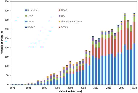
Figure 1.
Scopus database search results showing the number of articles published over the years (up to 2024) regarding the use of HAT-based methods for testing antioxidant properties.
The main aim of this review is to take a closer look at one of the oldest (the first mention of this method comes from Marco in 1968 [13]) and, at the same time, the most popular methods (compare the heights of the bars in Figure 1) for testing antioxidant properties based on the HAT mechanism–the β-carotene bleaching method (abbreviated as the β-carotene method). As mentioned earlier, the most popular and most commonly used methods include the DPPH and ABTS methods, respectively. Like the β-carotene method, these methods are spectrophotometric, with the difference that they are based on the SET mechanism. Numerous review articles have been devoted to both methods. However, a comprehensive study compiling information on the β-carotene method is lacking, hence the idea for this study.
This literature review was conducted by systematically collecting, reviewing, and collating information (with a significant share of recent papers) from available online databases, such as Google Scholar, Scopus, Web of Science, PubMed, and Science Direct. The search was limited to English. Article abstracts were pre-screened prior to full-text analysis. The collected information was analyzed in detail to ensure that this article serves as a compendium of general knowledge about the β-carotene bleaching method, its implementation procedure, factors to consider during its implementation, and examples of its application.
2. β-Carotene as Molecular Probe in β-Carotene Bleaching Assay
β-carotene, the 3D structure of which is shown in Figure 2, is a compound classified as a carotenoid, a naturally occurring pigment produced by photosynthetic organisms to protect against photodynamic damage [14]. In addition to plants, β-carotene is produced by certain microorganisms: fungi, microalgae, and bacteria [15]. Popular fungal strains include Blakeslea trispora and Rhodotorula glutinis, while microalgae such as Dunaliella salina are characterized by high beta-carotene production. Bacteria such as Erwinia uredovora, Pantoea agglomerans, and some strains of Serratia marcescens can also produce it.
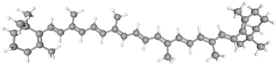
Figure 2.
Three-dimensional model of the chemical structure of the β-carotene conformer (IUPAC name: 1,3,3-trimethyl-2-[(1E,3E,5E,7E,9E,11E,13E,15E,17E)-3,7,12,16-tetramethyl-18-(2,6,6-trimethylcyclohexen-1-yl)octadeca-1,3,5,7,9,11,13,15,17-nonaenyl]cyclohexene) [16].
β-Carotene: naturally occurs as the trans isomer. Yet, under the influence of light (especially UV radiation), heat, and the presence of enzymes, one of the double bonds can isomerize to the cis position, which causes a change in the molecular structure. Both the cis and trans forms of β-carotene absorb light, but their absorption spectra differ due to differences in molecular structure [17,18]. The trans form is characterized by a strong yellow-orange color (absorption maximum 450–470 nm). The cis form shows a distinct additional peak at a shorter wavelength (around 330–340 nm). Additionally, the cis form is characterized by lower color intensity than trans-β-carotene due to the lower extinction coefficient.
This carotenoid is capable of quenching singlet oxygen. It is also characterized by its ability to be easily oxidized by free radicals. This is due to the long chain of conjugated double bonds present in the molecule, which give it an intense orange color (see Figure 2) [19,20]. These double bonds are susceptible to attack by free radicals. In a methodological context, its antioxidant activity can be easily monitored spectrophotometrically [21,22,23]. When β-carotene reacts with free radicals, the conjugated double bond system is broken, causing the molecule to lose color. This “bleaching” is a visual indicator of the consumption of β-carotene in the reaction with free radicals. It also allows researchers to effectively assess the antioxidant activity of the tested antioxidant(s) [21,22,23]. The extent to which a substance can inhibit β-carotene fading is directly related to its antioxidant activity. The weaker the bleaching, the stronger the antioxidant properties. Finally, the use of β-carotene as a molecular probe in antioxidant studies is supported by both economic (it is relatively cheap and readily available) and ecological considerations, as it is considered a substance generally recognized as safe (GRAS) [21,22,23]. Moreover, it can be used with equal effectiveness both in studies of processes occurring in living organisms, such as humans, animals, or plants (in vivo), and those conducted under controlled laboratory conditions (in vitro) [24,25]. However, it should be noted, as emphasized in many publications, that β-carotene is neither a preventive antioxidant nor a conventional chain reaction-terminating antioxidant. Its antioxidant activity is associated with low oxygen partial pressure, whereas at high oxygen partial pressure it acts as a pro-oxidant [26].
3. Principle of the Method
The main principle of the method is to monitor two competitive reactions in which β-carotene, as a model antioxidant, and a test antioxidant compete for the peroxide radical [27]. As a result, the antioxidant properties of the tested sample (containing β-carotene, the tested antioxidant(s), and peroxyl radicals) are always assessed against a control sample (containing β-carotene and peroxyl radicals but not the tested antioxidant). In both cases, changes in β-carotene absorbance value over time are monitored, and, as already noted, the color changes result from the rupture of the π-coupling by the addition reaction of radicals to the C=C bond of β-carotene [28]. The smaller the color change in the tested sample compared to the control system, the stronger its antioxidant activity. In other words, the tested antioxidant strongly protects the reference molecule, β-carotene, against oxidation (degradation).
β-carotene is a weak antioxidant; therefore, when other antioxidants are present in solution, they react first protecting the β-carotene structure by intercepting the attack of reactive oxygen species before they can attack and destroy the β-carotene. Only when these are depleted, and peroxyl radicals are still present in the measurement system, does β-carotene take over the antioxidant function. In this case, depending on the antioxidant’s potency, its consumption over time is lower (higher) than in the control sample, corresponding to the stronger (weaker) antioxidant–compare the positions of the curves in Figure 3 [29].
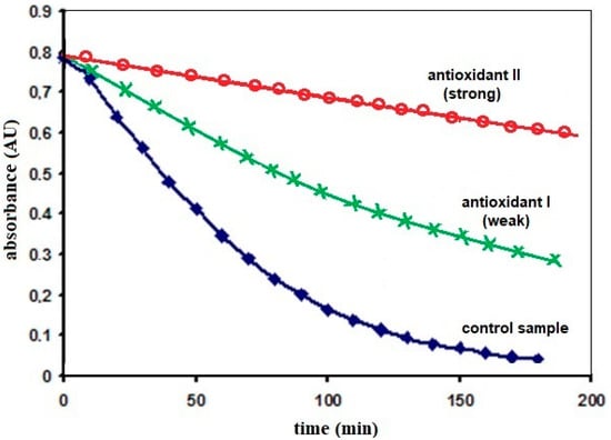
Figure 3.
Examples of possible changes in β-carotene absorbance values over time, monitored in a sample containing the model antioxidant and a neutralized peroxide radical (control sample, blue line) and test samples (in addition to the above-mentioned components, also containing the tested antioxidant) with weaker (green line) and stronger (green line) (red curve) antioxidant properties (based on the results of our own research, detailed description–see text above).
Considering the possible transformation of trans-β-carotene to the cis- form during the reaction, it should be added that the observed in this method degradation of β-carotene in both the control sample and the test sample may be additionally related not only to the process of β-carotene oxidation by linoleic acid but may also result from the above-mentioned isomerization, which results in a color change in the emulsion. It should be noted here that in both the control and test samples, the changes in the β-carotene structure caused by isomerization should occur to the same extent. This is related to the fact that both samples are heated in the same way (45 °C) for the same period of time.
Figure 4 illustrates the processes involved in the color change in β-carotene. The discoloration of the orange–yellow or dark yellow β-carotene solution is caused by radicals (lipids or lipid peroxide radicals) generated during the immediate oxidation of fatty acids present in an aqueous emulsion of linoleic acid and β-carotene (a lipophilic oxidizable substrate) (part A) [30,31]. Once formed, lipids or lipid peroxide radicals attack β-carotene (part B), causing discoloration. This discoloration is caused by the “loss” of a conjugated double bond system through an addition reaction of the radicals to the C=C bond or by the substitution of a radical to the β-ionone ring located at each end of the long chain of 11 conjugated double bonds in the β-carotene molecule [32,33]. The reaction results in the formation of radical adducts, examples of which are provided in the figure. Each of these adducts is a colorless form stabilized by resonance with electrons located on the carbon atom. However, it should be noted that the exact position of the electron (its delocalization in the resulting radical structure) is unknown (hence, the drawing shows intermediate bonds instead of double bonds, indicating charge dispersion). The radical can be located on any secondary C in the conjugated system. Undoubtedly, as a result of adduct formation, the concentration of β-carotene in the test solution (referred to as the control sample) decreases. In the presence of the antioxidant, β-carotene competes for the formation of the mentioned adducts [24,28].
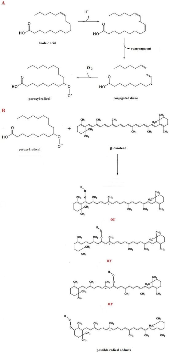
Figure 4.
Reactions showing the oxidation of linoleic acid (A) and the possible formation of various radical adducts between β-carotene and a peroxyl radical (B) [30,31,32,33].
4. Procedure
The β-carotene bleaching method was first proposed by Marco (1968) [13] to prevent autoxidation of emulsified linoleic acid in extracts. Over time, the technique has undergone several modifications aimed at facilitating its application (simplified the operation) [34] or converting it to microplates form [35]. Regardless of the modification, the β-carotene bleaching method is carried out in several stages, which are presented in Scheme 1 [36]. The most important step is the preparation of the test reagent See Step I in the scheme). For this purpose, a chloroform solution of β-carotene is prepared; the chloroform is evaporated under reduced pressure in the presence of linoleic acid and Tween (an emulsion stabilizer). During this time, the water is saturated with oxygen (usually 100 mL of water is saturated with oxygen for 30 min at a controlled flow of 100 mL/min), because the presence of oxygen and the elevated measurement temperature favor the oxidation of linoleic acid and the production of peroxide radicals. The resulting radical can either bleach the β-carotene or abstract a proton from the radical scavenger (antioxidant) [37]. Finally, the residue left after evaporation of the chloroform is gently combined with oxygenated water.
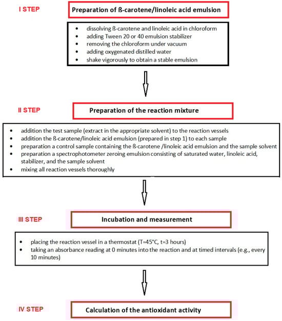
Scheme 1.
Stages for testing the antioxidant properties of solutions using the β-carotene bleaching method.
It should be noted that there is no standardization of antioxidant testing methods, including the β-carotene method. This means that studies using the same method are conducted under various, sometimes significantly different, conditions (e.g., different reaction monitoring times and temperatures). These factors, along with the different ways of presenting the obtained results, make direct comparisons difficult. Table 1 summarizes examples from the literature illustrating how measurements are performed and results interpreted using the β-carotene bleaching method. Table 2, in turn, proposes the optimal conditions for the procedure, according to the authors.

Table 1.
Literature examples of measurement conditions and results presentation for the β-carotene bleaching method.
Table 2.
Proposed measurement conditions for the β-carotene bleaching assay.
As the examples presented (see Table 1) demonstrate, the β-carotene bleaching method is used under various measurement conditions, which undoubtedly makes it difficult, if not impossible, to compare the results of different research teams. However, the lack of a uniform presentation of results is not the only problem with this method. Its infamous reputation also stems from the low reproducibility of the obtained results. Furthermore, emulsion preparation is time-consuming and labor-intensive, and susceptible to interference from various factors (including the solvent used, the influence of metal ions, pH, and temperature) [20,41]. As a result, as Figure 1 shows, the applicability of the β-carotene method, measured by the number of publications indexed in the Scopus database, has not increased over the last five years and remains at a comparable level of approximately 330 publications per year.
5. Presentation of the Final Results
The question arises how to interpret the obtained results and correctly express antioxidant activity using this method. The literature provides various methods for presenting final results [56], including for the β-carotene bleaching method. This leads to results that are difficult to interpret and compare, especially when the same antioxidant is tested using the same method but a different method for presenting final results. This is a significant problem not only for the β-carotene method. Below, we summarize the most well-known equations, collected from the literature and used to present final results. They are presented in order of popularity. However, the authors’ experience indicates that Equations (1) and (2) are more reliable.
The basic manner of presenting results is to determine the rate of β-carotene bleaching (R), both in the control and in the tested samples, according to the following equation [57,58]:
where ln = natural log, a = absorbance at time “0”, and b = absorbance at time “t”.
R = [ln(a/b)/t],
In turn, the antioxidant activity (expressed as inhibition percent, I%) can be determined from the below equation, where R is the bleaching rate [57,58]:
I (%) = (Rcontrol − Rsample)/Rcontrol × 100%.
The higher the value of this parameter, the better the antioxidant properties of the tested sample. When interpreting the results, it can also be assumed that the higher the % inhibition, the greater the degree of protection of β-carotene from degradation in the test sample compared to its degradation in the control sample.
In addition to the method mentioned above (Equations (1) and (2)), antioxidant activity determined by this method is calculated directly from absorbance alone [44,59,60,61]:
where %AOA—the percentage antioxidant activity, Ac(0) is the absorbance of the control sample at t = 0 min, Ac(t) is the absorbance of the control sample at t, As(0) is the absorbance of the test sample at t = 0 min, and As(t) is the absorbance of the test sample at t. The total measurement time (t) ranges from 60 min to 120, 150, 180 or 210 min, and absorbance measurements are performed at different time intervals (every 10 min, every 15 min, every 25 min) [44,59,60,61].
%AOA = [1 − (As(0) − As(t))/(Ac(0) − Ac(t))] × 100%,
You can also find papers [62,63,64] in which the antioxidant activity (percent inhibition, I%) of the samples was calculated using the following equation:
where Aβ-carotene (t) is the absorbance of β-carotene remaining in the samples after definite time of testing (t), and Aβ-carotene (0) is the absorbance of β-carotene at the beginning of the experiments.
I% = (Aβ-carotene (t)/Aβ-carotene (0)) × 100%,
Using each of the methods presented, based on the relationship between the determined % inhibition values and time, the so-called IC50 value can be determined. It should be noted that the IC50 is the concentration of an antioxidant/sample that, in this case, provides 50% protection against β-carotene oxidation. The lower the IC50 value when comparing different samples based on this parameter, the better the antioxidant properties.
In the literature, there are also other approaches to expressing test results using the β-carotene bleaching method, for example, using standards of compounds with known antioxidant properties [56,65]. This approach was used by Dapkevicius et al. in [66], who reported relative antioxidant activities (RAA) calculated based on the dependencies:
RAA = Absorbance of sample/Absorbance of BHT.
These researchers used butylhydroxytoluene (BHT) as the antioxidant standard, and the changes in β-carotene absorption in the tested sample and in the sample containing BHT were measured both after 28 and 56 h, noting that the values they obtained were smaller when the period after which the measurement was made was longer. It should be noted that the higher the RAA value, the greater the antioxidant properties.
Yet another, relatively new way of determining antioxidant properties is the so-called antioxidant activity coefficient (AAC) proposed in the work of Andrade et al. [67]. It is calculated based on the following equation:
where As(t) is the absorbance of the sample at definite time, Ac(t) is the absorbance of the control at definite time, and Ac(0) is the absorbance of the control at 0 min. The higher the value of this parameter, the better the antioxidant properties.
AAC = [As(t) − Ac(t)/Ac(0) − Ac(t)] × 1000,
6. Factors Influencing the Assessment of Antioxidant Properties Estimated by the β-Carotene Method
As mentioned above, a number of factors influence the assessment of antioxidant capacity using the β-carotene bleaching method [20,41]. Among the most frequently encountered in the literature are temperature, hydrogen ion concentration (pH), solvent, and metal ions. These are discussed in the individual paragraphs below.
Temperature is an important factor that should be controlled both during reagent preparation and the measurement itself. Temperature has been observed to increase the rate of β-carotene bleaching (regardless of whether linoleic acid is present in the measurement system) and also influence solvent evaporation. In the original description of the method presented by Marco, a measurement temperature range of 50–55 °C was used. Today, most applications of this method use a temperature of 45 °C. According to Prieto et al. [20], using this temperature significantly reduces spontaneous β-carotene oxidation, leads to better differentiation between different antioxidant levels, is significantly more sensitive to prooxidant effects, and significantly reduces temperature gradients, which is extremely important in microplate applications. It should be noted that using a lower temperature than originally proposed (50–55 °C) extends the analysis time but reduces side effects. At the expense of extending the measurement time, statistically better results are obtained. On the other hand, using higher temperatures shortens the analysis time but is subject to the possibility of errors resulting from excessive β-carotene oxidation, and furthermore, the reaction conditions move away from those in which antioxidants have practical significance.
The β-carotene method is often used to assess the antioxidant properties of plant extracts, which may differ in the content and type of natural acids. Studies conducted on BHT have shown that, in environments with varying pH, antioxidant activity of BHT is higher in more acidic environments [55]. It should be noted that these results were obtained relative to a control sample that did not contain hydrogen ions. As mentioned in the introduction, antioxidant activity in the β-carotene method is determined by comparing two competing chemical reactions involving the tested antioxidant and/or β-carotene (a model antioxidant) [68]. The main component of the reaction medium is an emulsion of β-carotene/linoleic acid in water with the generated peroxide radicals (LOO•). During the test, changes in β-carotene concentration are monitored in the control sample and in the sample containing the tested antioxidant. β-carotene reacts with peroxide radicals in the sample immediately after the antioxidant is depleted. Given the above, to determine whether the presence of hydrogen ions affects BHT or β-carotene (this was not fully known from monitoring the test sample), subsequent studies were conducted to monitor changes in beta-carotene in a control sample (without BHT). The data indicated that increasing hydrogen ion concentration reduced the loss of β-carotene in the control sample. This observation contradicts Valgimigli’s explanation [69], who reported that the presence of small amounts of acid accelerates the reaction between antioxidants and peroxide radicals. The results indicate that increasing hydrogen ion concentration leads to the neutralization of peroxide radicals, which cannot be true. The most likely explanation for the observed correlations is that the presence of acid in the measurement system slows down the formation of peroxide radicals. This process occurs both in the presence and absence of an antioxidant. Furthermore, in [55] it was demonstrated that the type of cations and anions (buffer components) also influences the observed antioxidant properties. This is also confirmed by the results of studies conducted by Prieto et al. [20], in which the effect of pH on β-carotene was studied in buffered solutions (pH 3.5–(0.5)–11.0) at various concentrations of BHA, 0–(0.5)–5 μM, in the presence and absence of linoleic acid. They show that no hypso- or bathochromic shifts in the β-carotene absorption spectrum were observed within the pH range studied. In the control sample, however, the time required to achieve a given degree of whitening was very long at low pH, confirming that hydrogen ions influence the oxidation of linoleic acid. The authors of the aforementioned article further state that the best range for testing this method, which guarantees stable results within a reasonable time, is a pH range of 5.5 to 7.5.
The solvent plays an important role in assessing the antioxidant properties of compounds. In the β-carotene method, the reaction medium is an aqueous emulsion, and when the sample is poorly soluble in water, it is necessary to use other solvents, such as alcohols (methanol, ethanol), acetone, or 1,4-dioxane. The solvent not only affects solubility but also participates in the peroxyl radical neutralization reaction, which, as mentioned earlier, is historically considered the HAT mechanism. According to the literature [2,70], the addition of hydrogen from the antioxidant to the peroxyl radical precedes the formation of a complex between the antioxidant and the peroxide radical, resulting in the phenolic hydrogen being hydrogen bonded to the peroxide radical. This hydrogen is then transferred as a proton from the phenol to the ion pair on the peroxide radical, along with the conjugate movement of an electron from the phenolic oxygen 2p ion pair to the orbital containing the unpaired electron on the peroxide radical, so that the electron moves between two formally non-bonding orbitals [71]. A solvent that is a stronger hydrogen acceptor (e.g., dioxane) can hinder this process and thus slow down the reaction rate, which may translate into the observed lower antioxidant properties. Furthermore, the type of antioxidant solvent, due to its different hydrophobicity, can affect the dispersion (or solubility) of the emulsion or its components, which can consequently change the reaction rate. An important aspect concerns the solvent is the so-called “polar paradox” [72] related to the fact that due to hydrophobic repulsion, non-polar antioxidants readily accumulate closer to the lipid environment in which oxidation takes place, hence their observed higher antioxidant activity in oil-water emulsion. Regarding the effect of solvent on antioxidant properties assessed using the β-carotene method, another aspect must be considered. The lack of standardized methods for measuring antioxidant properties means that tests are performed using varying volume ratios of the reagents. Such a variable volume ratio of the tested antioxidant solution to the β-carotene/linoleic acid emulsion also significantly impacts the assessment of antioxidant activity [41].
The presence of metal ions in plant extracts is a consequence of their natural occurrence in plants. Studies also show that the presence of metals in the measurement system affects antioxidant properties estimated using the β-carotene method. In most cases, increasing metal ion concentration accelerates the oxidation process [20,41]. However, none of the tested metals promoted β-carotene bleaching (in the absence of linoleic acid). This clearly suggests that in all cases the observed activity was not related to β-carotene bleaching, but to the production of radicals by the fatty acid. One possible explanation for this phenomenon is the decomposition of hydroperoxides formed after the neutralization reaction, observed in the presence of metal ions [73,74].
Typical factors that significantly modify the assessment of antioxidant properties using the β-carotene method are presented above. However, it is important to realize that there are more factors and controversies surrounding them. For example, some authors use initiators to initiate the radical formation reaction. However, according to Prieto et al. [20], their use is unnecessary, as the reaction proceeds smoothly without them. Others cite the size of the formed micelles as a factor differentiating the results. However, according to [20], this parameter is irrelevant if centrifugation or ultraviolet irradiation is used after reagent preparation (before measurement). Similarly, according to [20], initial oxygen saturation does not cause significant differences in the bleaching process, and the saturation step can be omitted.
7. Application
As mentioned, the β-carotene method was first used to test the autoxidation of emulsified linoleic acid. However, as the procedure was refined and simplified, its application range expanded significantly. Currently, it is used to study various matrices, and while liquid samples are directly analyzed, in the case of solid matrices, in accordance with the methodology of their processing, after preliminary grinding, most often using a mill, they are subjected to extraction. Each case requires an individual and differentiated approach, as demonstrated, among others, in Table 1. A literature review shows that this method is used to determine the antioxidant properties of, among others,
- Plant (including herbs, spices, vegetables, fruits and seaweed) extracts obtained from roots [32,75], leaves [76,77,78,79], stems [78], flowers [80], buds [81], seed [82,83], fruits [84,85] and seaweed species [86] are used for research.
- Essential oils [87,88,89,90];
- Food products (for example honey, propolis, beans) [91,92,93,94,95] and enriched food product [96,97];
- Beverages (functional drinks, juice) [98,99,100];
- Food waste and industrial waste [44,101,102,103,104,105,106,107];
- Synthetic compound [108,109,110,111,112];
- Components so called “active packing” [113,114,115,116];
- Mushrooms [117,118];
- Microorganisms (cyanobacteria) [119];
- Cosmetic products [120];
- Natural pigments [121];
- Edible oils [122];
- Animal proteins [123,124].
8. Conclusions
The aim of this review article is to systematize and update knowledge on the use of the β-carotene bleaching method in antioxidant studies based on the HAT mechanism. This article explains the basics of β-carotene’s reaction with free radicals and the causes of discoloration of this molecular probe. It is emphasized that the use of this molecule in antioxidant studies is supported by both economic and ecological considerations. β-carotene is considered a substance generally recognized as safe (GRAS). Furthermore, it can be used with equal effectiveness both in studies of processes occurring in living organisms (in vivo) and in studies conducted under controlled laboratory conditions (in vitro). Given the lack of standardized conditions for conducting antioxidant studies using this method, the authors propose an optimal methodology, in their opinion, that will help obtain consistent, reliable, and easily comparable results between research groups. The authors present the influence of relevant parameters that significantly differentiate the final analytical results. Therefore, this methodology should be considered a specific guide for studying the antioxidant properties of simple and complex systems using the β-carotene method, ensuring the effective identification of sources of naturally occurring antioxidants and the design of new antioxidant compounds for future use as health-promoting agents and/or safe food additives.
Author Contributions
Conceptualization and methodology, M.O.-T.; writing—original draft preparation, M.O.-T.; writing—review and editing, D.W.; supervision, D.W. All authors have read and agreed to the published version of the manuscript.
Funding
This research received no external funding.
Data Availability Statement
No new data were created or analyzed in this study. Data sharing is not applicable to this article.
Acknowledgments
The authors would like to thank the Institute of Chemical Sciences of the Maria Curie-Skłodowska University in Lublin for creating the research infrastructure, without which this research would not be possible.
Conflicts of Interest
The authors declare no conflicts of interest.
Abbreviations
The following abbreviations are used in this manuscript:
| AAC | Antioxidant Activity Coefficient |
| ABTS | 2,2′-azino-bis(3-ethylbenzothiazoline-6-sulfonic acid |
| AH | Antioxidant |
| AOA | Antioxidant Activity |
| BDE | bond dissociation energy |
| BHA | Butylhydroxyanisole |
| BHT | Butylhydroxytoluene |
| CUPRAC | cupric ion reducing antioxidant capacity method |
| DMSO | Dimethyl sulfoxide |
| DMPD | dimethyl-p-phenylenediamine dihydrochloride |
| DPPH | 2,2-diphenyl-1-picrylhydrazyl |
| EtOH | Ethanol |
| FRAP | ferric ion reducing antioxidant parameter method |
| GRAS | generally recognized as safe |
| HAT | Hydrogen Atom Transfer |
| HORAC | Hydroxyl Radical Antioxidant Capacity |
| IC | Inhibition Concentration |
| IP | ionization potential |
| LDL | Low Density Lipoprotein |
| MeOH | Methanol |
| ORAC | Oxygen Radical Absorbance Capacity |
| RAA | Relative Antioxidant Activities |
| ROS | Reactive Oxygen Species |
| SET | Single Electron Transfer |
| TOSCA | Total Radical Scavenging Capacity Assay |
| TRAP | Total Radical Trapping Antioxidant Parameter |
References
- Losada-Barreiro, S.; Sezgin-Bayindir, Z.; Paiva-Martins, F.; Bravo-Díaz, C. Biochemistry of Antioxidants: Mechanisms and Pharmaceutical Applications. Biomedicines 2022, 10, 3051. [Google Scholar] [CrossRef]
- Olszowy, M. What is responsible for antioxidant properties of polyphenolic compounds from plants? Plant Physiol. Biochem. 2019, 144, 135–143. [Google Scholar] [CrossRef]
- Siddeeg, A.; Al Kehayez, N.M.; Abu-Hiamed, H.A.; Al-Sanea, E.A.; AL-Farga, A.M. Mode of action and determination of antioxidant activity in the dietary sources: An overview. Saudi J. Biol. Sci. 2021, 28, 1633–1644. [Google Scholar] [CrossRef] [PubMed]
- Platzer, M.; Kiese, S.; Tybussek, T.; Herfellner, T.; Schneider, F.; Schweiggert-Weisz, U.; Eisner, P. Radical Scavenging Mechanisms of Phenolic Compounds: A Quantitative Structure-Property Relationship (QSPR) Study. Front. Nutr. 2022, 9, 882458. [Google Scholar] [CrossRef] [PubMed]
- Charlton, N.C.; Mastyugin, M.; Török, B.; Török, M. Structural Features of Small Molecule Antioxidants and Strategic Modifications to Improve Potential Bioactivity. Molecules 2023, 28, 1057. [Google Scholar] [CrossRef] [PubMed]
- Huang, D.; Boxin, O.; Prior, R.L. The Chemistry behind Antioxidant Capacity Assays. J. Agric. Food Chem. 2005, 53, 1841–1856. [Google Scholar] [CrossRef]
- Apak, R.; Özyürek, M.; Güçclü, K.; Çapanoğlu, E. Antioxidant Activity/Capacity Measurement. 1. Classification, Physicochemical Principles, Mechanisms, and Electron Transfer (ET)-Based Assays. J. Agric. Food Chem. 2016, 64, 997–1027. [Google Scholar]
- Prior, R.L.; Wu, X.; Schaich, K. Standardized methods for the determination of antioxidant capacity and phenolics in foods and dietary supplements. J. Agric. Food Chem. 2005, 53, 4290–4302. [Google Scholar] [CrossRef]
- Jiménez-Morales, W.A.; del Pilar Cañizares-Macias, M.; Pedraza-Chaver, J. Fast ORAC-SIA method for antioxidant capacity determination in food samples. Food Chem. 2022, 384, 132524. [Google Scholar] [CrossRef]
- Knez, E.; Kadac-Czapska, K.; Grembecka, M. Evaluation of Spectrophotometric Methods for Assessing Antioxidant Potential in Plant Food Samples—A Critical Approach. Appl. Sci. 2025, 15, 5925. [Google Scholar] [CrossRef]
- Apak, R.; Özyürek, M.; Güçclü, K.; Çapanoğlu, E. Antioxidant Activity/Capacity Measurement. 2. Hydrogen Atom Transfer (HAT)-Based, Mixed-Mode (Electron Transfer (ET)/HAT), and Lipid Peroxidation Assays. J. Agric. Food Chem. 2016, 64, 1028–1045. [Google Scholar]
- Apak, R. Current Issues in Antioxidant Measurement. J. Agric. Food Chem. 2019, 67, 9187–9202. [Google Scholar] [CrossRef]
- Marco, G.J. A rapid method for evaluation of antioxidants. J. Am. Oil Chem. Soc. 1968, 45, 594–598. [Google Scholar] [CrossRef]
- Amengual, J. Bioactive Properties of Carotenoids in Human Health. Nutrients 2019, 11, 2388. [Google Scholar] [CrossRef]
- Wang, L.; Liu, Z.; Jiang, H.; Mao, X. Biotechnology advances in β-carotene production by microorganisms. Trends Food Sci. Technol. 2021, 111, 322–332. [Google Scholar] [CrossRef]
- Available online: https://pubchem.ncbi.nlm.nih.gov/compound/Beta-Carotene#section=Names-and-Identifiers (accessed on 14 November 2025).
- Telegina, T.A.; Vechtomova, Y.L.; Aybush, A.V.; Buglak, A.A.; Kritsky, M.S. Isomerization of carotenoids in photosynthesis and metabolic adaptation. Biophys. Rev. 2023, 15, 887–906. [Google Scholar] [CrossRef] [PubMed]
- Khoo, H.E.; Prasad, K.N.; Kong, K.W.; Jiang, Y.; Ismail, A. Carotenoids and their isomers: Color pigments in fruits and vegetables. Molecules 2011, 16, 1710–1738. [Google Scholar] [CrossRef]
- Srivastava, R. Physicochemical, antioxidant properties of carotenoids and its optoelectronic and interaction studies with chlorophyll pigments. Sci. Rep. 2021, 11, 18365. [Google Scholar] [CrossRef]
- Prieto, M.A.; Vázquez, J.A.; Murado, M.A. β-Carotene Assay Revisited. Application To Characterize and Quantify Antioxidant and Prooxidant Activities in a Microplate. J. Agric. Food Chem. 2012, 60, 8983–8993. [Google Scholar] [CrossRef]
- Loffredo, C.; Pires, P.A.R.; Imran, M.; El Seoud, O.A. β-Carotene: A green, inexpensive, and convenient solvatochromic probe for the determination of solvent polarizability. Dye. Pigment. 2013, 96, 16–24. [Google Scholar] [CrossRef]
- Lu, Y.; Teng-Jin, K.; Wiart, C. Phytochemical Analysis and Antioxidant Activity Determination on Crude Extracts of Melodinus eugeniifolus Barks and Leaves from Malaysia. Pharmacol. Pharm. 2014, 5, 773–780. [Google Scholar] [CrossRef]
- Pérez-Gálvez, A.; Viera, I.; Roca, M. Carotenoids and Chlorophylls as Antioxidants. Antioxidants 2020, 9, 505. [Google Scholar] [CrossRef]
- Miazek, K.; Beton, K.; Śliwińska, A.; Brożek-Płuska, B. The Effect of ß-Carotene, Tocopherols and Ascorbic Acid as Anti-Oxidant Molecules on Human and Animal In Vitro/In Vivo Studies: A Review of Research Design and Analytical Techniques Used. Biomolecules 2022, 12, 1087. [Google Scholar] [CrossRef]
- Nimbalkar, V.; Joshi, U.; Shinde, S.; Pawar, G. In-vivo and in-vitro evaluation of therapeutic potential of β-Carotene in diabetes. J. Diabetes Metab. Disord. 2021, 20, 1621–1630. [Google Scholar] [CrossRef]
- Ahmed, D.; Malik, W.; Maqsood, M.; Atique, I.; Qamar, M.T. Study of anti-diabetic, beta-carotene-bleaching inhibiting and iron chelating properties of Carissa opaca root extracts. Braz. J. Pharm. Sci. 2022, 58, e18628. [Google Scholar] [CrossRef]
- Roginsky, V.; Lissi, E.A. Review of methods to determine chain-breaking antioxidant activity in food. Food Chem. 2005, 92, 235–254. [Google Scholar] [CrossRef]
- Abeyrathne, E.D.N.S.; Nam, K.; Ahn, D.U. Analytical Methods for Lipid Oxidation and Antioxidant Capacity in Food Systems. Antioxidants 2021, 10, 1587. [Google Scholar] [CrossRef] [PubMed]
- Gupta, D. Methods for determination of antioxidant capacity: A review. Int. J. Pharm. Sci. Res. 2015, 6, 546–566. [Google Scholar]
- Sadeer, N.B.; Montesano, D.; Albrizio, S.; Zengin, G.; Mahomoodally, M.F. The Versatility of Antioxidant Assays in Food Science and Safety—Chemistry, Applications, Strengths, and Limitations. Antioxidants 2020, 9, 709. [Google Scholar] [CrossRef]
- Christodoulou, M.C.; Orellana Palacios, J.C.; Hesami, G.; Jafarzadeh, S.; Lorenzo, J.M.; Domínguez, R.; Moreno, A.; Hadidi, M. Spectrophotometric Methods for Measurement of Antioxidant Activity in Food and Pharmaceuticals. Antioxidants 2022, 11, 2213. [Google Scholar] [CrossRef]
- Bryshten, I.; Paprotny, Ł.; Olszowy-Tomczyk, M.; Wianowska, D. Antioxidant Properties of Green Plants with Different Vitamin K Contents. Molecules 2024, 29, 3655. [Google Scholar] [CrossRef]
- Mordi, R.C.; Ademosun, O.T.; Ajanaku, C.O.; Olanrewaju, I.O.; Walton, J.C. Free Radical Mediated Oxidative Degradation of Carotenes and Xanthophylls. Molecules 2020, 25, 1038. [Google Scholar] [CrossRef] [PubMed]
- Miller, H. A simplified method for the evaluation of antioxidants. J. Am. Oil Chem. Soc. 1971, 48, 91. [Google Scholar] [CrossRef]
- Koleva, I.I.; van Beek, T.A.; Linssen, J.P.H.; de Groot, A.; Evstatieva, L.N. Screening of Plant Extracts for Antioxidant Activity: A Comparative Study on Three Testing Methods. Phytochem. Anal. 2022, 13, 8–17. [Google Scholar] [CrossRef]
- Gulluc, M.; Sahin, F.; Sokmen, M.; Ozer, H.; Daferera, D.; Sokmen, A.; Polissiou, M.; Adiguzel, A.; Ozkan, H. Antimicrobial and antioxidant properties of the essential oils and methanol extract from Mentha longfolia L. ssp. longifolia. Food Chem. 2007, 103, 1449–1456. [Google Scholar] [CrossRef]
- Ismail, A.; Marjan, Z.M.; Foong, C.W. Total antioxidant activity and phenolic content in selected vegetables. Food Chem. 2004, 87, 581–586. [Google Scholar] [CrossRef]
- Tepe, B.; Daferera, D.; Sokmen, A.; Sokmen, M.; Polissiou, M. Antimicrobial and antioxidant activities of the essential oil and various extracts of Salvia tomentosa Miller (Lamiaceae). Food Chem. 2005, 90, 333–340. [Google Scholar] [CrossRef]
- Mata, A.T.; Proença, C.; Ferreira, A.R.; Serralheiro, M.L.M.; Nogueira, J.M.F.; Araújo, M.E.M. Antioxidant and antiacetylcholinesterase activities of five plantsused as Portuguese food spices. Food Chem. 2007, 103, 778–786. [Google Scholar] [CrossRef]
- Wang, W.; Wu, N.; Zu, Y.G.; Fu, Y.J. Antioxidative activity of Rosmarinus officinalis L. essential oil compared to its main components. Food Chem. 2008, 108, 1019–1022. [Google Scholar] [CrossRef] [PubMed]
- Dawidowicz, A.L.; Olszowy, M. Influence of some experimental variables and matrix components in the determination of antioxidant properties by β-carotene bleaching assay: Experiments with BHT used as standard antioxidant. Eur. Food Res. Technol. 2010, 231, 835–840. [Google Scholar] [CrossRef]
- Nickavar, B.; Esbati, N. Evaluation of the Antioxidant Capacity and Phenolic Content of Three Thymus Species. J. Acupunct. Meridian Stud. 2012, 5, 119–125. [Google Scholar] [CrossRef]
- Loucif, K.; Benabdallah, H.; Benchikh, F.; Mehlous, S.; Souici, C.B.; Amira, S. Total Phenolic Contents, DPPH Radical Scavenging and β-Carotene Bleaching Activities of Aqueous Extract from Ammoides atlantica. J. Drug Deliv. Ther. 2020, 10, 196–198. [Google Scholar] [CrossRef]
- Chintong, S.; Phatvej, W.; Rerk-Am, U.; Waiprib, Y.; Klaypradit, W. In Vitro Antioxidant, Antityrosinase, and Cytotoxic Activities of Astaxanthin from Shrimp Waste. Antioxidants 2019, 8, 128. [Google Scholar] [CrossRef]
- Amiri, H. Chemical composition and antioxidant activity of essential oil and methanolic extracts of Ferula microcolea (boiss.) boiss (apiaceae). Int. J. Food Prop. 2014, 17, 722–730. [Google Scholar] [CrossRef]
- Moualek, I.; Lahcene, S.; Salem-Bekhit, M.M.; Khan, A.A.; Alanazi, A.M.; Bariz, K.; Msela, A.; Sebbane, H.; Park, H.K.; Jeon, B.H.; et al. Assessment of the Antioxidant and Anti-inflammatory Properties of Aqueous Extract of Rosa sempervirens Leaves. Cell Mol. Biol. 2023, 69, 214–222. [Google Scholar] [CrossRef] [PubMed]
- Öğüt, K.; Özek, G.; Oztürk, N.; Yaylaci, Ö.K.; Özek, T. Chemical composition, α-amylase inhibition, and antioxidant activities of Scabiosa hololeuca Bornm. Biochem. Syst. Ecol. 2025, 122, 105028. [Google Scholar] [CrossRef]
- Soares Mateus, A.R.; Pena, A.; Sanches Silva, A. Development of functional muffins enriched with lemon by-products as sources of Bioactive compounds. Food Chem. Adv. 2025, 7, 100972. [Google Scholar] [CrossRef]
- Azzouni, D.; Alaoui Mrani, S.; Bahij, F.; Zejli, H.; Alanazi, M.M.; Fadili, D.; El Moussaoui, A.; Mahmoud, A.M.; Taleb, M. Comprehensive Phytochemical, Antioxidant, and Antibacterial Analysis of Vitex agnus-castus L. Essential Oil (VACEO): Insights from ADMET and Molecular Docking Studies. Pharmaceuticals 2025, 18, 462. [Google Scholar] [CrossRef]
- Faidi, A.; El Hédi Becheikh, M.; Ali Lassoued, M.; Stumbé, J.F.; Safta, F.; Sfar, S. Isolation of sodium alginate-like polysaccharide from Padina pavonica: Optimization, characterization and antioxidant properties. J. Mol. Struct. 2025, 1321, 139737. [Google Scholar] [CrossRef]
- Ebrahimi, M.; Ballester, A.R.; Lopez-Rubiò, A.; Acha, V.; Aussenac, T.; Martínez-Abad, A. Antimicrobial and antioxidant properties of water-soluble lignin extracts obtained from ozonation of Miscanthus giganteus and Vitis vinifera in a pilot-scale reactor. Ind. Crops Prod. 2025, 223, 120092. [Google Scholar] [CrossRef]
- Solorzano, E.L.; Roverso, M.; Bogialli, S.; Bortoli, M.; Orian, L.; Badocco, D.; Pettenuzzo, S.; Favaro, G.; Pastore, P. Antioxidant activity of Zuccagnia-type propolis: A combined approach based on LC-HRMS analysis of bioanalyical-guided fractions and computational investigation. Food Chem. 2024, 461, 140827. [Google Scholar] [CrossRef] [PubMed]
- Prabakarans, S.; Saad, H.M.; Tan, C.H.; Rahman, S.N.S.A.; Sim, K.S. Investigation of Phytochemical Composition, Radical Scavenging Potential, Anti-Obesogenic Effects, and Anti-Diabetic Activities of Kaempferia parviflora Rhizomes. Chem. Biodivers. 2025, 22, e202401086. [Google Scholar] [CrossRef]
- Altaf, L.; Wani, S.A.; Hussain, P.R.; Suradkar, P.; Baqual, M.F.; Bhat, A.A. Bioactive compounds and antioxidant activity in various parts of Morus alba L. Cv. ichinose: A comparative analysis. Discov. Life 2024, 54, 7. [Google Scholar] [CrossRef]
- Dawidowicz, A.L.; Olszowy, M. Depletion/protection of β-carotene in estimating antioxidant activity by β-carotene bleaching assay. J. Food Sci. Technol. 2015, 52, 7321–7328. [Google Scholar] [CrossRef]
- Olszowy-Tomczyk, M. How to express the antioxidant properties of substances properly? Chem. Pap. 2021, 75, 6157–6167. [Google Scholar] [CrossRef]
- Gül, A.; Pehlivan, T. Antioxidant activities of some monofloral honey types produced across Turkey. Saudi J. Biol. Sci. 2018, 25, 1056–1065. [Google Scholar] [CrossRef] [PubMed]
- Morales, G.; Paredes, A. Antioxidant activities of Lampaya medicinalis extracts and their main chemical constituents. BMC Complement. Altern. Med. 2014, 21, 259. [Google Scholar] [CrossRef]
- Dawidowicz, A.; Wianowska, D.; Baraniak, B. The antioxidant properties of alcoholic extracts from Sambucus nigra L. (antioxidant properties of extracts). LWT-Food Sci. Technol. 2006, 39, 308–315. [Google Scholar] [CrossRef]
- Yang, J.; Guo, J.; Yuan, J. In vitro antioxidant properties of rutin. LWT-Food Sci. Technol. 2008, 41, 1060–1066. [Google Scholar] [CrossRef]
- Gouveia-Figueira, S.C.; Gouveia, C.A.; Carvalho, M.J.; Rodrigues, A.I.; Nording, M.L.; Castilho, P.C. Antioxidant Capacity, Cytotoxicity and Antimycobacterial Activity of Madeira Archipelago Endemic Helichrysum Dietary and Medicinal Plants. Antioxidants 2014, 3, 713–729. [Google Scholar] [CrossRef] [PubMed]
- Hamzeh, A. Essential Oils Composition and Antioxidant Properties of Three Thymus Species. Evid. Based Complement. Altern. Med. 2012, 2012, 728065. [Google Scholar]
- Khouya, T.; Ramchoun, M.; Hmidani, A.; El moualij, B.; Amrani, S.; Harnafi, H.; Benlyas, M.; Zegzouti, Y.F.; Nazih, E.H.; Ouguerram, K.; et al. Acute toxicity and antiproliferative and procoagulant activities of fractions derived from Thymus satureioides of the Moroccan High Atlas. S. Afr. J. Bot. 2019, 121, 568–576. [Google Scholar] [CrossRef]
- Khouya, T.; Ramchoun, M.; Hmidani, A.; Khouyaa, T.; dine Tariq Bouhlali, E.; Alem, C. Phytochemical analysis and bioactivity evaluation of Moroccan Thymus atlanticus (Ball) fractions. Sci. Afr. 2021, 11, e00716. [Google Scholar] [CrossRef]
- Hwang, S.J.; Lee, J.H. Comparison of antioxidant activities expressed as equivalents of standard antioxidant. Food Sci. Technol. 2023, 43, e121522. [Google Scholar] [CrossRef]
- Dapkevicius, A.; Venskutonis, R.; van Beek, T.A.; Linssen, J.P.H. Antioxidant activity of extracts obtained by different isolation procedures from some aromatic herbs grown in Lithuania. J. Sci. Food Agric. 1998, 77, 140–146. [Google Scholar] [CrossRef]
- Andrade, M.A.; Ribeiro-Santos, R.; Costa Bonito, M.C.; Saraiva, M.; Sanches-Silva, A. Characterization of rosemary and thyme extracts for incorporation into a whey protein based film. LWT 2018, 92, 497–508. [Google Scholar] [CrossRef]
- Moon, J.; Shibamoto, T. Antioxidant assays for plant and food components. J. Agric. Food Chem. 2009, 57, 1655–1660. [Google Scholar] [CrossRef]
- Valgimigli, L.; Amorati, R.; Petrucci, R.; Peduli, G.F.; Hu, D.; Hanthorn, J.J.; Pratt, D.A. Unexpected acid catalysis in reaction of peroxyl radicals with phenols. Angew. Chem. Int. Ed. 2009, 48, 8348–8351. [Google Scholar] [CrossRef]
- Olszowy-Tomczyk, M.; Wianowska, D. Comparison of the Antioxidant Properties of Extracts Obtained from Walnut Husks as well as the Influence of Juglone on Their Evaluation. Appl. Sci. 2024, 14, 2972. [Google Scholar] [CrossRef]
- Marković, Z. Study of the mechanisms of antioxidative action of different antioxidants. J. Serbian Soc. Comput. Mech. 2016, 10, 135–150. [Google Scholar] [CrossRef]
- Zhong, Y.; Shahidi, F. Antioxidant Behavior in Bulk Oil: Limitations of Polar Paradox Theory. J. Agric. Food Chem. 2012, 60, 4–6. [Google Scholar] [CrossRef]
- Laguerre, M.; Lecomte, J.; Villeneuve, P. Evaluation of the ability of antioxidants to counteract lipid oxidation: Existing methods, new trends and challenges. Prog. Lipid Res. 2007, 46, 244–282. [Google Scholar] [CrossRef]
- Durand, E.; Laguerre, M.; Bourlieu-Lacanal, C.; Lecomte, J.; Villeneuve, P. Navigating the complexity of lipid oxidation and antioxidation: A review of evaluation methods and emerging approaches. Progress. Lipid Res. 2025, 97, 101317. [Google Scholar] [CrossRef]
- Chirouf, S.; Belahcene, N.; Barhouchi, B.; Zenati, N.; Daira, A.; Bensouici, C.; Bendif, H. Bioactive Metabolites from Aristolochia navicularis of Souk Ahras: Therapeutic Insights. Trop. J. Nat. Prod. Res. 2025, 9, 3140–3149. [Google Scholar] [CrossRef]
- Norhaiza, M.; Maziah, M.; Hakiman, M. Antioxidative properties of leaf extracts of a popular Malaysian herb, Labisia pumila. J. Med. Plants Res. 2009, 3, 217–223. [Google Scholar]
- Tighilet, K.; Adjebli, A.; Messis, A. Inhibition of enzymatic browning and antioxidant activities of Marrubium vulgare L extracts: A promising natural solution. Pharmacol. Res. Mod. Chin. Med. 2025, 16, 100658. [Google Scholar] [CrossRef]
- Leporini, M.; Catinella, G.; Bruno, M.; Falco, T.; Tundis, R.; Loizzo, M.R. Investigating the Antiproliferative and Antioxidant Properties of Pancratium maritimum L. (Amaryllidaceae) Stems, Flowers, Bulbs, and Fruits Extracts. Evid. Based Complement. Altern. Med. 2018, 2018, 9301247. [Google Scholar] [CrossRef]
- Lahcene, S.; Moualek, I.; Bariz, K.; Benramdane, E.; Alenazy, R.; Alqasmi, M.; Almufarriji, F.M.; Thabet, M.; Fallata, G.; Alqurainy, N.; et al. Antioxidant and Antibacterial Activities and Phytochemical Analysis of Olea europaea subsp. laperrinei Leaves Extracts. Processes 2025, 13, 1113. [Google Scholar]
- Indrianingsih, A.W.; Windarsih, A.; Noviana, E.; Suratno; Asari, S.M.; Pratiw, S.I. In vitro evaluation of antioxidant, α-glucosidase inhibitor, and antibacterial activities of frangipani flower and the principal component analysis of its constituents. Process Biochem. 2023, 130, 346–357. [Google Scholar] [CrossRef]
- Atia, A.; Atmani-Kilani, D.; Atmani, D.; Ayouni, K.; Belkhir, S.; Benloukil, M.; Saidene, N.; Moulaoui, K.; Kasmi, S.; Medjahed, Z.; et al. Wound healing potential of a formula based on Populus nigra L. flower buds extract with anti-inflammatory activity. J. Ethnopharmacol. 2024, 331, 118319. [Google Scholar] [CrossRef]
- Azam, M.; Sana, A.; Saleem, R.; Faizi, S.; Razzaq, A.; Ul-Haq, Z.; Sarwar, H. Chemical characterization, antioxidant and molecular docking studies on Ajwa pulp and seed concert extracts as potential antihemolytic agent. Pak. J. Pharm. Sci. 2025, 38, 1240–1253. [Google Scholar] [CrossRef]
- Daoudi, N.E.; Houmy, N.; Melhaoui, R.; Bnouham, M. Effect of Heating Temperatures on the Physicochemical Parameters and Antihyperglycemic Activity of Argan seeds oil In vitro and In vivo. Trop. J. Nat. Prod. Res. 2025, 9, 2543–2552. [Google Scholar] [CrossRef]
- Zitouni, H.; Hssaini, L.H.; Ouaabou, R.; Viuda-Martos, M.; Hernandez, F.; Ercisli, S.; Hachimi, H.; Zerhoune, M.; Hanine, H. Functionnal and Technological Properties of Five Strawberry (Arbutus unedo L.) Fruit as Bioactive Ingredients in Functional Foods. Int. J. Food Prop. 2021, 24, 380–399. [Google Scholar] [CrossRef]
- Djenidi, H.; Khennouf, S.; Bouaziz, A. Antioxidant activity and phenolic content of commonly consumed fruits and vegetables in Algeria. Prog. Nutr. 2020, 22, 224–235. [Google Scholar]
- Hamid, M.A.; Yeap, C.H.; Mustapha, W.A.W.; Martony, O.; Fatmawati, F. Effects of Different Solvents on the Antioxidant Activity of Several Seaweed Species from Semporna, Sabah, Malaysia. Ilmu Kelaut Indones. J. Mar. Sci. 2024, 29, 29–36. [Google Scholar] [CrossRef]
- Boghozian, A.; Shiran, H.R.; Choopani, A.; Choopani, A.; Amin, N.G. Extraction of Artemisia aucheri Essential Oils and Evaluation of their Chemical Composition and Antioxidant Activity. J. Appl. Biotechnol. Rep. 2025, 12, 1678–1685. [Google Scholar]
- Ranjbar, F.; Abedpour, M.; Abdosheikhi, M.; Ahmadi, E.; Abedi, E. An investigation of anti-oxidant properties of salvia, conducting beta-carotene bleaching assay. Sci. J. 2015, 36, 1–6. [Google Scholar]
- Budiarto, R.; Khalisha, A.; Sari, D.N.; Ujilestari, T.; Wahyono, T.; Azmi, A.F.M.; Adli, D.N.; Lusiana, E.D.; Sitaresmi, P.I.; Sholikin, M.M. Antioxidant properties of lemon essential oils: A meta-analysis of plant parts, extraction methods, dominant compounds, and antioxidant assay categories. Chem. Biol. Technol. Agric. 2024, 11, 147. [Google Scholar] [CrossRef]
- EL Moussaoui, A.; Bourhia, M.; Jawhari, F.Z.; Salamatullah, A.M.; Ullah, R.; Bari, A.; Majid Mahmood, H.; Sohaib, M.; Serhii, B.; Rozhenko, A.; et al. Chemical Profiling, Antioxidant, and Antimicrobial Activity against Drug-Resistant Microbes of Essential Oil from Withania frutescens L. Appl. Sci. 2021, 11, 5168. [Google Scholar]
- Smeriglio, A.; Imbesi, M.; Ingegneri, M.; Rando, R.; Mandrone, M.; Chiocchio, I.; Poli, F.; Trombetta, D. From Waste to Resource: Nutritional and Functional Potential of Borlotto Bean Pods (Phaseolus vulgaris L.). Antioxidants 2025, 14, 625. [Google Scholar] [CrossRef]
- Lakhmili, H.; Warda, K.; El-Abbassi, A.; Hafidi, A. Antioxidant and anti-glycation activity of eight Moroccan honeys from different botanical origins. Discov. Food 2024, 4, 6. [Google Scholar] [CrossRef]
- Bazaid, A.S.; Alamri, A.; Almashjary, M.N.; Qanash, H.; Almishaal, A.A.; Amin, J.; Binsaleh, N.K.; Kraiem, J.; Aldarhami, A.; Alafnan, A. Antioxidant, Anticancer, Antibacterial, Antibiofilm Properties and Gas Chromatography and Mass Spectrometry Analysis of Manuka Honey: A Nature’s Bioactive Honey. Appl. Sci. 2022, 12, 9928. [Google Scholar] [CrossRef]
- Ali Haimoud, S.; Allem, R.; Benyahla Djeffaland, K.; Lembarki, N.E. Evaluation in Vitro and in Vivo of Biological Activities of Propolis and Bee Pollen Extracts. Phytothérapie 2022, 20, 63–71. [Google Scholar] [CrossRef]
- Belmehdi, O.; Bouyahya, A.; Jekő, J.; Cziáky, Z.; Zengin, G.; Sotkó, G.; El baaboua, A.; Skali SenhajI, N.; Abrini, J. Chemical analysis, antibacterial, and antioxidant activitiesof flavonoid-rich extracts from four Moroccan propolis. J. Food Process Preserv. 2021, 45, e15816. [Google Scholar] [CrossRef]
- Byczkiewicz, S.; Szwajgier, D.; Baranowska-Wójcik, E.; Telichowska, A.; Szymandera-Buszka, K.; Wojtczak, J.; Kobus-Cisowska, J. Research on Application of Japanese Quince (Chaenomeles L.) and Pork Collagen in Dark Chocolate—Benefits in Prevention of Inflammation In Vitro Model. Nutrients 2024, 16, 1758. [Google Scholar] [CrossRef] [PubMed]
- Dasdemir, Y.; Tuba Findik, B.; Yildiz, H.; Birisci, E. Blueberry-added black tea: Effects of infusion temperature, drying method«fruit concentration on the iron-polyphenol complex formation, polyphenols profile, antioxidant activity, and sensory properties. Food Chem. 2023, 410, 135463. [Google Scholar] [CrossRef]
- dos Santos Lima, M.; Ferreira, E.T.J.; de Souza, M.E.A.O.; Pereira, G.E.; Toaldo Fedrigo, I.M. Artificial neural network: A powerful tool in associating phenolic compounds with antioxidant activity of grape juices. Food Anal. Methods 2022, 15, 527–540. [Google Scholar] [CrossRef]
- Luěs, Ȃ.; Sousȃ, S.; Duarte, A.P.; Pereira, L.; Domingues, F. Phytochemical characterization, and evaluation of rheological and antioxidant properties of commercially available juices of berries. J. Berry Res. 2018, 8, 11–23. [Google Scholar] [CrossRef]
- Imran, A.; Quispe, C.; Zeeshan, A.; Imran, M.; Nadeem, M.; Gilani, S.A.; Gondal, T.A.; Tufail, T.; Aslam, F.; Rodrigues, C.F.; et al. Development and antioxidant characterization of Ginger-Mint drink prepared through different extraction techniques. J. Food Meas. Charact. 2021, 15, 2576–2590. [Google Scholar] [CrossRef]
- Olszowy-Tomczyk, M.; Garbaczewska, S.; Wianowska, D. Correlation Study of Biological Activity with Quercetin and Phenolics Content in Onion Extracts. Molecules 2022, 27, 8164. [Google Scholar] [CrossRef]
- Lasunon, P.; Phonkerd, N.; Tettawong, P.; Sengkhamparn, N. Total phenolic compound and its antioxidant activity of by-product from pineapple. Food Res. 2022, 6, 107–112. [Google Scholar] [CrossRef]
- Barbosa, C.H.; Andrade, M.A.; Séndon, R.; Silva, A.S.; Ramos, F.; Vilarinho, F.; Khwaldia, K.; Barbosa-Pereira, L. Industrial Fruits By-Products and Their Antioxidant Profile: Can They Be Exploited for Industrial Food Applications? Foods 2021, 10, 272. [Google Scholar] [CrossRef]
- Haddar, A. Polysaccharides extracted from pistachio external hull: Characterization, antioxidant activity and potential application on meat as preservative. Ind. Crops Prod. 2020, 148, 112315. [Google Scholar]
- Samotyja, U. Potato Peel as a Sustainable Resource of Natural Antioxidants for the Food Industry. Potato Res. 2019, 62, 435–451. [Google Scholar] [CrossRef]
- Yusof, A.H.M.; Gani, S.S.A.; Zaidan, U.H.; Halmi, M.I.E. Central Composite Design as a Tools for Optimization of Antioxidant Activity on Cocoa Shell Extract. Int. J. Eng. Adv. Technol. 2019, 8, 1028. [Google Scholar] [CrossRef]
- Fawwaz, M.; Pratama, M.; Hasrawati, A.; Sukmawati; Widiastuti, H.; Rahmawati; Abidin, Z. Total carotenoids, antioxidant and anticancer effect of penaeus monodon shells extract. Biointerface Res. Appl. Chem. 2020, 11, 11293–11302. [Google Scholar]
- Ueno, H.; Yamakura, S.; Arastoo, R.S.; Oshima, T.; Kokubo, K. Systematic Evaluation and Mechanistic Investigation of Antioxidant Activity of Fullerenols Using β-Carotene Bleaching Assay. J. Nanomater. 2014, 2014, 802596. [Google Scholar] [CrossRef]
- Kop, T.J.; Bjelaković, M.S.; Živković, L.; Žekić, A.; Milić, D.R. Stable colloidal dispersions of fullerene C60, curcumin and C60-curcumin in water as potential antioxidants. Colloids Surf. A Physicochem. Eng. Asp. 2022, 648, 129379. [Google Scholar] [CrossRef]
- Baba, Y.F.; Sert, Y.; Rodi, Y.K.; Hayani, S.; Mague, J.T.; Prim, D.; Marrot, J.; Chahdi, F.O.; Sebbar, N.K.; Essassi, E.M. Synthesis, crystal structure, spectroscopic characterization, Hirshfeld surface analysis, molecular docking studies and DFT calculations, and antioxidant activity of 2-oxo-1,2-dihydroquinoline-4-carboxylate derivatives. J. Mol. Struct. 2019, 1188, 255–268. [Google Scholar] [CrossRef]
- Shehzadi, N.; Hussain, K.; Khan, M.T.; Bukhari, N.I.; Islam, M.; Salman, M.; Qamar, S.; Iqbal, A.A.; Siddiqui, S.Z.; Rehman, A.; et al. Radical Scavenging and Endogenous Defence System Inducing Activities of 5-[(4-Chlorophenoxy)methyl]-1,3,4-oxadiazole-2-thiol: A Novel Antioxidant. Indian. J. Pharm. Sci. 2018, 80, 1125–1135. [Google Scholar] [CrossRef]
- Khan, M.A.; Kola, V.B.; Noor, B.; Acco, J. The Antioxidant Activity of Dihydropyridine Derivatives. Curr. Res. Bioorg. Org. Chem. 2020, 3, 124. [Google Scholar]
- Liu, J.; Chen, B.; Hu, Q.; Zhang, Q.; Huang, B.; Fei, P. Pectin grafted with resorcinol and 4-hexylresorcinol: Preparation, characterization and application in meat preservation. Int. J. Biol. Macromol. 2023, 237, 124212. [Google Scholar] [CrossRef]
- Andrade, M.A.; Barbosa, C.H.; Souza, V.G.L.; Coelhoso, I.M.; Reboleira, J.; Bernardino, S.; Ganhão, R.; Mendes, S.; Fernando, A.L.; Vilarinho, F.; et al. Novel Active Food Packaging Films Based on Whey Protein Incorporated with Seaweed Extract: Development, Characterization, and Application in Fresh Poultry Meat. Coatings 2021, 11, 229. [Google Scholar] [CrossRef]
- Mouna, S.; Hajji, S.; Tounsi, H. Waste to health: Green synthesis of Zn loaded LTA zeolite prepared from waste glass and aluminum scrap with high antioxidant and antimicrobial activities. J. Clean. Prod. 2024, 434, 139946. [Google Scholar] [CrossRef]
- Sayehi, M.; Hajji, S.; Boudjema, L.; Kazemian, H.; Nasri, M.; Tounsi, H. Using a zeolite produced from glass waste and aluminum scraps to develop a novel gelatin-based biodegradable composites films: Antibacterial and antioxidant properties of a potential food packaging material. Inorg. Chem. Commun. 2022, 140, 109415. [Google Scholar] [CrossRef]
- Khramchenkova, O.M. Spectrophotometric evaluation of photoprotective and antioxidant properties of ex-tracts from the cultured mushrooms fruit bodies. Chem. Plant Raw Mater. 2024, 4, 268–277. [Google Scholar] [CrossRef]
- Tel-Çayan, G.; Deveci, E.; Çayan, F. Study on Phenolic and Organic Acid Compositions and Antioxidant and Enzyme Inhibition Activities of Agaricomycetes Mushroom Species from Turkey. Int. J. Med. Mushrooms 2023, 25, 11–25. [Google Scholar] [CrossRef]
- Foo, S.C.; Lee, Z.S.; Yap, M.K.K.; Tan, J.W. The antioxidant, wound healing properties and proteomic analysis of water extracts from the tropical cyanobacteria, Nostoc NIES-2111_MUM004. 3 Biotech 2023, 13, 71. [Google Scholar] [CrossRef]
- Gani, S.S.A.; Halim, A.N.A.; Zaidan, U.H.; Halmi, M.I.E.; Wahab, N.A. Antioxidants and Characterization of Stability and Organoleptic Properties of Cocoa Facial Mask. J. Phys. Conf. Ser. 2021, 1860, 012023. [Google Scholar] [CrossRef]
- Pangestuti, R.; Kim, S.K. Biological activities and health benefit effects of natural pigments derived from marine algae. J. Func. Foods 2011, 3, 255–266. [Google Scholar] [CrossRef]
- Maghsoudlou, E.; Raftani Amiri, Z.; Esmaeilzadeh kenari, R. Determination and correlation analysis of phytochemical compounds, antioxidant activity, and oxidative stability of different edible oils. Food Meas. 2024, 18, 714–726. [Google Scholar] [CrossRef]
- Zouari, O.; Deracinois, B.; Flahaut, C.; Przyblski, R.; Nedjar, N. Poultry Cruor Hydrolysate is a New Potential Source of Hemoglobin: Obtaining of Active Peptides. Waste Biomass Valor. 2024, 15, 3323–3337. [Google Scholar] [CrossRef]
- Hamed, F.; Elgaoud, I.; Eljoudi, S.; Deracinois, B.; Flahaut, C.; Nedjar, N.; Barkia, A. Diplodus Protein Hydrolysates: Antioxidant and Antibacterial Properties and Identification of Biopeptides. Waste Biomass Valor. 2024, 15, 4309–4323. [Google Scholar] [CrossRef]


Disclaimer/Publisher’s Note: The statements, opinions and data contained in all publications are solely those of the individual author(s) and contributor(s) and not of MDPI and/or the editor(s). MDPI and/or the editor(s) disclaim responsibility for any injury to people or property resulting from any ideas, methods, instructions or products referred to in the content. |
© 2025 by the authors. Licensee MDPI, Basel, Switzerland. This article is an open access article distributed under the terms and conditions of the Creative Commons Attribution (CC BY) license (https://creativecommons.org/licenses/by/4.0/).