

Utility of Non-Invasive Monitoring of Cardiac Output and Cerebral Oximetry during Pain Management of Children with Sickle Cell Disease in the Pediatric Emergency Department


Abstract
1. Introduction
2. Materials and Methods
Statistical Analysis
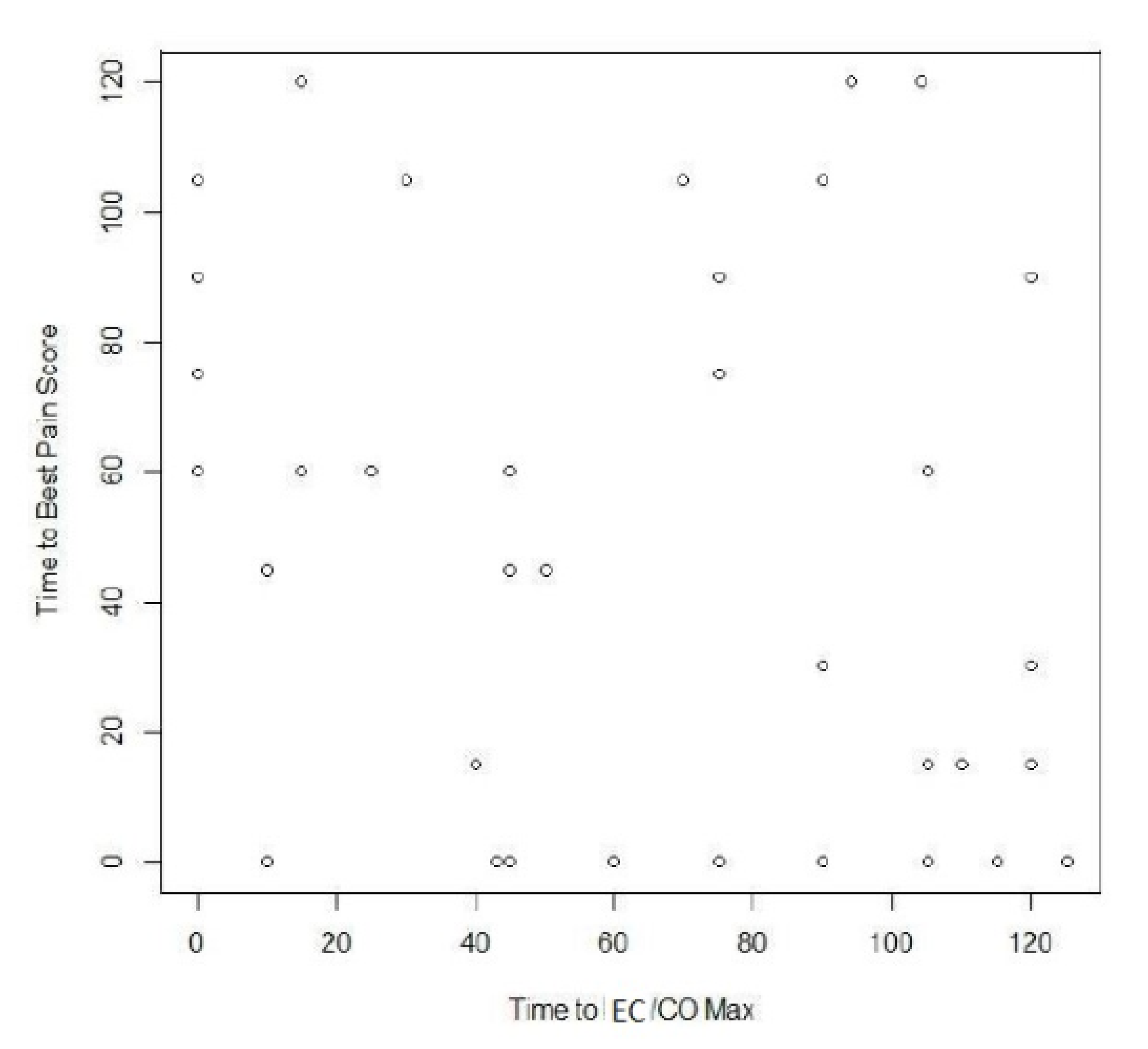
3. Results
4. Discussion
Acknowledgments
Author Contributions
Conflicts of Interest
References
- Jacob, E.; Mueller, B.U. Pain experience of children with sickle cell disease who had prolonged hospitalizations for acute painful episodes. Pain Med. 2008, 9, 13–21. [Google Scholar] [CrossRef] [Scilit] [PubMed]
- Agency for Health Care Policy and Research (US); Steiner, C.A.; Miller, J.L. Sickle Cell Disease Patients in US Hospitals, 2004: Statistical Brief #21. Healthcare Cost and Utilization Project (HCUP) Statistical Briefs (February 2006–December 2006). Available online: http://www.ncbi.nlm.nih.gov/books/NBK63489 (accessed on 13 August 2016).
- Plarr, O.S.; Thorington, B.D.; Brambilla, D.J.; Milner, P.F.; Rosse, W.F.; Vichinsky, E.; Kinney, T.R. Pain in sickle cell disease. Rates and risk factors. N. Engl. J. Med. 1991, 325, 1–16. [Google Scholar]
- Embury, S.H. Sickle Cell Disease: Basic Principles and Clinical Practice; Raven Press: New York, NY, USA, 1994. [Google Scholar]
- Poludasu, S.; Ramkissoon, K.; Salciccioli, L.; Kamran, H.; Lazar, J.M. Left ventricular systolic function in sickle cell anemia: A meta-analysis. J. Card. Fail. 2013, 19, 333–341. [Google Scholar] [CrossRef] [Scilit] [PubMed]
- Das, B.B.; Raj, A.; Recto, M.; Kong, M.; Bertolone, S. Utility of impedance cardiography for the detection of hemodynamic changes in stable patients with sickle cell disease. J. Pediatr. Hematol. Oncol. 2012, 34, 336–339. [Google Scholar] [CrossRef] [Scilit] [PubMed]
- Norozi, K.; Beck, C.; Osthaus, W.A.; Wille, I.; Wessel, A.; Bertram, H. Electrical velocimetry for measuring cardiac output in children with congenital heart disease. Br. J. Anaesth. 2008, 100, 88–94. [Google Scholar] [CrossRef] [Scilit] [PubMed]
- Raj, A.; Bertolone, S.J.; Mangold, S.; Edmonds, H.L., Jr. Assessment of cerebral tissue oxygenation in patients with sickle cell disease: effect of transfusion therapy. J. Pediatr. Hematol. Oncol. 2004, 26, 279–283. [Google Scholar] [CrossRef] [Scilit] [PubMed]
- Jensen, M.P.; Turner, J.A.; Romano, J.M. What is the maximum number of levels needed in pain intensity measurement? Pain 1994, 58, 387–392. [Google Scholar] [CrossRef] [Scilit]
- Pain Intensity Instruments. National Institutes of Health—Warren Grant Magnuson Clinical Center, July 2013. Available online: http://www.mvltca.net/presentations/mvltca.pdf (accessed on 12 February 2017).
- Buchanan, G.; Vichinsky, E.; Krishnamurti, L.; Shenoy, S. Severe sickle cell disease-Pathophysiology and therapy. Biol. Blood Marrow Transplant. 2010, 16, S64–S67. [Google Scholar] [CrossRef] [Scilit] [PubMed]
- Ballas, S.K. Pain management of sickle cell disease. Hematol. Oncol. Clin. N. Am. 2005, 19, 785–802. [Google Scholar] [CrossRef] [Scilit] [PubMed]
- Platt, O.S.; Brambilla, D.J.; Rosse, W.F.; Milner, P.F.; Castro, O.; Steinberg, M.H.; Klug, P.P. Mortality in sickle cell disease. Life expectancy and risk factors for early death. N. Engl. J. Med. 1994, 330, 1639–1644. [Google Scholar] [PubMed]
- Todd, K.H.; Ducharme, J.; Choiniere, M.; Crandall, C.S.; Fosnocht, D.E.; Homel, P.; Tanabe, P. PEMI Study Group. Pain in the emergency department: Results of the pain and emergency medicine initiative (PEMI) multicenter study. J. Pain 2007, 8, 460–466. [Google Scholar] [CrossRef] [Scilit] [PubMed]
- Padmanabhan, P.; Berkenbosch, J.W.; Lorenz, D.; Pierce, M.C. Evaluation of cerebral oxygenation during procedural sedation in children using near infrared spectroscopy. Ann. Emerg. Med. 2009, 54, 205–213. [Google Scholar] [CrossRef] [Scilit] [PubMed]
- Eddine, A.C.; Alvarez, O.; Lipshultz, S.E.; Kardon, R.; Arheart, K.; Swaminathan, S. Ventricular structure and function in children with sickle cell disease using conventional and tissue echocardiography. Am. J. Cardiol. 2012, 109, 1358–1364. [Google Scholar] [CrossRef] [Scilit] [PubMed]
- Coté, C.J.; Sui, J.; Anderson, T.A.; Bhattacharya, S.T.; Shank, E.S.; Tuason, P.M.; August, D.A.; Zibaitis, A.; Firth, P.G.; Fuzaylov, G. Continuous noninvasive cardiac output in children: Is this the next generation of operating room monitors? Initial experience in 402 pediatric patients. Paediatr. Anaesth. 2015, 25, 150–159. [Google Scholar] [CrossRef] [Scilit] [PubMed]

| Variable | Baseline Mean (SD) | Maximum Level Mean (SD) | p-Value |
|---|---|---|---|
| Cardiac Output | 5.15 (2.5) | 6.87 (2.3) | <0.01 |
| Cardiac Index | 3.70 (1.3) | 5.01 (1.2) | <0.01 |
| Cerebral O2 | 49.8 (8.8) | 59.1 (8.5) | <0.01 |
| Splanchnic O2 | 60.4 (15.6) | 72.0 (14.8) | <0.01 |
| Hemoglobin | 9.25 (1.9) | - | - |
| Hematocrit | 26.63 (5.9) | - | - |
| Fluids Administered | 0.85 (0.5) | - | - |
| Variable | Initial Cerebral Oxygenation | Minimum Cerebral Oxygenation | Maximum Cerebral Oxygenation | Time to Cerebral Oxygenation Max | Initial Splanchnic Oxygenation | Minimum Splanchnic Oxygenation | Maximum Splanchnic Oxygenation |
|---|---|---|---|---|---|---|---|
| Initial Cardiac Output | −0.125 | −0.070 | 0.158 | 0.158 | 0.151 | 0.147 | 0.182 |
| Minimum Cardiac Output | −0.058 | −0.007 | 0.113 | 0.15 | 0.147 | 0.182 | −0.064 |
| Maximum Cardiac Output | −0.156 | 0.019 | −0.067 | −0.038 | 0.162 | 0.198 | 0.067 |
| Time to Max Cardiac Output | −0.199 | 0.100 | −0.016 | −0.088 | 0.185 | 0.120 | −0.021 |
| Initial Cardiac Index | −0.193 | −0.059 | −0.048 | −0.109 | 0.143 | 0.090 | −0.006 |
| Minimum Cardiac Index | 0.088 | 0.037 | 0.100 | −0.145 | −0.014 | −0.086 | −0.123 |
| Maximum Cardiac Index | −0.362 | 0.146 | −0.350 * | 0.006 | 0.141 | 0.111 | −0.359 |
| Time to Max Cardiac Index | −0.300 | 0.129 | 0.027 | 0.044 | 0.126 | −0.128 | −0.162 |
| HB/HCT | −0.090 | 0.024 | 0.141 | −0.005 | −0.025 | −0.150 | 0.019 |
| HB | −0.107 | −0.058 | −0.068 | 0.148 | −0.006 | 0.088 | −0.048 |
| HCT | −0.088 | 0.158 | 0.176 | 0.007 | −0.193 | −0.007 | −0.109 |
| Reticulocyte count | −0.166 | 0.170 | 0.161 | −0.199 | −0.070 | −0.016 | 0.185 |
| Fluids | −0.231 | 0.123 | −0.156 | 0.165 | −0.067 | −0.088 | 0.198 |
| Total White Blood Count | −0.083 | 0.073 | 0.123 | 0.213 | −0.038 | 0.162 | −0.115 |
© 2018 by the authors. Licensee MDPI, Basel, Switzerland. This article is an open access article distributed under the terms and conditions of the Creative Commons Attribution (CC BY) license (http://creativecommons.org/licenses/by/4.0/).
Share and Cite
Padmanabhan, P.; Oragwu, C.; Das, B.; Myers, J.A.; Raj, A. Utility of Non-Invasive Monitoring of Cardiac Output and Cerebral Oximetry during Pain Management of Children with Sickle Cell Disease in the Pediatric Emergency Department. Children 2018, 5, 17. https://doi.org/10.3390/children5020017
Padmanabhan P, Oragwu C, Das B, Myers JA, Raj A. Utility of Non-Invasive Monitoring of Cardiac Output and Cerebral Oximetry during Pain Management of Children with Sickle Cell Disease in the Pediatric Emergency Department. Children. 2018; 5(2):17. https://doi.org/10.3390/children5020017
Chicago/Turabian StylePadmanabhan, Pradeep, Chikelue Oragwu, Bibhuti Das, John A. Myers, and Ashok Raj. 2018. "Utility of Non-Invasive Monitoring of Cardiac Output and Cerebral Oximetry during Pain Management of Children with Sickle Cell Disease in the Pediatric Emergency Department" Children 5, no. 2: 17. https://doi.org/10.3390/children5020017
APA StylePadmanabhan, P., Oragwu, C., Das, B., Myers, J. A., & Raj, A. (2018). Utility of Non-Invasive Monitoring of Cardiac Output and Cerebral Oximetry during Pain Management of Children with Sickle Cell Disease in the Pediatric Emergency Department. Children, 5(2), 17. https://doi.org/10.3390/children5020017

