Abstract
Biodetection, the basis of many biotechnologies, has rapidly developed in recent years. Among various biodetection methods, the photoelectrochemical (PEC) sensor is an emerging analytical method and has been applied in biodetection widely because of its high sensitivity, low cost, expandability into multichannel sensor arrays, and many other superior properties. Unlike conventional electrochemical aptasensors, the PEC aptasensor uses light as the excitation and an electrical photocurrent as the readout, which separates the stimulus from the measurement and reduces the excitation-related background. By modulating the light and demodulating the current, the PEC aptasensor improves the signal-to-noise ratio and lowers the limit of detection in complex matrices. Compared with optical aptasensors, the PEC aptasensor relies on simple light sources and electrodes rather than bulky imaging optics, enabling easier miniaturization and light-addressed multiplexed arrays. Therefore, aptamer-based PEC aptasensors have become a new hotspot in the field of biodetection. In this review, the development history of PEC aptasensors was presented. Then, this paper focuses on the photoactive nanomaterials, aptamers as sensing films, and sensing strategies of PEC aptasensors. The applications of PEC aptasensors in biodetection were also discussed. Finally, current challenges are discussed and opportunities in the future are prospected.
1. Introduction
With the increasing demand of early diagnosis of cancer, gene sequencing, protein detection, drug screening, and environmental monitoring, highly sensitive biodetection for biochemical analytes has drawn much attention in recent years. Conventional electrochemical and optical sensors have demonstrated high specificity, mature workflows, and reliable quantitative performance in many routine applications. However, previous detection method faces much challenges due to the complex biological environment, diverse types of analytes, and extremely low levels of the target [1,2,3]. Therefore, many efforts have been made in biodetection technology to solve these problems, and numerous novel biosensors are emerging.
Generally, the performance enhancement of biosensors focuses on two primary aspects—the sensing element and the transducer—which represent the core structural components of the sensor [4,5]. The sensing element usually refers to the part which can achieve the specific recognition of target analytes. This recognition process usually causes changes in physical or chemical properties of the sensor. Then, the transducer part of the sensor can produce related output signals according to the changes in physical or chemical properties caused by target analytes. The output signals from the transducer are usually required to be easily collected and processed, such as optical and electrical signals.
Traditional biosensors typically rely on sensing elements composed of biological enzymes, nanozymes, or specific antigen–antibody interactions with analytes. Enzyme- or nanozyme-based biosensors achieve high selectivity and catalytic efficiency when detecting small biomolecules (such as glucose, hydrogen peroxide, and glutathione), through catalytic redox reactions [6]. However, these biosensors are often ineffective for the detection of macromolecules, as the active sites of macromolecules are shielded by insulating layers, hindering effective electron transfer between the sensing element and the target. Therefore, redox-based detection becomes unfeasible for such analytes.
To address this limitation, biosensors based on antigen–antibody specificity, usually defined as immunosensors, have been extensively developed for macromolecule detection. Through antibody functionalization, such sensors can capture the corresponding antigens and modulate the charge transfer resistance at the electrode interface via steric hindrance, thereby producing a measurable output signal [7]. However, this method shows inferior sensitivity and specificity for detection of small molecule. This is primarily because small molecules usually contain only a few antigenic epitopes, resulting in weak and unstable binding, minimal conformational change, and consequently, limited impact on interfacial electron transfer.
Moreover, antibodies and enzymes, as complex natural proteins, lack uniform and well-defined modification sites, and are highly sensitive to environmental conditions such as temperature, pH, and ionic strength. These inherent drawbacks result in complex surface modification process, poor long-term stability, and high production costs. Therefore, developing sensing elements that are broadly applicable to both small molecules and macromolecules, while offering excellent performance and reliability, remains a hotspot in biosensor research.
Beyond antibody–antigen immunosensors, multiple strategies have been explored to enable electrochemical detection of macromolecules by enhancing electronic coupling between large biomolecules and the electrode, thereby improving sensor performance. One effective approach is to use electrochemically active mediators—such as conductive polymers, metal nanoparticles, and two-dimensional (2D) materials—as interlayers that facilitate electron transfer. In addition, the design of functional probes is critical. By introducing electroactive moieties (e.g., pyrrolic groups, phthalocyanines, or aromatic thiols) into the probe molecules, π-π stacking, electrostatic interactions, or covalent bonding with the electrode surface can be achieved, which further reinforces probe-electrode coupling. This strategy not only improves sensor stability but also expands applicability to complex biological samples. Moreover, molecularly imprinted polymers (MIPs)—as artificial receptor materials with high selectivity and stability—have been widely used in biosensors. MIPs recognize and capture specific macromolecules via complementary cavities formed in the polymer matrix. Compared with antibodies, MIPs are lower-cost, easier to synthesize and reusable, making them particularly suitable for macromolecule detection in complex samples.
Nevertheless, these approaches also face limitations, including restricted electron-transfer distances, insufficient stability and manufacturability/scalability, and suboptimal tolerance to interferents and specificity. MIPs further contend with binding-site heterogeneity, incomplete template removal, and a relatively high barrier to multiplexing.
In recent years, aptamers have emerged as a promising recognition element in biosensors [8,9]. Aptamers are single-stranded DNA or RNA oligonucleotides that can fold into unique three-dimensional structures to bind target molecules with high affinity and specificity. Compared with conventional biochemical recognition elements, aptamers offer several distinct advantages. Enzyme sensors rely on catalysis to convert the target into an electroactive product or to relay electrons from the enzyme’s redox center through a mediator to the electrode. By contrast, aptamer sensors do not require the target to be intrinsically electroactive. Specific binding at the electrode triggers an aptamer conformational change that alters probe orientation and electron-transfer pathways, thereby modulating the interfacial charge-transfer resistance and the double-layer capacitance (impedance signal) and yielding a stable, quantifiable electrical response. This mechanism helps overcome limitations commonly encountered by enzyme sensors in macromolecule detection. Compared with antibodies, aptamers undergo significant structural rearrangements—such as folding into G-quadruplexes or hairpin loops—upon binding small molecules. These conformational changes can modulate their spatial orientation and electron transport pathways on electrode surfaces, leading to measurable changes in impedance and enabling highly sensitive detection of small molecules. This effectively overcomes the poor sensitivity of immunosensors for small-molecule analysis. Then, aptamers exhibit high chemical and thermal stability, withstanding harsh conditions such as high temperatures, broad pH ranges, and high ionic strengths. Their small molecular size, well-defined sequence, and structurally simple backbone allow for the rational design of both binding domains and functional modification sites, greatly facilitating the construction of sensing interfaces. Additionally, they can be precisely selected through in vitro techniques such as systematic evolution of ligands by exponential enrichment (SELEX) according to different target molecules of various sizes and structural complexities, including small molecules, proteins, viruses, and cells, thus offering exceptional versatility and design flexibility [10].
Overall, aptamers not only address the detection range limitation of enzyme- and antibody-based biosensors, but also provides a more flexible and efficient solution for biosensors with high sensitivity, excellent stability, as well as easy modification and expandability.
Following the recognition of target molecules by the sensing element, biosensors generate corresponding measurable signals via a transducer. Apart from the sensing element, the transducer of a biosensor can also influence the sensing performance to target analytes. To date, the transducers of newly developed biosensors are usually based on three types of method: the optical method, electrochemical method, and photoelectrochemical (PEC) method. The optical method usually adopts a light source as an excitation signal, and the output is also an optical signal. The optical method has high sensitivity but usually requires sophisticated equipment and a professional operator [11,12]. In optical sensors, interference arises when the excitation light is not fully separated from the detected signal in wavelength, time, space, or polarization, allowing leaked or scattered excitation to reach the detector and elevate the background, which harms sensitivity and accuracy. In electrochemical sensors, the input signal is the electrical stimulus applied by the potentiostat, and the output signal is the measured electrode current. Because the excitation (applied potential/current) and the response (measured current/potential) are both electrical and occur at the same interface, within the same circuit and frequency range, they can overlap and interfere with one another. Therefore, the electrochemical method owns the advantages of inexpensive equipment and simple operation, but suffers from the interference between input and output electric signals, leading to low sensitivity and stability [12,13].
As a combination of the optical and electrochemical methods, photoelectrochemical (PEC) sensing has attracted considerable attention in recent years. PEC sensors typically employ a light source as the excitation signal and generate a measurable photocurrent or photovoltage through photoactive materials as the output signal. This configuration retains the high sensitivity characteristic of optical methods while effectively avoiding the signal overlap between the input and the output that often occurs in the electrochemical method. In photoelectrochemical sensors, the input is an optical signal and the output is an electrical current; placing the optical input and the electrical output in different domains makes mutual interference less likely than in purely electrochemical or optical formats. In addition, by modulating the light source at a fixed frequency while applying only a DC bias to the electrode and demodulating the photocurrent with a lock-in amplifier, only the component at the modulation frequency is retained, effectively removing DC drift, 1/f (low-frequency flicker) noise, and other off-frequency electrical perturbations. In addition, PEC systems exhibit excellent expandability and can be further developed into light-addressable multi-channel sensors. By illuminating different regions of the sensor, such light-addressable sensors enable simultaneous multi-analyte detection and spatially resolved imaging, demonstrating strong integrability and high-throughput potential [14,15]. Moreover, PEC methods require relatively simple and low-cost instrumentation, facilitating device miniaturization and user-friendly operation, which is highly promising for practical applications.
In the design of PEC transducers, the photoactive material which converts optical signals into electrical signals plays a crucial role. Early PEC sensors rely on bulk semiconductor materials, such as silicon wafers or thin films like TiO2 and ZnO. These materials possess inherent photoinduced electron-hole separation capabilities and are capable of fulfilling basic photoelectric conversion functions. However, their practical performance is constrained by several intrinsic limitations, including low specific surface area, narrow light absorption range, high charge recombination rates, and poor spatial resolution [16,17]. These shortcomings not only restrict their sensitivity and response efficiency but also hinder their expandability for sensor arrays. To address these limitations, researchers have increasingly incorporated nanomaterials as photoelectric conversion materials, leading to the development of nano-PEC sensors. Nanomaterials such as quantum dots, carbon-based nanostructures, and metal-organic frameworks (MOFs) offer superior optical absorption, enlarged surface areas, enhanced charge separation and transport properties, and facile surface functionalization. These features collectively contribute to significantly improved photoelectrochemical response, enhanced detection sensitivity, and higher spatial resolution. As a result, nano-PEC technology has enabled PEC sensing systems to evolve toward high-performance, multifunctional, and array-based platforms. Therefore, nano-PEC sensors have become the mainstream direction in PEC sensor development.
Due to the advantages of aptamer as the sensing element and PEC as the transducer, PEC biosensors based on aptamers have drawn much attention. Sensors using aptamers as a sensing element are usually called aptasensors, so PEC sensors using aptamers are defined as photoelectrochemical aptasensors. The PEC aptasensor combines the advantages of the PEC method, such as high sensitivity, low background noise, and integrability, with the remarkable specificity, stability, and ease of modification inherent to aptamers. This synergistic combination enhances the performance of the sensor, including its detection sensitivity, selectivity, and range of detectable targets. Furthermore, the PEC aptasensor exhibits excellent stability, reusability, and miniaturization potential, making it an ideal candidate for practical biosensing applications. From the perspective of device miniaturization, PEC aptasensors are inherently well suited for integration into lab-on-chip systems. The optical input can be remotely addressed, while the electrical readout requires only a low voltage and a compact transimpedance amplifier, enabling assays to be performed at the microliter scale within simple microfluidic channels. By employing LED modulation and bright/dark referencing, PEC aptasensors enable drift-resistant, low-power point-of-care testing (POCT) that can be powered by batteries or smartphones, facilitating rapid analysis. Additionally, the use of transparent or flexible electrodes (e.g., ITO/FTO, printed Au/carbon, and graphene) in conjunction with micro light sources (e.g., µLEDs or fiber-coupled emitters) allows for the development of wearable devices. These devices can be conformally deployed on curved surfaces, enabling light-addressed, multiplexed sensing for enhanced performance and user convenience.
This review focuses on the current advances in PEC aptasensors used for biosensing and provides insights into published papers in recent years. The fundamental principle and basic structure of the PEC aptasensor are presented. Nanomaterials and aptamers used in PEC aptasensors are also introduced. Then, various sensing mechanisms and enhancing strategies in different papers are thoroughly discussed and compared. Next, the target analytes of PEC aptasensors are classified, and their applications in biodetection are discussed. Finally, challenges faced by present PEC aptasensors and prospective chances in future are presented. The main contents are shown as Scheme 1.
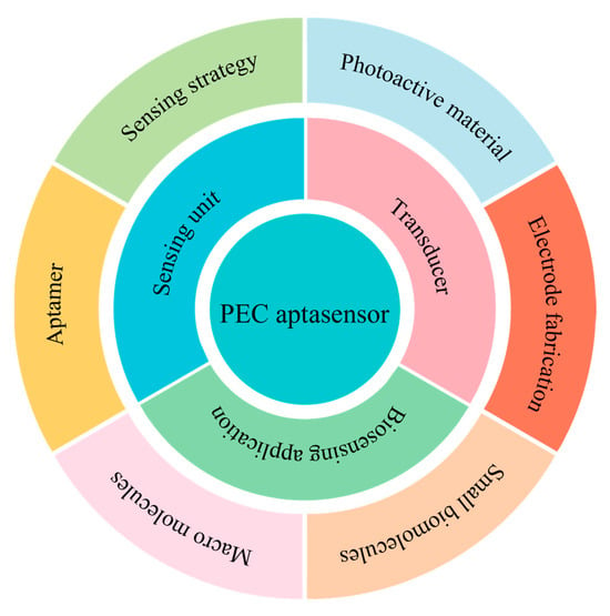
Scheme 1.
Illustration and structure of the PEC aptasensor for biosensing.
2. Construction of the Photoelectrochemical Aptasensor
2.1. Structure of the Photoelectrochemical Aptasensor Sensing System
A typical PEC aptasensor sensing system can be illustrated as Figure 1A. It can be divided in three parts: a signal transducer, a sensitive element, and an external measurement circuit. The signal transducer of the PEC aptasensor refers to the photoelectric conversion process, which converts the input optical signal into the output electrical signal through photoactive materials. The process of photoelectric conversion is illustrated in Figure 1B. Upon illumination of the PEC sensor by the light source, photoactive materials with a discrete band gap are excited, leading to the generation of photoinduced electron-hole pairs at the Lowest Unoccupied Molecular Orbital (LUMO). These electrons then travel through nanomaterials and reach the electrodes via tunneling (Ke). Simultaneously, electrons from the electrodes can tunnel into the Highest Occupied Molecular Orbital (HOMO) of the nanomaterials, recombining with photoexcited holes (Kb). The interplay between these two electron transfer processes (Ke and Kb) governs both the direction and magnitude of the photocurrent in the external circuit. In the absence of oxidizing or reducing agents in phosphate-buffered saline (PBS), the rates of Ke and Kb are influenced by the HOMO, LUMO of the nanomaterials, and the Fermi level of the electrodes, which is affected by the applied potential. No measurable photocurrent is observed when Ke and Kb are in a dynamic equilibrium state. Under a fixed bias, illumination creates electron–hole pairs in the photoactive material. The applied (and built-in) electric fields separate these carriers and drive them to the interface, where they transfer charge to or from species in solutions. Photogenerated holes can oxidize reducing agents or sacrificial donors in the electrolyte, whereas photogenerated electrons can reduce dissolved oxygen or other electron acceptors. If a redox mediator is present, the photoactive material can also exchange electrons with that mediator. These interfacial pathways decrease the charge-transfer resistance and increase the measured current. We define the baseline current measured without illumination as the dark current and the current under illumination as the light current, and we take their difference as the analytical photocurrent signal. In subsequent data analysis, all photocurrent values refer to this difference.
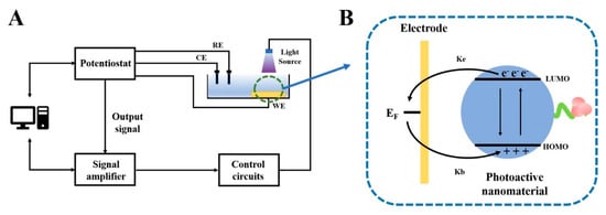
Figure 1.
(A). Schematic illustration of a typical PEC aptasensor sensing system. (B) The photoelectric conversion process of the PEC aptasensor.
The measurable signal of the PEC aptasensor depends on interfacial electron transfer and photocarrier collection, while noise and drift primarily arise from the dark current, capacitance, and 1/f noise. Several key trade-offs influence performance. The bias voltage, for example, plays a critical role. Stronger band bending enhances carrier collection but also increases the dark current, requiring the selection of a narrow bias window near the sensitivity peak. Light intensity and modulation frequency are also important factors, as the photocurrent increases linearly with light intensity, while shot noise increases with the square root of intensity. Thus, lock-in detection at frequencies higher than the 1/f corner effectively helps to suppress drift. In addition, the thickness and porosity of the photoactive film are crucial. Thicker films can enhance absorption but may also increase transport resistance and recombination. Therefore, the optimal film thickness is typically near the absorption length or at the point where the modulated photocurrent levels off.
When the transducer part of the PEC aptasensor is achieved by photoactive materials, the sensitive part is realized by the aptamer as the sensing element. Through the complementary shape interactions and three-dimensional folding, aptamers can bind with their targets with high affinity and specificity. Thus, aptamers are immobilized on the electrodes as the sensing element. When there are target molecules in the solution, aptamers can capture them. The interactions between aptamers and target molecules will influence the optical and electrical properties of the PEC aptasensor, such as light adsorption or impedance of the whole system. Moreover, upon target binding, the aptamer undergoes a conformational rearrangement that changes its distance and orientation with respect to the electrode and modifies the local dielectric constant and surface charge distribution of the molecular layer. Because tunneling transport decays exponentially with separation, nanometer-scale reorganization can produce large variations in current or impedance. At the same time, the interfacial reconfiguration alters band bending and the width of depletion/accumulation regions, which in turn affects the separation and collection of photogenerated carriers and the rate of surface recombination. Together, these effects account for the differences in photocurrent observed during sensing. Therefore, the binding of aptamers with target analytes will affect the charge transfer of the photoelectric conversion process. The charge transfer can be measured as a photocurrent by an external circuit, and the output photocurrent is usually proportional to the concentration of analytes in the solution.
The external circuit of the PEC detection system can generally be classified into two types. The first type is the direct-current (DC) PEC detection system, based on a constant light source and a potentiostat. In this system, the working electrode in a three-electrode setup is subjected to a bias voltage from the potentiostat under continuous, constant intensity light irradiation, and the current signal is recorded. The detection result is obtained by measuring the current difference before and after illumination, which reflects the light response intensity. This system has a simple structure, and the potentiostat can be directly implemented using analog-to-digital/digital-to-analog (AD/DA) or analog front end (AFE) chips, making it suitable for integration and miniaturization. However, the detection signal in this system is susceptible to interference from the 1/f noise, dark current, and baseline drift, which leads to low signal-to-noise ratios, reduced sensitivity, and insufficient accuracy.
The second type is the PEC detection system based on modulation/demodulation mechanisms. The main structure involves using a lock-in amplifier to output a modulation signal at a specific frequency to drive the excitation light source, generating modulated light to illuminate the PEC sensor. Under modulated light, the sensor generates a high-frequency modulated photocurrent, which is then collected by the potentiostat and sent to the lock-in amplifier for synchronous demodulation. The demodulation process converts the high-frequency signal into its DC component, filtering out low-frequency noise and background interference (such as 1/f noise and baseline drift), thereby significantly improving the system’s anti-interference capability, signal-to-noise ratio, and measurement accuracy. However, due to the inherent complexity of the lock-in amplifier, the large computational load of the algorithm, and high-power consumption, this system is difficult to achieve miniaturization and field deployment. To address the miniaturization challenges of this system, some groups have replaced the traditional lock-in algorithm with a Discrete Fourier Transform (DFT) algorithm, combined with the Coordinate Rotation Digital Computer (CORDIC) architecture and Hanning window functions for performance optimization, resulting in a high-precision, low-power, and resource-efficient miniaturized PEC detection system [18].
The signal transducer, sensing element and external measurement circuit constitute a typical PEC aptasensor system. As mentioned above, the external measurement circuits can be primarily divided into two types and have little developments or updates in recent years. Current studies of the PEC aptasensor mostly focused on the signal transducer and sensing element to improve the sensing properties. In the part of the signal transducer, photoactive nanomaterials play the core role, so finding new nanomaterials with higher photoelectric conversion property and proposing new strategies to enhance photoelectric conversion property are always hot spots. As for the sensing element, as the aptamer shows many advantages in building sensing materials, recent researches make much effort in developing novel sensing mechanisms and enhancing strategies to improve the sensitivity of the PEC aptasensor. Therefore, in the following sections, we will discuss and compare the nanomaterials used for building PEC transducers and aptamers used for sensing elements in PEC aptasensors. Then, the sensing mechanisms, enhancement strategies, and electrode fabrication methods will be studied and compared thoroughly and comprehensively. Finally, the application of PEC aptasensors in biosensing will be summarized.
2.2. Photoactive Nanomaterials
The most important feature of nanomaterials used in PEC aptasensors is the photoelectric conversion property, which achieves the transducer part of the sensor. The PEC aptasensor constructed with excellent photoelectric property can usually obtain obvious output photocurrent signals. Typically, a PEC aptasensor with a larger output photocurrent signal can provide a better sensitivity for detection of target analytes. In addition, photoactive nanomaterials immobilized on electrodes provide a place for the modification of aptamers and subsequent recognition processes for target analytes.
Compared to nanomaterials used in electrochemical and optical sensors, nanomaterials employed in PEC aptasensors require distinct properties. Electrochemical aptasensors typically focus on the catalytic properties, conductivity, and charge transfer capabilities of the materials. On the other hand, optical aptasensors require optical characteristics such as fluorescence, surface-enhanced Raman scattering (SERS), or light absorption. In PEC aptasensors, photoelectric nanomaterials not only need to exhibit excellent light absorption and electron transfer performance but also must possess efficient photoinduced electron−hole pair separation efficiency. Furthermore, these materials must be able to effectively respond to subtle interfacial disturbances at the electrode surface, such as spatial steric hindrance, charge distribution, or conformational changes induced by aptamer−target interactions.
Compared with traditional PEC sensors based on redox reactions or antigen–antibody recognition-induced impedance changes, PEC aptasensors offer greater flexibility in the selection of photoelectronic nanomaterials, due to the inherent advantages of aptamers. In redox-based PEC sensors, signal generation depends on the electron donor–acceptor behavior of target analytes during redox processes, typically leading to distinct photocurrent variations. Such sensors, therefore, require photoactive semiconductors with stable energy levels and strong catalytic activity toward the analyte or its intermediates.
Antibody-based PEC immunosensors, on the other hand, rely on steric hindrance effects induced by specific binding between protein molecules. This mechanism primarily affects the overall interfacial resistance of the electrode and is characterized by rigid recognition behavior and limited conformational dynamics. As a result, these sensors require a relatively high charge separation efficiency and interfacial electron transfer rate of the photoactive material. Moreover, the preservation of antibody bioactivity under mild environmental conditions further constrains material selection, especially in terms of surface functionalization, pH tolerance, and biocompatibility.
In contrast, PEC aptasensors utilize sensing mechanisms such as aptamer conformational changes, charge redistribution, and steric effects upon target binding, which can modulate interfacial charge transfer either directly or indirectly. These mechanisms enable even subtle physical disturbances at the electrode interface to be converted into detectable PEC signals, which reduces the performance requirements on the material. In addition, aptamers can be immobilized onto photoactive materials through diverse and controllable methods, including thiol–gold interactions, self-assembled monolayers, and π–π stacking, while maintaining excellent stability under a wide range of environmental conditions. These features broaden the range of suitable photoactive nanomaterials for PEC aptasensor development and facilitate the design of more flexible, robust, and adaptable sensors.
Up to now, various photoactive nanomaterials have been applied in fabricating PEC aptasensors for high-performance biosensing. They can be classified in semiconductors and heterojunction nanocomposites.
2.2.1. Single-Component Semiconductors
The most widely used photoactive nanomaterials are various semiconductors, which can generally be classified as inorganic semiconductors and organic semiconductors.
Among them, inorganic semiconductors have laid the foundational framework for the early development of PEC aptasensors. The earliest inorganic semiconductors used for PEC aptasensing were CdS quantum dots (QDs), which originated from the work of Willner et al. They functionalized CdS QDs with one of the anticocaine aptamers subunits and modified a Au electrode with another anticocaine aptamer subunit. In the presence of cocaine, the formation of supramolecular complexes between the aptamer subunits and cocaine would confine CdS QDs onto the electrode surface. Due to the photoelectric conversion property of CdS QDs, the electron transfer between the Au electrode and CdS QDs under illumination would generate a photocurrent. The intensity of the photocurrent was controlled by the amount of supramolecular cocaine−aptamer complexes associated with the electrode, thus achieving PEC aptasensing of cocaine [19].
Following this pioneering study, a variety of inorganic semiconductor materials have been explored to improve sensitivity and broaden target applicability, such as metal oxides, metal chalcogenides, and various QDs. For example, Zhang et al. used negatively charged CdSe semiconductor nanoparticles capped with mercaptoacetic acid as photoactive nanomaterials on the ITO electrode and achieved highly sensitive and specific aptasensing of Ramos cells [20]. To expand applicability to diverse biomolecules, alternative quantum dots were investigated. Wang et al. proposed a label-free “switch-on” PEC aptasensor based on PbS QDs to construct a PEC-sensing platform toward versatile biomolecular targets such as DNA and thrombin [21]. The photoelectric conversion process of PbS QDs is illustrated in Figure 2A. Other QDs were also widely used in fabricating PEC aptasensors, such as CdTe QDs, CdSeTe QDs, and g-C3N4 QDs [22,23,24]. Despite the excellent photoelectric performance of QDs, concerns regarding toxicity and long-term stability prompted a shift toward exploring metal oxide semiconductors with better biocompatibility and environmental robustness. For example, Cao et al. constructed a competitive PEC aptasensor for thrombin (TB) detection using a TiO2 photoactive electrode, coupled with TB-Au nanoparticle (AuNP)–GOx labels [25]. Li et al. used a Fe2O3 nanorod photoelectrode to fabricate a PEC aptasensor for highly sensitive and selective detection of lysozyme [26]. Many other inorganic semiconductors were also used in PEC aptasensors, such as BiOI, BiVO4, and Bi4NbO8Cl [27,28,29].
While inorganic semiconductors have made significant contributions, their limited structural flexibility and relatively high fabrication temperatures have motivated researchers to turn attention toward organic semiconductors. Organic semiconductors have the advantages of functional adjustability, easy of processing, low cost, and good photoelectric conversion efficiency, which are beneficial to the construction of PEC aptasensors [30,31]. Yao et al. reported the synthesis and electropolymerization of a photoactive bifunctional copolymer using copyrenebutyric acid Nα′, N-bis(carboxymethyl)-L-lysine amide (NTA-pyrene), and [tris-(2,2′-bipyridine) (4,4′-bis(4-pyrenyl-1-ylbutyloxy)-2,2′-bipyridine] ruthenium (II) hexafluorophosphate (Ru(II)–pyrene) complex. This copolymer was used to fabricate a PEC aptasensor for detection of thrombin and anti-cholera toxin antibodies [32]. To enhance molecular recognition compatibility and light adsorption efficiency, researchers further explored covalent organic frameworks (COFs), which feature ordered π-conjugated structures and high porosity. Zhang et al. presented a novel photoelectrochemical (PEC) aptasensor based on two-dimensional (2D) porphyrinic covalent organic frameworks (COFs) for the label-free detection of C-reactive protein [33]. The photoelectric conversion process of COFs is illustrated in Figure 2B. More recently, to address the need for higher quantum yield and better dispersibility, polymer-based quantum dots have been proposed. Chen et al. used [poly[(9,9-dioctylfluorenyl-2,7-diyl)-co-(1,4-benzo-(2,1,3)-thiadazole)] polymer quantum dots as photoactive materials for PEC aptasensors and achieved the detection of tetracycline [34]. Beyond the materials discussed above, two-dimensional (2D) materials have attracted broad interest in photoelectrochemical (PEC) aptasensors. Their very high surface area and short carrier-diffusion paths enable efficient separation and interfacial transfer of photogenerated charges at low bias. In addition, tunable layer terminations and energy levels facilitate the formation of uniform, thin interfaces that suppress dark current and allow higher-density, better-oriented aptamer immobilization—features that suit sensitive, low-power, and miniaturizable PEC aptasensors. Li and co-workers used water-dispersible graphitic carbon nitride (g-C3N4) as a single photoactive component to build a PEC aptasensor for kanamycin. Under visible light, binding-induced changes in interfacial charge and mass transfer were converted into a readable current signal [35]. Subsequently, Peng et al. implemented a self-powered cathodic PEC aptamer platform based on g-C3N4 for oxytetracycline detection, illustrating the advantages of 2D carbon nitrides in achieving low dark current and low-power readout [36]. Zhou et al. reported a chloramphenicol PEC aptasensor using tungsten disulfide (WS2) nanosheets: the aptamer first adsorbed onto WS2 via van der Waals/π interactions to suppress the photocurrent, and target binding followed by DNase I (deoxyribonuclease I)-assisted cyclic release restored the photocurrent, providing sensitive amplification [37]. In addition, Bu et al. employed black phosphorus quantum dots (BPQDs, obtained by exfoliating 2D black phosphorus) as a single-phase photocathode, together with hemin as an electron acceptor, to realize detection of amyloid-β (Aβ) peptide, highlighting the promise of 2D phosphorus materials for biomacromolecule analysis [38].
Despite the diverse material choices, single-phase semiconductors still face intrinsic limitations such as a suboptimal light absorption range and fast photoinduced electron−hole recombination. With the rapid development of nanotechnology, many methods were proposed to improve the photoelectric conversion efficiency of single-phase photoactive nanomaterials. The most used method is element doping, which is a simple but effective way to improve the photoelectric properties of semiconductors. Element doping usually introduces localized electronic states between the conduction and valence bands of the host, thus regulating the separation and recombination process of photogenerated electron−hole pairs. It should be noted that element doping did not result in a formation of new crystallographic phase. Liu et al. synthesized nitrogen-doped graphene quantum dots (GQDs) as a photoactive nanomaterial and fabricated a PEC aptasensor for chloramphenicol detection [39]. To further improve charge separation and light response, transition metal doping was also investigated. Li et al. fabricated a simple and label-free PEC aptasensor for detection of acetamiprid based on Co-doped ZnO, a diluted magnetic semiconductor, as a photoelectric beacon [40]. In addition, non-metal dopants such as phosphorus have been introduced to modulate defect states and surface energy. Peng et al. presented a PEC aptasensor using phosphorous-doped ultrathin g-C3N4 porous nanosheets as the photoactive nanomaterial and achieved detection of oxytetracycline.
In summary, although a wide range of single-phase semiconductors and doping strategies have been explored to enhance PEC performance of photoactive nanomaterials, their improvements remain fundamentally limited by intrinsic material properties. To further boost PEC performance, the construction of semiconductor heterojunctions has emerged as an effective and promising approach. The following section will focus on recent advances in heterojunction photoactive materials for PEC aptasensing.
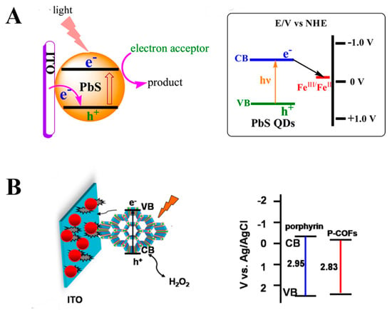
Figure 2.
Schematics of the photoelectric conversion process of the PEC aptasensor based on the elements: (A) inorganic semiconductor: PbS QDs (reprinted with permission from Ref. [21]. Copyright 2015 American Chemical Society); (B) organic semiconductor: p-COFs (reprinted with permission from Ref. [33] Copyright 2019 Elsevier).
2.2.2. Heterojunction Nanohybrids
In recent years, constructing heterojunction has been demonstrated to be an effective way to improve the photoelectric conversion efficiency of photoactive nanomaterials. A heterojunction is the junction formed at the interface between two solids with dissimilar electronic structures—most commonly two semiconductors, or a semiconductor and a metal—that differ in band gap, electron affinity, and/or work function. After Fermi-level equilibration, band bending and a built-in electric field arise at the interface; these provide a driving force for directional separation and transport of photogenerated carriers and, in turn, modulate the kinetics of charge transfer across the interface. This significantly reduces the charge carrier recombination. In addition, heterojunction can broaden the light absorption range, optimize band alignment and enhance interfacial charge transfer kinetics, thereby exhibiting superior performance in PEC sensors. As a result, heterojunctions have been widely adopted in the development of high-performance PEC aptasensors and are becoming a mainstream design strategy. Up to now, heterojunction nanohybrids for PEC asptasensor construction can be classified as inorganic–inorganic, organic–inorganic, and organic–organic heterojunctions, and multicomponent heterojunction.
Inorganic–Inorganic Heterojunction
Compared to other types of heterojunctions, inorganic–inorganic heterojunctions offer significant advantages in PEC aptasensors due to the superior chemical stability, thermal robustness, and tunable electronic structures. These heterojunctions are generally classified into two main categories: semiconductor–semiconductor and metal–semiconductor heterojunctions. Semiconductor–semiconductor heterojunctions can be engineered via band structure alignment to form internal electric fields at the interface, which effectively promote the separation and directional transfer of photogenerated electron–hole pairs, thereby enhancing photoelectric conversion efficiency. In contrast, metal–semiconductor heterojunctions leverage the excellent conductivity of metals and surface plasmon resonance (SPR) effects, which not only intensify local electric fields and improve light-harvesting efficiency but also generate hot electrons to further boost PEC sensitivity. As a result, inorganic–inorganic heterojunctions play a vital role in the construction of high-performance PEC aptasensors, contributing to improved stability, signal-to-noise ratio, and interfacial charge regulation.
Semiconductor–semiconductor heterojunctions are usually constructed by two kinds of semiconductor with matching band structures and energy levels. Under illumination, the photogenerated electrons are transferred from the higher conduction band (CB) level of a semiconductor to the lower CB of another semiconductor, while the photogenerated holes are transferred from the lower valence band (VB) of a semiconductor to the higher VB of another semiconductor. By this way, the recombination of photoinduced electron-hole pairs is effectively suppressed, resulting in a better photoelectric conversion property [41,42]. There have been many studies on semiconductor–semiconductor heterojunctions used in constructing PEC aptasensors. Yang et al. designed a MgIn2S4-TiO2 heterojunction by growing MgIn2S4 nanoplates on a TiO2 nanoarray and applied it in developing a PEC aptasensor for detection of adenosine triphosphate [43]. Chen et al. presented a selective and stable PEC aptasensor based on Co3O4 nanoparticles/g-CN heterojunction to detect oxytetracycline [44]. The mechanism of enhanced PEC property of this heterojunction is shown in Figure 3A. Many other semiconductor–semiconductor heterojunctions were synthesized for PEC aptasensors, such as GO/w-g-C3N4, WO3/Fe2O3, CdS-Cu2O, CdTe-Bi2S3, and Bi24O31Cl10/BiOCl [35,45,46,47,48].
To further improve the PEC property of photoactive nanomaterials, heterojunctions constructed with three semiconductors through a ternary Z-scheme structure were proposed. For example, Chen et al. designed NiS2/NiFe-layered double/graphitic carbon nitride ternary heterostructure by the surface vulcanization of flower-like NiFe LDH/g-C3N4 and it was used for developing a label-free PEC aptasensing platform to detect enrofloxacin [49]. Peng et al. constructed a WO3/g-C3N4/MnO2 photoanode for PEC aptasensors and applied in oxytetracycline detection [50]. Other dual Z-scheme structure heterojunction nanohybrids were also widely used in PEC aptasensors, such as MoS2 QDs-BiOI, Ag2CrO4/g-C3N4/GO, AgBr/AgI-Ag-CNTs, and Ag2S/Ag/ZnIn2S4/C3N4 [51,52,53,54].
In addition to semiconductor–semiconductor heterojunctions, researchers have increasingly turned to metal–semiconductor heterojunctions as a promising alternative strategy to further enhance the PEC performance of nanomaterials. Due to the Schottky junction formed by metal and semiconductor, the photogenerated electrons will flow from the CB of the semiconductor into the Fermi energy level of the metal, which will effectively promote the separation of photoinduced electron–hole pairs. Meanwhile, some noble metal nanomaterials have a localized surface plasmon resonance (LSPR) effect, which can drastically enhance the light absorption and promote the charge transfer of the semiconductor [55]. Based on these two main reasons, metal–semiconductor heterojunctions have been demonstrated their great potential in constructing PEC aptasensors. Hu et al. fabricated a Schottky junction based on gold nanoparticles (AuNPs)/gallium nitride (GaN) by growing AuNPs in situ on the surface of GaN, followed by etching with H2O2 to an appropriate diameter. Then, the AuNPs/GaN Schottky photoelectrode was applied to develop a novel photoelectrochemical (PEC) aptasensor for the detection of the epithelial ovarian cancer marker—CA125 [56]. In another example, Li et al. utilized the LSPR effect of Au nanoparticles to enhance the PEC property of TiO2 nanoparticles and fabricated a label-free PEC aptasensor for tetracycline detection [57]. As research in this area progressed, more complex hybrid systems were developed to further integrate Schottky and LSPR effects. Zhang et al. synthesized CuO/Pd nanocomposites, which exhibited excellent PEC property due to the Schottky junction and the LSPR effect and were applied in developing a PEC aptasensor for kanamycin detection [58]. Then, they used AuNPs to fabricate a CuO/AuNPs Schottky junction. Combined with the LSPR effect of AuNPS, the CuO/AuNPs Schottky junction was used to construct a PEC aptasensor for 3,3′,4,4′-tetrachlorobiphenyl in fish [59]. Beyond these examples, a variety of other metal–semiconductor heterojunctions have also been explored as photoactive nanomaterials in PEC aptasensors, such as Bi/CN, (AuNPs)2/GR-TiO2, and Au/BiVO4 [60,61,62].
In summary, inorganic–inorganic heterojunctions have demonstrated remarkable capabilities in enhancing charge separation, light absorption, and interfacial electron transfer, thereby significantly improving the performance of PEC aptasensors.
Organic–Inorganic Heterojunction
To further expand the design flexibility and functional tunability of photoactive interfaces, researchers have begun exploring organic–inorganic heterojunctions. Organic–inorganic heterojunctions in PEC aptasensors combine the structural tunability and excellent biocompatibility of organic materials with the high photoelectric property and stability of inorganic semiconductors. On the one hand, the organic components possess abundant functional groups and excellent surface modifiability, which facilitate aptamer immobilization and interfacial engineering, thereby enhancing the synergistic interaction between the sensing unit and the transducer interface. On the other hand, the inorganic semiconductor component offers efficient generation and transport of photogenerated charge carriers, significantly boosting photoelectric conversion efficiency. Through rational band structure design, organic–inorganic heterojunctions can establish effective electron transport pathways and built-in electric fields at the interface, enabling efficient electron–hole separation, reducing recombination probability, and improving the signal-to-noise ratio. As a result, such heterojunctions not only enhance the sensitivity and response speed of PEC aptasensors, but also expand the diversity of material design.
Organic–inorganic heterojunctions can generally be divided into organic semiconductor–inorganic semiconductor and organic semiconductor–metal heterojunctions.
The organic materials of organic semiconductor–inorganic semiconductor heterojunctions used in PEC aptasensors mainly refer to conjugated polymer semiconductors. Conjugated polymer semiconductors have attracted much attention in fabricating PEC aptasensors because of their charming optical, electronic capabilities, and adjustable molecular geometry by modifying the polymeric monomer. The way of conjugated polymer semiconductors-based organic–inorganic heterojunctions in improving PEC property is similar to that of the inorganic semiconductor–semiconductor heterojunctions. By the flow of photoinduced electrons and holes between conjugated polymer semiconductors and inorganic semiconductors, the charge separation of photoactive nanomaterials will be effectively promoted. To demonstrate these capabilities, a number of studies have reported conjugated polymer-based heterojunctions with enhanced PEC performance. For example, Zheng et al. proposed a poly(diphenylbutadiene)–BiOBr heterojunction. PDPB nanofibers are grown on BiOBr flower-like microspheres to form a PDPB-BiOBr nanocomposite dispersed in aqueous chitosan, which is then used to modify an indium tin oxide (ITO) electrode to fabricate a PEC aptasensor [63]. Qin et al. proposed a polydopamine/Ag2S/TiO2 NTs organic–inorganic heterojunction and applied it in PEC aptasensors for ofloxacin (OFL) detection [64].
While organic semiconductor–inorganic semiconductor systems offer promising charge transport behavior, researchers have also explored organic–metal heterojunctions to take advantage of additional mechanisms such as Schottky junction formation and LSPR. The organic–metal heterojunctions mainly contain polymer semiconductor–metal heterojunction, metal–organic frameworks (MOFs), and organic–inorganic hybrid perovskites. The organic–metal heterojunction-based photoactive materials usually exhibit excellent PEC property because of the Schottky junction and the LSPR effect, which is similar to the inorganic semiconductor–metal heterojunction. Many studies have made effort in developing organic semiconductor–metal heterojunction-based PEC aptasensors. For example, Wu et al. used a Au nano-flower/organic polymer (PTB7-Th) heterojunction as a photoelectric platform for ultrasensitive aptasensing of Hg2+ [65]. The mechanism of enhanced PEC property of this heterojunction is shown in Figure 3B. Further expanding on the organic–metal interface strategy, Jiang et al. constructed a PEC aptasensor using a two-dimensional metal–organic framework-based Schottky junction called Au NPs/Yb-TCPP and achieved the detection of SARS-CoV-2 [66]. In addition to MOF-based platforms, hybrid perovskites represent another emerging class of organic–metal heterostructures. For example, Shen et al. fabricated a photoelectrode based on CH3NH3PbI3 perovskite and achieved PEC aptasensing of dibutyl phthalat [67].
In summary, organic–inorganic heterojunctions effectively combine the tunable functionality and biocompatibility of organic materials with the superior photoelectric properties of inorganic components, resulting in enhanced charge separation, interfacial coupling, and overall PEC performance. These structures have shown great promise in improving sensitivity and versatility in PEC aptasensors.
Organic–Organic Heterojunction
Researchers have also begun to explore organic–organic heterojunctions as a novel strategy to construct fully organic PEC aptasensors with flexible design, low-cost processing, and excellent interfacial compatibility. This type of heterojunction is typically composed of two organic semiconductor materials, which form a built-in electric field at the interface through energy level alignment. This internal field facilitates efficient separation of photogenerated electron–hole pairs and enhances photoelectric conversion efficiency. Compared with inorganic materials, organic components offer superior processability, biocompatibility, and structural versatility. Their electronic band structures and surface functional groups can be precisely tailored through molecular engineering, enabling effective aptamer immobilization and sensitive modulation of the interfacial microenvironment. In addition, organic–organic heterojunctions generally exhibit excellent flexibility and solution processability, making them promising candidates for the development of flexible and wearable PEC aptasensors. Tian et al. firstly prepared a poly(5-formylindole)/poly(3,4-ethylenedioxythiophene) (P5FIn/PEDOT) organic heterojunction by simple two-step electrochemical polymerization, and it exhibited a strong photocurrent signal owing to the synergistic effect between P5FIn and PEDOT. Then, the organic polymer heterojunction was utilized to fabricate a novel photocathode to construct a PEC aptasensor for ultrasensitive and selective detection of thrombin [68]. The mechanism of enhanced PEC property of this heterojunction is shown in Figure 3C. Encouraged by the promising PEC enhancement observed in polymer-based heterojunctions, researchers began to explore more advanced donor–acceptor systems to further optimize charge separation and transport efficiency. Luo et al. used Y6 as an organic acceptor and combined it with the organic donor PM6 to form an organic–organic heterojunction, which promoted separation of photo-generated charges with high charge transfer efficiency, resulting in the enhancement of photocurrent intensity. The organic PM6:Y6 p-n heterojunction was used as photoactive materials to construct a PEC aptasensor for the detection of circulating tumor cells [69]. Compared to earlier systems, this design benefits from the advanced optoelectronic properties of non-fullerene acceptors like Y6, offering not only higher signal output but also improved selectivity and suitability for detecting low-abundance cellular targets.
In summary, organic–organic heterojunctions offer distinct advantages including tunable energy levels, excellent biocompatibility, and mechanical flexibility. However, to further enhance light absorption, charge transport, and overall PEC performance, recent research has increasingly focused on multicomponent hybrid systems, where two or more photoactive materials are integrated to achieve synergistic effects beyond single or binary heterojunctions.
Multicomponent Heterojunction
Multicomponent heterojunctions are typically constructed by integrating three or more nanomaterials with complementary properties onto a single electrode. On the one hand, rationally designed band structures among the different components help establish multilevel energy gradients and strengthen the interfacial built-in electric field, which more effectively drives the separation and directional transport of photogenerated electron–hole pairs. On the other hand, individual materials can be assigned specific roles—such as light harvesting, charge transport, or catalysis—forming a synergistic multi-pathway mechanism that enhances overall PEC performance. In addition, multicomponent systems can broaden the spectral absorption range, extend carrier lifetime and diffusion length, and provide more surface binding sites for aptamer immobilization. These features collectively contribute to improved sensitivity, stability, and selectivity of PEC aptasensors. Therefore, multicomponent heterojunctions represent a more flexible and efficient material design strategy for building high-performance PEC aptasensors, especially suited for multifunctional integration and detection in complex biological environments. For example, Xu et al. designed a metal–inorganic semiconductor–inorganic semiconductor heterojunction. They prepared a Bi/BiVO4/g-C3N4 heterojunction by in situ reduction of BiVO4/g-C3N4. Bi served as an electron conduction bridge between BiVO4 and g-C3N4 to form a Z-scheme structure, significantly accelerating the separation of photogenerated carriers. Meanwhile, the LSPR effect of Bi could greatly increase light harvesting. Therefore, the synergism of the Z-scheme heterojunction and the SPR effect of Bi efficiently boosted the photoelectric response of the Bi/BiVO4/g-C3N4 multicomponent heterojunction [70]. Inspired by the synergistic interplay of multiple functionalities, other studies have employed noble metal-based ternary heterojunctions for improved PEC performance. Zhu et al. successfully prepared AgI/Ag/BiOI nanosheets by simple ion-exchange and photo-reduction and fabricated a PEC aptasensor to detect chloramphenicol [71]. The mechanism of enhanced PEC property of this multicomponent heterojunction is shown in Figure 3D. While most early efforts focused on all-inorganic components, recent research has extended the multicomponent design strategy to include organic semiconductors, aiming to integrate their molecular tunability and interface flexibility. For example, Lv et al. used F8BT polymers as the donor–acceptor (D-A)-conjugated polymer semiconductors and g-C3N4 as the inorganic semiconductors to fabricate a D-A F8BT/g-C3N4 organic semiconductor–inorganic semiconductor heterojunction. Then, Au NPs were introduced to form a metal–organic semiconductor–inorganic semiconductor heterojunction. The organic semiconductor D-A type F8BT with the LUMO levels of −3.49 eV and HOMO levels of −5.74 eV versus vacuum could accelerate Frenkel exciton migration, reduce energy loss and enhance photoelectric conversion efficiency. Along with the LSPR effect of Au NPs, this multicomponent heterojunction exhibited excellent PEC property. PEC aptasensors based on this organic–inorganic heterojunction achieved the detection of carcinoembryonic antigen [72]. To further improve the photocurrent output and material surface area, researchers have also explored combinations of MOFs and noble metals. For example, Li et al. integrated gold nanoparticles onto an Er-MOFs surface to enhance the photocurrent response of Er-MOFs. Then, a PEC aptasensor based on Er-MOFs/AuNPs was developed for sensing Aflatoxin B1 [73].
In summary, these studies collectively demonstrate that multicomponent heterojunctions—by integrating semiconductors, noble metals, organic materials, and MOFs—enable a high degree of functional synergy. Such hybrid structures not only optimize charge separation and light absorption, but also expand the scope of material design, offering promising platforms for high-performance PEC aptasensors in complex sensing environments.
In PEC aptasensors, the evolution of photoactive material design has progressed from single-component semiconductors to binary heterojunctions and, more recently, to multicomponent heterostructures, each offering distinct advantages and limitations. Single-component semiconductors offer simple structures and easy fabrication but suffer from limited light absorption and rapid charge recombination. Binary heterojunctions improve charge separation and broaden the photoresponse range through energy band alignment or Schottky junction formation, striking a balance between performance and structural simplicity. Multicomponent heterojunctions further enhance PEC performance by integrating materials with complementary functions—such as light harvesting, charge transport, and catalysis—leading to higher sensitivity and stability. Although more complex to construct, they provide a flexible and powerful strategy for building high-performance PEC aptasensors suited for demanding biosensing applications. The advantages and disadvantages of current photoactive materials are summarized in Table 1.

Table 1.
Comparison of photoactive materials used in PEC aptasensors.
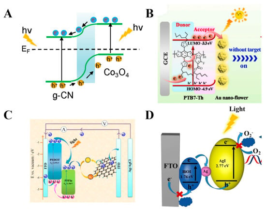
Figure 3.
Schematics of enhanced PEC property of typical heterojunctions: (A) inorganic–inorganic heterojunction Co3O4/g-CN (reprinted with permission from Ref. [44] Copyright 2020 Elsevier); (B) organic–inorganic heterojunction PTB7-Th/Au (reprinted with permission from Ref. [65] Copyright 2022 Elsevier); (C) organic–organic heterojunction P5FIn/PEDOT (reprinted with permission from Ref. [68] Copyright 2021 Elsevier); (D) multicomponent heterojunction AgI/Ag/BiOI (reprinted with permission from Ref. [71] Copyright 2021 Elsevier).
2.3. Aptamers
The term “aptamer” is derived from the Latin word “aptus”, meaning “to fit”, and the Greek word “meros” meaning “part”. The aptamer refers to single-stranded DNA (ssDNA) or RNA molecules that possess the ability to bind their targets with high specificity and affinity. In 1990, Tuerk and Gold were the first to select an RNA aptamer sequence that targeted T4 DNA polymerase [74]. In 1990, the first sequence of an RNA aptamer against T4 DNA polymerase was selected by Tuerk and Gold [75]. Almost simultaneously, another RNA sequence which can bind with organic dye specifically was selected through affinity chromatography by Ellington and Szostak [74]. Two years later, in 1992, the first DNA aptamer which inhibited the catalysis of human thrombin was obtained by John Toole et al. [76]. The method used to select aptamers through the in vitro screening method is termed as systematic evolution of ligands by exponential enrichment (SELEX) [74,75]. The selection process involves creating a library of random oligonucleotides, allowing the library to interact with the target, isolating the target–oligonucleotide complexes, and generating sub-libraries. Typically, 4 to 20 rounds of the selection process are performed. Finally, the aptamer sequences with high affinity for a specific target can be obtained by high throughput sequencing after selection [77,78,79]. The basic process of aptamer generation by conventional SELEX is shown in Figure 4.
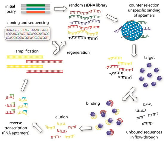
Figure 4.
The basic principle of the aptamer generation by conventional SELEX (reprinted with permission from Ref. [77] Copyright 2015 Elsevier).
During the past decades, there have been great advances in the conjugation of aptamers on various nanomaterials. The applications of aptamers in conjugation with nanomaterials, such as quantum dots, metallic nanoparticles, inorganic semiconductor nanoparticles, carbon nanotubes, and organic frameworks, provided a wide prospect for biosensors [80,81,82].
Electrochemical aptasensors primarily rely on electron donor–acceptor interactions or changes in electrochemical potential, while optical aptasensors focus on variations in optical signals such as fluorescence, surface plasmon resonance, or colorimetric responses. As a result, these systems are generally less dependent on aptamer conformational changes, allowing for a broader range of aptamer choices. Beyond redox-based electrochemical aptasensors, electrochemical aptasensors that employ electrochemical impedance spectroscopy (EIS) are also widely used. When the aptamer binds its target, the resulting steric hindrance, charge screening, and interfacial reorganization at the electrode–electrolyte interface alter the interfacial impedance (for example, the charge-transfer resistance and the double-layer capacitance). These impedance changes manifest as variations in the electrical readout, yielding a highly specific sensing signal [83,84,85].
In contrast, PEC aptasensors impose more specific and stringent requirements on aptamer selection due to their unique signal transduction mechanism. The signal output of PEC aptasensors is typically governed by changes in electron transfer pathways at the photoactive interface, which are modulated by aptamer-induced conformational rearrangements, charge redistribution, or steric hindrance upon target recognition. Therefore, aptamers used in PEC aptasensors must exhibit pronounced three-dimensional structural changes upon binding to the target analyte, ensuring that these conformational transitions are sufficient to influence interfacial charge migration and produce measurable changes in photocurrent. In addition, to maintain conformational flexibility while enabling stable interface construction, the selected aptamers should possess strong surface immobilization capabilities. Common immobilization strategies include thiol–gold bonding, self-assembled monolayers (SAMs), and π–π stacking interactions, which enable robust coupling with photoactive nanomaterials. In summary, ideal aptamers for PEC aptasensors not only require affinity and specificity to target analytes, but also trigger detectable modulation of photoelectric conversion processes. These combined characteristics ultimately guide the rational design and screening of aptamers for PEC aptasensors.
Generally, PEC sensors based on aptamers possess universal advantages compared to those using natural sensing elements such as antibodies and enzymes [86,87,88]. First, aptamers can be selected in vitro for any given target, ranging from small molecules to large proteins and even cells. Thus, it is possible to develop PEC aptasensors with a wide range of target analytes. Second, aptamers have significant conformational changes when binding with their target molecules. This can provide an obvious in photocurrent signals of PEC aptasensors during the detection process, resulting in an excellent sensitivity. Third, after the aptamer is selected through SELEX, this type of aptamer can be obtained in a large scale with high reproducibility and purity from commercial sources. In addition, aptamers usually possess high chemical stability compared to natural antibodies and enzymes. Therefore, PEC aptasensors have great potential for commercial application [8,9,88,89,90].
2.4. Sensing Strategies
Due to the excellent affinity and specificity of aptamers to target analytes, many PEC aptasensors have been presented for the detection of a large variety of biomolecules. Among these PEC aptasensors, there are many sensing mechanisms and signal-enhancing strategies proposed to improve the sensing properties. Up to now, the sensing mechanisms of PEC aptasensors can generally been divided in three formats: signal-off type, signal-on type, and complex type.
2.4.1. Signal-Off Type
A signal-off-type PEC aptasensor usually refers to the decrease in photocurrent signal when detecting target analytes. The key points of designing a signal-off PEC aptasensor are achieving a high initial photocurrent response and employing an efficient signal quenching strategy. The initial photocurrent response is achieved by utilizing photoactive nanomaterials with high photoelectric conversion efficiency, which have been thoroughly discussed in the above section on nanomaterials. As for the signal quenching strategy, there have been many methods proposed to achieve efficient signal quenching.
Steric Hindrance Effect
The most common and classic signal quenching strategy is based on the steric hindrance effect. The complex formed by the binding of target analytes with an aptamer often leads to an increase in steric hindrance effect on the surface of an electrode, which can affect the charge transfer of the sensor. The steric hindrance effect can be reflected by the impedance of the electrode, leading to an decrease in photocurrent signals. Many signal-off PEC aptasensors were proposed based on the steric hindrance effect. For example, Yan et al. constructed a PEC aptasensor by employing a p-type semiconductor BiOI doped with graphene as photoactive nanomaterials to provide a high initial photocurrent. Due to the steric hindrance effect caused by oxytetracycline captured by the aptamer, an obvious decline in photocurrent can be obtained when detecting oxytetracycline [91]. Qiao et al. used g-C3N4/WC/WO3 composites to provide an initial photocurrent and fabricated a PEC aptasensor. Tobramycin (TOB) can be recognized by the aptamer anchored on the g-C3N4/WC/WO3 composites. The qualitative and quantitative detection of TOB can be achieved by the decrease in photocurrent signal caused by the steric hindrance effect [92]. The schematic illustration of this signal-off PEC aptasensor is presented in Figure 5. Jiang et al. proposed a PEC aptasensor based on Au NPs/Yb-TCPP. The modified DNA aptamer on the surface of Au NPs/Yb-TCPP can bind with SARS-CoV-2 spike glycoprotein (S protein) with high selectivity, thus decreasing the photocurrent of the system due to the high steric hindrance from the S protein. Thus, a signal-off PEC aptasensor for SARS-CoV-2 spike glycoprotein detection was successfully developed [66].
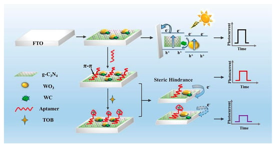
Figure 5.
Construction of a typical signal-off PEC aptasensor based on the steric hindrance effect (reprinted with permission from Ref. [92] Copyright 2022 Elsevier).
To improve the sensing properties of the signal-off PEC aptasensor based on the steric hindrance effect, some studies proposed enhancing strategies. For example, Tang et al. successfully synthesized a GO-CdS-MoS2-AuNPs heterojunction, which was used to fabricate a photoelectrode. Then, as the signal amplified element, porphyrin molecules was intercalated into the grooves of double-stranded DNA as an efficient photosensitizer to generate a strong initial photocurrent intensity. Upon the introduction of ochratoxin A (OTA), the aptamer–OTA complex was formed and detached from the electrode, causing the release of TMPyP molecules from the platform. This led to a significant decrease in photocurrent due to the target-induced removal of the photosensitizer. Subsequently, the SiO2@hDNA could be assembled onto the electrode via the re-exposed aDNA. As a result, the PEC aptasensor’s sensitivity for OTA detection was greatly enhanced due to the increased steric hindrance effect from SiO2 and hDNA [93].
In summary, steric hindrance-based signal-off strategies remain one of the most classic and widely used transduction mechanisms in PEC aptasensors. While this approach offers simplicity and broad applicability, its sensitivity is often limited by the extent of steric interference alone. To further improve detection performance, recent efforts have focused on signal amplification strategies, such as integrating nanostructured carriers or photoactive amplifiers to enhance steric effects and signal contrast.
Energy Transfer
Another widely used method to realize a signal-off PEC aptasensor is energy transfer (ET). Unlike steric hindrance, which relies on physical blocking of charge migration, ET-based strategies modulate the photocurrent by regulating photogenerated exciton behavior through non-radiative energy coupling between donor and acceptor materials. This approach offers a highly efficient and tunable method to suppress the photocurrent, especially when precise control over donor–acceptor proximity is achieved. Zhao et al. first presented the energy transfer phenomenon between CdS quantum dots and Au nanoparticles. The emission of the CdS QDs, resulting from the combination of photoinduced electron–hole pairs, was used to excite the surface plasmon resonance (SPR) of the proximal Au NPs. Then, local electric fields created by this process, in turn, modulate the exciton states in the CdS QDs by enhancing the radiative decay rate. Energy transfer from CdS CDs to Au NPs can significantly decrease the photogenerated electron–hole separation efficiency of CdS CDs, resulting in a decrease in photocurrent response. By immobilizing CD-modified probe DNA on an ITO electrode, the degree of energy transfer was directly related to the concentration of Au NP-labeled target DNA. Thus, a signal-off PEC aptasensor could be fabricated by tracking the decline in photocurrent to monitor the extent of DNA hybridization [94]. Building on this pioneering concept, subsequent studies have refined ET-based strategies by exploring different donor–acceptor configurations and target types. Zhan et al. developed a sandwich-type photoelectrochemical aptamer sensing platform for detection of target protein. A chitosan-modified CdS:Mn/TiO2/ITO electrode was used as a photoactive nanomaterial to immobilize capture DNA (S1). Once the capture DNA (S1) and target protein were immobilized on the electrode step-by-step, the specific recognition of the target protein by the aptamer enabled the immobilization of AuNP-labeled DNA (AuNPs-S2). AuNPs were far from the CdS:Mn/TiO2/ITO electrode and thus produces a strong quenched photocurrent due to the ET effect between the AuNPs and CdS:Mn QDs. This signal-off PEC aptasensor was applied to achieve thrombin detection [95]. The schematic illustration of this signal-off PEC aptasensor is presented in Figure 6. To further enhance signal modulation and broaden applicability, Dong et al. used a CdS@g-C3N4 heterojunction as a photoactive nanomaterial and Au@Ag nanoparticles as a quenching element. The energy transfer from the CdS@g-C3N4 heterojunction to Au@Ag nanoparticles occurred when the probe DNA-functionalized Au@Ag tags specifically interacted with the sheared hairpin-structured molecular beacon immobilized on the CdS@g-C3N4 nanowire. This energy transfer led to a sharp decrease in the photocurrent response, thus achieving the signal-off aptasensing of microRNA-21 detection [96].
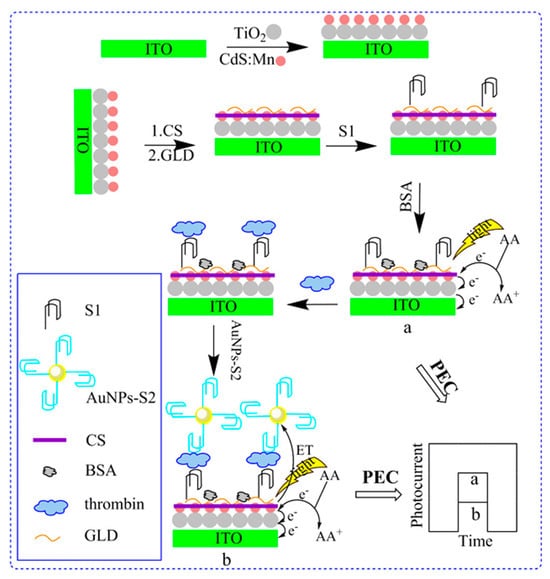
Figure 6.
Construction of a typical signal-off PEC aptasensor based on energy transfer (reprinted with permission from Ref. [95] Copyright 2019 Elsevier).
It should be noted that there are two requirements to construct a PEC aptasensor based on the ET effect. Firstly, the emission spectrum of photoactive nanomaterials should overlap with the absorption spectrum of quenching element nanomaterials. This can ensure the efficient energy transfer between the two nanomaterials. Secondly, a relatively far distance is required between the photoactive nanomaterials and quenching element nanomaterials. This is because the LSPR effect of noble metal nanomaterials will promote the separation of photogenerated electron–hole pairs of the semiconductor nanomaterials when they are close to each other. Therefore, a long distance between the two nanomaterials is required to make ET play a dominant role, thus further reducing the photocurrent significantly.
In summary, energy transfer-based PEC aptasensors provide a powerful alternative to steric hindrance strategies by enabling precise, distance-dependent modulation of exciton dynamics and photocurrent output. The use of plasmonic nanoparticles or hybrid nanostructures as quenching elements further amplifies signal responsiveness, offering high sensitivity and versatility for diverse analytes.
Electron Donor or Acceptor Competition
Many signal-off PEC aptasensors are based on competition between the nanomaterials and target analytes towards an electron donor or acceptor. This kind of signal-off PEC aptasensors usually involves oxidants or reductants which can undergo electron transfer with the photoactive electrode. Through the involvement of the electron donor or acceptor in redox reaction with nanomaterials, the oxidants or reductant can accelerate the electron transfer of photoactive nanomaterials and suppress the recombination of photoinduced electron–hole pairs, resulting in a large photocurrent response. Then, in the presence of target analytes, the electron exchange between the electron donor or acceptor and the photoactive electrode will be suppressed due to the competition redox reaction brought by target analytes. This competition towards the electron donor or acceptor finally can lead to a decline in photocurrent response. Thus, signal-off PEC aptasensors can be constructed by the electron donor or acceptor competition.
Li et al. fabricated a signal-off PEC aptasensor based on Co-doped ZnO as the photoactive nanomaterial and quercetin as the electron donor. After acetamiprid was captured by the aptamer, the electron transfer between quercetin and photoactive nanomaterials would be effectively suppressed. Then, the signal-off aptasensing of acetamiprid was achieved by the decreased photocurrent response [40]. Here, the aptamer played a crucial role in enabling specific molecular recognition and physically blocking the redox interface, which would not be achievable through simple non-specific adsorption or steric methods alone. Expanding this approach to more complex architectures, Zou et al. used ascorbic acid (AA) as the electron donor in the solution and CuInS2/b-TiO2 as the photoactive nanomaterial. After thrombin molecules was captured by the aptamer modified on the CuInS2/b-TiO2, the Au-rGO-CuS-labeled aptamer can be immobilized to the electrode face to form a sandwichlike structure. Au-rGO-CuS can competitively consume the electron donor with CuInS2/b-TiO2, leading to a significant drop in photocurrent response. Thus, a signal-off PEC aptasensor was successfully developed for thrombin detection [97]. Another signal-off PEC aptasensor for thrombin detection was also fabricated based on electron donor competition. Similarly, Yang et al. utilized the competition between Au nanoparticle-decorated perylene tetracarboxylic acid and C60@C3N4 nanocomposites towards AA to achieve aptasensing of thrombin [98]. The schematic illustration of this signal-off PEC aptasensor is presented in Figure 7. Li et al. used PTB7-Th as the photoactive material and H2O2 as the electron acceptor. When C-reactive protein (CRP) was captured by the aptamer, the obtained aptamer–CRP complex could block electron transfer, leading to a significantly depressed PEC signal. Thus, a signal-off PEC aptasensor was developed for quantitative determination of CRP [99].
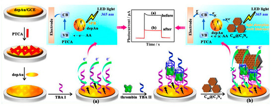
Figure 7.
Construction of a typical signal-off PEC aptasensor based on electron donor competition (reprinted with permission from Ref. [98] Copyright 2019 Elsevier).
In summary, competition-based signal-off PEC aptasensors provide a flexible and sensitive detection method by integrating redox reaction with aptamer-mediated target recognition. The aptamer’s unique ability to selectively bind targets and modulate electron transfer pathways is indispensable in these systems. This mechanism, therefore, highlights the synergistic relationship between molecular recognition and photoelectrochemical control, laying a strong foundation for developing highly responsive and selective PEC aptasensors.
Compound Signal-Off Strategy
In addition to PEC aptasensors based on the single signal-off strategy mentioned above, some researchers exerted their effort to construct a signal-off PEC aptasensor using multiple signal quenching strategies. Compared with the single signal-off strategy, the compound signal-off strategy can provide signal quenching from several combined quenching elements simultaneously, thus efficiently improving the sensitivity of the sensor. Wang et al. proposed a signal-off PEC aptasensor based on a p-n semiconductor competition and the steric hindrance effect simultaneously. They utilized Au NPs@MIS-GR as the photoactive nanomaterial to provide an initial photocurrent, and capture DNA (S1) was fixed on the electrode by Au–S. CIS QDs-labeled messenger DNA (S2) obtained by combining a strand displacement reaction was introduced into the electrode surface by the complementary base pairing. The immobilization of CIS QDs onto the electrode could form a p-n semiconductor competition and enhance the steric hindrance effect simultaneously, which quench the photocurrent signal significantly [100]. Huang et al. used Au-doped 3,4,9,10-perylene tetracarboxylic acid (PTCA) as the photoactive material to fabricate a PEC aptasensor. Then, manganese porphyrin (MnPP)-decorated 3D DNA networks were introduced after target analytes were captured by the electrode. MnPP could lead to an obvious drop in photocurrent response with two combined strategies: the competition process with the electrode towards the electrons derived from the excited state of PTCA and the steric hindrance effect caused by precipitate formed by MnPP as a mimic enzyme. The competition process involved the injection of photoinduced electrons from PTCA into the d-orbitals of Mn (III), which significantly reduced the number of electrons transmitted to the electrode, leading to a decrease in the photocurrent response. Additionally, MnPP, a well-known enzyme mimic of horseradish peroxidase (HRP), facilitates the breakdown of H2O2 and enhances the oxidation of 4-chloro-1-naphthol (4-CN), resulting in the formation of insoluble precipitates that block electron transfer. With MnPP ingeniously acting as two roles, this compound sign-off strategy-based PEC aptasensor exhibited excellent sensing property for thrombin detection [101]. This dual-functionality of MnPP enabled the construction of a highly sensitive and versatile PEC aptasensor for thrombin detection, demonstrating the strength of compound quenching strategies.
In summary, the development of signal-off PEC aptasensors based on multiple quenching strategies represents an important advancement in the field. By integrating mechanisms such as steric hindrance, charge competition, energy transfer, and catalytic precipitation, these approaches enhance signal suppression and reduce detection limits.
2.4.2. Signal-On Type
Although signal-off-type PEC aptasensors are widely used in biodetection, they still have some drawbacks. The most worrying thing is that many other factors, such as detachment of some nanomaterials from the electrode and non-specific binding of other substances in the solution onto the electrode, may also cause a decrease in photocurrent response of the sensor. These unexpected signal quenching sources are sometimes unavoidable and always affect the sensing properties of the signal-off PEC aptasensor. Therefore, some other researchers are interested in developing signal-on type PEC aptasensors. An increase of photocurrent response can be much difficult to be achieved by unexpected factors, so a signal-on type PEC aptasensor can show a better anti-interference property. The signal-on type PEC aptasensor can be divided into three types by the change in signal state: single-state “signal-on” type, double-state “signal off-on” type, and multiple-state “signal on-off-on” type.
Single-State “Signal-On” Type
The most common signal-on PEC aptasensor is based on a redox reaction which can accelerate the electron transfer of photoinduced electron–hole pairs of the photoactive nanomaterials.
At the early stage, the primary signal-on mechanism centered on direct oxidation of the captured target by photogenerated holes. Feng et al. fabricated a signal-on PEC aptasensor by using gold nanoparticles (Au NPs) coupled with MOF-derived In2O3@g-C3N4 nanoarchitectures (AuInCN) as the photoactive matrix. The cysteamine decorated on Au NPs could immobilize the SH-terminated aptamer as a biosensing element, and tetracycline (Tc) molecules would then be trapped through a specific interaction with the immobilized aptamer. aptamer. An increase in PEC photocurrent could be obtained under visible light illumination, due to the oxidation of captured Tc by photo-generated holes of the photoactive nanomaterial. Therefore, the concentration of Tc could be detected by the signal-on PEC aptasensor [102]. The schematic illustration of this signal-on PEC aptasensor is presented in Figure 8. To further strengthen the oxidative capacity and improve charge utilization, some researchers introduced artificial Z-scheme mechanisms to enable dual redox pathways. Wang et al. synthesized an artificial Z-scheme bipyridine ruthenium (Ru(bpy)32+) sensitizing narrow-gap bismuth oxy-iodide (BiOI) microspheres and applied it in fabricating a PEC aptasensor. Upon absorbing the light, Ru (II) was excited to Ru (II)*, which was very erratic to turn itself into Ru (III) and released electrons, which were powerful to reduced O2 to ·O2− directly. Meanwhile, abundant photogenerated holes in the valence band (VB) of BiOI would immediately oxidize the ampicillin molecules captured by the aptamer. These two redox reactions would lead to a significant rise in photocurrent response. Therefore, a signal-on-type PEC aptasensor was successfully fabricated for ampicillin detection [103]. Recognizing the need for high-throughput detection in real samples, dual-channel signal-on strategies were later proposed. Zhang et al. successfully achieved a signal-on PEC aptasensor for simultaneous detection of enrofloxacin (ENR) and ciprofloxacin (CIP) based on redox reactions. They synthesized two kinds of photoactive nanomaterials with excellent PEC performance: three-dimensional nitrogen-doped graphene-loaded copper indium disulfide (CuInS2/3DNG) and Bi3+-doped black anatase titania nanoparticles decorated with reduced graphene oxide (Bi3+/B-TiO2/rGO). The cathodic current generated by CuInS2/3DNG and the anodic current generated by Bi3+/B-TiO2/rGO could be clearly distinguished without interfering with each other, because different bias potentials were applied to the two photoactive nanomaterials on one ITO electrode. ENR and CIP aptamers were modified on the CuInS2/3DNG and Bi3+/B-TiO2/rGO, respectively, to capture ENR and CIP. Under different bias potentials, ENR and CIP would be oxidized by the photogenerated holes of CuInS2/3DNG and Bi3+/B-TiO2/rGO, respectively, under illumination. In this way, a novel signal-on PEC aptasensor for simultaneous detection of ENR and CIP was fabricated [104].
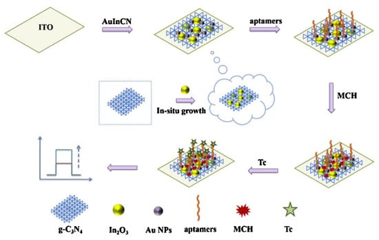
Figure 8.
Construction of a typical signal-on PEC aptasensor based on redox reaction (reprinted with permission from Ref. [102] Copyright 2019 Elsevier).
In summary, the signal-on PEC aptasensor strategy has evolved from single-target, single-reaction mechanisms to more sophisticated dual redox systems and multiplex detection formats. Throughout these developments, aptamers play a central role not only in molecular recognition, but also in facilitating selective electron-transfer reactions, directly linking molecular events to measurable PEC signals.
Single-State Signal Off-On Type
Compared with PEC aptasensors fabricated only based on signal-on or signal-off strategies, “off-on” PEC aptasensors can overcome their inherent drawbacks, such as the unneglected background signals, which may result in misleading positive PEC responses and influence the sensing properties of the PEC aptasensors [105]. A typical signal off-on-type PEC aptasensor usually requires an initial signal-off state, which is generally achieved by steric hindrance, energy transfer, or charge blocking, as previously discussed. Then, approaches to eliminating the signal quenching are applied to get a signal-on state of the PEC aptasensor. Generally, there are two main strategies to achieve the signal-on state of the photocurrent response. One is based on competitive binding, which usually utilizes the stronger affinity between analytes and the aptamer to detach the aptamer away from the electrode. The other one is to destroy the structure formed in the signal-off state to achieve the recovery of photocurrent response.
Zhang et al. developed a straightforward signal-on PEC aptasensor for detecting Aflatoxin B1 (AFB1) using electrochemically reduced graphene oxide/poly(5-formylindole)/Au (erGO/P5FIn/Au) nanocomposites as the photoactive material. The AFB1 aptamer is attached to the erGO through π-π stacking interactions between the carbon rings in graphene and the C-N heterocyclic rings of the nucleobases in ssDNA. The attachment of the insulating AFB1 aptamer chain to the electrode induces a signal-off state in the PEC aptasensor. Subsequently, in the presence of AFB1, the aptamer chain would detach from the surface of erGO, which would result in a recover state of the photocurrent. In this way, a “off-on”-type PEC aptasensor was constructed for AFB1 detection [106]. In a similar strategy, Qin et al. also achieved chloramphenicol detection by utilizing the competition binding between CAP and graphene quantum dots (GQDs) towards the CAP aptamer. GQDs/TiO2 NTs were used to fabricate a photoactive electrode, and the CAP aptamer can be connected to GQDs through π-π stacking interaction. The stronger affinity of CAP with the CAP aptamer would lead to dissociation of the aptamer from the surface of electrode. The recovery of photocurrent response during the dissociation of the aptamer enabled the signal-on aptasensing for CAP [107].
To further amplify signal modulation and achieve higher specificity, structural transformation mechanisms were introduced. Zhu et al. presented a “off-on” PEC aptasensor based on a 3D self-supporting Z-scheme AgI/Ag/BiOI heterojunction arrays subtly integrated with in situ formed biocatalytic precipitation (BCP). The introduced horseradish peroxidase (HRP) modified with a chloramphenicol (CAP) aptamer would bind with the ssDNA on the electrode surface and then catalyzed the oxidation of 4-chloro-1-naphthol (4-CN) in the solution to insoluble benzo-4-chlorohexadienone (4-CD) in the presence of H2O2. The 4-CD precipitates would block the active sites on the electrode surface, consequently inhibiting the electron transfer, leading to a significant decline in the photocurrent response. In the presence of CAP, the photocurrent signal was slowly enhanced, owing to the efficient prevention of the BCP reaction by the hybridization of the HRP-CAP aptamer with introduced CAP and the decline in the 4-CD precipitates. Thus, a “off-on” PEC aptasensor was achieved for CAP detection [71]. Wang et al. presented an alternative approach based on structural disruption, where they used the energy transfer quenching effect from CdS:Mn to Au nanoparticles (AuNPs) to achieve a signal-off state. In the absence of miRNA-21, the immobilized DNA remained in a hairpin conformation. This configuration resulted in a low photocurrent response, as the short distance between CdS:Mn and AuNPs facilitated efficient electron transfer. However, when miRNA-21 was present, it hybridized with the hairpin DNA, causing a transition to a more rigid, rod-like double helix. This structural change pushed the AuNPs away from the electrode surface, leading to a noticeable recovery of the photocurrent due to the reduced electron transfer effect. In this way of destroying the structure formed during the signal-off state, a “off-on” PEC aptasensor was successfully fabricated [108]. The schematic illustration of this “off-on” PEC aptasensor is presented in Figure 9.
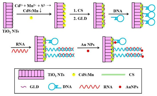
Figure 9.
Construction of a typical “off-on” PEC aptasensor (reprinted with permission from Ref. [108] Copyright 2017 Elsevier).
Combining multiple quenching mechanisms in one system offers another route to enhance detection sensitivity. Tang et al. combined the strategy based on the steric hindrance effect and energy transfer to construct a PEC aptasensor. They successfully synthesized Ce-TiO2@MoSe2 composites as the photoactive nanomaterial and then modified the photoactive electrode with an aminated aflatoxin B1 (AFB1) aptamer. An AuNP-labeled DNA sequence was subsequently hybridized with the aptamer to fabricate a sandwich structure, which led to a decline in photocurrent response due to the steric hindrance effect and energy transfer from Ce-TiO2@MoSe2 to AuNP. This structure would be destroyed in the presence of AFB1, decreasing the amount of the energy acceptor (AuNPs) at the electrode surface and weakening the steric hindrance effect, leading to a recover in photocurrent response [109].
In summary, “off-on” PEC aptasensors represent a versatile and powerful sensing strategy by effectively integrating aptamer–target recognition with reversible signal transduction mechanisms. Through strategies like target-induced aptamer dissociation or disruption of quenching assemblies, these sensors provide enhanced sensitivity, lower background, and stronger anti-interference capability. The design flexibility of “off-on” architectures also paves the way for constructing multiplex and switchable biosensing systems.
Signal On-Off-On Type
In addition to signal off-on-type PEC aptasensors, there are some “on-off-on” PEC aptasensors emerging for biodetection in recent years. The initial “on” state can provide a higher photocurrent for the “off” state, thus leading to a higher recovery in the final “off” state during the sensing process. Compared to conventional signal-on or signal-off sensors, this triphasic format provides enhanced signal contrast, reduced background interference, and improved detection sensitivity. For example, Xu et al. constructed a “on-off-on” PEC aptasensor for detection of cancer markers. They employed the formation of a heterojunction between Cd:Sb2S3 and La2Ti2O7 to establish the initial “signal-on” state, enhancing the stability of the PEC platform. Next, V2O5 nanospheres were conjugated with aptamer DNA and utilized as a catalyst for H2O2, which consumed the electron donor and triggered the “signal-off” state. Finally, the target analyte was modified onto the surface of the PEC platform, causing some of the V2O5–aptamer DNA complexes to detach from the electrode surface. The detachment of V2O5–aptamer from the electrode surface led to a recovery of photocurrent, so the “signal-on” state was realized again. This “on-off-on” PEC aptasensor was applied in PSA detection [110]. This work integrates catalytic quenching and aptamer release to achieve reversible signal modulation with strong recovery, making it suitable for stable, sensitive PSA detection.
To address the challenge of precise control over signal quenching, Xin et al. developed an ultrasensitive “on-off-on” PEC aptasensor for carcinoembryonic antigen (CEA). Xin et al. proposed an ultrasensitive “on-off-on” PEC aptasensor for detection of carcinoembryonic antigen based on BiOI/α-Fe2O3 nanosheet arrays (BiOI/α-Fe2O3 NSAs) by in-situ regulation of electron donor distance. Ferrocene-modified hairpin DNA (Fc-hDNA), which could offer electrons (Fc part), was immobilized on the electrode surface. This introduction of Fc-hDNA provided the initial “on” state of PEC signals. The distance between the electron donor, Fc, and the BiOI/α-Fe2O3 NSAs interface can be precisely controlled through the specific interaction between Fc-hDNA and the CEA aptamer. When the CEA aptamer binds to the Fc-hDNA, a stable hybrid structure is formed, causing the hairpin structure of Fc-hDNA to open and move Fc away from the electrode surface, resulting in a “signal-off” state. However, in the presence of CEA, the Fc-hDNA reverts to its original closed-loop structure. This occurs because the strong affinity between CEA and its aptamer causes the aptamer to detach from the hybrid, restoring the “signal-on” state. Therefore, a “on-off-on” PEC aptasensor was developed for detection of CEA [111]. The schematic illustration of this “on-off-on” PEC aptasensor is presented in Figure 10. This work demonstrates precise molecular-level regulation of donor–acceptor distance, achieving highly sensitive and controlled target detection.
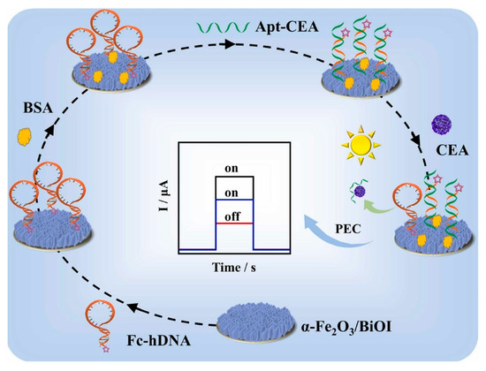
Figure 10.
Construction of a typical “on-off-on” PEC aptasensor (reprinted with permission from Ref. [111] Copyright 2022 Elsevier).
In summary, “on-off-on” PEC aptasensors represent a powerful evolution of PEC biosensing by combining dynamic signal regulation, specific aptamer recognition, and PEC signal recovery. These designs not only enhance detection sensitivity and specificity, but also offer flexible platforms adaptable to a wide range of targets through tailored aptamer and material selection.
2.5. Fabrication Methods of Electrodes
After selecting the appropriate photoactive materials, sensing elements, and corresponding sensing strategies, these components need to be assembled into a functional sensing electrode. A typical PEC aptasensing electrode consists of three main parts: a conductive substrate, a photoactive material, and recognition components. Upon specific binding of the sensing element to the target analyte, the PEC properties of the photoactive material are altered, and the resulting photocurrent response can be collected through the conductive substrate in an external circuit.
Common conductive substrates used in PEC aptasensors include metal oxide-based conductive glasses (such as indium tin oxide (ITO) and fluorine-doped tin oxide (FTO)), metal substrates (such as gold and titanium electrodes), and carbon-based materials (such as glassy carbon electrodes). The photoactive materials are as described in Section 2.2. Aptamers or aptamer-functionalized complexes are used as recognition components for the sensing unit in PEC aptasensors, as described in Section 2.4.
During the construction of PEC aptasensors, the electrode fabrication strategy not only determines the recognition efficiency and stability of the aptamer toward its target molecule but also directly influences the PEC performance of the photoactive component. Typically, there are two main fabrication routes for preparing the sensing electrode. In the first approach, the photoactive material is immobilized onto the conductive substrate first, followed by the attachment of the recognition components. In the second approach, the recognition element is first conjugated to the photoactive material, and the resulting composite is then immobilized onto the conductive substrate. In both strategies, the immobilization of the photoactive material onto the electrode surface is essentially similar to that used in traditional PEC sensors based on redox reactions or antigen–antibody recognition. However, in PEC aptasensors, the methods for coupling aptamers with photoactive nanomaterials are more diverse and flexible compared to conventional PEC systems, offering greater adaptability in sensor design.
In the fabrication of PEC aptasensor electrodes, the immobilization of aptamers onto photoactive nanomaterials can be achieved through several main strategies, including physical adsorption, covalent bonding, biotin–streptavidin interaction, and self-assembled monolayer (SAM) formation.
2.5.1. Physical Adsorption
Physical adsorption relies primarily on electrostatic interactions, van der Waals forces, or hydrophobic interactions to anchor aptamers onto the surface of photoactive materials. A notable advantage of this method is the excellent tolerance of aptamers to aggressive surface treatments, such as alkaline washing, electrochemical activation, or plasma etching, which can significantly enhance the surface hydrophilicity and interfacial binding activity of PEC electrodes. In contrast, antibodies are prone to denaturation and functional loss under such conditions. For example, Li et al. developed a PEC aptasensor for ultrasensitive tetracycline detection by employing π–π stacking interactions between MoS2 nanosheets and aptamers, thus eliminating the need for conventional chemical conjugation [112]. In another study, Sun et al. designed a “label-free” PEC aptasensor that bypassed the need for electrode-bound recognition elements by constructing a homogeneous recognition system through strong π–π interactions between graphene and aptamers. This design allowed for reusable electrodes and detection of the herbicide atrazine (ATZ) at picomolar levels in environmental water samples [113]. Another physical adsorption fabrication method is polymer entrapment. Polymer entrapment involves the in situ generation of a thin, permeable polymer network (e.g., dopamine, chitosan, polypyrrole, or o-phenylenediamine) on the electrode surface, which physically or chemically crosslinks to immobilize and protect the aptamer. This coating can be formed by electropolymerization or spontaneous deposition onto Au, ITO/FTO, or carbon-based substrates to create uniform films. These coatings exhibit good mechanical adhesion, reducing aptamer loss during repeated washing, flow washing, or exposure to environments containing enzymes or proteolytic proteins, thereby enhancing long-term and field application stability. This strategy also allows for the addition of antifouling top layers (e.g., PEG/zwitterionic polymers, BSA) or functional fillers, maintaining low dark current and stable photocurrent baselines, and facilitating the printing and miniaturization of electrodes. These studies clearly demonstrate the advantages of physical adsorption in constructing aptamer interfaces, particularly in enhancing PEC response intensity, interfacial stability, and reusability.
2.5.2. Covalent Bonding
Covalent linkage involves forming a chemical bond between functionalized aptamers (e.g., thiol, amine, and azide/alkyne groups) and reactive sites on the electrode or intermediate layer. In this strategy, aptamers can be covalently linked to various substrates via functional groups such as –SH, –NH2, or –N3. The excellent chemical stability of covalent bonds allows the immobilization process to be carried out under harsh conditions, including acidic or basic environments, organic solvents, and elevated temperatures, thereby improving surface modification efficiency and electrode reproducibility. This strong linkage resists detachment caused by storage, repeated washing, or potential scanning, significantly reducing baseline drift and supporting gentle regeneration (e.g., low ionic strength or urea treatment). Overall, covalent coupling is more conducive to obtaining long-term stable, electrode-consistent PEC sensors with good reproducibility and consistent readouts in complex matrices. Notably, covalent coupling between aptamers and photoactive materials (e.g., metal oxides and quantum dots) enables precise modulation of electron transfer pathways and interfacial spatial impedance. This facilitates the effective separation of photogenerated electron–hole pairs, leading to enhanced PEC signal intensity and operational stability.
For instance, Cheng et al. covalently grafted carboxyl-modified aptamers onto TiO2 quantum dot-sensitized ITO electrodes to construct a PEC aptasensor for thrombin detection. The sensor exhibited a wide linear range of 1.0–250 pM and an ultralow detection limit of 0.83 pM, along with excellent signal stability and reproducibility [114].
In summary, covalent immobilization not only confers high interfacial stability but also improves the surface coverage and spatial distribution of aptamers, minimizing probe–probe interference. These features significantly enhance target binding efficiency and signal output, underscoring the value of covalent coupling in the development of highly sensitive and stable PEC aptasensors.
2.5.3. Biotin–Streptavidin Interaction
The biotin–streptavidin coupling strategy relies on the strong non-covalent affinity between biotin-modified aptamers and streptavidin to form a highly stable interfacial linkage. In this method, the chemical stability and structural reversibility of aptamers allow them to retain their bioactivity during the immobilization process. The resulting aptamer–streptavidin interface exhibits excellent specificity and structural integrity, which is critical for reliable target recognition. More importantly, the binding between aptamers and target molecules is inherently reversible. This enables the regeneration of PEC electrodes through mild elution treatments such as salt washing or thermal denaturation, thereby allowing multiple reuse cycles. Such reusability substantially reduces device cost and ensures long-term stability, which is difficult to achieve with enzyme- or antibody-based PEC sensors.
For example, Kamila Malecka et al. electrochemically deposited a AuNP/graphene-doped PEDOT nanofilm on an FTO electrode and immobilized biotinylated aptamers using the biotin–streptavidin system. The resulting PEC aptasensor achieved ultrasensitive detection of the tumor marker MUC1, with a detection limit as low as 1 fg/mL (0.03 fM) measured by differential pulse voltammetry [115]. The aptamer interface demonstrated excellent specificity, and upon high-salt treatment, the signal nearly returned to baseline, enabling multiple cycles of reuse without significant loss of performance. These results highlight the unique advantage of aptamer-based PEC sensors in terms of sensor regeneration and cost-effectiveness.
2.5.4. SAM
The SAM strategy utilizes thiol-modified aptamers to spontaneously form Au–S bonds with metal electrode surfaces such as gold, leading to the formation of a uniform monolayer. In this approach, the thiolated aptamers arrange themselves into a highly ordered and compact monolayer through strong Au-S interactions, creating a low-defect, highly reproducible sensing interface that facilitates well-regulated charge transfer pathways. This ordered structure not only enhances interfacial consistency but also allows for in situ thermal annealing of the immobilized aptamers. Such post-modification treatment helps refold the aptamers into their stable secondary structures while reducing nonspecific folding, thereby improving both the binding efficiency toward target analytes and the magnitude of PEC photocurrent responses. Moreover, the small molecular size of aptamers enables the formation of dense receptor layers on the electrode surface, effectively broadening the linear detection range and amplifying signal output. A common and effective strategy involves the co-assembly of thiolated aptamers with 6-mercapto-1-hexanol on gold electrodes to form a mixed monolayer with high surface density, reduced interfacial impedance, and improved electron transfer efficiency [116]. This self-assembled aptamer interface demonstrates excellent photoelectrochemical stability and signal-to-noise enhancement, making it a widely adopted technique for constructing high-performance PEC aptasensors.
The advantages and disadvantages of the existing aptamer immobilization methods are summarized in Table 2.

Table 2.
Comparison of aptamer immobilization methods.
It is worth noting that aptamers inherently possess excellent chemical stability, including resistance to high temperatures, extreme pH environments, and organic solvents. Compared with traditional protein-based recognition elements such as enzymes and antibodies, aptamers can retain their structural integrity and molecular recognition capabilities even under harsh conditions. This unique resilience opens up broad possibilities for innovation in electrode fabrication processes. Previous studies have demonstrated that aptamers can be stably immobilized and maintain their binding functionality in organic solvents, and still exhibit good affinity after exposure to elevated temperatures or repeated washing cycles. These findings suggest that the robust tolerance of aptamers can be fully leveraged to develop more aggressive and efficient strategies for electrode pretreatment and sensor construction. However, to date, there are few reported examples of PEC aptasensors constructed under extreme conditions such as high-temperature annealing, electrochemical activation, or organic solvent exposure. Therefore, future efforts in PEC aptasensor fabrication could explore the use of electrochemical activation, plasma treatment, alkaline washing, or high-temperature annealing to enhance the surface properties of the electrode. Simultaneously, in situ aptamer immobilization and thermally induced conformational reconstruction under high temperature or extreme pH could be employed to improve interfacial binding efficiency and structural stability of the recognition element.
In summary, the construction methodology of PEC aptasensors holds significant theoretical and practical potential. By harnessing the unique stability of aptamers, it is feasible to overcome the conventional limitations associated with the fabrication of biological recognition interfaces. Such “aggressive” pretreatment strategies could be boldly introduced into PEC aptasensor preparation workflows to further enhance electrode surface properties without compromising aptamer recognition performance.
3. Applications of PEC Aptasensors in Biosensing
The combination of PEC detection technology and aptamers brings PEC aptasensors many advantages, such as high sensitivity, low cost, easy modification, low toxicity, and high reproducibility. These superiority of PEC aptasensors make it suitable for biosensing application.
Firstly, photoelectrochemical (PEC) technology utilizes light as the excitation source and electrical signals as the output, thereby achieving physical separation between the input and output signals. This unique configuration effectively reduces background interference and significantly enhances both the signal-to-noise ratio and detection sensitivity. The PEC approach not only inherits the high sensitivity of optical detection but also integrates the cost-effectiveness, simplicity, and portability of electrochemical sensing. Secondly, aptamers, as recognition elements, possess several inherent advantages, such as small molecular size, ease of modification, high affinity, and excellent specificity. Their nucleotide sequences can be precisely tailored through in vitro selection techniques like SELEX to bind various targets including proteins, small molecules, cells, and pathogens, demonstrating remarkable versatility and target adaptability. Importantly, aptamers are structurally flexible and chemically stable, which allows them to be adapted to multiple signal transduction mechanisms, including steric hindrance, redox interaction, energy transfer, and electron donor/acceptor competition. This compatibility with diverse sensing principles not only broadens the types of detectable analytes but also enables the design of sensors with enhanced sensitivity and tunable signal response. In addition, aptamers exhibit outstanding chemical and thermal stability, making them suitable for a wide array of electrode modification strategies, including covalent bonding, self-assembled monolayers, physical adsorption, and biotin–streptavidin coupling. These flexible immobilization approaches enable the efficient and reproducible fabrication of PEC aptasensors, while maintaining the structural integrity and recognition performance of the aptamers under various operating conditions.
In conclusion, these advantages have fueled the rapid development and broad application of PEC aptasensors in biosensing. To date, a substantial number of studies have demonstrated their successful implementation for the detection of a wide range of analytes, including tumor markers, antibiotics, anticancer drugs, bacteria, and viruses. Notably, the ability of aptamers to match different PEC sensing mechanisms allows for the rational design of customized sensors targeting specific biological events, which significantly improves analytical performance. Beyond academic research, PEC aptasensors have also shown considerable promise for practical applications in clinical diagnostics, environmental monitoring, and food safety.
In the following sections, we will classify the applications of PEC aptasensors according to target analytes and systematically summarize and compare their analytical performance across different biosensing contexts.
3.1. Biomarkers
Biomarkers are chemical compounds that can be produced by cancer cells or other body cells as a reaction to the presence of a tumor [117]. Typically, abnormal levels of biomarkers can be detected in blood, urine, tissues, or bodily fluids of individuals affected by various diseases. While proteins are the most commonly found biomarkers, other types include DNA and RNA. In recent years, the highly sensitive detection of biomarkers has been demonstrated to be an effective method in diagnosis of various diseases. Compared with other biosensors for biomarker detection, PEC aptasensors have unique advantages. Due to the high signal-to-noise ratio, PEC aptasensors can achieve detection of biomarkers with an extremely low expression level. Therefore, many researchers exerted their effort in constructing PEC aptasensors for detection of biomarkers.
Hu et al. achieved highly sensitive and selective detection of CA125 by a signal-off PEC aptasensor. They used a gold nanoparticle (AuNP)/gallium nitride (GaN) Schottky junction as the photoactive nanomaterial. Then, the DNA aptamer of CA125 was modified on the surface of the AuNPs via Au−S bonds. The photoelectron transfer process of the system can be blocked by the CA125 protein, which can be captured by the DNA aptamer of CA125, resulting in the decrease in the photocurrent of the system [56]. The aptamer’s selective binding and conformational impact were essential for effective signal modulation.
To target nucleic acid biomarkers, Niu et al. constructed a PEC aptasensing platform for ultrasensitive miRNA-141 detection. They used CdS QDs in the presence of a 5,10,15,20-tetrakis (4-aminophenyl)-21H,23H-porphine (Tph-2H)-coated glassy carbon electrode (GCE) as the photoactive electrode. Based on the aptamer modified on the GCE, miRNA-141 could be captured, thus achieving sensitive and selective detection [118]. Liu et al. also developed a PEC aptasensor for detecting target DNA. This aptasensor was fabricated by sequentially modifying fluorine-doped tin oxide (FTO) electrodes with TiO2 nanoparticles, gold (Au) nanoparticles, hairpin DNA (DNA1), and CdSe-COOH quantum dots (QDs), which served as signal amplifiers. In the absence of target DNA, the hairpin structure of DNA1 and the CdSe-COOH QDs remained in close proximity to the TiO2/Au electrode surface, resulting in an increased photocurrent due to the sensitization effect. However, in the presence of target DNA, DNA1 transitioned into a double-helix structure, causing the CdSe QDs to move away from the electrode surface. This displacement weakened the sensitization effect, leading to a reduction in the PEC signal. Therefore, a signal-off PEC aptasensor was successfully created for target DNA detection in tumor diagnosis [119]. This example highlights the value of aptamer-based conformational switching in dynamic signal control.
To address the need for detecting multiple biomarkers simultaneously, some studies have expanded PEC aptasensors to multiplexed platforms. For example, Sun et al. achieved simultaneous detection of tumor marker protein and microRNA. They used a WO3/Fe2O3 heterojunction as a transducer nanomaterial to fabricate a PEC aptasensor. Recognition sites of protein and microRNA were formed when the integrated signal probe (ISP) composed of signal probe 1 (sP1) and signal probe 2 (sP2) was introduced on paper-based working units modified with gold nanoparticles (AuNPs). They applied this aptasensor for the simultaneous detection of mucin 1 (MUC1) and microRNA 21 (miRNA-21) [48]. Aptamers specific to each biomarker enabled orthogonal detection of both targets, demonstrating the programmability and multitarget compatibility of aptamer-based PEC platforms.
In summary, PEC aptasensors have shown remarkable versatility and sensitivity for tumor biomarker detection. The ability of aptamers to recognize diverse targets while integrating with a range of PEC signal mechanisms makes them highly adaptable and powerful components in biosensing platforms. Representative studies on PEC aptasensors for biomarker detection in recent years are summarized in Table 3.

Table 3.
Performances of the developed PEC apsensors for tumor marker detection in recent years.
3.2. Pharmaceuticals
Bioanalysis of pharmaceuticals in biological samples provides valuable insights into the absorption, distribution, metabolism, excretion, and other processes associated with drugs in living organisms. This field is crucial for advancing drug discovery, monitoring therapeutic drug levels, and evaluating the effectiveness of treatments in the pharmaceutical industry [133,134,135].
While pharmaceutical analysis has a positive impact on biomedicine, the extensive use of pharmaceuticals has also resulted in significant environmental issues. Substantial quantities of pharmaceuticals have been found in environmental soil, groundwater, and drinking water, leading to severe pollution that can negatively affect both the ecosystem and public health [136,137]. As a result, detecting pharmaceuticals in biological samples is crucial for both biomedical and environmental applications.
With the rapid development of PEC aptasensors, they have shown great potential in detecting various pharmaceuticals. Compared with traditional electrochemical and optical methods, PEC aptasensors offer enhanced signal-to-noise ratios, low background interference, cost-effective instrumentation, and excellent adaptability across different analytes. In particular, the use of aptamers enables high specificity toward diverse pharmaceutical compounds, while allowing integration with flexible sensing mechanisms such as steric hindrance, conformational switching, and electron transfer regulation. This makes PEC aptasensors highly versatile and suitable for detecting trace levels of pharmaceutical contaminants. For example, Qin et al. designed a PEC aptasensor for sensitive CAP detection, utilizing graphene quantum dot (GQD)-sensitized TiO2 nanotube arrays. The CAP aptamers were immobilized onto the GQDs/TiO2 NT photoelectrode through π–π stacking interactions between the GQDs and the nucleobases of the aptamers. The aptasensor functioned by monitoring the increase in photocurrent, which resulted from the formation of aptamer–CAP bioaffinity complexes. Ascorbic acid was used as an efficient electron donor to scavenge photogenerated holes, enabling the detection of CAP through aptasensing [107]. Liu et al. used SnO2/Bi2S3 as the photoactive nanomaterial to fabricate a PEC aptasensor. TOB can be selectively and sensitively determined by the decline in the photocurrent caused by TOB binding with the aptamer modified on the electrode [138].
These examples demonstrate how the rational design of PEC aptasensors enables sensitive, specific, and portable detection of pharmaceutical analytes by leveraging both the tunable electronic properties of photoactive nanomaterials and the high-affinity, flexible recognition ability of aptamers. Representative PEC aptasensors for pharmaceutical detection reported in recent years are summarized in Table 4.

Table 4.
Performances of the developed PEC apsensors for pharmaceutical detection in recent years.
3.3. Viruses, Bacteria, and Cells
Detection of viruses, bacteria, and cells have great significance in biological diagnosis and clinical treatment. However, biosensing of viruses, bacteria, and cells is still difficult for many biosensors due to their great volume and mass. To overcome these limitations, PEC aptasensors have emerged as a powerful platform by combining the high signal-to-noise ratio of photoelectrochemical techniques with the selective and flexible recognition capabilities of aptamers. Aptamers can be specifically designed to target unique membrane proteins or nucleic acid sequences on pathogens, enabling precise and efficient capture of large biomolecules or whole cells. Tabrizi et al. developed a PEC aptasensor for the quantitative detection of the receptor-binding domain (RBD) of the severe acute respiratory syndrome coronavirus-2 (SARS-CoV-2). The ITO electrode was modified with Chitosan/CdS-gC3N4 as a photoactive electrode, and aptamers were mobilized as the recognition element. The results obtained showed that the proposed PEC aptasensor is capable of detecting the SARS-CoV-2 RBD. Building on this, efforts have been made to translate PEC aptasensing into field applications for pathogen monitoring. Zheng et al. fabricated a portable signal-off PEC aptasensor based on CdS/AuNPs for field environment monitoring of pathogen Escherichia coli O157:H7 (E. coli O157:H7) [160]. In the context of cell detection, conventional signal-off strategies may suffer from high background noise and limited sensitivity due to cellular size and complexity. To address this, Meng et al. developed a signal-on PEC aptasensor. They used CNTs/SnSe as the photoactive nanomaterial to generate a strong PEC signal. Next, methionine (Met), AuNPs, and probe DNA are combined to create a nano-probe that functions as a quencher, inhibiting the PEC signal of CNTs/SnSe. When metastatic breast cancer cells are present, the nanoprobe is released from the electrode surface, eliminating the quenching effect between the nanoprobe and CNTs/SnSe. As a result, the PEC aptasensor for cell detection was effectively developed [161].
These studies collectively highlight the versatility and adaptability of PEC aptasensors in detecting structurally diverse and complex biological targets. By leveraging aptamer programmability and PEC signal amplification, these systems offer enhanced detection performance across a wide range of viruses, bacteria, and cells.
The reported PEC aptasensors for viruses, bacteria, and cells are summarized in Table 5.

Table 5.
Performances of the developed PEC apsensors for detection of viruses, bacteria, and cells in recent years.
3.4. Contaminants in Environment and Food
The pervasive presence of contaminants, such as pesticide residues and toxins, in agro-food systems and aquatic environments poses severe threats to public health and ecological stability. Rapid and sensitive monitoring technologies are needed to detect these contaminants. Traditional methods like chromatography and ELISA suffer from limitations including costly instrumentation, prolonged detection time, and operational complexity. In recent years, PEC aptasensors emerge as a potential solution with the advantages of high-sensitivity, rapid detection time, low cost, and simple operation. For example, Ding et al. reported an ultrasensitive MoS2-based “signal-off” PEC aptasensor for acetamiprid assays and studied the charge separation mechanism in PEC aptasensing [171]. Wen et al. successfully prepared a Pd NPs/CdS Schottky junction to improve photoelectric conversion property and constructed a PEC aptasensor for Carbendazim detection [172]. These examples reflect the potential PEC aptasensor in real-world food samples. Many other PEC aptasensors have been widely used in food safety, shown in Table 6.
In addition to food safety, PEC aptasensors have also shown excellent potential in detecting environmental contaminants. The flexible design of aptamers enables recognition of various pollutants, including heavy metals and algal toxins, which are difficult to monitor using traditional immunosensors. For example, Guo et al. used H-TiO2/Ti2COX as a photoactive material to fabricate a universal “signal-off” aptasensor, achieving MC-LR detection with high sensitivity [173]. Chen et al. employed Fe2O3/g-C3N4 with excellent photoelectric property to construct a PEC aptasensor for sensitive detection of Pb2+ [174]. These advancements highlight the significant role of PEC aptasensors in enhancing contaminant detection capabilities in food and environment. Their inherent advantages, including high specificity from aptamers, enhanced PEC signal modulation, and operational simplicity, make them a competitive choice for next-generation sensing technologies.
The typical PEC aptasensors for detection of environmental contaminants are also summarized in Table 6.

Table 6.
Performances of the developed PEC apsensors for detection of contaminants in environment and food in recent years.
Table 6.
Performances of the developed PEC apsensors for detection of contaminants in environment and food in recent years.
| Analytes | Photoactive Nanomaterial | Signal Type | Immobilization Method | Linear Range | Limit of Detection | Matrix and Recovery Rate (%) | References |
|---|---|---|---|---|---|---|---|
| Acetamiprid | NGQDs/MoS2 | off | Covalent bonding | 0.1 pM–0.1 nM | 0.033 pM | Tomato and cucumber: 89.7–108.3 | Ding et al. [171] |
| Acetamiprid | t-SG/ZnO | on-off-on | Physical adsorption | 0.001–1 μg/mL | 6.7 × 10⁻⁹ μg/mL | Cucumber: 97.8–101.2 | Yan et al. [175] |
| Malathion | BiOBr/Bi2S3 | on | Physical adsorption | 1 × 10⁻⁶–1 μg/mL | 1.2 × 10⁻⁷ μg/mL | Milk: 95.2–106.8 | Li et al. [176] |
| Malathion | AuNPs/Ag2S | on | Covalent bonding | 6 × 10⁻⁶–0.6 μg/mL | 2×10⁻⁶ μg/mL | Apple juice: 5–103.5 | Zeng et al. [177] |
| Malathion | MnO2 NF@CdS | off | Biotin−streptavidin interaction | 1 × 10⁻⁶–0.1 μg/mL | 6.8 × 10⁻⁷ μg/mL | Milk: 97.8–100.4 | Tang et al. [178] |
| Profenofos | MoTe2 NPs/RGO | on | Physical adsorption | 1 × 10⁻⁶–10 μg/mL | 3.3 × 10⁻⁷ μg/mL | Chinese chive, and potato: 95.3–114 | Ding et al. [179] |
| Chlorpyrifos | CuS QDs/Co3O4 | on | Covalent bonding | 1 × 10⁻⁴–1 × 10⁴ μg/mL | 0.34 × 10⁻⁴ μg/mL | River water: 98.27–103.47 | Tan et al. [180] |
| microcystin-LR (MC-LR) | NiO | on-off | Covalent bonding | 0.005–10 nM | 1.7 pM | Tap water: 96.6–104 | Xu et al. [181] |
| MC-LR | In2O3−CaIn2S4 | on | Covalent bonding | 0.5 × 10²–4 × 10⁵ pM | 19.45 pM | Water: 99.04–100.11 | Wang et al. [182] |
| Pb2+ | Fe2O3/g-C3N4 | on | Covalent bonding | 62 pg/mL–1 μg/mL | 7.9 pg/mL | Lake water, tap water, and serum: 93–102.2 | Chen et al. [174] |
| Pb2+ | MoS2-CdS | on | Covalent bonding | 50 fM–100 nM | 16.7 fM | Lake water: 102.5–108.0 | Shi et al. [183] |
| Hg2+ | Cu2O@Ag@Ag3PO | on | Covalent bonding | 1 fM–1 mM | 0.4 fM | Lake water and tap water: / | Zhang et al. [184] |
| Hg2+ | Au NPs@g-C3N4 | on | Covalent bonding | 1 pM–1000 nM | 0.33 pM | Water: 94.6–105.6 | Li et al. [185] |
4. Conclusions and Future Perspectives
The combination of PEC detection technology with the aptamer as the sensing element provides obvious advantages for the biodetection, including excellent specificity, sensitivity, ease of operation, low cost, and high stability in diverse conditions. In this review, recent advances in PEC aptasensors for biosensing were introduced comprehensively. The PEC aptasensor can generally divided into two parts: a transducer and a sensing element. The transducer of a PEC aptasensor is based on photoactive nanomaterials, ranging from inorganic and organic semiconductors to various semiconductor heterojunctions. A PEC aptasensor fabricated based on nanomaterials which own excellent PEC properties can provide high photocurrent response for sensitive detection of target analytes. Thus, synthesis of nanomaterial with high PEC conversion efficiency is always a hot spot for PEC aptasensors. In addition to the photoactive nanomaterial, another part of the PEC aptasensor is the sensing strategy, which mainly is based on the affinity of the aptamer with target analytes. Generally, the sensing strategy of PEC aptasensors for biodetection can classified as “signal-off”, “signal-on”, and complex strategies, such as “off-on” and “on-off-on”. With different sensing strategies, the sensitivity of PEC aptasensors can be efficiently improved. With the combination of photoactive nanomaterials, aptamers and conductive substrates, an electrode can be fabricated for PEC aptasensors. Up to now, PEC aptasensors have been widely used to achieve highly sensitive and specific detection of various markers, pharmaceuticals, bacteria, viruses, cells, and contaminants in food and environment. It is undoubtedly that PEC aptasensors have been demonstrated to be a potential and prospective biodetection technology.
Although PEC aptasensors have exhibited unique advantages for biosensing, there are some aspects to improve for future research in PEC aptasensing. Firstly, novel photoactive materials with excellent PEC conversion efficiency should be synthesized to construct a sensing interface, which can support rapid electron transfer and provide a sufficient effective area for detection. Secondly, new sensing strategies should be proposed for high-sensitive detection of more biomolecules. With the growing need for biosensing, more PEC aptasensors should be constructed for different kinds of biomolecules. Thirdly, a new combination of novel nanomaterials and the sensing structure of aptamers should be considered to fully develop the potential of PEC aptasensors and improve the sensing performance. Novel sensing structures constructed by aptamers should fully utilize the difference between the input optical signal and the output electrical signal, and further improve the sensing performance of PEC aptasensors. For example, most PEC aptasensors achieved detection by the signal on or off state separately. Novel sensing strategies which can simultaneously utilize the on and off state may greatly improve the detection efficiency or realize the cycle detection. Fourthly, simultaneous assays for multiple target analytes should be developed to construct high-throughput PEC aptasensors.
In terms of scaling and miniaturization, the use of screen and inkjet printing for electrodes, wafer-scale ITO/FTO substrates, and roll-to-roll processing will enable high-volume production of disposable chips. Micro LEDs, simplified optical paths, and capillary/microfluidic systems can be integrated at the device level, along with flexible and transparent electrodes for wearable or conformal applications. These innovations will allow for low bias and low power consumption, reducing system costs. The integration with readout electronics is another critical aspect. Incorporating low-noise AFEs and light modulation with lock-in detection will effectively separate input and output signals in the frequency domain, improving signal-to-noise ratios (SNRs) and suppressing drift and 1/f noise. Additionally, mobile phone power or wireless connectivity will support point-of-care (POC) applications. To ensure reproducibility, standardized immobilization chemistries should be employed, with clearly defined aptamers and linker ratios. Quality control throughout the production process will help establish traceable standard operating procedures. Long-term stability depends on robust surface chemistry and packaging. Covalent bonding and antifouling coatings will help minimize probe loss and biofouling, enhancing durability and reliability over time. Finally, in situ calibration and data analysis should be systematically integrated into the sensor operation. This includes dual-channel ratiometric referencing (active/non-binding probes), standard addition or on-chip calibration, and periodic diagnostics through EIS or dark current tracking. Such approaches will allow automatic correction for matrix effects and device aging, ensuring reliable and consistent performance in real-world applications. These integrated strategies will help transform laboratory-developed PEC aptasensors into manufacturable, stable, and field-deployable platforms.
With the rapid development of PEC aptasensors driven by the growing interest and endeavor from researchers, we believe the current challenges faced by PEC aptasensors used for biosensing will be solved in the near future.
Funding
This work was supported by the National Natural Science Foundation of China (Grant NOs. 61871240) and the Open Project of Institute of Optometry and Vision Science in Nankai University (Grant No. NKSGY202409).
Institutional Review Board Statement
Not applicable.
Informed Consent Statement
Not applicable.
Data Availability Statement
Data sharing is not applicable to this article as no new data were created or analyzed in this study.
Conflicts of Interest
The authors declare no conflict of interest.
References
- Turner, A.P. Biosensors: Sense and sensibility. Chem. Soc. Rev. 2013, 42, 3184–3196. [Google Scholar] [CrossRef] [PubMed]
- Cooper, M.A. Optical biosensors in drug discovery. Nat. Rev. Drug Discov. 2002, 1, 515–528. [Google Scholar] [CrossRef]
- Ronkainen, N.J.; Halsall, H.B.; Heineman, W.R. Electrochemical biosensors. Chem. Soc. Rev. 2010, 39, 1747–1763. [Google Scholar] [CrossRef]
- Wu, J.; Liu, H.; Chen, W.; Ma, B.; Ju, H. Device integration of electrochemical biosensors. Nat. Rev. Bioeng. 2023, 1, 346–360. [Google Scholar] [CrossRef]
- Turner, A.P. Biosensors--sense and sensitivity. Science 2000, 290, 1315–1317. [Google Scholar] [CrossRef]
- Wilson, G.S.; Hu, Y. Enzyme-based biosensors for in vivo measurements. Chem. Rev. 2000, 100, 2693–2704. [Google Scholar] [CrossRef]
- Prodromidis, M.I. Impedimetric immunosensors—A review. Electrochim. Acta 2010, 55, 4227–4233. [Google Scholar] [CrossRef]
- Song, S.; Wang, L.; Li, J.; Fan, C.; Zhao, J. Aptamer-based biosensors. TrAC Trends Anal. Chem. 2008, 27, 108–117. [Google Scholar] [CrossRef]
- Zhou, W.; Huang, P.-J.J.; Ding, J.; Liu, J. Aptamer-based biosensors for biomedical diagnostics. Analyst 2014, 139, 2627–2640. [Google Scholar] [CrossRef] [PubMed]
- Dunn, M.R.; Jimenez, R.M.; Chaput, J.C. Analysis of aptamer discovery and technology. Nat. Rev. Chem. 2017, 1, 0076. [Google Scholar] [CrossRef]
- Damborský, P.; Švitel, J.; Katrlík, J. Optical biosensors. Essays Biochem. 2016, 60, 91–100. [Google Scholar] [CrossRef]
- Yue, Z.; Lisdat, F.; Parak, W.J.; Hickey, S.G.; Tu, L.; Sabir, N.; Dorfs, D.; Bigall, N.C. Quantum-dot-based photoelectrochemical sensors for chemical and biological detection. ACS Appl. Mater. Interfaces 2013, 5, 2800–2814. [Google Scholar] [CrossRef]
- Hammond, J.L.; Formisano, N.; Estrela, P.; Carrara, S.; Tkac, J. Electrochemical biosensors and nanobiosensors. Essays Biochem. 2016, 60, 69–80. [Google Scholar] [CrossRef]
- Xiao, G.; Ge, H.; Yang, Q.; Zhang, Z.; Cheng, L.; Cao, S.; Ji, J.; Zhang, J.; Yue, Z. Light-addressable photoelectrochemical sensors for multichannel detections of GPC1, CEA and GSH and its applications in early diagnosis of pancreatic cancer. Sens. Actuators B Chem. 2022, 372, 132663. [Google Scholar] [CrossRef]
- Wang, J.; Long, J.; Liu, Z.; Wu, W.; Hu, C. Label-free and high-throughput biosensing of multiple tumor markers on a single light-addressable photoelectrochemical sensor. Biosens. Bioelectron. 2017, 91, 53–59. [Google Scholar] [CrossRef]
- Qureshi, A.; Shaikh, T.; Niazi, J.H. Semiconductor quantum dots in photoelectrochemical sensors from fabrication to biosensing applications. Analyst 2023, 148, 1633–1652. [Google Scholar] [CrossRef] [PubMed]
- Mo, F.; Li, W.; Zhao, J.; Zheng, Y.; Sun, Q.; Huang, X.; Wu, G.; Zhang, Y.; Shen, Y. Recent Advances in Photoelectroanalysis: Carbon-Containing Materials for Enhanced Sensing Performance. Adv. Funct. Mater. 2025, 2504679. [Google Scholar] [CrossRef]
- Cao, S.; Jiao, Y.; Xiao, G.; Wu, W.; Xie, Z.; Li, J.; Liu, X.; Zhao, E.; Yue, Z. Miniaturized photoelectrochemical sensing system for reusable detection of macromolecules and its applications for unattended environmental monitoring. Sens. Actuators B Chem. 2024, 421, 136515. [Google Scholar] [CrossRef]
- Golub, E.; Pelossof, G.; Freeman, R.; Zhang, H.; Willner, I. Electrochemical, photoelectrochemical, and surface plasmon resonance detection of cocaine using supramolecular aptamer complexes and metallic or semiconductor nanoparticles. Anal. Chem. 2009, 81, 9291–9298. [Google Scholar] [CrossRef]
- Zhang, X.; Li, S.; Jin, X.; Li, X. Aptamer based photoelectrochemical cytosensor with layer-by-layer assembly of CdSe semiconductor nanoparticles as photoelectrochemically active species. Biosens. Bioelectron. 2011, 26, 3674–3678. [Google Scholar] [CrossRef]
- Wang, G.-L.; Shu, J.-X.; Dong, Y.-M.; Wu, X.-M.; Zhao, W.-W.; Xu, J.-J.; Chen, H.-Y. Using G-quadruplex/hemin to “switch-on” the cathodic photocurrent of p-type PbS quantum dots: Toward a versatile platform for photoelectrochemical aptasensing. Anal. Chem. 2015, 87, 2892–2900. [Google Scholar] [CrossRef]
- Zeng, X.; Ma, S.; Bao, J.; Tu, W.; Dai, Z. Using graphene-based plasmonic nanocomposites to quench energy from quantum dots for signal-on photoelectrochemical aptasensing. Anal. Chem. 2013, 85, 11720–11724. [Google Scholar] [CrossRef]
- Fan, G.-C.; Zhu, H.; Shen, Q.; Han, L.; Zhao, M.; Zhang, J.-R.; Zhu, J.-J. Enhanced photoelectrochemical aptasensing platform based on exciton energy transfer between CdSeTe alloyed quantum dots and SiO2@Au nanocomposites. Chem. Commun. 2015, 51, 7023–7026. [Google Scholar] [CrossRef] [PubMed]
- Dang, X.; Zhao, H.; Wang, X.; Sailijiang, T.; Chen, S.; Quan, X. Photoelectrochemical aptasensor for sulfadimethoxine using g-C3N4 quantum dots modified with reduced graphene oxide. Microchim. Acta 2018, 185, 345. [Google Scholar] [CrossRef] [PubMed]
- Cao, J.-T.; Zhang, J.-J.; Gong, Y.; Ruan, X.-J.; Liu, Y.-M.; Chen, Y.-H.; Ren, S.-W. A competitive photoelectrochemical aptasensor for thrombin detection based on the use of TiO2 electrode and glucose oxidase label. J. Electroanal. Chem. 2015, 759, 46–50. [Google Scholar] [CrossRef]
- Li, Z.; Su, C.; Wu, D.; Zhang, Z. Gold nanoparticles decorated hematite photoelectrode for sensitive and selective photoelectrochemical aptasensing of lysozyme. Anal. Chem. 2018, 90, 961–967. [Google Scholar] [CrossRef]
- Wang, H.; Li, F.; Dong, Y.; Li, Z.; Wang, G.-L. Ferricyanide stimulated cathodic photoelectrochemistry of flower-like bismuth oxyiodide under ambient air: A general strategy for robust bioanalysis. Sens. Actuators B Chem. 2019, 288, 683–690. [Google Scholar] [CrossRef]
- Ruan, Y.-F.; Zhang, N.; Zhu, Y.-C.; Zhao, W.-W.; Xu, J.-J.; Chen, H.-Y. Photoelectrochemical bioanalysis platform of gold nanoparticles equipped perovskite Bi4NbO8Cl. Anal. Chem. 2017, 89, 7869–7875. [Google Scholar] [CrossRef]
- Okoth, O.K.; Yan, K.; Zhang, J. Mo-doped BiVO4 and graphene nanocomposites with enhanced photoelectrochemical performance for aptasensing of streptomycin. Carbon 2017, 120, 194–202. [Google Scholar] [CrossRef]
- Yang, D.; Ma, D. Development of organic semiconductor photodetectors: From mechanism to applications. Adv. Opt. Mater. 2019, 7, 1800522. [Google Scholar] [CrossRef]
- Borges-González, J.; Kousseff, C.J.; Nielsen, C.B. Organic semiconductors for biological sensing. J. Mater. Chem. C 2019, 7, 1111–1130. [Google Scholar] [CrossRef]
- Wenjuan, Y.; Le Goff, A.; Spinelli, N.; Holzinger, M.; Diao, G.-W.; Shan, D.; Defrancq, E.; Cosnier, S. Electrogenerated trisbipyridyl Ru (II)-/nitrilotriacetic-polypyrene copolymer for the easy fabrication of label-free photoelectrochemical immunosensor and aptasensor: Application to the determination of thrombin and anti-cholera toxinantibody. Biosens. Bioelectron. 2013, 42, 556–562. [Google Scholar]
- Zhang, X.; Chi, K.-N.; Li, D.-L.; Deng, Y.; Ma, Y.-C.; Xu, Q.-Q.; Hu, R.; Yang, Y.-H. 2D-porphrinic covalent organic framework-based aptasensor with enhanced photoelectrochemical response for the detection of C-reactive protein. Biosens. Bioelectron. 2019, 129, 64–71. [Google Scholar] [CrossRef] [PubMed]
- Piaopiao, C.; Yichen, X.; Xiaoxiao, C.; Shan, Z.; Yang, L.; Chaobiao, H. A “signal on” photoelectrochemical aptasensor for tetracycline detection based on semiconductor polymer quantum dots. J. Electrochem. Soc. 2020, 167, 067516. [Google Scholar] [CrossRef]
- Li, R.; Liu, Y.; Cheng, L.; Yang, C.; Zhang, J. Photoelectrochemical aptasensing of kanamycin using visible light-activated carbon nitride and graphene oxide nanocomposites. Anal. Chem. 2014, 86, 9372–9375. [Google Scholar] [CrossRef]
- Peng, B.; Tang, L.; Zeng, G.; Fang, S.; Ouyang, X.; Long, B.; Zhou, Y.; Deng, Y.; Liu, Y.; Wang, J. Self-powered photoelectrochemical aptasensor based on phosphorus doped porous ultrathin g-C3N4 nanosheets enhanced by surface plasmon resonance effect. Biosens. Bioelectron. 2018, 121, 19–26. [Google Scholar]
- Zhou, Y.; Sui, C.; Yin, H.; Wang, Y.; Wang, M.; Ai, S. Tungsten disulfide (WS2) nanosheet-based photoelectrochemical aptasensing of chloramphenicol. Microchim. Acta 2018, 185, 453. [Google Scholar] [CrossRef] [PubMed]
- Bu, Y.; Zhang, M.; Fu, J.; Yang, X.; Liu, S. Black phosphorous quantum dots for signal-on cathodic photoelectrochemical aptasensor monoitoring amyloid β peptide. Anal. Chim. Acta 2022, 1189, 339200. [Google Scholar] [CrossRef]
- Liu, Y.; Yan, K.; Okoth, O.K.; Zhang, J. A label-free photoelectrochemical aptasensor based on nitrogen-doped graphene quantum dots for chloramphenicol determination. Biosens. Bioelectron. 2015, 74, 1016–1021. [Google Scholar] [PubMed]
- Li, H.; Qiao, Y.; Li, J.; Fang, H.; Fan, D.; Wang, W. A sensitive and label-free photoelectrochemical aptasensor using Co-doped ZnO diluted magnetic semiconductor nanoparticles. Biosens. Bioelectron. 2016, 77, 378–384. [Google Scholar]
- Shu, J.; Tang, D. Recent advances in photoelectrochemical sensing: From engineered photoactive materials to sensing devices and detection modes. Anal. Chem. 2019, 92, 363–377. [Google Scholar] [CrossRef]
- Low, J.; Yu, J.; Jaroniec, M.; Wageh, S.; Al-Ghamdi, A.A. Heterojunction photocatalysts. Adv. Mater. 2017, 29, 1601694. [Google Scholar] [CrossRef]
- Yang, L.; Liu, X.; Li, L.; Zhang, S.; Zheng, H.; Tang, Y.; Ju, H. A visible light photoelectrochemical sandwich aptasensor for adenosine triphosphate based on MgIn2S4-TiO2 nanoarray heterojunction. Biosens. Bioelectron. 2019, 142, 111487. [Google Scholar]
- Chen, Y.; Wang, Y.; Yan, P.; Ouyang, Q.; Dong, J.; Qian, J.; Chen, J.; Xu, L.; Li, H. Co3O4 nanoparticles/graphitic carbon nitride heterojunction for photoelectrochemical aptasensor of oxytetracycline. Anal. Chim. Acta 2020, 1125, 299–307. [Google Scholar] [CrossRef] [PubMed]
- Yang, M.; Chen, Y.; Yan, P.; Dong, J.; Duan, W.; Xu, L.; Li, H. A photoelectrochemical aptasensor of ciprofloxacin based on Bi24O31Cl10/BiOCl heterojunction. Microchim. Acta 2021, 188, 289. [Google Scholar] [CrossRef] [PubMed]
- Liu, Q.; Huan, J.; Hao, N.; Qian, J.; Mao, H.; Wang, K. Engineering of heterojunction-mediated biointerface for photoelectrochemical aptasensing: Case of direct Z-scheme CdTe-Bi2S3 heterojunction with improved visible-light-driven photoelectrical conversion efficiency. ACS Appl. Mater. Interfaces 2017, 9, 18369–18376. [Google Scholar] [CrossRef]
- Kong, W.; Qu, F.; Lu, L. A photoelectrochemical aptasensor based on pn heterojunction CdS-Cu2O nanorod arrays with enhanced photocurrent for the detection of prostate-specific antigen. Anal. Bioanal. Chem. 2020, 412, 841–848. [Google Scholar]
- Sun, J.; Li, L.; Ge, S.; Zhao, P.; Zhu, P.; Wang, M.; Yu, J. Dual-mode aptasensor assembled by a WO3/Fe2O3 heterojunction for paper-based colorimetric prediction/photoelectrochemical multicomponent analysis. ACS Appl. Mater. Interfaces 2021, 13, 3645–3652. [Google Scholar]
- Chen, Y.; Yang, M.; Jia, Y.; Yan, P.; Chen, F.; Qian, J.; Xu, L.; Li, H. NiS2/NiFe LDH/g-C3N4 ternary heterostructure-based label-free photoelectrochemical aptasensing for highly sensitive determination of enrofloxacin. Mater. Today Chem. 2022, 24, 100845. [Google Scholar]
- Peng, B.; Zhang, Z.; Tang, L.; Ouyang, X.; Zhu, X.; Chen, L.; Fan, X.; Zhou, Z.; Wang, J. Self-powered photoelectrochemical aptasensor for oxytetracycline cathodic detection based on a dual Z-scheme WO3/g-C3N4/MnO2 photoanode. Anal. Chem. 2021, 93, 9129–9138. [Google Scholar] [CrossRef]
- Peng, B.; Lu, Y.; Luo, J.; Zhang, Z.; Zhu, X.; Tang, L.; Wang, L.; Deng, Y.; Ouyang, X.; Tan, J. Visible light-activated self-powered photoelectrochemical aptasensor for ultrasensitive chloramphenicol detection based on DFT-proved Z-scheme Ag2CrO4/g-C3N4/graphene oxide. J. Hazard. Mater. 2021, 401, 123395. [Google Scholar] [CrossRef]
- Liu, Z.; Deng, K.; Zhang, H.; Li, C.; Wang, J.; Huang, H.; Yi, Q.; Zhou, H. Dual-mode photoelectrochemical/electrochemical sensor based on Z-scheme AgBr/AgI-Ag-CNTs and aptamer structure switch for the determination of kanamycin. Microchim. Acta 2022, 189, 417. [Google Scholar]
- Zhu, J.-H.; Gou, H.; Zhao, T.; Mei, L.-P.; Wang, A.-J.; Feng, J.-J. Ultrasensitive photoelectrochemical aptasensor for detecting telomerase activity based on Ag2S/Ag decorated ZnIn2S4/C3N4 3D/2D Z-scheme heterostructures and amplified by Au/Cu2+-boron-nitride nanozyme. Biosens. Bioelectron. 2022, 203, 114048. [Google Scholar]
- Zhang, J.; Gao, Y.; Liu, P.; Yan, J.; Zhang, X.; Xing, Y.; Song, W. Charge transfer accelerated by internal electric field of MoS2 QDs-BiOI pn heterojunction for high performance cathodic PEC aptasensing. Electrochim. Acta 2021, 365, 137392. [Google Scholar]
- Zhang, X.; Chen, Y.L.; Liu, R.-S.; Tsai, D.P. Plasmonic photocatalysis. Rep. Prog. Phys. 2013, 76, 046401. [Google Scholar] [CrossRef] [PubMed]
- Hu, D.; Liang, H.; Wang, X.; Luo, F.; Qiu, B.; Lin, Z.; Wang, J. Highly sensitive and selective photoelectrochemical aptasensor for cancer biomarker CA125 based on AuNPs/GaN Schottky junction. Anal. Chem. 2020, 92, 10114–10120. [Google Scholar]
- Li, H.; Li, J.; Qiao, Y.; Fang, H.; Fan, D.; Wang, W. Nano-gold plasmon coupled with dual-function quercetin for enhanced photoelectrochemical aptasensor of tetracycline. Sens. Actuators B Chem. 2017, 243, 1027–1033. [Google Scholar]
- Zhang, C.; Zhou, L.; Peng, J. Blue-light photoelectrochemical aptasensor for kanamycin based on synergistic strategy by Schottky junction and sensitization. Sens. Actuators B Chem. 2021, 340, 129898. [Google Scholar]
- Zhang, C.; Chen, P.; Zhou, L.; Peng, J. Photoelectrochemical detection for 3, 3′, 4, 4′-tetrachlorobiphenyl in fish based on synergistic effects by Schottky junction and sensitization. Food Chem. 2022, 366, 130490. [Google Scholar] [CrossRef]
- Yan, P.; Mo, Z.; Xu, L.; Pang, J.; Qian, J.; Zhao, L.; Zhang, J.; Chen, J.; Li, H. Plasmonic Bi microspheres doped carbon nitride heterojunction: Intensive photoelectrochemical aptasensor for bisphenol A. Electrochim. Acta 2019, 319, 10–17. [Google Scholar]
- Wang, L.; Meng, Y.; Zhang, Y.; Zhang, C.; Xie, Q.; Yao, S. Photoelectrochemical aptasensing of thrombin based on multilayered gold nanoparticle/graphene-TiO2 and enzyme functionalized graphene oxide nanocomposites. Electrochim. Acta 2017, 249, 243–252. [Google Scholar] [CrossRef]
- Zhou, N.; Xu, X.; Li, X.; Yao, W.; He, X.; Dong, Y.; Liu, D.; Hu, X.; Lin, Y.; Xie, Z. A sandwich-type photoelectrochemical aptasensor using Au/BiVO4 and CdS quantum dots for carcinoembryonic antigen assay. Analyst 2021, 146, 5904–5912. [Google Scholar] [CrossRef] [PubMed]
- Zheng, H.; Zhang, S.; Yuan, J.; Qin, T.; Li, T.; Sun, Y.; Liu, X.; Wong, D.K. Amplified detection signal at a photoelectrochemical aptasensor with a poly (diphenylbutadiene)-BiOBr heterojunction and Au-modified CeO2 octahedrons. Biosens. Bioelectron. 2022, 197, 113742. [Google Scholar] [CrossRef] [PubMed]
- Qin, X.; Geng, L.; Wang, Q.; Wang, Y. Photoelectrochemical aptasensing of ofloxacin based on the use of a TiO2 nanotube array co-sensitized with a nanocomposite prepared from polydopamine and Ag2S nanoparticles. Microchim. Acta 2019, 186, 430. [Google Scholar] [CrossRef]
- Wu, C.; Deng, H.; Ding, Q.; Yuan, R.; Yuan, Y. Au nano-flower/organic polymer heterojunction-based cathode photochemical biosensor with reduction-accelerated quenching effect of porphyrin manganese. J. Hazard. Mater. 2022, 445, 130510. [Google Scholar] [CrossRef]
- Jiang, Z.W.; Zhao, T.T.; Li, C.M.; Li, Y.F.; Huang, C.Z. 2D MOF-based photoelectrochemical aptasensor for SARS-CoV-2 spike glycoprotein detection. ACS Appl. Mater. Interfaces 2021, 13, 49754–49761. [Google Scholar] [CrossRef] [PubMed]
- Shen, Y.-Z.; Guan, J.; Ma, C.; Shu, Y.; Xu, Q.; Hu, X.-Y. Competitive Displacement Triggering DBP Photoelectrochemical Aptasensor via Cetyltrimethylammonium Bromide Bridging Aptamer and Perovskite. Anal. Chem. 2022, 94, 1742–1751. [Google Scholar] [CrossRef]
- Tian, Y.; Wang, B.; Wang, J.; Guo, Q.; Yang, X.; Nie, G. A separated type cathode photoelectrochemical aptasensor for thrombin detection based on novel organic polymer heterojunction photoelectric material. Microchem. J. 2022, 175, 107140. [Google Scholar] [CrossRef]
- Luo, J.; Zeng, Q.; Liu, S.; Wei, Q.; Wang, Z.; Yang, M.; Zou, Y.; Lu, L. Highly sensitive photoelectrochemical sensing platform based on PM6: Y6 pn heterojunction for detection of MCF-7 cells. Sens. Actuators B Chem. 2022, 363, 131814. [Google Scholar] [CrossRef]
- Xu, Y.; Wen, Z.; Wang, T.; Zhang, M.; Ding, C.; Guo, Y.; Jiang, D.; Wang, K. Ternary Z-scheme heterojunction of Bi SPR-promoted BiVO4/g-C3N4 with effectively boosted photoelectrochemical activity for constructing oxytetracycline aptasensor. Biosens. Bioelectron. 2020, 166, 112453. [Google Scholar] [CrossRef]
- Zhu, J.-H.; Feng, Y.-G.; Wang, A.-J.; Mei, L.-P.; Luo, X.; Feng, J.-J. A signal-on photoelectrochemical aptasensor for chloramphenicol assay based on 3D self-supporting AgI/Ag/BiOI Z-scheme heterojunction arrays. Biosens. Bioelectron. 2021, 181, 113158. [Google Scholar] [CrossRef]
- Lv, S.; Zhang, K.; Zhou, Q.; Tang, D. Plasmonic enhanced photoelectrochemical aptasensor with DA F8BT/g-C3N4 heterojunction and AuNPs on a 3D-printed device. Sens. Actuators B Chem. 2020, 310, 127874. [Google Scholar] [CrossRef]
- Li, W.; Xu, L.; Zhang, X.; Ding, Z.; Xu, X.; Cai, X.; Wang, Y.; Li, C.; Sun, D. Fabrication of a high-performance photoelectrochemical aptamer sensor based on Er-MOF nanoballs functionalized with ionic liquid and gold nanoparticles for aflatoxin B1 detection. Sens. Actuators B Chem. 2022, 378, 133153. [Google Scholar] [CrossRef]
- Ellington, A.D.; Szostak, J.W. In vitro selection of RNA molecules that bind specific ligands. Nature 1990, 346, 818–822. [Google Scholar] [CrossRef]
- Tuerk, C.; Gold, L. Systematic evolution of ligands by exponential enrichment: RNA ligands to bacteriophage T4 DNA polymerase. Science 1990, 249, 505–510. [Google Scholar] [CrossRef]
- Bock, L.C.; Griffin, L.C.; Latham, J.A.; Vermaas, E.H.; Toole, J.J. Selection of single-stranded DNA molecules that bind and inhibit human thrombin. Nature 1992, 355, 564–566. [Google Scholar] [CrossRef]
- Darmostuk, M.; Rimpelova, S.; Gbelcova, H.; Ruml, T. Current approaches in SELEX: An update to aptamer selection technology. Biotechnol. Adv. 2015, 33, 1141–1161. [Google Scholar] [CrossRef] [PubMed]
- Zhuo, Z.; Yu, Y.; Wang, M.; Li, J.; Zhang, Z.; Liu, J.; Wu, X.; Lu, A.; Zhang, G.; Zhang, B. Recent advances in SELEX technology and aptamer applications in biomedicine. Int. J. Mol. Sci. 2017, 18, 2142. [Google Scholar] [CrossRef]
- Sampson, T. Aptamers and SELEX: The technology. World Pat. Inf. 2003, 25, 123–129. [Google Scholar] [CrossRef]
- Xie, S.; Ai, L.; Cui, C.; Fu, T.; Cheng, X.; Qu, F.; Tan, W. Functional aptamer-embedded nanomaterials for diagnostics and therapeutics. ACS Appl. Mater. Interfaces 2021, 13, 9542–9560. [Google Scholar] [CrossRef] [PubMed]
- Kim, Y.S.; Raston, N.H.A.; Gu, M.B. Aptamer-based nanobiosensors. Biosens. Bioelectron. 2016, 76, 2–19. [Google Scholar] [CrossRef] [PubMed]
- Sarkar, D.J.; Behera, B.K.; Parida, P.; Aralappamavar, V.K.; Mondal, S.; Dei, J.; Das, B.K.; Mukherjee, S.; Pal, S.; Weerathunge, P. Aptamer-based NanoBioSensors for seafood safety. Biosens. Bioelectron. 2022, 219, 114771. [Google Scholar] [CrossRef]
- Fan, L.; Zhao, G.; Shi, H.; Liu, M.; Li, Z. A highly selective electrochemical impedance spectroscopy-based aptasensor for sensitive detection of acetamiprid. Biosens. Bioelectron. 2013, 43, 12–18. [Google Scholar] [CrossRef] [PubMed]
- Wang, Y.; Feng, J.; Tan, Z.; Wang, H. Electrochemical impedance spectroscopy aptasensor for ultrasensitive detection of adenosine with dual backfillers. Biosens. Bioelectron. 2014, 60, 218–223. [Google Scholar] [CrossRef]
- Formisano, N.; Jolly, P.; Bhalla, N.; Cromhout, M.; Flanagan, S.P.; Fogel, R.; Limson, J.L.; Estrela, P. Optimisation of an electrochemical impedance spectroscopy aptasensor by exploiting quartz crystal microbalance with dissipation signals. Sens. Actuators B Chem. 2015, 220, 369–375. [Google Scholar] [CrossRef]
- Lupoi, T.; Leroux, Y.R.; Feier, B.; Cristea, C.; Geneste, F. Electrochemical and photoelectrochemical aptasensors for the detection of environmental pharmaceutical pollutants. Electrochim. Acta 2025, 530, 146396. [Google Scholar] [CrossRef]
- Svitková, V.; Konderíková, K.; Nemčeková, K. Photoelectrochemical aptasensors for detection of viruses. Monatshefte Für Chem.-Chem. Mon. 2022, 153, 963–970. [Google Scholar] [CrossRef]
- Zhao, W.-W.; Xu, J.-J.; Chen, H.-Y. Photoelectrochemical aptasensing. TrAC Trends Anal. Chem. 2016, 82, 307–315. [Google Scholar] [CrossRef]
- Mehlhorn, A.; Rahimi, P.; Joseph, Y. Aptamer-based biosensors for antibiotic detection: A review. Biosensors 2018, 8, 54. [Google Scholar] [CrossRef]
- Stanciu, L.A.; Wei, Q.; Barui, A.K.; Mohammad, N. Recent advances in aptamer-based biosensors for global health applications. Annu. Rev. Biomed. Eng. 2021, 23, 433–459. [Google Scholar] [CrossRef]
- Yan, K.; Liu, Y.; Yang, Y.; Zhang, J. A cathodic “signal-off” photoelectrochemical aptasensor for ultrasensitive and selective detection of oxytetracycline. Anal. Chem. 2015, 87, 12215–12220. [Google Scholar] [CrossRef]
- Qiao, L.; Zhu, Y.; Zeng, T.; Zhang, Y.; Zhang, M.; Song, K.; Yin, N.; Tao, Y.; Zhao, Y.; Zhang, Y. “Turn-off” photoelectrochemical aptasensor based on g-C3N4/WC/WO3 composites for tobramycin detection. Food Chem. 2023, 403, 134287. [Google Scholar] [CrossRef]
- Tang, J.; Xiong, P.; Li, J.; Cheng, Y.; Tian, X.; Chen, Y.; Sun, Y.; Tang, D. Target-induced elimination of photosensitizer and formation insulation layer enabling ultrasensitive photoelectrochemical detection of ochratoxin A. Sens. Actuators B Chem. 2019, 297, 126707. [Google Scholar] [CrossRef]
- Zhao, W.-W.; Wang, J.; Xu, J.-J.; Chen, H.-Y. Energy transfer between CdS quantum dots and Au nanoparticles in photoelectrochemical detection. Chem. Commun. 2011, 47, 10990–10992. [Google Scholar] [CrossRef]
- Zhan, Y.; Tang, J.; Huang, D.; Zou, L.; Ye, B. Quenched sandwich-type photoelectrochemical aptasensor for protein detection based on exciton energy transfer. Talanta 2019, 198, 302–309. [Google Scholar] [CrossRef] [PubMed]
- Dong, Y.-X.; Cao, J.-T.; Wang, B.; Ma, S.-H.; Liu, Y.-M. Exciton–plasmon interactions between CdS@ g-C3N4 heterojunction and Au@ Ag nanoparticles coupled with DNAase-triggered signal amplification: Toward highly sensitive photoelectrochemical bioanalysis of MicroRNA. ACS Sustain. Chem. Eng. 2017, 5, 10840–10848. [Google Scholar] [CrossRef]
- Zou, L.; Yang, L.; Zhan, Y.; Huang, D.; Ye, B. Photoelectrochemical aptasensor for thrombin based on Au-rGO-CuS as signal amplification elements. Microchim. Acta 2020, 187, 433. [Google Scholar] [CrossRef]
- Yang, L.; Zhong, X.; Huang, L.; Deng, H.; Yuan, R.; Yuan, Y. C60@ C3N4 nanocomposites as quencher for signal-off photoelectrochemical aptasensor with Au nanoparticle decorated perylene tetracarboxylic acid as platform. Anal. Chim. Acta 2019, 1077, 281–287. [Google Scholar] [CrossRef]
- Li, M.-J.; Wang, H.-J.; Yuan, R.; Chai, Y.-Q. A sensitive label-free photoelectrochemical aptasensor based on a novel PTB7-Th/H2O2 system with unexpected photoelectric performance for C-reactive protein analysis. Biosens. Bioelectron. 2021, 181, 113162. [Google Scholar] [CrossRef] [PubMed]
- Wang, H.; Zhang, C.; An, X.; Li, G.; Ye, B.; Zou, L. Signal-off photoelectrochemical aptasensor for kanamycin: Strand displacement reaction combines pn competition. Anal. Chim. Acta 2021, 1181, 338927. [Google Scholar] [CrossRef]
- Huang, L.; Zhang, L.; Yang, L.; Yuan, R.; Yuan, Y. Manganese porphyrin decorated on DNA networks as quencher and mimicking enzyme for construction of ultrasensitive photoelectrochemistry aptasensor. Biosens. Bioelectron. 2018, 104, 21–26. [Google Scholar] [CrossRef] [PubMed]
- Feng, Y.; Yan, T.; Wu, T.; Zhang, N.; Yang, Q.; Sun, M.; Yan, L.; Du, B.; Wei, Q. A label-free photoelectrochemical aptasensing platform base on plasmon Au coupling with MOF-derived In2O3@ g-C3N4 nanoarchitectures for tetracycline detection. Sens. Actuators B Chem. 2019, 298, 126817. [Google Scholar] [CrossRef]
- Wang, Y.; Liu, Q.; Wei, J.; Dai, Z.; Ding, L.; Yuan, R.; Wen, Z.; Wang, K. Visible light-driven photoelectrochemical ampicillin aptasensor based on an artificial Z-scheme constructed from Ru (bpy) 32+-sensitized BiOI microspheres. Biosens. Bioelectron. 2021, 173, 112771. [Google Scholar] [CrossRef] [PubMed]
- Zhang, Z.; Liu, Q.; Zhang, M.; You, F.; Hao, N.; Ding, C.; Wang, K. Simultaneous detection of enrofloxacin and ciprofloxacin in milk using a bias potentials controlling-based photoelectrochemical aptasensor. J. Hazard. Mater. 2021, 416, 125988. [Google Scholar] [CrossRef]
- Li, M.; Zheng, Y.; Liang, W.; Yuan, Y.; Chai, Y.; Yuan, R. An ultrasensitive “on–off–on” photoelectrochemical aptasensor based on signal amplification of a fullerene/CdTe quantum dots sensitized structure and efficient quenching by manganese porphyrin. Chem. Commun. 2016, 52, 8138–8141. [Google Scholar] [CrossRef] [PubMed]
- Zhang, B.; Lu, Y.; Yang, C.; Guo, Q.; Nie, G. Simple “signal-on” photoelectrochemical aptasensor for ultrasensitive detecting AFB1 based on electrochemically reduced graphene oxide/poly (5-formylindole)/Au nanocomposites. Biosens. Bioelectron. 2019, 134, 42–48. [Google Scholar] [CrossRef]
- Qin, X.; Wang, Q.; Geng, L.; Shu, X.; Wang, Y. A “signal-on” photoelectrochemical aptasensor based on graphene quantum dots-sensitized TiO2 nanotube arrays for sensitive detection of chloramphenicol. Talanta 2019, 197, 28–35. [Google Scholar] [CrossRef]
- Wang, B.; Dong, Y.-X.; Wang, Y.-L.; Cao, J.-T.; Ma, S.-H.; Liu, Y.-M. Quenching effect of exciton energy transfer from CdS: Mn to Au nanoparticles: A highly efficient photoelectrochemical strategy for microRNA-21 detection. Sens. Actuators B Chem. 2018, 254, 159–165. [Google Scholar] [CrossRef]
- Tang, Y.; Liu, X.; Zheng, H.; Yang, L.; Li, L.; Zhang, S.; Zhou, Y.; Alwarappan, S. A photoelectrochemical aptasensor for aflatoxin B1 detection based on an energy transfer strategy between Ce-TiO2@MoSe2 and Au nanoparticles. Nanoscale 2019, 11, 9115–9124. [Google Scholar] [CrossRef]
- Xu, R.; Du, Y.; Ma, H.; Wu, D.; Ren, X.; Sun, X.; Wei, Q.; Ju, H. Photoelectrochemical aptasensor based on La2Ti2O7/Sb2S3 and V2O5 for effectively signal change strategy for cancer marker detection. Biosens. Bioelectron. 2021, 192, 113528. [Google Scholar] [CrossRef]
- Xin, F.-F.; Xue, Y.; Xu, B.-F.; Wang, A.-J.; Song, P.; Mei, L.-P.; Feng, J.-J. Ultrasensitive “on-off-on” photoelectrochemical aptasensor for tumor biomarker using BiOI/α-Fe2O3 pn heterojunction arrays by in-situ regulation of electron donor distance. Sens. Actuators B Chem. 2023, 375, 132933. [Google Scholar] [CrossRef]
- Li, W.; Wang, X.; Chen, L.; Luo, F.; Guo, L.; Lin, C.; Wang, J.; Qiu, B.; Lin, Z. A photoelectrochemical aptasensor for tetracycline based on the self-assembly of 2D MoS2 on a 3D ZnO/Au/ITO electrode. Analyst 2023, 148, 4995–5001. [Google Scholar] [CrossRef]
- Sun, H.; Feng, Y.; Zhang, Z.; Du, B.; Lu, G.; Liu, M. Immobilization-free photoelectrochemical aptasensor for atrazine based on bifunctional graphene signal amplification and a controllable sulfhydryl-assembled BiOBr/Ag NP microinterface. Anal. Chem. 2023, 95, 15736–15744. [Google Scholar] [CrossRef] [PubMed]
- Cheng, W.; Pan, J.; Yang, J.; Zheng, Z.; Lu, F.; Chen, Y.; Gao, W. A photoelectrochemical aptasensor for thrombin based on the use of carbon quantum dot-sensitized TiO2 and visible-light photoelectrochemical activity. Microchim. Acta 2018, 185, 263. [Google Scholar] [CrossRef]
- Malecka, K.; Mikuła, E.; Ferapontova, E.E. Design strategies for electrochemical aptasensors for cancer diagnostic devices. Sensors 2021, 21, 736. [Google Scholar] [CrossRef] [PubMed]
- Li, H.; Bai, X.; Wang, N.; Chen, X.; Li, J.; Zhang, Z.; Tang, J. Aptamer-based microcantilever biosensor for ultrasensitive detection of tumor marker nucleolin. Talanta 2016, 146, 727–731. [Google Scholar] [CrossRef] [PubMed]
- Pirsaheb, M.; Mohammadi, S.; Salimi, A. Current advances of carbon dots based biosensors for tumor marker detection, cancer cells analysis and bioimaging. TrAC Trends Anal. Chem. 2019, 115, 83–99. [Google Scholar] [CrossRef]
- Niu, X.; Lu, C.; Su, D.; Wang, F.; Tan, W.; Qu, F. Construction of a polarity-switchable photoelectrochemical biosensor for ultrasensitive detection of miRNA-141. Anal. Chem. 2021, 93, 13727–13733. [Google Scholar] [CrossRef]
- Liu, X.-P.; Chen, J.-S.; Mao, C.-J.; Niu, H.-L.; Song, J.-M.; Jin, B.-K. Enhanced photoelectrochemical DNA sensor based on TiO2/Au hybrid structure. Biosens. Bioelectron. 2018, 116, 23–29. [Google Scholar] [CrossRef]
- Feng, J.; Dai, L.; Ren, X.; Ma, H.; Wang, X.; Fan, D.; Wei, Q.; Wu, R. Self-Powered Cathodic Photoelectrochemical Aptasensor Comprising a Photocathode and a Photoanode in Microfluidic Analysis Systems. Anal. Chem. 2021, 93, 7125–7132. [Google Scholar] [CrossRef]
- Cai, G.; Yu, Z.; Ren, R.; Tang, D. Exciton–plasmon interaction between AuNPs/graphene nanohybrids and CdS quantum dots/TiO2 for photoelectrochemical aptasensing of prostate-specific antigen. ACS Sens. 2018, 3, 632–639. [Google Scholar] [CrossRef] [PubMed]
- Liu, F.; Huang, W.; Geng, L.; Zhao, S.; Ye, F. Highly sensitive photoelectrochemical detection of cancer biomarkers based on CdS/Ni-CAT-1 nanorod arrays Z-scheme heterojunction with spherical nucleic acids-templated copper nanoclusters as signal amplification. Sens. Actuators B Chem. 2023, 374, 132786. [Google Scholar] [CrossRef]
- Zhang, S.; Zheng, H.; Jiang, R.; Yuan, J.; Li, F.; Qin, T.; Sakthivel, A.; Liu, X.; Alwarappan, S. Ultrasensitive PEC aptasensor based on one dimensional hierarchical SnS2|oxygen vacancy-WO3 co-sensitized by formation of a cascade structure for signal amplification. Sens. Actuators B Chem. 2022, 351, 130966. [Google Scholar] [CrossRef]
- Hu, D.; Cui, H.; Wang, X.; Luo, F.; Qiu, B.; Cai, W.; Huang, H.; Wang, J.; Lin, Z. Highly sensitive and selective photoelectrochemical aptasensors for cancer biomarkers based on MoS2/Au/GaN photoelectrodes. Anal. Chem. 2021, 93, 7341–7347. [Google Scholar] [CrossRef]
- Zhong, Y.; Wang, X.; Zha, R.; Wang, C.; Zhang, H.; Wang, Y.; Li, C. Dual-wavelength responsive photoelectrochemical aptasensor based on ionic liquid functionalized Zn-MOFs and noble metal nanoparticles for the simultaneous detection of multiple tumor markers. Nanoscale 2021, 13, 19066–19075. [Google Scholar] [CrossRef]
- Zhao, J.; Chen, L.; Liu, F.; Liu, Y.; Ji, J.; Chen, G.; Yang, G.; Dong, X.; Qu, L.-L. Porous organic polymers assisted aptamer signal amplification for enhanced photoeletrochemical detection of MUC1. Anal. Chim. Acta 2024, 1312, 342762. [Google Scholar] [CrossRef]
- Zhang, M.; Bu, Y.; Zhang, M.; Lin, J.; Zhou, H.; Yang, X. A cathodic photoelectrochemical dual aptamer-based biosensor for detection of β-amyloid peptide. Microchem. J. 2025, 210, 112997. [Google Scholar] [CrossRef]
- Cui, Y.B.; Sun, Z.; Qing, M.; Yu, L.D.; Yan, H.; Luo, H.Q.; Li, N.B. A “signal on” photoelectrochemical biosensor for miRNA-21 detection based on layer-by-layer assembled nn type Bi2S3@ CdIn2S4 heterojunction. Sens. Actuators B Chem. 2022, 373, 132702. [Google Scholar] [CrossRef]
- Pang, X.; Qi, J.; Zhang, Y.; Ren, Y.; Su, M.; Jia, B.; Wang, Y.; Wei, Q.; Du, B. Ultrasensitive photoelectrochemical aptasensing of miR-155 using efficient and stable CH3NH3PbI3 quantum dots sensitized ZnO nanosheets as light harvester. Biosens. Bioelectron. 2016, 85, 142–150. [Google Scholar] [CrossRef]
- Li, B.; Yin, H.; Zhou, Y.; Wang, M.; Wang, J.; Ai, S. Photoelectrochemical detection of miRNA-319a in rice leaf responding to phytohormones treatment based on CuO-CuWO4 and rolling circle amplification. Sens. Actuators B Chem. 2018, 255, 1744–1752. [Google Scholar] [CrossRef]
- Fan, G.-C.; Han, L.; Zhang, J.-R.; Zhu, J.-J. Enhanced photoelectrochemical strategy for ultrasensitive DNA detection based on two different sizes of CdTe quantum dots cosensitized TiO2/CdS: Mn hybrid structure. Anal. Chem. 2014, 86, 10877–10884. [Google Scholar] [CrossRef]
- Zhao, W.-W.; Yu, P.-P.; Shan, Y.; Wang, J.; Xu, J.-J.; Chen, H.-Y. Exciton-plasmon interactions between CdS quantum dots and Ag nanoparticles in photoelectrochemical system and its biosensing application. Anal. Chem. 2012, 84, 5892–5897. [Google Scholar] [CrossRef] [PubMed]
- Li, N.; Zhang, T.; Chen, G.; Xu, J.; Ouyang, G.; Zhu, F. Recent advances in sample preparation techniques for quantitative detection of pharmaceuticals in biological samples. TrAC Trends Anal. Chem. 2021, 142, 116318. [Google Scholar] [CrossRef]
- Siddiqui, M.R.; AlOthman, Z.A.; Rahman, N. Analytical techniques in pharmaceutical analysis: A review. Arab. J. Chem. 2017, 10, S1409–S1421. [Google Scholar] [CrossRef]
- Cui, P.; Wang, S. Application of microfluidic chip technology in pharmaceutical analysis: A review. J. Pharm. Anal. 2019, 9, 238–247. [Google Scholar] [CrossRef]
- Vasquez, M.I.; Lambrianides, A.; Schneider, M.; Kümmerer, K.; Fatta-Kassinos, D. Environmental side effects of pharmaceutical cocktails: What we know and what we should know. J. Hazard. Mater. 2014, 279, 169–189. [Google Scholar] [CrossRef]
- Kümmerer, K. Pharmaceuticals in the environment. Annu. Rev. Environ. Resour. 2010, 35, 57–75. [Google Scholar] [CrossRef]
- Liu, X.; Jiang, Y.; Luo, J.; Guo, X.; Ying, Y.; Wen, Y.; Yang, H.; Wu, Y. A SnO2/Bi2S3-based photoelectrochemical aptasensor for sensitive detection of tobramycin in milk. Food Chem. 2021, 344, 128716. [Google Scholar] [CrossRef]
- Chen, Y.; Xu, L.; Dong, J.; Yan, P.; Chen, F.; Qian, J.; Li, H. An enhanced photoelectrochemical ofloxacin aptasensor using NiFe layered double hydroxide/graphitic carbon nitride heterojunction. Electrochim. Acta 2021, 368, 137595. [Google Scholar] [CrossRef]
- Chen, Y.; Xu, L.; Yang, M.; Jia, Y.; Yan, Y.; Qian, J.; Chen, F.; Li, H. Design of 2D/2D CoAl LDH/g-C3N4 heterojunction-driven signal amplification: Fabrication and assay for photoelectrochemical aptasensor of ofloxacin. Sens. Actuators B Chem. 2022, 353, 131187. [Google Scholar] [CrossRef]
- Feng, R.; Zhang, X.; Xue, X.; Xu, Y.; Ding, H.; Yan, T.; Yan, L.; Wei, Q. [Ru(bpy)3]2+@ Ce-UiO-66/Mn: Bi2S3 Heterojunction and Its Exceptional Photoelectrochemical Aptasensing Properties for Ofloxacin Detection. ACS Appl. Bio Mater. 2021, 4, 7186–7194. [Google Scholar] [CrossRef]
- Zhu, Y.; Yan, K.; Xu, Z.; Wu, J.; Zhang, J. Cathodic “signal-on” photoelectrochemical aptasensor for chloramphenicol detection using hierarchical porous flower-like Bi-BiOI@ C composite. Biosens. Bioelectron. 2019, 131, 79–87. [Google Scholar] [CrossRef]
- Shang, M.; Zhang, J.; Qi, H.; Gao, Y.; Yan, J.; Song, W. All-electrodeposited amorphous MoSx@ ZnO core-shell nanorod arrays for self-powered visible-light-activated photoelectrochemical tobramycin aptasensing. Biosens. Bioelectron. 2019, 136, 53–59. [Google Scholar] [CrossRef]
- Zhang, Z.; Zhang, M.; Xu, Y.; Wen, Z.; Ding, C.; Guo, Y.; Hao, N.; Wang, K. Bi3+ engineered black anatase titania coupled with graphene for effective tobramycin photoelectrochemical detection. Sens. Actuators B Chem. 2020, 321, 128464. [Google Scholar] [CrossRef]
- Zhao, Z.; Wu, Z.; Lin, X.; Han, F.; Liang, Z.; Huang, L.; Dai, M.; Han, D.; Han, L.; Niu, L. A label-free PEC aptasensor platform based on g-C3N4/BiVO4 heterojunction for tetracycline detection in food analysis. Food Chem. 2023, 402, 134258. [Google Scholar] [CrossRef] [PubMed]
- Dong, J.; Li, H.; Yan, P.; Xu, L.; Zhang, J.; Qian, J.; Chen, J.; Li, H. A label-free photoelectrochemical aptasensor for tetracycline based on Au/BiOI composites. Inorg. Chem. Commun. 2019, 109, 107557. [Google Scholar] [CrossRef]
- Gao, J.; Chen, Y.; Ji, W.; Gao, Z.; Zhang, J. Synthesis of a CdS-decorated Eu-MOF nanocomposite for the construction of a self-powered photoelectrochemical aptasensor. Analyst 2019, 144, 6617–6624. [Google Scholar] [CrossRef]
- Yan, T.; Zhang, X.; Ren, X.; Lu, Y.; Li, J.; Sun, M.; Yan, L.; Wei, Q.; Ju, H. Fabrication of N-GQDs and AgBiS2 dual-sensitized ZIFs-derived hollow ZnxCo3−xO4 dodecahedron for sensitive photoelectrochemical aptasensing of ampicillin. Sens. Actuators B Chem. 2020, 320, 128387. [Google Scholar] [CrossRef]
- Wang, H.; Wang, M.; Liu, J.; Li, G.; Ye, B.; Zou, L. An ultrasensitive label-free photoelectrochemical aptasensor based on terminal deoxynucleotidyl transferase amplification and catalytic reaction of G-quadruplex/hemin. Anal. Chim. Acta 2022, 1211, 339912. [Google Scholar] [CrossRef]
- Yang, M.; Jia, Y.; Chen, Y.; Yan, P.; Xu, L.; Qian, J.; Chen, F.; Li, H. Fabrication of a photoelectrochemical aptasensor for sensitively detecting enrofloxacin antibiotic based on g-C3N4/Bi24O31Cl10 heterojunction. J. Environ. Chem. Eng. 2022, 10, 107208. [Google Scholar] [CrossRef]
- Xu, Y.; Jiang, D.; Zhang, M.; Zhang, Z.; Qian, J.; Hao, N.; Ding, C.; Wang, K. High-performance photoelectrochemical aptasensor for enrofloxacin based on Bi-doped ultrathin polymeric carbon nitride nanocomposites with SPR effect and carbon vacancies. Sens. Actuators B Chem. 2020, 316, 128142. [Google Scholar] [CrossRef]
- Dong, J.; Xu, L.; Dang, S.; Sun, S.; Zhou, Y.; Yan, P.; Yan, Y.; Li, H. A sensitive photoelectrochemical aptasensor for enrofloxacin detection based on plasmon-sensitized bismuth-rich bismuth oxyhalide. Talanta 2022, 246, 123515. [Google Scholar] [CrossRef]
- Song, P.; Wang, M.; Xue, Y.; Wang, A.-J.; Mei, L.-P.; Feng, J.-J. Bimetallic PtNi nanozyme-driven dual-amplified photoelectrochemical aptasensor for ultrasensitive detection of sulfamethazine based on Z-scheme heterostructured Co9S8@ In-CdS nanotubes. Sens. Actuators B Chem. 2022, 371, 132519. [Google Scholar] [CrossRef]
- Liu, W.; Zhang, M.; Guo, L.a.; Peng, K.; Man, Z.; Xie, S.; Liu, P.; Xie, D.; Wang, S.; Cheng, F. Photoelectrochemical aptasensor based on nanocomposite of CdSe@ SnS2 for ultrasensitive and selective detection of sulfamethazine. Microchim. Acta 2022, 189, 453. [Google Scholar] [CrossRef]
- Xing, Y.; Chen, X.; Jin, B.; Chen, P.; Huang, C.; Jin, Z. Photoelectrochemical Aptasensors Constructed with Photosensitive PbS Quantum Dots/TiO2 Nanoparticles for Detection of Kanamycin. Langmuir 2021, 37, 3612–3619. [Google Scholar] [CrossRef] [PubMed]
- Liu, X.; Liu, P.; Tang, Y.; Yang, L.; Li, L.; Qi, Z.; Li, D.; Wong, D.K. A photoelectrochemical aptasensor based on a 3D flower-like TiO2-MoS2-gold nanoparticle heterostructure for detection of kanamycin. Biosens. Bioelectron. 2018, 112, 193–201. [Google Scholar] [CrossRef]
- Sun, Z.; Zhao, L.; Li, C.; Jiang, Y.; Wang, F. Direct Z-scheme In2S3/Bi2S3 heterojunction-based photoelectrochemical aptasensor for detecting oxytetracycline in water. J. Environ. Chem. Eng. 2022, 10, 107404. [Google Scholar] [CrossRef]
- Ding, H.; Feng, Y.; Xu, Y.; Xue, X.; Feng, R.; Yan, T.; Yan, L.; Wei, Q. Self-powered photoelectrochemical aptasensor based on MIL-68 (In) derived In2O3 hollow nanotubes and Ag doped ZnIn2S4 quantum dots for oxytetracycline detection. Talanta 2022, 240, 123153. [Google Scholar] [CrossRef] [PubMed]
- He, X.; Ying, Y.; Zhao, X.; Deng, W.; Tan, Y.; Xie, Q. Cobalt-doped tungsten trioxide nanorods decorated with Au nanoparticles for ultrasensitive photoelectrochemical detection of aflatoxin B1 based on aptamer structure switch. Sens. Actuators B Chem. 2021, 332, 129528. [Google Scholar] [CrossRef]
- Zheng, T.; Jiang, X.; Li, N.; Jiang, X.; Liu, C.; Xu, J.-J.; Wu, P. A portable, battery-powered photoelectrochemical aptasesor for field environment monitoring of E. coli O157: H7. Sens. Actuators B Chem. 2021, 346, 130520. [Google Scholar] [CrossRef]
- Meng, Y.; Wang, S.; Zhao, J.; Hun, X. Photoelectrochemical aptasensor with low background noise. Microchim. Acta 2020, 187, 622. [Google Scholar] [CrossRef]
- Tabrizi, M.A.; Nazari, L.; Acedo, P. A photo-electrochemical aptasensor for the determination of severe acute respiratory syndrome coronavirus 2 receptor-binding domain by using graphitic carbon nitride-cadmium sulfide quantum dots nanocomposite. Sens. Actuators B Chem. 2021, 345, 130377. [Google Scholar] [CrossRef] [PubMed]
- Wang, S.; Zhao, J.; Zhang, Y.; Yan, M.; Zhang, L.; Ge, S.; Yu, J. Photoelectrochemical biosensor of HIV-1 based on cascaded photoactive materials and triple-helix molecular switch. Biosens. Bioelectron. 2019, 139, 111325. [Google Scholar] [CrossRef] [PubMed]
- Shen, Q.; Han, L.; Fan, G.; Zhang, J.-R.; Jiang, L.; Zhu, J.-J. “Signal-on” photoelectrochemical biosensor for sensitive detection of human T-cell lymphotropic virus type II DNA: Dual signal amplification strategy integrating enzymatic amplification with terminal deoxynucleotidyl transferase-mediated extension. Anal. Chem. 2015, 87, 4949–4956. [Google Scholar] [CrossRef]
- Dong, X.; Shi, Z.; Xu, C.; Yang, C.; Chen, F.; Lei, M.; Wang, J.; Cui, Q. CdS quantum dots/Au nanoparticles/ZnO nanowire array for self-powered photoelectrochemical detection of Escherichia coli O157: H7. Biosens. Bioelectron. 2020, 149, 111843. [Google Scholar] [CrossRef]
- Bakhshandeh, F.; Saha, S.; Sen, P.; Sakib, S.; MacLachlan, R.; Kanji, F.; Osman, E.; Soleymani, L. A universal bacterial sensor created by integrating a light modulating aptamer complex with photoelectrochemical signal readout. Biosens. Bioelectron. 2023, 235, 115359. [Google Scholar] [CrossRef] [PubMed]
- Chen, X.; Yang, Z.; Ai, L.; Zhou, S.; Fan, H.; Ai, S. Signal-off Photoelectrochemical Aptasensor for S. aureus Detection Based on Graphite-like Carbon Nitride Decorated with Nickel Oxide. Electroanalysis 2022, 34, 310–315. [Google Scholar] [CrossRef]
- Cui, A.; Dong, L.; Hou, Y.; Mu, X.; Sun, Y.; Wang, H.; Zhong, X.; Shan, G. NIR-driven multifunctional PEC biosensor based on aptamer-modified PDA/MnO2 photoelectrode for bacterial detection and inactivation. Biosens. Bioelectron. 2024, 257, 116320. [Google Scholar] [CrossRef] [PubMed]
- Liu, F.; Zhang, Y.; Yu, J.; Wang, S.; Ge, S.; Song, X. Application of ZnO/graphene and S6 aptamers for sensitive photoelectrochemical detection of SK-BR-3 breast cancer cells based on a disposable indium tin oxide device. Biosens. Bioelectron. 2014, 51, 413–420. [Google Scholar] [CrossRef]
- Wang, K.; Zhang, R.; Sun, N.; Li, X.; Wang, J.; Cao, Y.; Pei, R. Near-infrared light-driven photoelectrochemical aptasensor based on the upconversion nanoparticles and TiO2/CdTe heterostructure for detection of cancer cells. ACS Appl. Mater. Interfaces 2016, 8, 25834–25839. [Google Scholar] [CrossRef]
- Jiang, D.; Du, X.; Zhou, L.; Li, H.; Wang, K. New insights toward efficient charge-separation mechanism for high-performance photoelectrochemical aptasensing: Enhanced charge-carrier lifetime via coupling ultrathin MoS2 nanoplates with nitrogen-doped graphene quantum dots. Anal. Chem. 2017, 89, 4525–4531. [Google Scholar] [CrossRef]
- Wen, Z.; Zhu, W.; You, F.; Yuan, R.; Ding, L.; Hao, N.; Wei, J.; Wang, K. Ultrasensitive photoelectrochemical aptasensor for carbendazim detection based on in-situ constructing Schottky junction via photoreducing Pd nanoparticles onto CdS microsphere. Biosens. Bioelectron. 2022, 203, 114036. [Google Scholar] [CrossRef] [PubMed]
- Guo, Y.; Dai, X.; Zhang, Y.; Ma, S.; Yang, L.; Bu, Y.; Hao, Y. Universal Hydrogen-Treated TiO2 Nanorod Array/Ti2CO X MXene PEC Aptamer Sensor Modulated by the Transport Characteristic of Photogenerated Holes. Anal. Chem. 2023, 95, 7560–7568. [Google Scholar] [CrossRef]
- Chen, J.; Zhao, J.; Feng, R.; Ma, H.; Wang, H.; Ren, X.; Wei, Q.; Ju, H. Competitive photoelectrochemical aptamer sensor based on a Z-scheme Fe2O3/g-C3N4 heterojunction for sensitive detection of lead ions. J. Hazard. Mater. 2023, 459, 132122. [Google Scholar] [CrossRef] [PubMed]
- Yan, Y.; Li, H.; Liu, Q.; Hao, N.; Mao, H.; Wang, K. A facile strategy to construct pure thiophene-sulfur-doped graphene/ZnO nanoplates sensitized structure for fabricating a novel “on-off-on” switch photoelectrochemical aptasensor. Sens. Actuators B Chem. 2017, 251, 99–107. [Google Scholar] [CrossRef]
- Li, J.; Xiong, P.; Tang, J.; Liu, L.; Gao, S.; Zeng, Z.; Xie, H.; Tang, D.; Zhuang, J. Biocatalysis-induced formation of BiOBr/Bi2S3 semiconductor heterostructures: A highly efficient strategy for establishing sensitive photoelectrochemical sensing system for organophosphorus pesticide detection. Sens. Actuators B Chem. 2021, 331, 129451. [Google Scholar] [CrossRef]
- Zeng, Z.; Tang, J.; Zhang, M.; Pu, S.; Tang, D. Ultrasensitive zero-background photoelectrochemical biosensor for analysis of organophosphorus pesticide based on in situ formation of DNA-templated Ag2S photoactive materials. Anal. Bioanal. Chem. 2021, 413, 6279–6288. [Google Scholar] [CrossRef]
- Tang, J.; Li, J.; Xiong, P.; Sun, Y.; Zeng, Z.; Tian, X.; Tang, D. Rolling circle amplification promoted magneto-controlled photoelectrochemical biosensor for organophosphorus pesticides based on dissolution of core-shell MnO2 nanoflower@ CdS mediated by butyrylcholinesterase. Microchim. Acta 2020, 187, 450. [Google Scholar] [CrossRef]
- Ding, L.; Wei, J.; Qiu, Y.; Wang, Y.; Wen, Z.; Qian, J.; Hao, N.; Ding, C.; Li, Y.; Wang, K. One-step hydrothermal synthesis of telluride molybdenum/reduced graphene oxide with Schottky barrier for fabricating label-free photoelectrochemical profenofos aptasensor. Chem. Eng. J. 2021, 407, 127213. [Google Scholar] [CrossRef]
- Tan, J.; Peng, B.; Tang, L.; Zeng, G.; Lu, Y.; Wang, J.; Ouyang, X.; Zhu, X.; Chen, Y.; Feng, H. CuS QDs/Co3O4 polyhedra-driven multiple signal amplifications activated h-BN photoeletrochemical biosensing platform. Anal. Chem. 2020, 92, 13073–13083. [Google Scholar] [CrossRef]
- Xu, R.; Yu, X.; Jiang, C.; Wei, Q.; Wang, L. Dye-sensitized NiO photocathode sensor based on signal-sensitive change strategy for MC-LR detection. Microchim. Acta 2024, 191, 567. [Google Scholar] [CrossRef]
- Wang, Y.; Wang, Y.; Wang, L.; Feng, R.; Sun, M.; Zhao, Y.; Yan, T.; Wei, Q. A self-powered PFC sensing platform via integrating aptamer-modified photoanode and Z-scheme signal amplifying photocathode for microcystin-LR detection. Anal. Chim. Acta 2025, 1347, 343792. [Google Scholar] [CrossRef] [PubMed]
- Shi, J.-J.; Zhu, J.-C.; Zhao, M.; Wang, Y.; Yang, P.; He, J. Ultrasensitive photoelectrochemical aptasensor for lead ion detection based on sensitization effect of CdTe QDs on MoS2-CdS: Mn nanocomposites by the formation of G-quadruplex structure. Talanta 2018, 183, 237–244. [Google Scholar] [CrossRef] [PubMed]
- Zhang, Y.; Luo, J.; Wang, L.; Zhang, Y.; Luan, W.; Wang, H.; Yang, H.; Fan, Y.; Fan, D.; Wei, Q. Self-powered photochemical cathode aptamer sensor based on ZnIn2S4 photoanode and Cu2O@ Ag@ Ag3PO4 photocathode for the sensitive detection of Hg2+. Microchim. Acta 2024, 191, 392. [Google Scholar] [CrossRef] [PubMed]
- Li, M.; Wu, Y.; An, S.; Yan, Z. Au NP-decorated g-C3N4-based photoelectochemical biosensor for sensitive mercury ions analysis. ACS Omega 2022, 7, 19622–19630. [Google Scholar] [CrossRef]
Disclaimer/Publisher’s Note: The statements, opinions and data contained in all publications are solely those of the individual author(s) and contributor(s) and not of MDPI and/or the editor(s). MDPI and/or the editor(s) disclaim responsibility for any injury to people or property resulting from any ideas, methods, instructions or products referred to in the content. |
© 2025 by the authors. Licensee MDPI, Basel, Switzerland. This article is an open access article distributed under the terms and conditions of the Creative Commons Attribution (CC BY) license (https://creativecommons.org/licenses/by/4.0/).