Impact of Copper Oxide Nanoparticles on Adventitious Shoot Regeneration, Axillary Shoot Multiplication, Rooting, and Bioactive Compounds in Ajuga multiflora Bunge

 ,
,  ,
,  and
and 
Abstract
1. Introduction
2. Results
2.1. Effect of CuO NPs on In Vitro Propagation
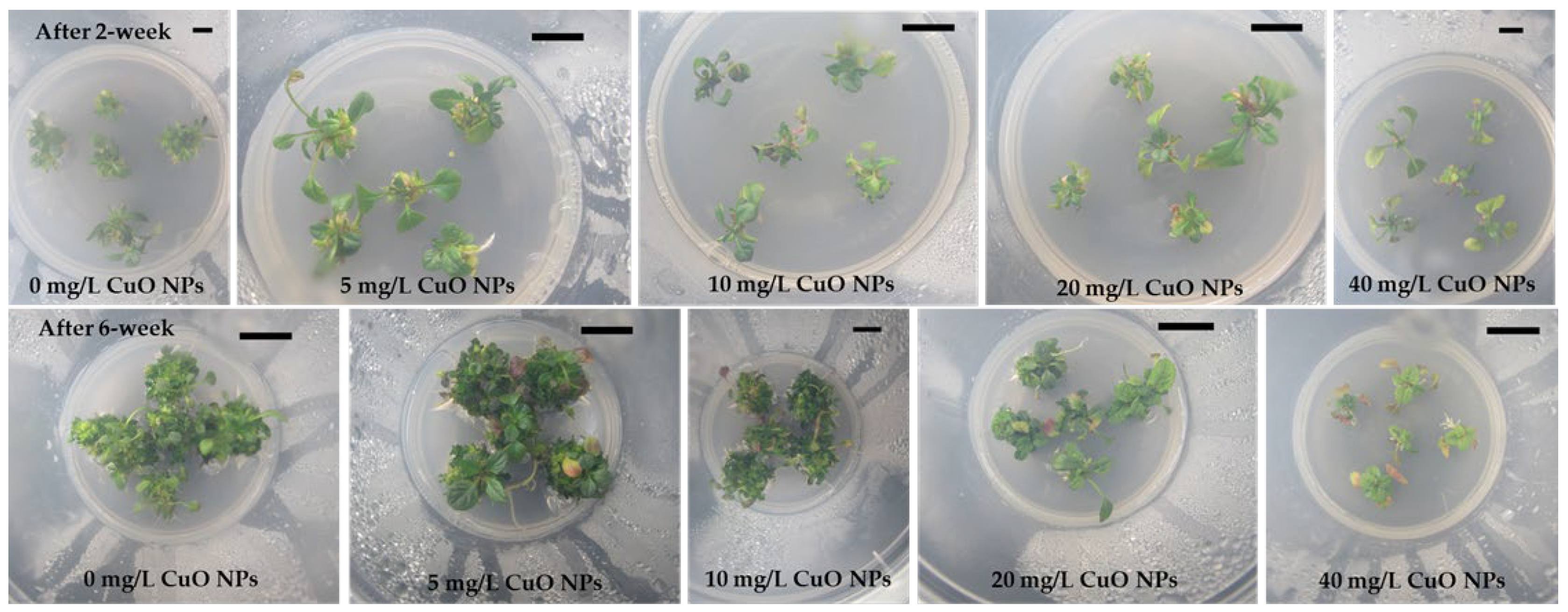
2.1.1. AS Regeneration from Leaf Explants
2.1.2. ASM from Shoot Tips
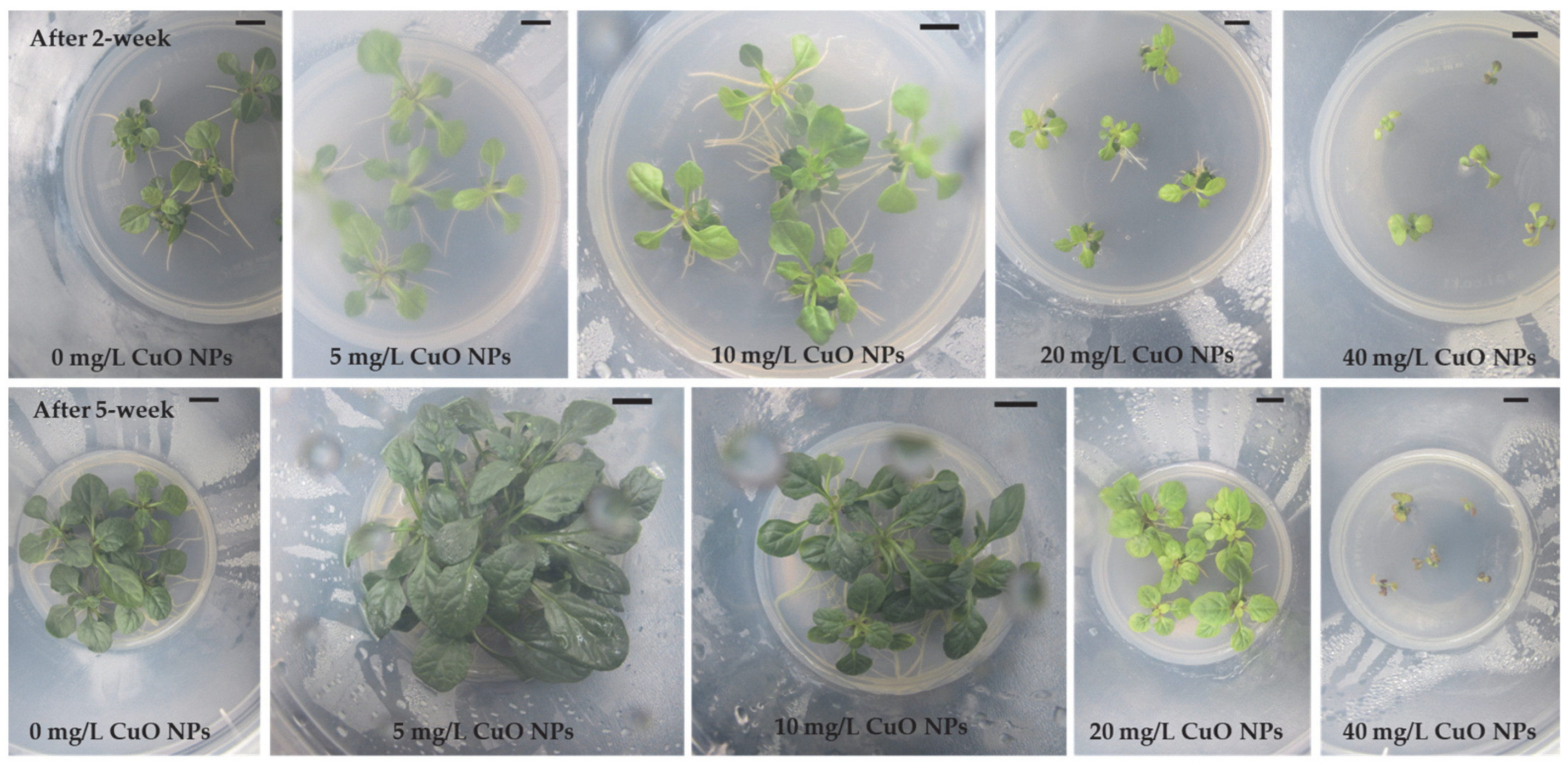
2.1.3. Rooting and Plantlet Development
2.2. Effect of CuO NPs on Lipophilic Metabolites
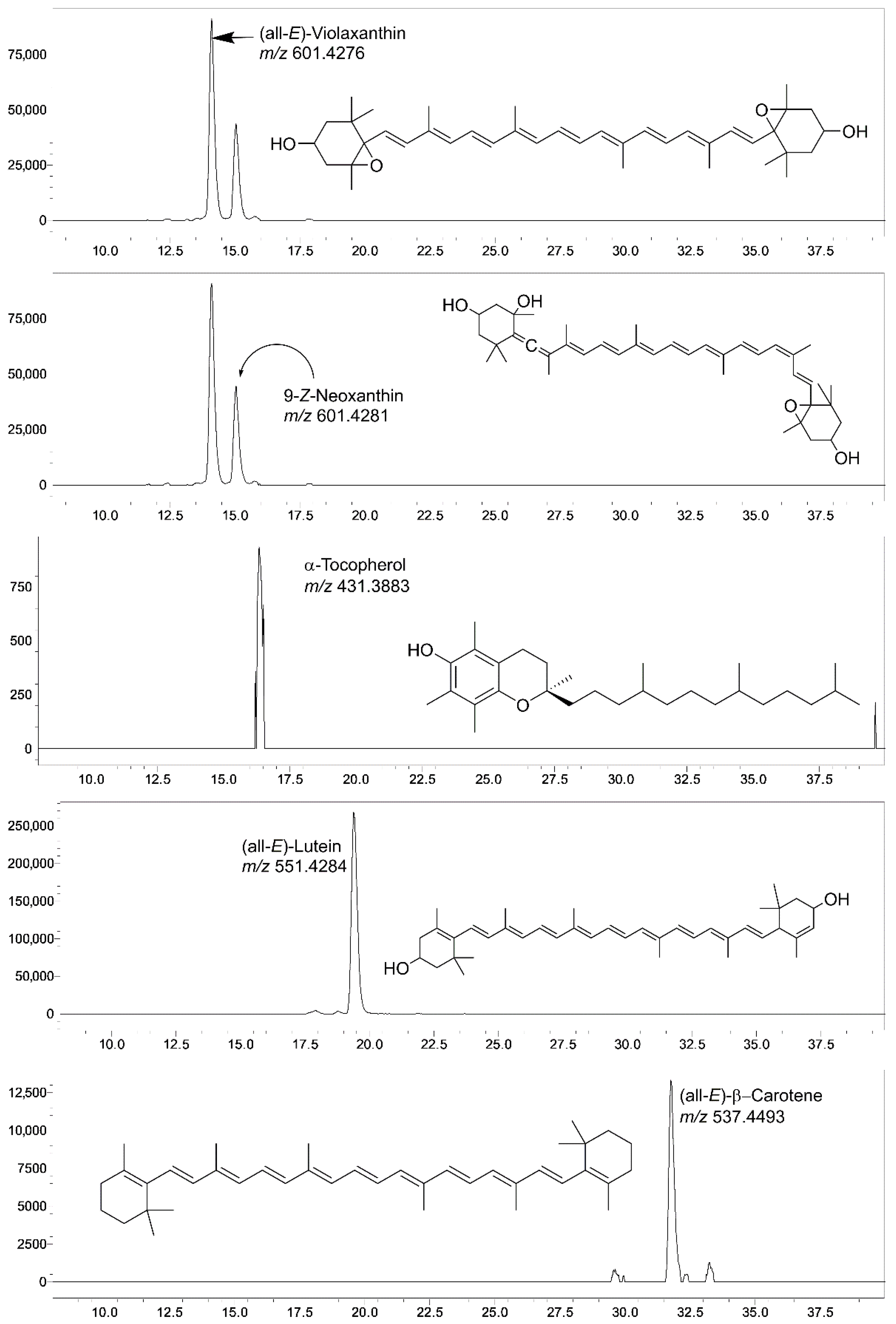
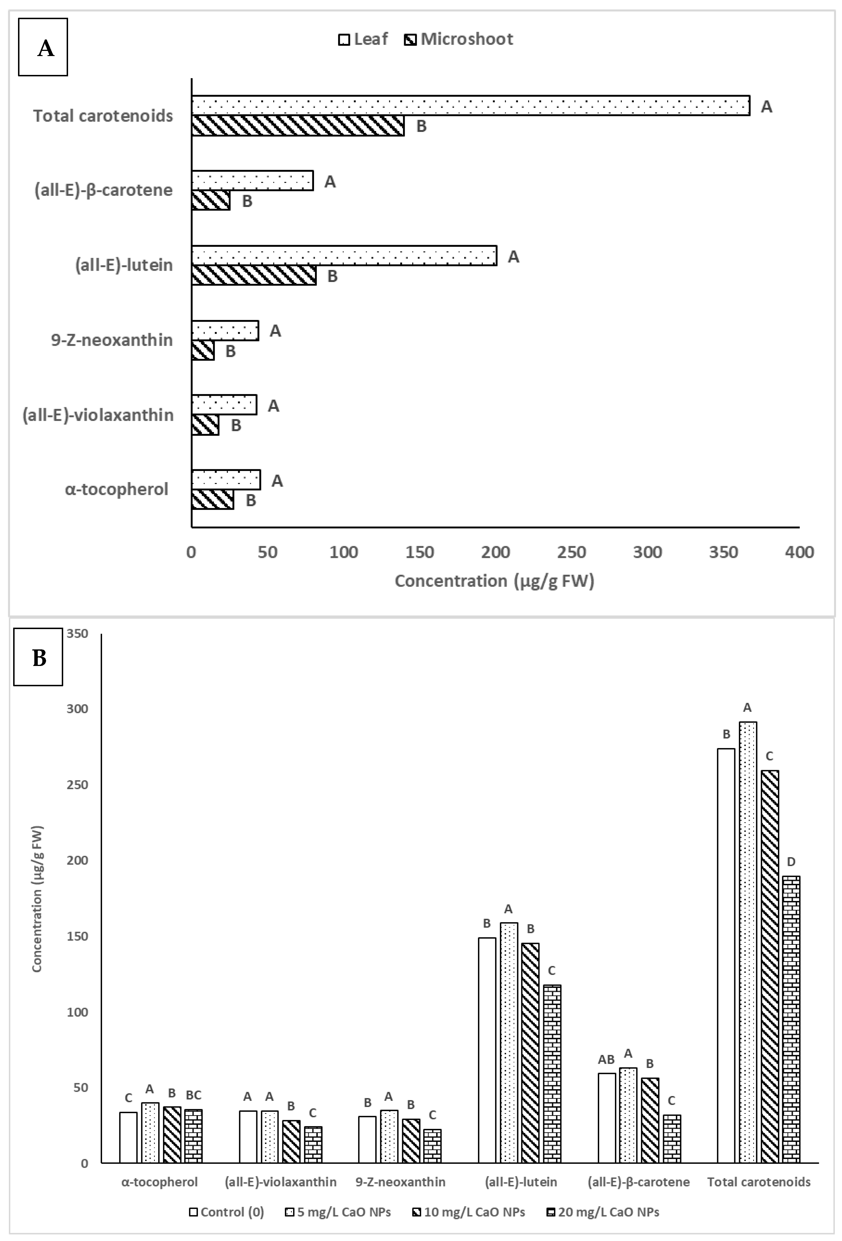
2.2.1. Impact of CuO NPs Levels on α-Tocopherol and Carotenoid Contents in Microshoot and Leaf Tissues of A. multiflora
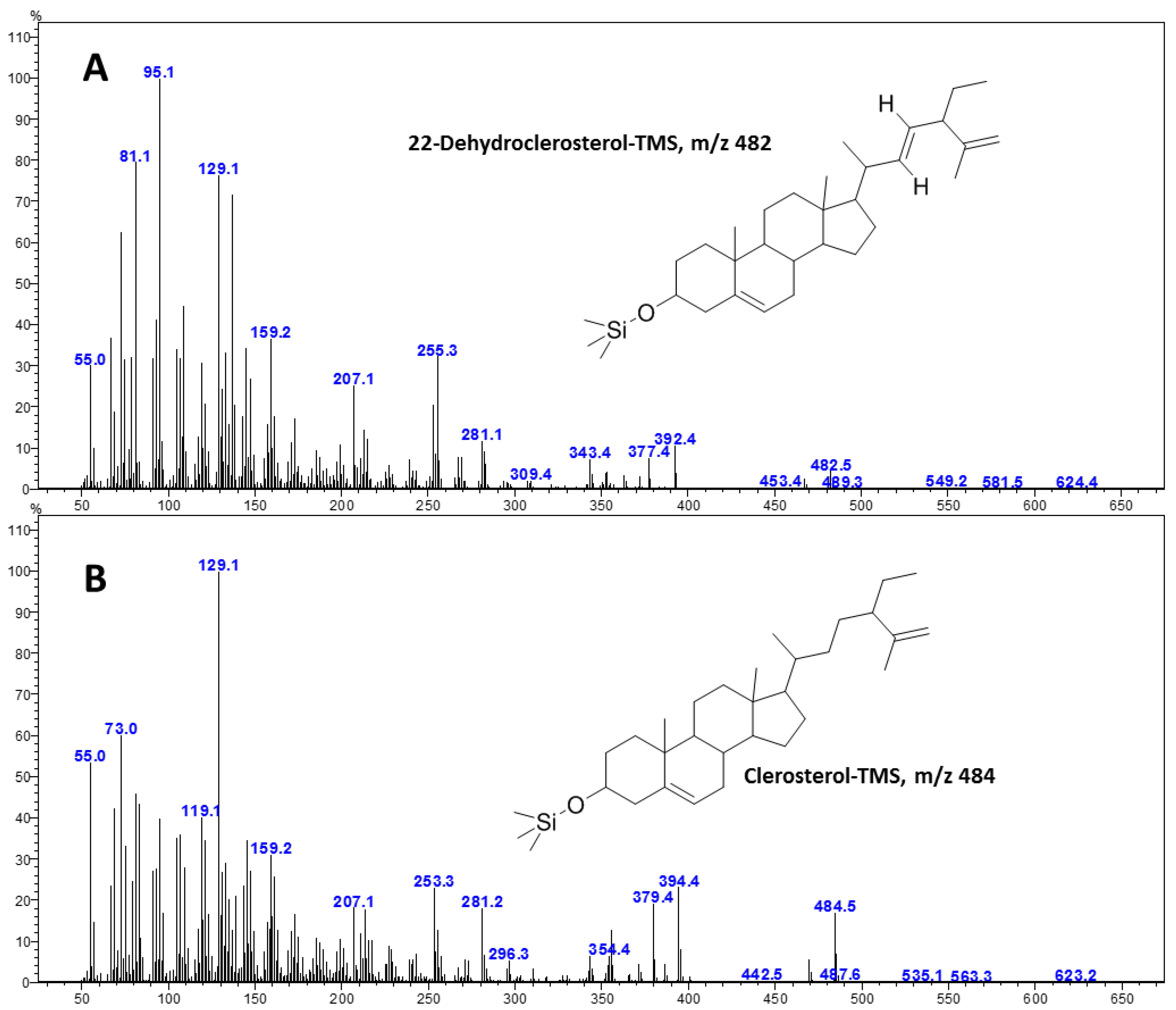
2.2.2. Impact of CuO NPs Levels on Phytosterols Content in Microshoot and Leaf Tissues of A. multiflora
2.2.3. Impact of CuO NPs Levels on Fatty Acids Content in Microshoot and Leaf Tissues of A. multiflora
3. Discussion
4. Materials and Methods
4.1. Impact of CuO NPs on Micropropagation
4.1.1. Plant Materials
4.1.2. Adventitious Shoot Induction (ASI)
4.1.3. Axillary Shoot Multiplication (ASM)
4.1.4. Root Induction (RI)
4.2. Impact of CuO NPs on the Content of Lipophilic Compounds
4.2.1. Reagents and Standards
4.2.2. Extraction of Lipophilic Compounds (Crude Lipids)
4.2.3. LC-SIM-MS Analysis of Carotenoids and Tocols
4.2.4. Qualitative Analysis of FAMEs Utilizing GC-FID and GC-MS
4.2.5. Qualitative Analysis of Sterols Utilizing GC-MS
4.3. Statistical Analysis
5. Conclusions
Supplementary Materials
Author Contributions
Funding
Data Availability Statement
Acknowledgments
Conflicts of Interest
References
- Lakhani, K.G.; Hamid, R.; Motamedi, E.; Marviya, G.V. A review on plant metabolite-mediated nanoparticle synthesis: Sustainable applications in horticultural crops. Front. Nanotechnol. 2025, 7, 1545413. [Google Scholar] [CrossRef] [Scilit]
- Mosqueda-Frómeta, O.; Andujar, I.; Mosqueda-Rodríguez, G.M.; Pestryakov, A.; Companioni, B.; Tapia y Figueroa, M.d.L.; Concepción, O.; Escalona, M.; Bogdanchikova, N.; Lorenzo, J.C. Mini-review: Application of nanoparticles in plant biotechnology and agriculture. Vitr. Cell. Dev. Biol.-Plant 2025, 61, 659–663. [Google Scholar] [CrossRef] [Scilit]
- Kang, I.; Sivanesan, I. Micropropagation of Philodendron ‘White Knight’ via Shoot Regeneration from Petiole Explants. Plants 2025, 14, 1714. [Google Scholar] [CrossRef] [Scilit]
- Chun, S.C.; Seob, P.K.; Kang, K.W.; Sivanesan, I. Micropropagation of Ardisia mamillata Hance through axillary shoot multiplication. Propag. Ornam. Plants 2024, 24, 63–70. [Google Scholar]
- Sivanesan, I.; Lee, Y.K.; Kang, K.W.; Park, H.Y. Micropropagation of Monstera deliciosa Liebm.‘Thai Constellation’. Propag. Ornam. Plants 2023, 23, 31–38. [Google Scholar]
- Soni, S.; Singh, K.M.; Jha, A.B.; Dubey, R.S.; Sharma, P. Size-based dynamics of nanoparticles in plant growth and environmental stress tolerance: Potential benefits and hazards. Environ. Sci. Nano 2026. [Google Scholar] [CrossRef] [Scilit]
- Kaleem, M.; Shah, A.A.; Usman, S.; Xu, W.; Alfarhan, A.H. Foliar Application of Calcium Phosphate Nanoparticles Improves Photosynthetic Efficiency, Nutrient Uptake, and Antioxidant Capacity in Coriandrum sativum L. under Salinity Stress. ACS Omega 2025, 10, 46393–46409. [Google Scholar] [CrossRef] [Scilit]
- Djanaguiraman, M.; Anbazhagan, V.; Dhankher, O.P.; Prasad, P.V.V. Uptake, Translocation, Toxicity, and Impact of Nanoparticles on Plant Physiological Processes. Plants 2024, 13, 3137. [Google Scholar] [CrossRef] [Scilit]
- Alabdallah, N.M.; Kotb, E. Nanoparticles: Unveiling the impact on biochemical reactions in plants. In Molecular Impacts of Nanoparticles on Plants and Algae; Tombuloglu, H., Tombuloglu, G., Al-Suhaimi, E., Baykal, A., Hakeem, K.R., Eds.; Academic Press: Cambridge, MA, USA, 2024; pp. 195–209. [Google Scholar]
- Prasad, A.; Sidhic, J.; Sarbadhikary, P.; Narayanankutty, A.; George, S.; George, B.P.; Abrahamse, H. Role of metal nanoparticles in organogenesis, secondary metabolite production and genetic transformation of plants under in vitro condition: A comprehensive review. Plant Cell Tissue Organ Cult. (PCTOC) 2024, 158, 33. [Google Scholar] [CrossRef] [Scilit]
- Al-Khayri, J.M.; Rashmi, R.; Surya Ulhas, R.; Sudheer, W.N.; Banadka, A.; Nagella, P.; Aldaej, M.I.; Rezk, A.A.-S.; Shehata, W.F.; Almaghasla, M.I. The Role of Nanoparticles in Response of Plants to Abiotic Stress at Physiological, Biochemical, and Molecular Levels. Plants 2023, 12, 292. [Google Scholar] [CrossRef] [Scilit]
- Lala, S. Nanoparticles as elicitors and harvesters of economically important secondary metabolites in higher plants: A review. IET Nanobiotechnol. 2021, 15, 28–57. [Google Scholar] [CrossRef] [Scilit]
- Rohela, G.K.; Saini, P.; Aziz, D.; Rafiq, S.; Jogam, P.; Zhang, B. Nanoparticles as elicitors and stimulators for plant tissue culture, transgenics, and genome editing: A comprehensive review. Ind. Crops Prod. 2024, 222, 120097. [Google Scholar] [CrossRef] [Scilit]
- Mustafa, M.; Azam, M.; Nawaz Bhatti, H.; Khan, A.; Zafar, L.; Rehan Abbasi, A.M. Green fabrication of copper nano-fertilizer for enhanced crop yield in cowpea cultivar: A sustainable approach. Biocatal. Agric. Biotechnol. 2024, 56, 102994. [Google Scholar] [CrossRef] [Scilit]
- Ogunyemi, S.O.; Luo, J.; Abdallah, Y.; Yu, S.; Wang, X.; Alkhalifah, D.H.M.; Hozzein, W.N.; Wang, F.; Bi, J.; Yan, C.; et al. Copper oxide nanoparticles: An effective suppression tool against bacterial leaf blight of rice and its impacts on plants. Pest Manag. Sci. 2024, 80, 1279–1288. [Google Scholar] [CrossRef] [Scilit] [PubMed]
- Khaldari, I.; Naghavi, M.R.; Motamedi, E.; Zargar, M. The effects of green and chemically-synthesized copper oxide nanoparticles on the production and gene expression of morphinan alkaloids in Oriental poppy. Sci. Rep. 2024, 14, 6000. [Google Scholar] [CrossRef] [Scilit] [PubMed]
- Gupta, R.; Verma, N.; Tewari, R.K. Micronutrient deficiency-induced oxidative stress in plants. Plant Cell Rep. 2024, 43, 213. [Google Scholar] [CrossRef] [Scilit]
- Xu, E.; Liu, Y.; Gu, D.; Zhan, X.; Li, J.; Zhou, K.; Zhang, P.; Zou, Y. Molecular Mechanisms of Plant Responses to Copper: From Deficiency to Excess. Int. J. Mol. Sci. 2024, 25, 6993. [Google Scholar] [CrossRef] [Scilit]
- Rakhimol, K.R.; Ashitha, A.; Thomas, S.; Kalarikkal, N.; Jayachandran, K. Casein stabilized metal and metal oxide nanoparticle to eradicate the endophytic bacterial contamination from in vitro culturing of Scoparia dulcis L. Plant Cell Tissue Organ Cult. (PCTOC) 2023, 155, 521–529. [Google Scholar] [CrossRef] [Scilit]
- Fatima, K.; Abbas, S.R.; Zia, M.; Sabir, S.M.; Khan, R.T.; Khan, A.A.; Hassan, Z.; Zaman, R. Induction of secondary metabolites on nanoparticles stress in callus culture of Artemisia annua L. Braz. J. Biol. 2021, 81, 474–483. [Google Scholar] [CrossRef] [Scilit]
- Nazir, S.; Jan, H.; Zaman, G.; Khan, T.; Ashraf, H.; Meer, B.; Zia, M.; Drouet, S.; Hano, C.; Abbasi, B.H. Copper oxide (CuO) and manganese oxide (MnO) nanoparticles induced biomass accumulation, antioxidants biosynthesis and abiotic elicitation of bioactive compounds in callus cultures of Ocimum basilicum (Thai basil). Artif. Cells Nanomed. Biotechnol. 2021, 49, 626–634. [Google Scholar] [CrossRef] [Scilit]
- Nalci, O.B.; Nadaroglu, H.; Pour, A.H.; Gungor, A.A.; Haliloglu, K. Effects of ZnO, CuO and γ-Fe3O4 nanoparticles on mature embryo culture of wheat (Triticum aestivum L.). Plant Cell Tissue Organ Cult. (PCTOC) 2018, 136, 269–277. [Google Scholar] [CrossRef] [Scilit]
- Malik, W.A.; Mahmood, I.; Razzaq, A.; Afzal, M.; Shah, G.A.; Iqbal, A.; Zain, M.; Ditta, A.; Asad, S.A.; Ahmad, I.; et al. Exploring potential of copper and silver nano particles to establish efficient callogenesis and regeneration system for wheat (Triticum aestivum L.). GM Crops Food 2021, 12, 564–585. [Google Scholar] [CrossRef] [Scilit] [PubMed]
- Abdel-Wahab, D.A.; Othman, N.A.R.M.; Hamada, A.M. Effects of copper oxide nanoparticles to Solanum nigrum and its potential for phytoremediation. Plant Cell Tissue Organ Cult. (PCTOC) 2019, 137, 525–539. [Google Scholar] [CrossRef] [Scilit]
- Javed, R.; Yucesan, B.; Zia, M.; Gurel, E. Elicitation of Secondary Metabolites in Callus Cultures of Stevia rebaudiana Bertoni Grown Under ZnO and CuO Nanoparticles Stress. Sugar Tech. 2017, 20, 194–201. [Google Scholar] [CrossRef] [Scilit]
- Park, H.Y.; Saini, R.K.; Keum, Y.-S.; Sivanesan, I. Exploring somatic embryogenesis in Ajuga multiflora Bunge: Profiling lipophilic metabolites via HPLC, GC-FID, and GCMS– analysis. Sci. Hortic. 2024, 332, 113228. [Google Scholar] [CrossRef] [Scilit]
- Jeong, B.R.; Sivanesan, I. Impact of light quality and sucrose on adventitious shoot regeneration and bioactive compound accumulation in Ajuga multiflora Bunge. Sci. Hortic. 2018, 236, 222–228. [Google Scholar] [CrossRef] [Scilit]
- De-fu, C.; Ming-xue, S.; Wen-fu, X. Pesticidal character of phytoecdysteroids from Ajuga multiflora Bunge (Labiatae) on larvae of Cryptorrhynchus lapathi L. (Coleoptera: Curculionidae). J. For. Res. 2002, 13, 177–182. [Google Scholar] [CrossRef] [Scilit]
- Lee, Y.; Kim, H.M.; Kim, J.H.; Lee, J.H.; Zhang, K.X.; Gao, E.M.; Jeon, J.S.; Syed, A.S.; Son, R.H.; Kim, J.Y.; et al. Chemical constituents of the Ajuga multiflora bunge and their protective effects on dexamethasone-induced muscle atrophy in C2C12 myotubes. Nat. Prod. Res. 2023, 37, 1978–1985. [Google Scholar] [CrossRef] [Scilit]
- Popova, E.; Titova, M.; Tynykulov, M.; Zakirova, R.P.; Kulichenko, I.; Prudnikova, O.; Nosov, A. Sustainable Production of Ajuga Bioactive Metabolites Using Cell Culture Technologies: A Review. Nutrients 2023, 15, 1246. [Google Scholar] [CrossRef] [Scilit]
- Ryu MyengHwan, R.M.; Aeam YongDea, A.Y.; Byun JongHo, B.J.; Cho Hoon, C.H.; Yang EunYeong, Y.E.; Kang KilUng, K.K.; Shin MinKyo, S.M.; Baek SeungHwa, B.S. Studies on the cytotoxicity and antimicrobial effects of the extract of Ajuga multiflora Bunge. Korean J. Pharmacogn. 2000, 31, 72–76. [Google Scholar]
- Sivanesan, I.; Saini, R.K.; Noorzai, R.; Zamany, A.J.; Kim, D.H. In vitro propagation, carotenoid, fatty acid and tocopherol content of Ajuga multiflora Bunge. 3 Biotech 2016, 6, 91. [Google Scholar] [CrossRef] [Scilit]
- Yu, Y.-J.; Do, J.-C.; Jung, K.-Y.; Kwon, S.-Y.; Son, K.-H. Studies on the Constituents of the Herbs of Ajuga multiflora (I). Korean J. Pharmacogn. 1998, 29, 75–78. [Google Scholar]
- Park, H.Y.; Kim, D.H.; Sivanesan, I. Micropropagation of Ajuga species: A mini review. Biotechnol. Lett. 2017, 39, 1291–1298. [Google Scholar] [CrossRef] [Scilit] [PubMed]
- Sivanesan, I.; Ko ChungHo, K.C.; Lee JangPyung, L.J.; Jeong ByoungRyong, J.B. Influence of cytokinins on adventitious shoot regeneration from leaf and petiole explants of Ajuga multiflora bunge. Propag. Ornam. Plants 2011, 11, 156–158. [Google Scholar]
- Park, S.; Sivanesan, I. Effect of plant growth regulators on axillary shoot multiplication from nodal explants of Ajuga multiflora Bunge. Propag. Ornam. Plants 2015, 15, 42–44. [Google Scholar]
- Kim, O.T.; Manickavasagm, M.; Kim, Y.J.; Jin, M.R.; Kim, K.S.; Seong, N.S.; Hwang, B. Genetic transformation of Ajuga multiflora bunge with Agrobacterium rhizogenes and 20-hydroxyecdysone production in hairy roots. J. Plant Biol. 2005, 48, 258–262. [Google Scholar] [CrossRef] [Scilit]
- Wang, Y.C.; Zhao, J.J.; Chi, D.F. beta-Ecdysterone accumulation and regulation in Ajuga multiflora Bunge suspension culture. 3 Biotech 2018, 8, 87. [Google Scholar] [CrossRef] [Scilit]
- Ali, H.; Khan, M.A.; Ullah, N.; Khan, R.S. Impacts of hormonal elicitors and photoperiod regimes on elicitation of bioactive secondary volatiles in cell cultures of Ajuga bracteosa. J. Photochem. Photobiol. B Biol. 2018, 183, 242–250. [Google Scholar] [CrossRef] [Scilit] [PubMed]
- Kayani, W.K.; Palazòn, J.; Cusidò, R.M.; Mirza, B. Effect of pRi T-DNA genes and elicitation on morphology and phytoecdysteroid biosynthesis in Ajuga bracteosa hairy roots. RSC Adv. 2017, 7, 47945–47953. [Google Scholar] [CrossRef] [Scilit]
- Ullah, M.A.; Gul, F.Z.; Khan, T.; Bajwa, M.N.; Drouet, S.; Tungmunnithum, D.; Giglioli-Guivarc’h, N.; Liu, C.; Hano, C.; Abbasi, B.H. Differential induction of antioxidant and anti-inflammatory phytochemicals in agitated micro-shoot cultures of Ajuga integrifolia Buch. Ham. ex D.Don with biotic elicitors. AMB Express 2021, 11, 137. [Google Scholar] [CrossRef] [Scilit]
- Haider Abbasi, B.; Ullah, M.A.; Nadeem, M.; Tungmunnithum, D.; Hano, C. Exogenous application of salicylic acid and gibberellic acid on biomass accumulation, antioxidant and anti-inflammatory secondary metabolites production in multiple shoot culture of Ajuga integrifolia Buch. Ham. ex D.Don. Ind. Crops Prod. 2020, 145, 112098. [Google Scholar] [CrossRef] [Scilit]
- El-Naggar, H.M.; Osman, A.R. Enhancing growth and bioactive metabolites characteristics in Mentha pulegium L. via silicon nanoparticles during in vitro drought stress. BMC Plant Biol. 2024, 24, 657. [Google Scholar] [CrossRef] [Scilit]
- Gonçalves, S.; Mansinhos, I.; Rodríguez-Solana, R.; Pereira-Caro, G.; Moreno-Rojas, J.M.; Romano, A. Impact of Metallic Nanoparticles on In Vitro Culture, Phenolic Profile and Biological Activity of Two Mediterranean Lamiaceae Species: Lavandula viridis L’Hér and Thymus lotocephalus G. López and R. Morales. Molecules 2021, 26, 6427. [Google Scholar] [CrossRef] [Scilit]
- Khattab, S.; El Sherif, F.; AlDayel, M.; Yap, Y.-K.; Meligy, A.; Ibrahim, H.I.M. Silicon dioxide and silver nanoparticles elicit antimicrobial secondary metabolites while enhancing growth and multiplication of Lavandula officinalis in-vitro plantlets. Plant Cell Tissue Organ Cult. (PCTOC) 2022, 149, 411–421. [Google Scholar] [CrossRef] [Scilit]
- Razavizadeh, R.; Adabavazeh, F.; Mosayebi, Z. Titanium dioxide nanoparticles improve element uptake, antioxidant properties, and essential oil productivity of Melissa officinalis L. seedlings under in vitro drought stress. Environ. Sci. Pollut. Res. 2023, 30, 98020–98033. [Google Scholar] [CrossRef] [Scilit] [PubMed]
- Yılmaz Baydu, F.; Bektaş, E. Effects of Zinc Oxide Nanoparticles on Growth and Secondary Metabolite Accumulation in In Vitro Culture of Ocimum basilicum L. J. Apitherapy Nat. 2025, 8, 37–60. [Google Scholar] [CrossRef] [Scilit]
- Gunasena, M.D.K.M.; Alahakoon, A.M.P.D.; Polwaththa, K.P.G.D.M.; Galpaya, G.D.C.P.; Priyanjani, H.A.S.A.; Koswattage, K.R.; Senarath, W.T.P.S.K. Transforming plant tissue culture with nanoparticles: A review of current applications. Plant Nano Biol. 2024, 10, 100102. [Google Scholar] [CrossRef] [Scilit]
- Kim, D.H.; Gopal, J.; Sivanesan, I. Nanomaterials in plant tissue culture: The disclosed and undisclosed. RSC Adv. 2017, 7, 36492–36505. [Google Scholar] [CrossRef] [Scilit]
- Guha, P.S.; Gupta, S.D.; Saha, N. Nano-gardening: Harnessing Metal Nanoparticles for Enhanced In Vitro Plant Regeneration. BioNanoScience 2024, 14, 3555–3571. [Google Scholar] [CrossRef] [Scilit]
- Zafar, H.; Ali, A.; Zia, M. CuO Nanoparticles Inhibited Root Growth from Brassica nigra Seedlings but Induced Root from Stem and Leaf Explants. Appl. Biochem. Biotechnol. 2017, 181, 365–378. [Google Scholar] [CrossRef] [Scilit] [PubMed]
- Paramo, L.A.; Feregrino-Perez, A.A.; Guevara, R.; Mendoza, S.; Esquivel, K. Nanoparticles in Agroindustry: Applications, Toxicity, Challenges, and Trends. Nanomaterials 2020, 10, 1654. [Google Scholar] [CrossRef] [Scilit]
- Balamurugan, V.; Abdi, G.; Karthiksaran, C.; Thillaigovindhan, N.; Arulbalachandran, D. A review: Improvement of plant tissue culture applications by using nanoparticles. J. Nanoparticle Res. 2024, 26, 188. [Google Scholar] [CrossRef] [Scilit]
- Fedorova, O.; Grodetskaya, T.; Evtushenko, N.; Evlakov, P.; Gusev, A.; Zakharova, O. The impact of copper oxide and silver nanoparticles on woody plants obtained by in vitro method. IOP Conf. Ser. Earth Environ. Sci. 2021, 875, 012048. [Google Scholar] [CrossRef] [Scilit]
- Zakharova, O.; Gusev, A.; Strekalova, N.; Grigoriev, G.; Chebotareva, S.; Baranchikov, P.; Kataranova, A. Copper oxide nanoparticles in clonal micropropagation of red oak. In BIO Web of Conferences; EDP Sciences: Les Ulis, France, 2024; p. 02006. [Google Scholar]
- Nair, P.M.; Chung, I.M. Study on the correlation between copper oxide nanoparticles induced growth suppression and enhanced lignification in Indian mustard (Brassica juncea L.). Ecotoxicol. Environ. Saf. 2015, 113, 302–313. [Google Scholar] [CrossRef] [Scilit]
- Wang, Z.; Xie, X.; Zhao, J.; Liu, X.; Feng, W.; White, J.C.; Xing, B. Xylem- and phloem-based transport of CuO nanoparticles in maize (Zea mays L.). Environ. Sci. Technol. 2012, 46, 4434–4441. [Google Scholar] [CrossRef] [Scilit] [PubMed]
- Pathak, A.; Haq, S.; Meena, N.; Dwivedi, P.; Kothari, S.L.; Kachhwaha, S. Multifaceted Role of Nanomaterials in Modulating In Vitro Seed Germination, Plant Morphogenesis, Metabolism and Genetic Engineering. Plants 2023, 12, 3126. [Google Scholar] [CrossRef] [Scilit] [PubMed]
- Wang, X.; Wang, W.X. Cu(I)/Cu(II) Released by Cu Nanoparticles Revealed Differential Cellular Toxicity Related to Mitochondrial Dysfunction. Environ. Sci. Technol. 2023, 57, 9548–9558. [Google Scholar] [CrossRef] [Scilit]
- Xiong, T.; Zhang, S.; Kang, Z.; Zhang, T.; Li, S. Dose-Dependent Physiological and Transcriptomic Responses of Lettuce (Lactuca sativa L.) to Copper Oxide Nanoparticles—Insights into the Phytotoxicity Mechanisms. Int. J. Mol. Sci. 2021, 22, 3688. [Google Scholar] [CrossRef] [Scilit]
- Yruela, I. Copper in plants. Braz. J. Plant Physiol. 2005, 17, 145–156. [Google Scholar] [CrossRef] [Scilit]
- Iqbal, Z.; Javad, S.; Naz, S.; Shah, A.A.; Shah, A.N.; Paray, B.A.; Gulnaz, A.; Abdelsalam, N.R. Elicitation of the in vitro Cultures of Selected Varieties of Vigna radiata L. With Zinc Oxide and Copper Oxide Nanoparticles for Enhanced Phytochemicals Production. Front. Plant Sci. 2022, 13, 908532. [Google Scholar] [CrossRef] [Scilit]
- Bamal, D.; Singh, A.; Swami, N.; Sehrawat, A.R. Alhagi maurorum: A Medicinal Treasure Trove Empowered by Copper Oxide Nanoparticles for Enhanced Secondary Metabolite Synthesis. Appl. Biochem. Biotechnol. 2025, 197, 5221–5248. [Google Scholar] [CrossRef] [Scilit]
- Murashige, T.; Skoog, F. A Revised Medium for Rapid Growth and Bio Assays with Tobacco Tissue Cultures. Physiol. Plant. 2006, 15, 473–497. [Google Scholar] [CrossRef] [Scilit]
- Saini, R.K.; Moon, S.H.; Gansukh, E.; Keum, Y.S. An efficient one-step scheme for the purification of major xanthophyll carotenoids from lettuce, and assessment of their comparative anticancer potential. Food Chem. 2018, 266, 56–65. [Google Scholar] [CrossRef] [Scilit] [PubMed]
- Kim, D.E.; Shang, X.; Assefa, A.D.; Keum, Y.S.; Saini, R.K. Metabolite profiling of green, green/red, and red lettuce cultivars: Variation in health beneficial compounds and antioxidant potential. Food Res. Int. 2018, 105, 361–370. [Google Scholar] [CrossRef] [Scilit] [PubMed]
- Saini, R.K.; Keum, Y.S. Carotenoid extraction methods: A review of recent developments. Food Chem. 2018, 240, 90–103. [Google Scholar] [CrossRef] [Scilit] [PubMed]
- Cruz, R.; Casal, S.; Mendes, E.; Costa, A.; Santos, C.; Morais, S. Validation of a Single-Extraction Procedure for Sequential Analysis of Vitamin E, Cholesterol, Fatty Acids, and Total Fat in Seafood. Food Anal. Methods 2012, 6, 1196–1204. [Google Scholar] [CrossRef] [Scilit]
- Saini, R.K.; Rauf, A.; Khalil, A.A.; Ko, E.-Y.; Keum, Y.-S.; Anwar, S.; Alamri, A.; Rengasamy, K.R.R. Edible mushrooms show significant differences in sterols and fatty acid compositions. S. Afr. J. Bot. 2021, 141, 344–356. [Google Scholar] [CrossRef] [Scilit]





| CuO NP (mg/L) | Shoot Induction (%) | Number of Shoots/Explants | Shoot FW (g) |
|---|---|---|---|
| 0 | 100 ± 0.0 a | 23.7 ± 3.1 b | 0.105 ± 0.01 b |
| 5 | 100 ± 0.0 a | 29.4 ± 4.1 a | 0.107 ± 0.03 b |
| 10 | 100 ± 0.0 a | 20.4 ± 3.4 c | 0.115 ± 0.02 b |
| 20 | 100 ± 0.0 a | 13.1 ± 2.2 d | 0.151 ± 0.04 a |
| 40 | 8.3 ± 2.5 b | 1.1 ± 0.3 e | 0.013 ± 0.003 c |
| CuO NPs (mg/L) | Shoot Induction (%) | Number of Shoots/Explants | Shoot FW (g) |
|---|---|---|---|
| 0 | 100 ± 0.0 a | 17.3 ± 2.4 b | 0.211 ± 0.04 b |
| 5 | 100 ± 0.0 a | 22.4 ± 3.2 a | 0.247 ± 0.03 ab |
| 10 | 100 ± 0.0 a | 15.9 ± 1.6 b | 0.269 ± 0.02 a |
| 20 | 100 ± 0.0 a | 9.6 ± 2.8 c | 0.247 ± 0.06 ab |
| 40 | 71.1 ± 15.4 b | 1.0 ± 0.0 d | 0.106 ± 0.03 c |
| CuO NPs (mg/L) | Root Induction (%) | Number of Roots/Shoots | Plantlet FW (mg) |
|---|---|---|---|
| 0 | 100 ± 0.0 a | 5.7 ± 0.9 b | 1.71 ± 0.29 b |
| 5 | 100 ± 0.0 a | 10.2 ± 2.0 a | 2.26 ± 0.24 a |
| 10 | 100 ± 0.0 a | 11.1 ± 3.6 a | 1.35 ± 0.23 c |
| 20 | 80.6 ± 8.1 b | 4.8 ± 1.6 b | 0.52 ± 0.07 d |
| 40 | 0.0 ± 0.0 c | 0.0 ± 0.0 c | 0.0 ± 0.0 e |
| Organ | CuO NPs (mg/L) | α-Tocopherol (Retention Time (RT) 16.996 min) | (All-E)-Violaxanthin (RT 14.116 min) | 9-Z-Neoxanthin (RT 15.059 min) | (All-E)-Lutein (RT 19.398 min) | (All-E)-β-Carotene (RT 31.771 min) | Total Carotenoids |
|---|---|---|---|---|---|---|---|
| Microshoot | 0 | 26.5 ± 0.92 d | 15.8 ± 0.8 e | 12.1 ± 0.2 g | 77.3 ± 2.5 f | 25.5 ± 0.6 e | 130.8 ± 3.0 f |
| 5 | 29.4 ± 1.4 d | 17.6 ± 1.0 de | 15.5 ± 1.0 ef | 78.9 ± 1.3 f | 23.7 ± 2.0 e | 135.8 ± 5.4 ef | |
| 10 | 28.6 ± 1.0 d | 17.5 ± 1.3 de | 13.8 ± 0.4 fg | 80.7 ± 1.7 f | 28.0 ± 1.7 e | 140.0 ± 4.8 ef | |
| 20 | 26.7 ± 0.5 d | 21.1 ± 1.9 d | 17.3 ± 1.2 e | 90.6 ± 7.3 e | 24.1 ± 2.3 e | 153.1 ± 12.6 e | |
| 40 | - | - | - | - | - | - | |
| Leaf | 0 | 41.2 ± 2.6 c | 53.5 ± 4.6 a | 49.5 ± 0.8 b | 220.9 ± 5.4 b | 93.0 ± 5.0 b | 417.0 ± 6.1 b |
| 5 | 50.3 ± 1.8 a | 51.6 ± 1.2 a | 54.2 ± 0.6 a | 238.7 ± 2.0 a | 102.6 ± 1.5 a | 447.1 ± 2.1 a | |
| 10 | 45.7 ± 0.7 b | 39.2 ± 0.5 b | 44.8 ± 1.2 c | 210.1 ± 8.3 c | 84.8 ± 11.5 c | 378.9 ± 21.3 c | |
| 20 | 44.6 ± 2.7 b | 26.9 ± 2.3 c | 27.3 ± 2.9 d | 132.9 ± 7.7 d | 39.4 ± 0.9 f | 226.5 ± 13.8 d | |
| 40 | - | - | - | - | - | - | |
| F-test | Org | <0.0001 | <0.0001 | <0.0001 | <0.0001 | <0.0001 | <0.0001 |
| Con | <0.0001 | <0.0001 | <0.0001 | <0.0001 | <0.0001 | <0.0001 | |
| Org*Con | 0.0336 | <0.0001 | <0.0001 | <0.0001 | <0.0001 | <0.0001 |
| Organ | CuO NPs (mg/L) | 22-Dehydroclerosterol | Clerosterol | Total Phytosterol |
|---|---|---|---|---|
| Microshoot | 0 | 71.2 ± 0.3 f | 168.3 ± 2.7 d | 239.6 ± 2.4 e |
| 5 | 86.8 ± 4.5 d | 214.4 ± 3.5 a | 301.2 ± 8.0 b | |
| 10 | 78.3 ± 1.7 e | 201.3 ± 8.8 b | 279.6 ± 7.2 c | |
| 20 | 73.3 ± 2.7 ef | 189.6 ± 3.9 c | 262.9 ± 5.9 d | |
| 40 | - | - | - | |
| Leaf | 0 | 105.5 ± 4.8 ab | 177.6 ± 6.5 cd | 283.1 ± 11.3 c |
| 5 | 108.5 ± 3.6 a | 210.5 ± 7.7 ab | 319.1 ± 10.9 a | |
| 10 | 101.4 ± 5.6 bc | 181.6 ± 11.3 c | 283.0 ± 15.8 c | |
| 20 | 99.4 ± 0.9 c | 168.6 ± 2.6 d | 268.0 ± 3.5 cd | |
| 40 | - | - | - | |
| F-test | Org | <0.0001 | 0.0049 | 0.0002 |
| Con | 0.0001 | <0.0001 | <0.0001 | |
| Org*Con | 0.0214 | 0.0030 | 0.0053 |
| Organ | CuO NPs (mg/L) | Palmitic (RT 20.931 min) | Stearic (RT 24.562 min) | Oleic (RT 25.647 min) | Linoleic (RT 27.278 min) | Arachidic (RT 27.988 min) | α-Linolenic (RT 29.125 min) | Total SFAs | Total PUFAs | n-6/n-3 PUFAs |
|---|---|---|---|---|---|---|---|---|---|---|
| Microshoot | 0 | 21.7 ± 0.77 a | 3.8 ± 0.03 a | 8.2 ± 0.31 d | 25.7 ± 0.52 b | 1.2 ± 0.08 a | 39.5 ± 0.26 cd | 26.6 ± 0.69 a | 65.2 ± 0.39 c | 0.65 ± 0.01 a |
| 5 | 20.0 ± 0.80 b | 3.8 ± 0.31 a | 10.7 ± 0.36 b | 25.6 ± 2.27 b | 1.1 ± 0.13 a | 38.8 ± 3.14 d | 24.8 ± 1.22 ab | 64.4 ± 0.94 c | 0.66 ± 0.11 a | |
| 10 | 19.5 ± 0.33 b | 3.7 ± 0.11 a | 10.2 ± 0.53 bc | 25.9 ± 0.21 b | 1.1 ± 0.05 a | 39.7 ± 1.21 cd | 24.2 ± 0.48 b | 65.7 ± 1.01 c | 0.65 ± 0.03 a | |
| 20 | 19.0 ± 0.24 b | 3.8 ± 0.04 a | 7.1 ± 0.42 e | 28.3 ± 0.57 a | 1.1 ± 0.03 a | 40.6 ± 0.11 cd | 23.9 ± 0.24 b | 69.0 ± 0.64 b | 0.69 ± 0.02 a | |
| 40 | - | - | - | - | - | - | - | - | - | |
| Leaf | 0 | 11.2 ± 0.28 c | 1.8 ± 0.07 d | 6.2 ± 0.17 f | 18.7 ± 0.30 de | 0.3 ± 0.04 d | 61.6 ± 0.56 a | 13.2 ± 0.31 d | 80.3 ± 059 a | 0.30 ± 0.01 d |
| 5 | 18.4 ± 0.24 b | 2.7 ± 0.11 c | 8.2 ± 0.07 d | 19.8 ± 0.10 d | 0.4 ± 0.01 c | 50.4 ± 0.24 b | 21.5 ± 0.18 c | 70.2 ± 0.15 b | 0.39 ± 0.01 c | |
| 10 | 18.5 ± 0.88 b | 3.1 ± 0.05 b | 9.8 ± 0.24 c | 17.0 ± 0.44 e | 0.4 ± 0.05 c | 51.0 ± 0.18 b | 22.0 ± 0.87 c | 68.0 ± 0.62 b | 0.33 ± 0.01 cd | |
| 20 | 18.8 ± 1.98 b | 3.9 ± 0.35 a | 13.6 ± 0.87 a | 21.7 ± 1.2 c | 0.6 ± 0.02 b | 41.7 ± 1.96 c | 23.3 ± 2.36 bc | 63.4 ± 3.21 c | 0.52 ± 0.01 b | |
| 40 | - | - | - | - | - | - | - | - | - | |
| F-test | Org | <0.0001 | <0.0001 | 0.0394 | <0.0001 | <0.0001 | <0.0001 | <0.0001 | <0.0001 | <0.0001 |
| Con | 0.0002 | <0.0001 | <0.0001 | <0.0001 | 0.0068 | <0.0001 | <0.0001 | <0.0001 | 0.0002 | |
| Org*Con | <0.0001 | <0.0001 | <0.0001 | 0.0768 | 0.0003 | <0.0001 | <0.0001 | <0.0001 | 0.0125 |
Disclaimer/Publisher’s Note: The statements, opinions and data contained in all publications are solely those of the individual author(s) and contributor(s) and not of MDPI and/or the editor(s). MDPI and/or the editor(s) disclaim responsibility for any injury to people or property resulting from any ideas, methods, instructions or products referred to in the content. |
© 2025 by the authors. Licensee MDPI, Basel, Switzerland. This article is an open access article distributed under the terms and conditions of the Creative Commons Attribution (CC BY) license (https://creativecommons.org/licenses/by/4.0/).
Share and Cite
Sivanesan, I.; Upadhyay, S.; Keum, Y.-S.; Chun, S.C.; Saini, R.K. Impact of Copper Oxide Nanoparticles on Adventitious Shoot Regeneration, Axillary Shoot Multiplication, Rooting, and Bioactive Compounds in Ajuga multiflora Bunge. Plants 2025, 14, 3807. https://doi.org/10.3390/plants14243807
Sivanesan I, Upadhyay S, Keum Y-S, Chun SC, Saini RK. Impact of Copper Oxide Nanoparticles on Adventitious Shoot Regeneration, Axillary Shoot Multiplication, Rooting, and Bioactive Compounds in Ajuga multiflora Bunge. Plants. 2025; 14(24):3807. https://doi.org/10.3390/plants14243807
Chicago/Turabian StyleSivanesan, Iyyakkannu, Shuchi Upadhyay, Young-Soo Keum, Se Chul Chun, and Ramesh Kumar Saini. 2025. "Impact of Copper Oxide Nanoparticles on Adventitious Shoot Regeneration, Axillary Shoot Multiplication, Rooting, and Bioactive Compounds in Ajuga multiflora Bunge" Plants 14, no. 24: 3807. https://doi.org/10.3390/plants14243807
APA StyleSivanesan, I., Upadhyay, S., Keum, Y.-S., Chun, S. C., & Saini, R. K. (2025). Impact of Copper Oxide Nanoparticles on Adventitious Shoot Regeneration, Axillary Shoot Multiplication, Rooting, and Bioactive Compounds in Ajuga multiflora Bunge. Plants, 14(24), 3807. https://doi.org/10.3390/plants14243807