
Vitex Genus as a Source of Antimicrobial Agents


Abstract
1. Introduction
2. Results
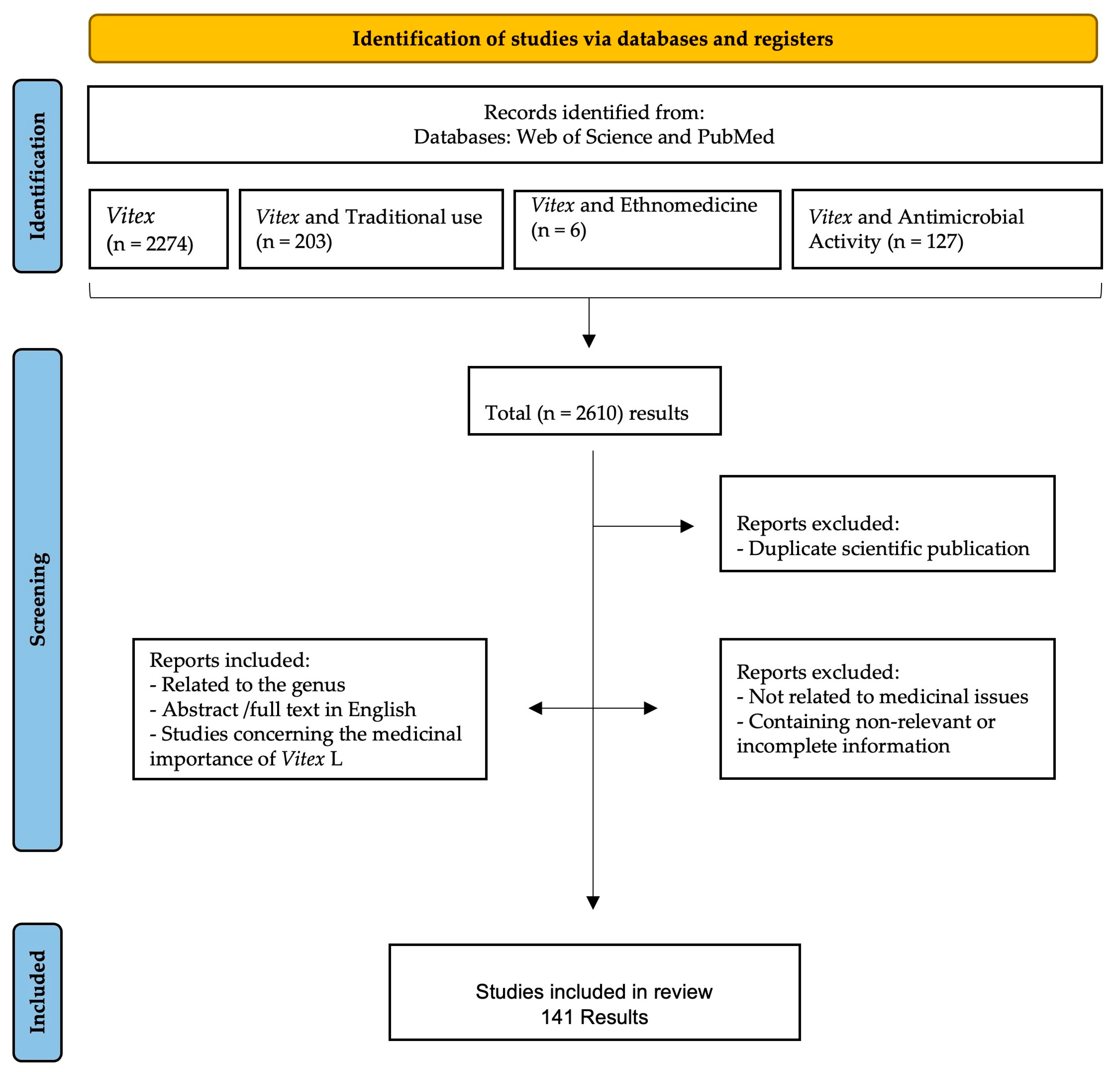
2.1. Selection of the Information
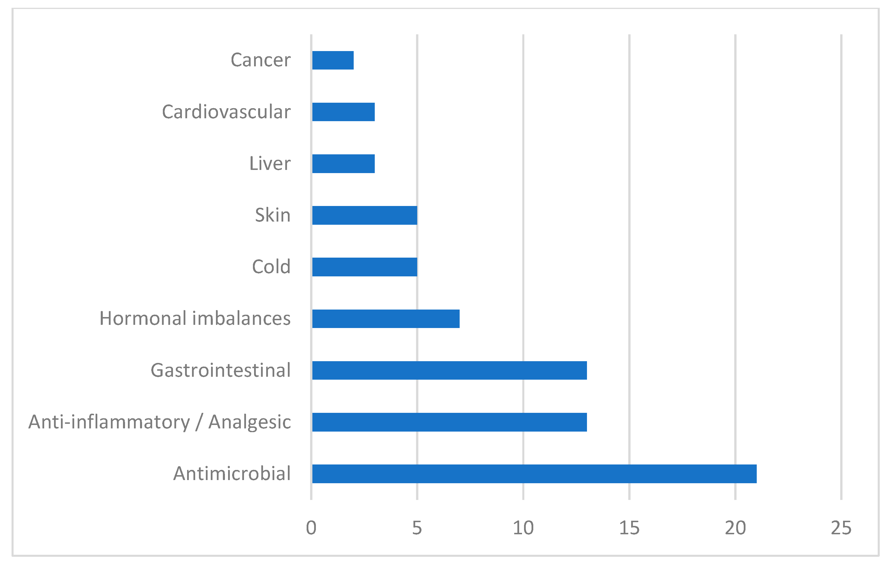
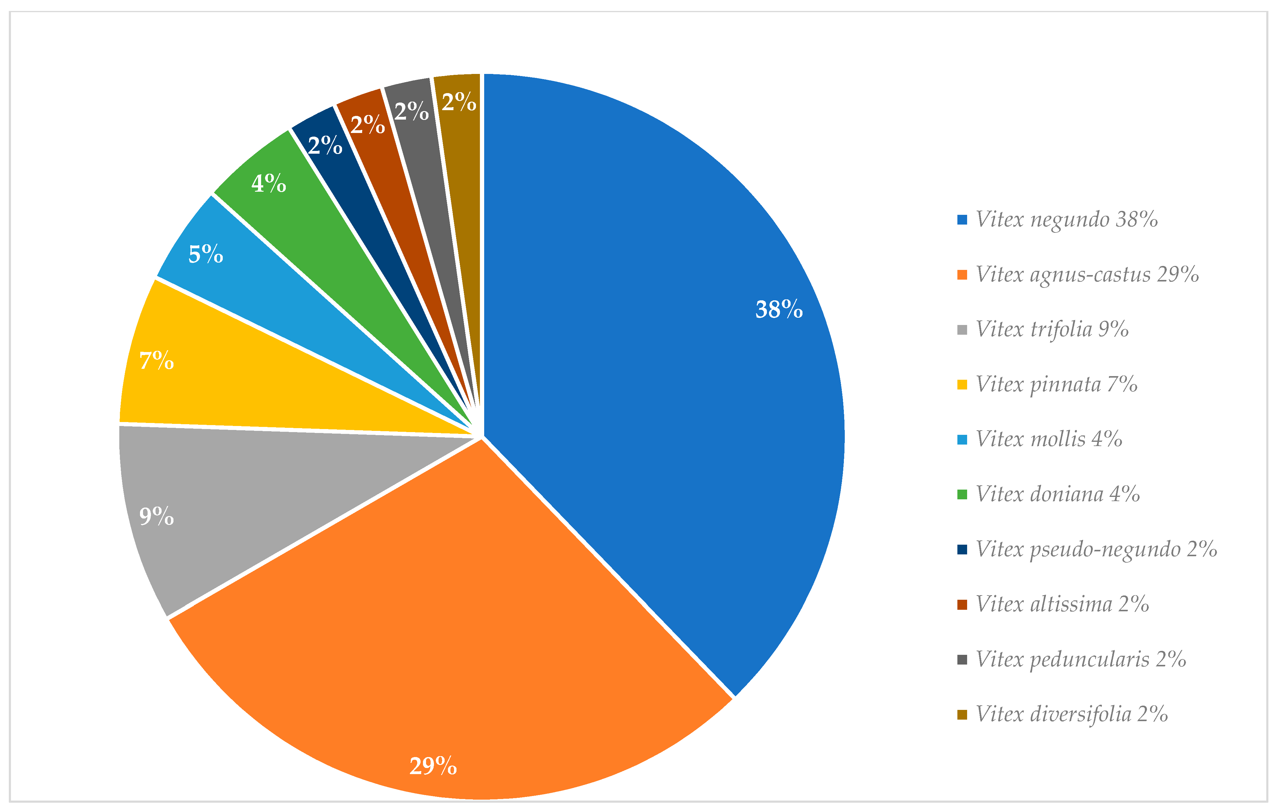
2.2. Traditional Uses
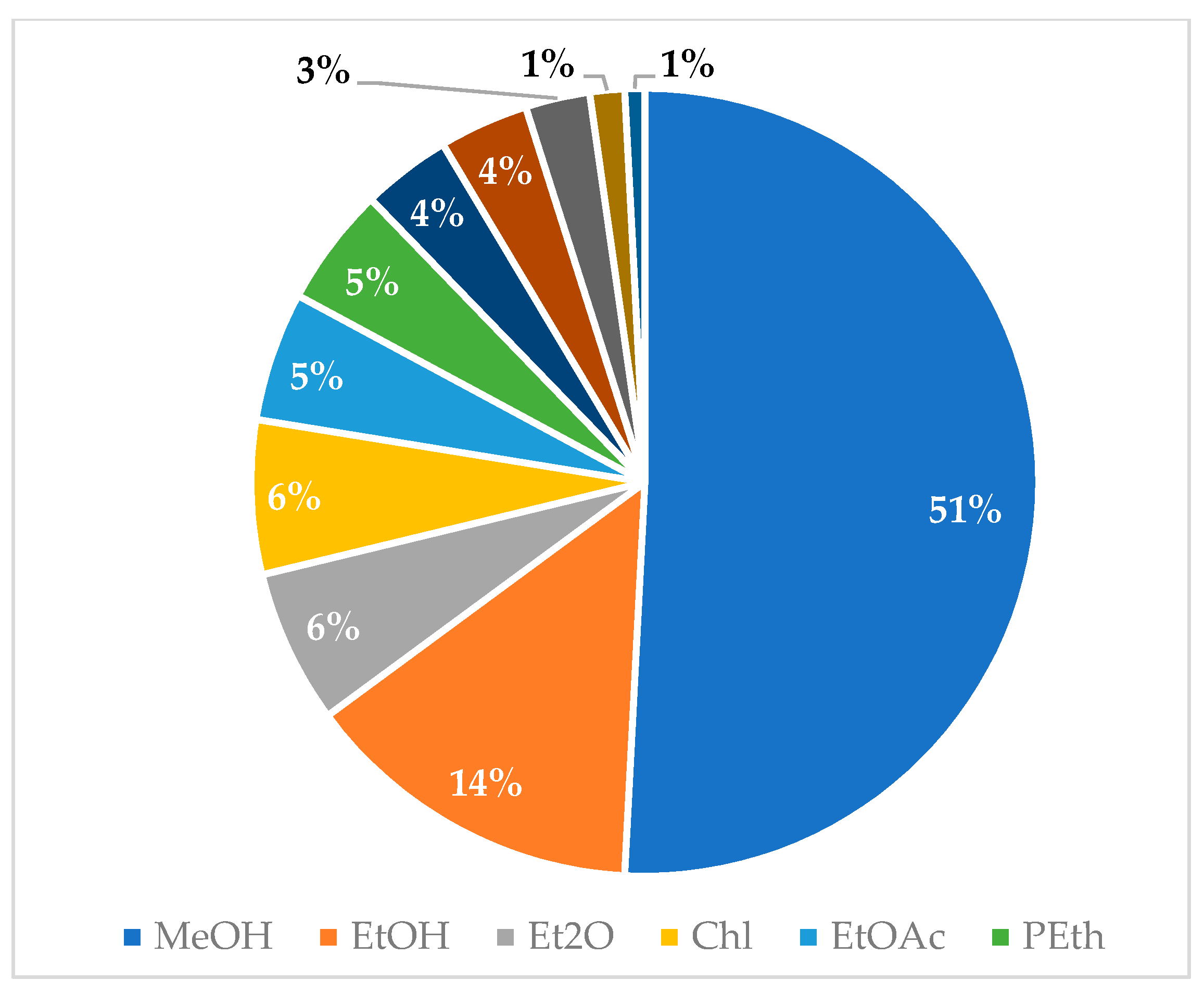
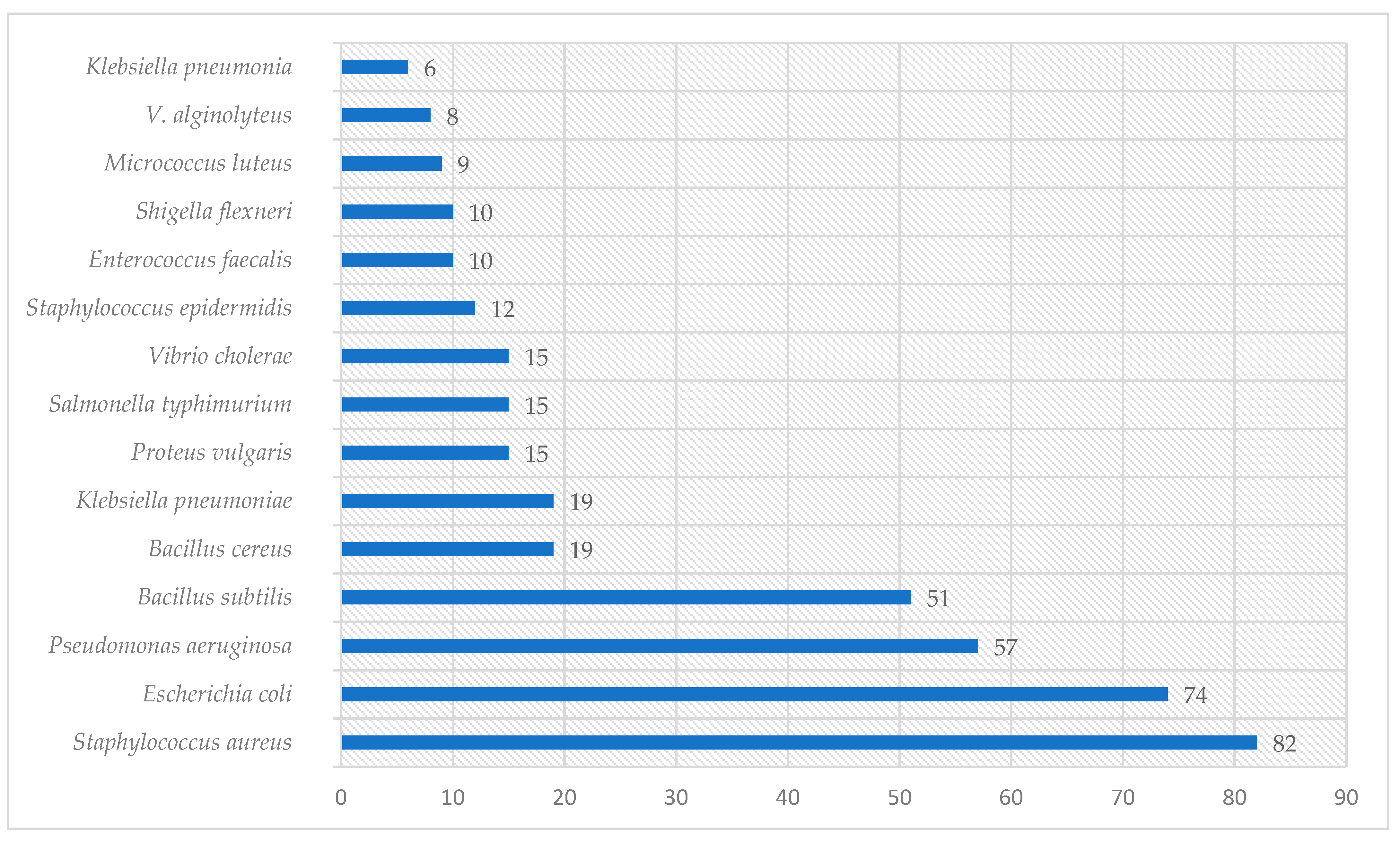
2.3. In Vitro Antibacterial Activity Studies
2.4. In Vitro Antifungal, Antiviral, and Antiprotozoal Activity
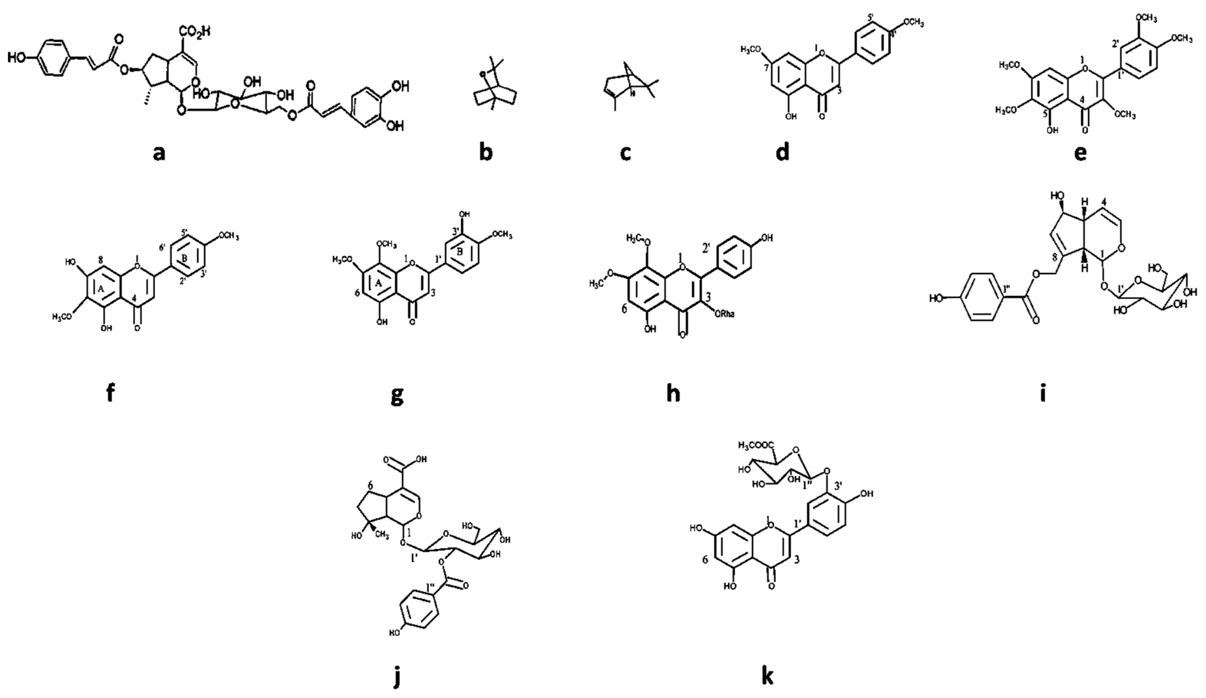
2.5. Characteristic Vitex Secondary Metabolites with Antimicrobial Activity
3. Materials and Methods
3.1. Search Strategy
3.2. Data Inclusion and Exclusion Criteria
3.2.1. Inclusion Criteria
- -
- Related to the Vitex genus.
- -
- Abstract or full text in English.
- -
- Studies on Vitex species concerning antimicrobial activity.
3.2.2. Exclusion Criteria
- -
- Duplicate scientific publications.
- -
- Not directly related to medicinal issues.
- -
- Containing irrelevant or incomplete information.
4. General Discussion
5. Conclusions
Author Contributions
Funding
Institutional Review Board Statement
Informed Consent Statement
Data Availability Statement
Conflicts of Interest
References
- Khameneh, B.; Diab, R.; Ghazvini, K.; Fazly Bazzaz, B.S. Breakthroughs in Bacterial Resistance Mechanisms and the Potential Ways to Combat Them. Microb. Pathog. 2016, 95, 32–42. [Google Scholar] [CrossRef]
- Pulingam, T.; Parumasivam, T.; Gazzali, A.M.; Sulaiman, A.M.; Chee, J.Y.; Lakshmanan, M.; Chin, C.F.; Sudesh, K. Antimicrobial Resistance: Prevalence, Economic Burden, Mechanisms of Resistance and Strategies to Overcome. Eur. J. Pharm. Sci. 2022, 170, 106103. [Google Scholar] [CrossRef]
- Guo, Y.; Song, G.; Sun, M.; Wang, J.; Wang, Y. Prevalence and Therapies of Antibiotic-Resistance in Staphylococcus aureus. Front. Cell. Infect. Microbiol. 2020, 10, 107. [Google Scholar] [CrossRef]
- Adu, F.; Boakye, Y.D.; Agyare, C.; Sam, G.H.; Boamah, V.E.; Osei, F.B. Antibacterial Resistance Modulatory Properties of Selected Medicinal Plants from Ghana. Afr. J. Pharm. Pharmacol. 2019, 13, 57–69. [Google Scholar] [CrossRef]
- Chambers, C.S.; Viktorová, J.; Řehořová, K.; Biedermann, D.; Turková, L.; Macek, T.; Křen, V.; Valentová, K. Defying Multidrug Resistance! Modulation of Related Transporters by Flavonoids and Flavonolignans. J. Agric. Food Chem. 2020, 68, 1763–1779. [Google Scholar] [CrossRef]
- Ganapaty, S.; Vidyadhar, K.N. Phytoconstituents and Biological Activities of Vitex—A Review. J. Nat. Remedies 2005, 5, 75–95. [Google Scholar]
- Bello, M.O.; Zaki, A.A.; Aloko, S.; Fasinu, P.S.; Bello, E.O.; Ajao, U.L.; Oguntoye, O.S. The Genus Vitex: An Overview of Iridoids as Chemotaxonomic Marker. Beni-Suef Univ. J. Basic Appl. Sci. 2018, 7, 414–419. [Google Scholar] [CrossRef]
- Li, B.; Cantino, P.D.; Olmstead, R.G.; Bramley, G.L.C.; Xiang, C.-L.; Ma, Z.-H.; Tan, Y.-H.; Zhang, D.-X. A Large-Scale Chloroplast Phylogeny of the Lamiaceae Sheds New Light on Its Subfamilial Classification. Sci. Rep. 2016, 6, 34343. [Google Scholar] [CrossRef] [PubMed]
- Whitten, T.; Soeriaatmadja, R.E.; Afiff, S.A. The Ecology of Indonesia Series Volume II: The Ecology of Java and Bali; Periplus: Hongkong, China, 1996. [Google Scholar]
- Chopra, R.N. 1882-Glossary of Indian Medicinal Plants; Council of Scientific & Industrial Research: New Delhi, India, 1956. [Google Scholar]
- Katiraee, F.; Mahmoudi, R.; Tahapour, K.; Hamidian, G.; Emami, S.J. Biological Properties of Vitex agnus-castus Essential Oil (Phytochemical Component, Antioxidant and Antifungal Activity). Biotechnol. Health Sci. 2015, 2, 26797. [Google Scholar] [CrossRef]
- Abdolahi, F.; Azadbakht, M.; Shabankhani, B.; Rezaie Abhari, F.; Moslemizadeh, N. Effect of Aqueous Glycyrrhza Globra Extract on Menopausal Symptoms. J. Maz. Univ. Med. Sci. 2007, 16, 75–82. [Google Scholar]
- Odenthal, K.P. Vitex agnus-castusVitex agnus-castus L.,—Traditional Drug and Actual Indications. Phytother. Res. 1998, 12, S160–S161. [Google Scholar] [CrossRef]
- Amaning Danquah, C.; Minkah, P.A.B.; Osei Duah Junior, I.; Amankwah, K.B.; Somuah, S.O. Antimicrobial Compounds from Microorganisms. Antibiotics 2022, 11, 285. [Google Scholar] [CrossRef]
- Meena, A.K.; Niranjan, U.S.; Rao, M.M.; Padhi, M.M.; Babu, R. A Review of the Important Chemical Constituents and Medicinal Uses of Vitex Genus. Asian J. Tradit. Med. 2011, 6, 54–60. [Google Scholar]
- Meena, A.K.; Perumal, A.; Kumar, N.; Singh, R.; Ilavarasan, R.; Srikanth, N.; Dhiman, K.S. Studies on Physicochemical, Phytochemicals, Chromatographic Profiling & Estimation and in-Silico Study of Negundoside in Roots & Small Branches of Vitex Negundo Plant. Phytomed. Plus 2022, 2, 100205. [Google Scholar] [CrossRef]
- Das, N.; Salgueiro, A.C.F.; Choudhury, D.R.; Mandal, S.K.; Logesh, R.; Hassan, M.M.; Devkota, H.P. Traditional Uses, Phytochemistry, and Pharmacology of Genus Vitex (Lamiaceae). Phytother. Res. 2022, 36, 571–671. [Google Scholar] [CrossRef]
- Azadbakht, M.; Baheddini, A.; Shorideh, S.M.; Naserzadeh, A. Effect of Vitex Agnus—Castus L. Leaf and Fruit Flavonoidal Extracts on Serum Prolactin Concentration. J. Med. Plants 2005, 4, 56–61. [Google Scholar]
- Dugoua, J.-J.; Seely, D.; Perri, D.; Koren, G.; Mills, E. Safety and Efficacy of Chastetree (Vitex agnus-castus) During Pregnancy and Lactation. J. Popul. Ther. Clin. Pharmacol. 2008, 15, 74–79. [Google Scholar]
- Atmaca, M.; Kumru, S.; Tezcan, E. Fluoxetine versus Vitex agnus-castusVitex agnus-castus Extract in the Treatment of Premenstrual Dysphoric Disorder. Hum. Psychopharmacol. Clin. Exp. 2003, 18, 191–195. [Google Scholar] [CrossRef] [PubMed]
- Saeed AL-Wajeeh, N.; Halabi, M.F.; Hajrezaie, M.; Dhiyaaldeen, S.M.; Abdulaziz Bardi, D.; Salama, S.M.; Rouhollahi, E.; Karimian, H.; Abdolmalaki, R.; Azizan, A.H.S.; et al. The Gastroprotective Effect of Vitex pubescens Leaf Extract against Ethanol-Provoked Gastric Mucosal Damage in Sprague-Dawley Rats. PLoS ONE 2016, 11, e0157431. [Google Scholar] [CrossRef] [PubMed]
- Goncalves, R.; Ayres, V.F.; Carvalho, C.E.; Souza, M.G.; Guimaraes, A.C.; Correa, G.M.; Martins, C.H.; Takeara, R.; Silva, E.O.; Crotti, A.E. Chemical Composition and Antibacterial Activity of the Essential Oil of Vitex agnus-castus L. (Lamiaceae). An. Acad. Bras. Ciênc. 2017, 89, 2825–2832. [Google Scholar] [CrossRef] [PubMed]
- Goh, M.P.Y.; Basri, A.M.; Yasin, H.; Taha, H.; Ahmad, N. Ethnobotanical Review and Pharmacological Properties of Selected Medicinal Plants in Brunei Darussalam: Litsea elliptica, Dillenia suffruticosa, Dillenia excelsa, Aidia racemosa, Vitex pinnata and Senna alata. Asian Pac. J. Trop. Biomed. 2017, 7, 173–180. [Google Scholar] [CrossRef]
- Ladda, P.; Magdum, C. Vitex negundo Linn.: Ethnobotany, Phytochemistry and Pharmacology—A Review. Int. J. Adv. Pharm. Biol. Chem. 2012, 1, 111–120. [Google Scholar]
- Kilani, A.M. Antibacterial Assessment of Whole Stem Bark of Vitex doniana against Some Enterobactriaceae. Afr. J. Biotechnol. 2006, 5. [Google Scholar] [CrossRef]
- Vishwanathan, A.S.; Basavaraju, R. A Review on Vitex negundo L.—A Medicinally Important Plant. Eur. J. Biol. Sci. 2010, 3, 30–42. [Google Scholar]
- Chhabra, G.S.; Kulkarni, K.S. Vitex agnus-castus—An Overview. J. Nat. Remedies 2011, 11, 90–97. [Google Scholar] [CrossRef]
- Pereira, E.J.P.; do Vale, J.P.C.; da Silva, P.T.; Lima, J.d.R.; Alves, D.R.; Costa, P.S.; Rodrigues, T.H.S.; de Menezes, J.E.S.A.; de Morais, S.M.; Bandeira, P.N. Circadian Rhythm, and Antimicrobial and Anticholinesterase Activities of Essential Oils from Vitex gardneriana. Nat. Prod. Commun. 2018, 13, 1934578X1801300528. [Google Scholar] [CrossRef]
- Hernández, M.M.; Heraso, C.; Villarreal, M.L.; Vargas-Arispuro, I.; Aranda, E. Biological Activities of Crude Plant Extracts from Vitex trifolia L. (Verbenaceae). J. Ethnopharmacol. 1999, 67, 37–44. [Google Scholar] [CrossRef] [PubMed]
- Tandon, V.R.; Khajuria, V.; Kapoor, B.; Kour, D.; Gupta, S. Hepatoprotective Activity of Vitex negundo Leaf Extract against Anti-Tubercular Drugs Induced Hepatotoxicity. Fitoterapia 2008, 79, 533–538. [Google Scholar] [CrossRef] [PubMed]
- Tasduq, S.A.; Kaiser, P.J.; Gupta, B.D.; Gupta, V.K.; Johri, R.K. Negundoside, an Irridiod Glycoside from Leaves of Vitex negundo, Protects Human Liver Cells against Calcium-Mediated Toxicity Induced by Carbon Tetrachloride. World J. Gastroenterol. 2008, 14, 3693–3709. [Google Scholar] [CrossRef] [PubMed]
- Mahmud, S.; Shareef, H.; Farrukh, U.; Kamil, A.; Rizwani, G.H. Antifungal Activities of Vitex negundo Linn. Pak. J. Bot. 2009, 41, 1941–1943. [Google Scholar]
- Sujanapal, P.; Sankaran, K.V. Common Plants of Maldives. Bangk. Food Agric. Organ. U. N. Kerala For. Res. Inst. 2016, 258, 9789251092958. [Google Scholar]
- Avadhoot, Y.; Rana, A.C. Hepatoprotective Effect of Vitex negundo against Carbon Tetrachloride-Induced Liver Damage. Arch. Pharm. Res. 1991, 14, 96–98. [Google Scholar] [CrossRef] [PubMed]
- Joshi, A.R.; Joshi, K. Indigenous Knowledge and Uses of Medicinal Plants by Local Communities of the Kali Gandaki Watershed Area, Nepal. J. Ethnopharmacol. 2000, 73, 175–183. [Google Scholar] [CrossRef] [PubMed]
- Au, D.T.; Wu, J.; Jiang, Z.; Chen, H.; Lu, G.; Zhao, Z. Ethnobotanical Study of Medicinal Plants Used by Hakka in Guangdong, China. J. Ethnopharmacol. 2008, 117, 41–50. [Google Scholar] [CrossRef] [PubMed]
- Rajadurai, M.; Vidhya, V.G.; Ramya, M.; Bhaskar, A. Ethno-Medicinal Plants Used by the Traditional Healers of Pachamalai Hills, Tamilnadu, India. Stud. Ethno Med. 2009, 3, 39–41. [Google Scholar] [CrossRef]
- Rana, S.; Rana, K.K. Review on Medicinal Usefulness of Vitex negundo Linn. OALib 2014, 1, 1–13. [Google Scholar] [CrossRef]
- Saikia, A.P.; Ryakala, V.K.; Sharma, P.; Goswami, P.; Bora, U. Ethnobotany of Medicinal Plants Used by Assamese People for Various Skin Ailments and Cosmetics. J. Ethnopharmacol. 2006, 106, 149–157. [Google Scholar] [CrossRef] [PubMed]
- Kirtikar, K.R. Indian Medicinal Plants: By K.R. Kirtikar, B.D. Basu, and An I.C.S. In 4 Volumes, 2nd ed.; Blatter, E., Caius, J.F., Mhaskar, K.S., Eds.; Lalit Mohan Basu: Allahabad, India, 1935. [Google Scholar]
- Graham, J.G.; Quinn, M.L.; Fabricant, D.S.; Farnsworth, N.R. Plants Used against Cancer—An Extension of the Work of Jonathan Hartwell. J. Ethnopharmacol. 2000, 73, 347–377. [Google Scholar] [CrossRef]
- Ullah, Z.; Ullah, R.; Shah, A.-H.A.; Ahmad, I.; Haider, S. Phytochemical and Biological Evaluation of Vitex negundo Linn: A Review. Int. J. Pharm. Sci. Res. 2012, 3, 2421. [Google Scholar]
- Naik, M.R.; Venugopalan, V.; Kumaravelayutham, P.; Krishnamurthy, Y.L. Ethnoveterinary Uses of Medicinal Plants among the Lambani Community in Chitradurga District, Karnataka, India. Asian Pac. J. Trop. Biomed. 2012, 2, S470–S476. [Google Scholar] [CrossRef]
- Chen, J.; Fan, C.-L.; Wang, Y.; Ye, W.-C. A New Triterpenoid Glycoside from Vitex negundo. Chin. J. Nat. Med. 2014, 12, 218–221. [Google Scholar] [CrossRef] [PubMed]
- Yao, J.-L.; Fang, S.-M.; Liu, R.; Oppong, M.; Liu, E.-W.; Fan, G.-W.; Zhang, H. A Review on the Terpenes from Genus Vitex. Molecules 2016, 21, 1179. [Google Scholar] [CrossRef] [PubMed]
- Zabihullah, Q.; Rashid, A.; Akhtar, N. Ethnobotanical Survey of Kot Manzary Baba Valley, Malakand Agency, Pakistan’. Pak. J. Pl. Sci. 2006, 12, 115–121. [Google Scholar]
- Katare, A.K.; Singh, B.; Kumar, S.; Roy, S.; Gupta, A.P.; Kumar, A.; Singh, B.; Tabassum, A.; Sharma, A.K. Optimisation of Extraction Process for Negundoside and Agnuside from Vitex negundo L. Leaves Using Soxhlet Extraction, HPLC–MS/MS, and CCD-RSM Methods. Chem. Afr. 2022, 5, 907–915. [Google Scholar] [CrossRef]
- Kamruzzaman, M.; Bari, S.M.N.; Faruque, S.M. In Vitro and In Vivo Bactericidal Activity of Vitex negundo Leaf Extract against Diverse Multidrug Resistant Enteric Bacterial Pathogens. Asian Pac. J. Trop. Med. 2013, 6, 352–359. [Google Scholar] [CrossRef]
- Koirala, N.; Dhakal, C.; Munankarmi, N.N.; Ali, S.W.; Hameed, A.; Martins, N.; Sharifi-Rad, J.; Imran, M.; Arif, A.M.; Hanif, M.S.; et al. Vitex negundo Linn.: Phytochemical Composition, Nutritional Analysis, and Antioxidant and Antimicrobial Activity. Cell. Mol. Biol. 2020, 66, 1–7. [Google Scholar] [CrossRef] [PubMed]
- Nyiligira, E.; Viljoen, A.M.; Van Heerden, F.R.; Van Zyl, R.L.; Van Vuuren, S.F.; Steenkamp, P.A. Phytochemistry and In Vitro Pharmacological Activities of South African Vitex (Verbenaceae) Species. J. Ethnopharmacol. 2008, 119, 680–685. [Google Scholar] [CrossRef]
- Rani, A.; Sharma, A. The Genus Vitex: A Review. Pharmacogn. Rev. 2013, 7, 188. [Google Scholar] [CrossRef]
- Suksamrarn, A.; Kumpun, S.; Kirtikara, K.; Yingyongnarongkul, B.; Suksamrarn, S. Iridoids with Anti-Inflammatory Activity from Vitex peduncularis. Planta Med. 2002, 68, 72–73. [Google Scholar] [CrossRef]
- Vaughan, J.C.S. A Preliminary Note on the Use of Vitex peduncularis in Malarial Fever and in Blackwater Fever. Br. Med. J. 1921, 1, 186. [Google Scholar] [CrossRef]
- Batubara, I.; Mitsunaga, T.; Ohashi, H. Screening Antiacne Potency of Indonesian Medicinal Plants: Antibacterial, Lipase Inhibition, and Antioxidant Activities. J. Wood Sci. 2009, 55, 230–235. [Google Scholar] [CrossRef]
- Ata, A.; Mbong, N.; Iverson, C.D.; Samarasekera, R. Minor Chemical Constituents of Vitex pinnata. Nat. Prod. Commun. 2009, 4, 1934578X0900400. [Google Scholar] [CrossRef]
- Oramahi, H.A.; Yoshimura, T. Antifungal and Antitermitic Activities of Wood Vinegar from Vitex pubescens Vahl. J. Wood Sci. 2013, 59, 344–350. [Google Scholar] [CrossRef]
- Anwar, L.; Efdi, M.; Ninomiya, M.; Ibrahim, S.; Putra, D.P.; Tanaka, K.; Koketsu, M. Labdane Diterpene Lactones of Vitex pubescens and Their Antileukemic Properties. Med. Chem. Res. 2017, 26, 2357–2362. [Google Scholar] [CrossRef]
- Leitão, S.G.; Delle Monache, F. 2″-O-Caffeoylorientin from Vitex polygama. Phytochemistry 1998, 49, 2167–2169. [Google Scholar] [CrossRef]
- Ahmadvand, H.; Hamadani, S.E.; Bagheri, S.; Moradi, H.; Khoramabadi, R.M.R.; Khosravi, P.; Cheraghi, A.; Cheraghi, R.A. Chemical Composition of Vitex pseudo-negundo Leavese Ssential Oil. J. Chem. Pharma. Res. 2014, 11, 300–304. [Google Scholar]
- Yan, C.-X.; Wei, Y.-W.; Li, H.; Xu, K.; Zhai, R.-X.; Meng, D.-C.; Fu, X.-J.; Ren, X. Vitex Rotundifolia L. f. and Vitex trifolia L.: A Review on Their Traditional Medicine, Phytochemistry, Pharmacology. J. Ethnopharmacol. 2023, 308, 116273. [Google Scholar] [CrossRef] [PubMed]
- Ahmed, R.; Anis, M. Rapid In Vitro Propagation System through Shoot Tip Cultures of Vitex trifolia L.—An Important Multipurpose Plant of the Pacific Traditional Medicine. Physiol. Mol. Biol. Plants 2014, 20, 385–392. [Google Scholar] [CrossRef]
- Xavier, T.F.; Kannan, M.; Lija, L.; Auxillia, A.; Rose, A.K.F. Ethnobotanical Study of Kani Tribes in Thoduhills of Kerala, South India. J. Ethnopharmacol. 2014, 152, 78–90. [Google Scholar] [CrossRef]
- Ong, H.G.; Kim, Y.-D. Quantitative Ethnobotanical Study of the Medicinal Plants Used by the Ati Negrito Indigenous Group in Guimaras Island, Philippines. J. Ethnopharmacol. 2014, 157, 228–242. [Google Scholar] [CrossRef]
- Weiner, M.A. Secrets of Fijian Medicine; Ministry of Health: Wellington, New Zealand, 1984. [Google Scholar]
- Whistler, W.A. Polynesian Herbal Medicine; National Tropical Botanical Garden: Lawai, HI, USA, 1992. [Google Scholar]
- Boiteau, P.; Allorge, L. Plantes Médicinales de Madagascar; Lune Rouge: Ibiza, Spain, 1993. [Google Scholar]
- Panthong, A.; Kanjanapothi, D.; Taylor, W.C. Ethnobotanical Review of Medicinal Plants from Thai Traditional Books, Part I: Plants with Anti-Inflammatory, Anti-Asthmatic and Antihypertensive Properties. J. Ethnopharmacol. 1986, 18, 213–228. [Google Scholar] [CrossRef] [PubMed]
- Ghani, S.; Rafiee, B.; Bahrami, S.; Mokhtari, A.; Aghamiri, S.; Yarian, F. Green Synthesis of Silver Nanoparticles Using the Plant Extracts of Vitex agnus-castusVitex agnus-castus L: An Ecofriendly Approach to Overcome Antibiotic Resistance. Int. J. Prev. Med. 2022, 13, 13–133. [Google Scholar] [CrossRef]
- Badawy, M.E.I.; Abdelgaleil, S.A.M. Composition and Antimicrobial Activity of Essential Oils Isolated from Egyptian Plants against Plant Pathogenic Bacteria and Fungi. Ind. Crops Prod. 2014, 52, 776–782. [Google Scholar] [CrossRef]
- Zhelev, I.; Petkova, Z.; Kostova, I.; Damyanova, S.; Stoyanova, A.; Dimitrova-Dyulgerova, I.; Antova, G.; Ercisli, S.; Assouguem, A.; Kara, M.; et al. Chemical Composition and Antimicrobial Activity of Essential Oil of Fruits from Vitex agnus-castus L., Growing in Two Regions in Bulgaria. Plants 2022, 11, 896. [Google Scholar] [CrossRef] [PubMed]
- Bakr, R.O.; Zaghloul, S.S.; Hassan, R.A.; Sonousi, A.; Wasfi, R.; Fayed, M.A. Antimicrobial Activity of Vitex agnus-castus Essential Oil and Molecular Docking Study of Its Major Constituents. J. Essent. Oil Bear. Plants 2020, 23, 184–193. [Google Scholar] [CrossRef]
- Balpınar, N.; Ökmen, G.; Vurkun, M. Antibacterial and Antioxidant Activities of Vitex agnus-castus L. Against Mastitis Pathogens. Fresenius Environ. Bull. 2019, 28, 278. [Google Scholar]
- Bayraktar, O.; Altıok, E.; Yılmazer, Ö.; Rusçuklu, D.; Büyüköz, M. Antioxidant, Antimicrobial and Cytotoxic Activities of Extracts from Some Selected Mediterranean Shrub Species (Maquis). Biointerface Res. Appl. Chem. 2016, 6, 1437–1444. [Google Scholar]
- Berrani, A.; Marmouzi, I.; Bouyahya, A.; Kharbach, M.; El Hamdani, M.; El Jemli, M.; Lrhorfi, A.; Zouarhi, M.; Faouzi, M.E.A.; Bengueddour, R. Phenolic Compound Analysis and Pharmacological Screening of Vitex agnus-castus Functional Parts. BioMed Res. Int. 2021, 2021, e6695311. [Google Scholar] [CrossRef]
- Eryigit, T.; Çig, A.; Okut, N.; Yildirim, B.; Ekici, K. Evaluation of Chemical Composition and Antimicrobial Activity of Vitex agnus-castusVitex agnus-castus L. Fruits’ Essential Oils from West Anatolia, Turkey. J. Essent. Oil Bear. Plants 2015, 18, 208–214. [Google Scholar] [CrossRef]
- Kadak, A.E.; Salem, M.O.A. Antibacterial Activity of Chitosan, Some Plant Seed Extracts and Oils against Pathogenic Organisms Escherichia coli and Staphylococcus aureus. Alinteri J. Agric. Sci. 2020, 35, 144–150. [Google Scholar] [CrossRef]
- Khoury, M.; Stien, D.; Eparvier, V.; Ouaini, N.; El Beyrouthy, M. Report on the Medicinal Use of Eleven Lamiaceae Species in Lebanon and Rationalization of Their Antimicrobial Potential by Examination of the Chemical Composition and Antimicrobial Activity of Their Essential Oils. Evid. Based Complement. Alternat. Med. 2016, 2016, 2547169. [Google Scholar] [CrossRef]
- Stojković, D.; Soković, M.; Glamočlija, J.; Džamić, A.; Ćirić, A.; Ristić, M.; Grubišić, D. Chemical Composition and Antimicrobial Activity of Vitex agnus-castus L. Fruits and Leaves Essential Oils. Food Chem. 2011, 128, 1017–1022. [Google Scholar] [CrossRef]
- Tanhaeian, A.; Sekhavati, M.H.; Moghaddam, M. Antimicrobial Activity of Some Plant Essential Oils and an Antimicrobial-Peptide against Some Clinically Isolated Pathogens. Chem. Biol. Technol. Agric. 2020, 7, 13. [Google Scholar] [CrossRef]
- Ulukanli, Z.; Çenet, M.; Öztürk, B.; Bozok, F.; Karabörklü, S.; Demirci, S.C. Chemical Characterization, Phytotoxic, Antimicrobial and Insecticidal Activities of Vitex agnus-castus’ Essential Oil from East Mediterranean Region. J. Essent. Oil Bear. Plants 2015, 18, 1500–1507. [Google Scholar] [CrossRef]
- Zazharskyi, V.V.; Davydenko, P.; Kulishenko, O.; Borovik, I.V.; Zazharska, N.M.; Brygadyrenko, V.V. Antibacterial and Fungicidal Activities of Ethanol Extracts of 38 Species of Plants. Biosyst. Divers. 2020, 28, 281–289. [Google Scholar] [CrossRef]
- Kannathasan, K.; Senthilkumar, A.; Venkatesalu, V. In Vitro Antibacterial Potential of Some Vitex Species against Human Pathogenic Bacteria. Asian Pac. J. Trop. Med. 2011, 4, 645–648. [Google Scholar] [CrossRef]
- Bunu, M.I.; Ndinteh, D.T.; Macdonald, J.R.; Langat, M.K.; Isyaka, S.M.; Sadgrove, N.J.; Melnikovova, I.; Fernandez-Cusimamani, E. Ecdysteroids from the Stem Bark of Vitex doniana sweet (Lamiaceae; Ex. Verbenaceae): A Geographically Variable African Medicinal Species. Antibiotics 2021, 10, 937. [Google Scholar] [CrossRef] [PubMed]
- Sonibare, O.O.; Effiong, I.; Oladosu, I.A.; Ekundayo, O. Chemical Constituents and Antimicrobial Activity of the Essential Oil of Vitex doniana sweet (Verbernaceae). J. Essent. Oil Bear. Plants 2009, 12, 185–188. [Google Scholar] [CrossRef]
- Vale, J.P.C.d.; Ribeiro, L.H.d.F.; de Vasconcelos, M.A.; Sá-Firmino, N.C.; Pereira, A.L.; do Nascimento, M.F.; Rodrigues, T.H.S.; da Silva, P.T.; de Sousa, K.C.; da Silva, R.B.; et al. Chemical Composition, Antioxidant, Antimicrobial and Antibiofilm Activities of Vitex gardneriana schauer Leaves’s Essential Oil. Microb. Pathog. 2019, 135, 103608. [Google Scholar] [CrossRef]
- Montes-Avila, J.; López-Angulo, G.; Duarte-de-la-Peña, G.; Díaz-Camacho, S.P.; Osuna-Galindo, V.C.; López-Valenzuela, J.Á.; Delgado-Vargas, F. Antioxidant, Antibacterial, and Antiparasitary Activities of Green Nanoparticles Synthesized Using Water-Soluble Melanins of Fruits. BioNanoScience 2022, 12, 228–240. [Google Scholar] [CrossRef]
- Medina, M.F.E.; Alaba, P.A.; Estrada-Zuñiga, M.E.; Velázquez-Ordoñez, V.; Barbabosa-Pliego, A.; Salem, M.Z.M.; Alonso-Fresán, M.U.; Camacho-Díaz, L.M.; Salem, A.Z.M. Anti-Staphylococcal Properties of Four Plant Extracts against Sensitive and Multi-Resistant Bacterial Strains Isolated from Cattle and Rabbits. Microb. Pathog. 2017, 113, 286–294. [Google Scholar] [CrossRef]
- Uribe-Beltrán, M.d.J.; Ahumada-Santos, Y.P.; Díaz-Camacho, S.P.; Eslava-Campos, C.A.; Reyes-Valenzuela, J.E.; Báez-Flores, M.E.; Osuna-Ramírez, I.; Delgado-Vargas, F. High Prevalence of Multidrug-Resistant Escherichia coli Isolates from Children with and without Diarrhoea and Their Susceptibility to the Antibacterial Activity of Extracts/Fractions of Fruits Native to Mexico. J. Med. Microbiol. 2017, 66, 972–980. [Google Scholar] [CrossRef]
- Dogra, S.; Sharma, M.D.; Tabassum, S.; Mishra, P.; Bhatt, A.K.; Bhuyar, P. Green Biosynthesis of Silver Nanoparticles (Agnps) From Vitex negundo Plant Extract and Its Phytochemical Screening and Antimicrobial Assessment Next to Pathogenic Microbes. J. Microbiol. Biotechnol. Food Sci. 2022, 12, e5993. [Google Scholar] [CrossRef]
- Ahmad, N.; Khan, M.I.; Ahmed, S.; Javed, S.B.; Faisal, M.; Anis, M.; Rehman, S.; Umair, S.M. Change in Total Phenolic Content and Antibacterial Activity in Regenerants of Vitex negundo L. Acta Physiol. Plant. 2013, 35, 791–800. [Google Scholar] [CrossRef]
- Arivudainambi, U.S.E.; Anand, T.D.; Shanmugaiah, V.; Karunakaran, C.; Rajendran, A. Novel Bioactive Metabolites Producing Endophytic Fungus Colletotrichum Gloeosporioides against Multidrug-Resistant Staphylococcus aureus. FEMS Immunol. Med. Microbiol. 2011, 61, 340–345. [Google Scholar] [CrossRef] [PubMed]
- Arumanayagam, S.; Arunmani, M. Antibacterial Activity of Vitex negundo Linn. J. Microbiol. Biotechnol. Food Sci. 2021, 2021, 829–834. [Google Scholar] [CrossRef]
- Balasubramani, S.; Rajendhiran, T.; Moola, A.K.; Diana, R.K.B. Development of Nanoemulsion from Vitex negundo L. Essential Oil and Their Efficacy of Antioxidant, Antimicrobial and Larvicidal Activities (Aedes aegypti L.). Environ. Sci. Pollut. Res. 2017, 24, 15125–15133. [Google Scholar] [CrossRef] [PubMed]
- Islam, S.; Akhtar, M.; Parvez, S.; Alam, J.; Alam, F.M. Antitumor and Antibacterial Activity of a Crude Methanol Leaf Extract of Vitex negundo L. Arch. Biol. Sci. 2013, 65, 229–238. [Google Scholar]
- Perumal Samy, R.; Ignacimuthu, S.; Sen, A. Screening of 34 Indian Medicinal Plants for Antibacterial Properties. J. Ethnopharmacol. 1998, 62, 173–182. [Google Scholar] [CrossRef]
- Sichaem, J.; Nguyen, H.-H.; Nguyen, V.-H.; Mac, D.-H.; Mai, D.-T.; Nguyen, H.-C.; Tran, T.-N.-M.; Pham, N.-K.-T.; Nguyen, H.-H.; Niamnont, N.; et al. A New Labdane-Type Diterpenoid from the Leaves of Vitex negundo L. Nat. Prod. Res. 2021, 35, 2329–2334. [Google Scholar] [CrossRef]
- Mamatha, G.; Sowmya, P.; Madhuri, D.; Mohan Babu, N.; Suresh Kumar, D.; Vijaya Charan, G.; Varaprasad, K.; Madhukar, K. Antimicrobial Cellulose Nanocomposite Films with In Situ Generations of Bimetallic (Ag and Cu) Nanoparticles Using Vitex negundo Leaves Extract. J. Inorg. Organomet. Polym. Mater. 2021, 31, 802–815. [Google Scholar] [CrossRef]
- Khan, A.M.; Qureshi, R.A.; Gillani, S.A.; Ullah, F. Antimicrobial Activity of Selected Medicinal Plants of Margalla Hills, Islamabad, Pakistan. J. Med. Plant Res. 2011, 5, 4665–4670. [Google Scholar]
- Kumar, S.; Singh, A.K.; Verma, S.K.; Misra, R.; Seniya, C. Antibacterial and Phyto-Chemical Analysis of Some Medicinal Plants and Their Efficacy on Multidrug Resistant Bacteria. J. Pure Appl. Microbiol. 2013, 7, 2191–2204. [Google Scholar]
- Mishra, S.; Mekap, S.K.; Patra, S.; Dhal, N.K.; Sahoo, S. Antioxidant and Anti Infective Potential of Oleanolic Acid Acetate Vis-à-Vis Vitex negundo Linn. and Oroxylum Indicum Vent. against Human Pathogens Causing Infections of UT, GIT and Skin. Orient. Pharm. Exp. Med. 2015, 15, 73–82. [Google Scholar] [CrossRef]
- Nagarsekar, K.S.; Nagarsenker, M.S.; Kulkarni, S.R. Evaluation of Composition and Antimicrobial Activity of Supercritical Fluid Extract of Leaves of Vitex negundo. Indian J. Pharm. Sci. 2010, 72, 641–643. [Google Scholar] [CrossRef] [PubMed]
- Naidu, G.K.; Sujatha, B. Screening of Preliminary Phytochemical Analysis and In-Vitro Antimicrobial Activity of Stem Bark Extracts of Vitex negundo L. Res. J. Pharm. Biol. Chem. Sci. 2015, 6, 1001–1006. [Google Scholar]
- Panda, S.K.; Mohanta, Y.K.; Padhi, L.; Park, Y.-H.; Mohanta, T.K.; Bae, H. Large Scale Screening of Ethnomedicinal Plants for Identification of Potential Antibacterial Compounds. Molecules 2016, 21, 293. [Google Scholar] [CrossRef]
- Panda, S.K.; Thatoi, H.N.; Dutta, S.K. Antibacterial Activity and Phytochemical Screening of Leaf and Bark Extracts of Vitex negundo L. from Similipal Biosphere Reserve, Orissa. J. Med. Plant Res. 2009, 3, 294–300. [Google Scholar]
- Prabhu, N.; Raj, D.T.; Yamuna, G.K.; Ayisha, S.S.; Joseph Puspha, I.D. Synthesis of Silver Phyto Nanoparticles and Their Antibacterial Efficacy. Dig. J. Nanomater. Biostructures DJNB 2010, 5, 185–189. [Google Scholar]
- Prakash, S.; Ramasubburayan, R.; Ramkumar, V.S.; Kannapiran, E.; Palavesam, A.; Immanuel, G. In Vitro—Scientific Evaluation on Antimicrobial, Antioxidant, Cytotoxic Properties and Phytochemical Constituents of Traditional Coastal Medicinal Plants. Biomed. Pharmacother. 2016, 83, 648–657. [Google Scholar] [CrossRef] [PubMed]
- Sathiamoorthy, B.; Gupta, P.; Kumar, M.; Chaturvedi, A.K.; Shukla, P.K.; Maurya, R. New Antifungal Flavonoid Glycoside from Vitex negundo. Bioorg. Med. Chem. Lett. 2007, 17, 239–242. [Google Scholar] [CrossRef]
- Sharma, A.; Tyagi, S.; Nag, R.; Chaturvedi, A.; Nag, T.N. Antimicrobial Activity and Cellular Toxicity of Flavonoid Extracts from Pongamia pinnata and Vitex negundo. Rom. Biotechnol. Lett. 2011, 16, 6396–6400. [Google Scholar]
- Sharma, K.; Guleria, S.; Razdan, V.K.; Babu, V. Synergistic Antioxidant and Antimicrobial Activities of Essential Oils of Some Selected Medicinal Plants in Combination and with Synthetic Compounds. Ind. Crops Prod. 2020, 154, 112569. [Google Scholar] [CrossRef]
- Sumathi, P.; Parvathi, A. Antimicrobial Activity of Some Traditional Medicinal Plants. J. Med. Plants Res. 2010, 4, 316–321. [Google Scholar]
- Zargar, M.; Hamid, A.A.; Bakar, F.A.; Shamsudin, M.N.; Shameli, K.; Jahanshiri, F.; Farahani, F. Green Synthesis and Antibacterial Effect of Silver Nanoparticles Using Vitex negundo L. Molecules 2011, 16, 6667–6676. [Google Scholar] [CrossRef] [PubMed]
- Nuraskin, C.A.; Marlina, M.; Idroes, R.; Soraya, C.; Djufri, D. Antibacterial Activity Tests of N-Hexane, Ethyl Acetate, and Methanol Leaves (Vitex) Extract (Pinnata) against Streptococcus mutans. Open Access Maced. J. Med. Sci. 2020, 8, 181–184. [Google Scholar] [CrossRef]
- Shafie, N.A.; Suhaili, N.A.; Taha, H.; Ahmad, N. Evaluation of Antioxidant, Antibacterial and Wound Healing Activities of Vitex pinnata. F1000Research 2020, 9, 187. [Google Scholar] [CrossRef] [PubMed]
- Nyiligira, E.; Viljoen, A.M.; Başer, K.H.C.; Õzek, T.; van Vuuren, S.F.; Houghton, P.J. Essential Oil Composition and In Vitro Antimicrobial and Anti-Inflammatory Activity of South African Vitex Species. South Afr. J. Bot. 2004, 70, 611–617. [Google Scholar] [CrossRef]
- Hashemi, S.M.B.; Kamani, M.H.; Amani, H.; Mousavi Khaneghah, A. Voltage and NaCl Concentration on Extraction of Essential Oil from Vitex pseudo negundo Using Ohmic-Hydrodistillation. Ind. Crops Prod. 2019, 141, 111734. [Google Scholar] [CrossRef]
- Geetha, V.; Doss, A.; Doss, A.P.A. Antimicrobial Potential of Vitex trifolia Linn. Anc. Sci. Life 2004, 23, 30. [Google Scholar]
- Ali, A.M.; El-Sharhawy, S.H.; Hamid, J.A.; Ismail, N.H.; Lajis, N.H. Antimicrobial Activity of Selected Malaysian Plants. Pertanika J. Trop. Agric. Sci. 1995, 18, 57–62. [Google Scholar]
- Ababutain, I.M.; Alghamdi, A.I. In Vitro Anticandidal Activity and Gas Chromatography-Mass Spectrometry (GC-MS) Screening of Vitex agnus-castus Leaf Extracts. PeerJ 2021, 9, e10561. [Google Scholar] [CrossRef] [PubMed]
- Keikha, N.; Shafaghat, M.; Mousavi, S.M.; Moudi, M.; Keshavarzi, F. Antifungal Effects of Ethanolic and Aqueous Extracts of Vitex agnus-castus against Vaginal Isolates of Candida Albicans. Curr. Med. Mycol. 2018, 4, 30186986. [Google Scholar] [CrossRef] [PubMed]
- Abu-Tahon, M.A.; Mogazy, A.M.; Isaac, G.S. Resistance Assessment and Enzymatic Responses of Common Bean(Phaseolus vulgaris L) against Rhizoctonia solani Damping-off in Response to Seed Presoaking in Vitex agnus-castus L. Oils and Foliar Spray with Zinc Oxide Nanoparticles. South Afr. J. Bot. 2022, 146, 77–89. [Google Scholar] [CrossRef]
- Maltaş, E.; Uysal, A.; Yildiz, S.; Durak, Y. Evaluation of Antioxidant and Antimicrobial Activity of Vitex agnus-castus L. Fresen Env. Bull 2010, 19, 3094–3099. [Google Scholar]
- Mansour, M.M.A.; EL-Hefny, M.; Salem, M.Z.M.; Ali, H.M. The Biofungicide Activity of Some Plant Essential Oils for the Cleaner Production of Model Linen Fibers Similar to Those Used in Ancient Egyptian Mummification. Processes 2020, 8, 79. [Google Scholar] [CrossRef]
- López-Velázquez, J.G.; Delgado-Vargas, F.; Ayón-Reyna, L.E.; López-Angulo, G.; Bautista-Baños, S.; Uriarte-Gastelum, Y.G.; López-López, M.E.; Vega-García, M.O. Postharvest Application of Partitioned Plant Extracts from Sinaloa, Mexico for Controlling Papaya Pathogenic Fungus Colletotrichum Gloeosporioides. J. Plant Pathol. 2021, 103, 831–842. [Google Scholar] [CrossRef]
- Aiyaz, M.; Divakara, S.T.; Chandranayaka, S.; Niranjana, S.R. Efficacy of Seed Hydropriming with Phytoextracts on Plant Growth Promotion and Antifungal Activity in Maize. Int. J. Pest Manag. 2015, 61, 153–160. [Google Scholar] [CrossRef]
- Guleria, S.; Kumar, A. Antifungal Activity of Some Himalayan Medicinal Plants Using Direct Bioautography. J. Cell Mol. Biol. 2006, 5, 95–98. [Google Scholar]
- Panda, S.K.; Behera, B.; Dutta, S.K. Anti-Candidal Activity of Vitex negundo L.: An Ethnomedicinal Plant. J. Pure Appl. Microbiol. 2009, 3, 777–784. [Google Scholar]
- Bello, O.M.; Zaki, A.A.; Khan, S.I.; Fasinu, P.S.; Ali, Z.; Khan, I.A.; Usman, L.A.; Oguntoye, O.S. Assessment of Selected Medicinal Plants Indigenous to West Africa for Antiprotozoal Activity. South Afr. J. Bot. 2017, 113, 200–211. [Google Scholar] [CrossRef]
- Pan, W.; Liu, K.; Guan, Y.; Tan, G.T.; Hung, N.V.; Cuong, N.M.; Soejarto, D.D.; Pezzuto, J.M.; Fong, H.H.S.; Zhang, H. Bioactive Compounds from Vitex leptobotrys. J. Nat. Prod. 2014, 77, 663–667. [Google Scholar] [CrossRef]
- Ban, N.K.; Thoa, N.T.K.; Linh, T.M.; Trang, D.T.; Van Kiem, P.; Nhiem, N.X.; Tai, B.H.; Van Minh, C.; Song, J.-H.; Ko, H.-J.; et al. Labdane-Type Diterpenoids from Vitex limonifolia and Their Antivirus Activities. J. Nat. Med. 2018, 72, 290–297. [Google Scholar] [CrossRef]
- Kannan, M.; Rajendran, P.; Vedha, V.; Ashok, G.; Anushka, S.; Chandran, P.; Nair, R. HIV-1 Reverse Transcriptase Inhibition by Vitex negundo L. Leaf Extract and Quantification of Flavonoids in Relation to Anti-HIV Activity. J. Cell. Mol. Biol. 2012, 10, 53–59. [Google Scholar]
- Gonçalves, J.L.S.; Leitão, S.G.; Monache, F.D.; Miranda, M.M.F.S.; Santos, M.G.M.; Romanos, M.T.V.; Wigg, M.D. In Vitro Antiviral Effect of Flavonoid-Rich Extracts of Vitex polygama (Verbenaceae) against Acyclovir-Resistant Herpes Simplex Virus Type 1. Phytomedicine 2001, 8, 477–480. [Google Scholar] [CrossRef]
- Vimalanathan, S.; Ignacimuthu, S.; Hudson, J.B. Medicinal Plants of Tamil Nadu (Southern India) Are a Rich Source of Antiviral Activities. Pharm. Biol. 2009, 47, 422–429. [Google Scholar] [CrossRef]
- Kuruüzüm-Uz, A.; Ströch, K.; Demirezer, L.Ö.; Zeeck, A. Glucosides from Vitex agnus-castus. Phytochemistry 2003, 63, 959–964. [Google Scholar] [CrossRef] [PubMed]
- Zheng, C.-J.; Lan, X.-P.; Wang, Y.; Huang, B.-K.; Han, T.; Zhang, Q.-Y.; Qin, L.-P. A New Labdane Diterpene from Vitex negundo. Pharm. Biol. 2012, 50, 687–690. [Google Scholar] [CrossRef] [PubMed]
- Gautam, L.N.; Shrestha, S.; Wagle, P.; Tamrakar, B. Chemical Constituents from Vitex negundo Linn. of Nepalese Origin. Sci. World 2010, 6, 27–32. [Google Scholar] [CrossRef]
- Rudrapaul, P.; Sarma, I.S.; Das, N.; De, U.C.; Bhattacharjee, S.; Dinda, B. New Flavonol Methyl Ether from the Leaves of Vitex peduncularis Exhibits Potential Inhibitory Activity against Leishmania Donovani through Activation of iNOS Expression. Eur. J. Med. Chem. 2014, 87, 328–335. [Google Scholar] [CrossRef] [PubMed]
- Amikacin. In Meyler’s Side Effects of Drugs, 6th ed.; Aronson, J.K., Ed.; Elsevier: Oxford, UK, 2016; pp. 207–209. ISBN 978-0-444-53716-4. [Google Scholar]
- Mrugała, B.; Miłaczewska, A.; Porebski, P.J.; Niedzialkowska, E.; Guzik, M.; Minor, W.; Borowski, T. A Study on the Structure, Mechanism, and Biochemistry of Kanamycin B Dioxygenase (KanJ)—An Enzyme with a Broad Range of Substrates. FEBS J. 2021, 288, 1366–1386. [Google Scholar] [CrossRef] [PubMed]
- Scholar, E. Amikacin. In xPharm: The Comprehensive Pharmacology Reference; Enna, S.J., Bylund, D.B., Eds.; Elsevier: New York, NY, USA, 2007; pp. 1–5. ISBN 978-0-08-055232-3. [Google Scholar]
- Danish, P.; Ali, Q.; Hafeez, M.M.; Malik, A. Antifungal and Antibacterial Activity of Aloe vera Plant Extract. Biol. Clin. Sci. Res. J. 2020, 4, 1. [Google Scholar] [CrossRef]
- Bordean, M.-E.; Ungur, R.A.; Toc, D.A.; Borda, I.M.; Marțiș, G.S.; Pop, C.R.; Filip, M.; Vlassa, M.; Nasui, B.A.; Pop, A.; et al. Antibacterial and Phytochemical Screening of Artemisia Species. Antioxidants 2023, 12, 596. [Google Scholar] [CrossRef] [PubMed]


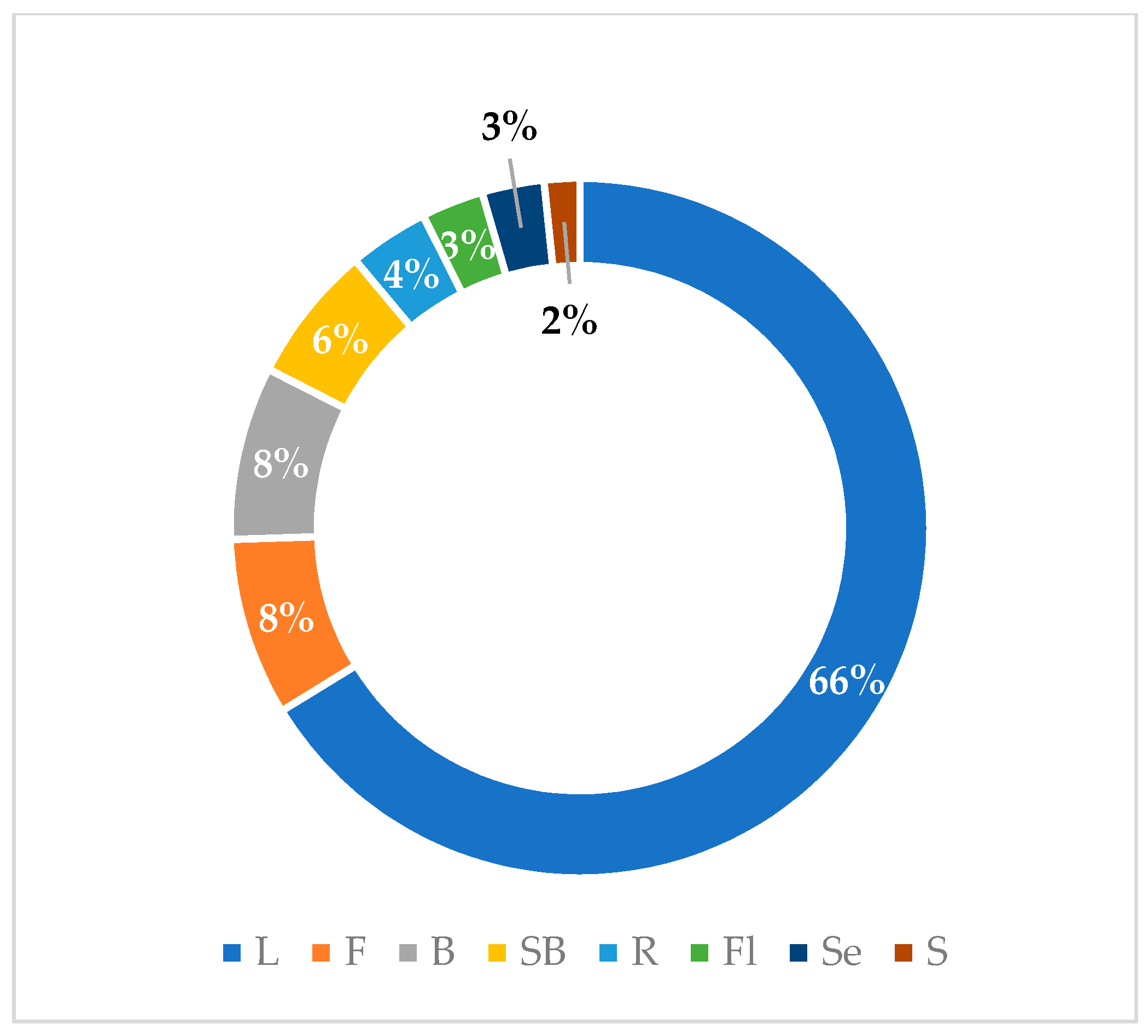
| Species | Part Used | Country | Signals or Symptoms or Pathology | Bib. References |
|---|---|---|---|---|
| V. agnus-castus | L | Iran | increasing breast milk | [18] |
| F | Turkey | corpus luteum insufficiency, hyperprolactinemia, infertility, menstrual disorders, premenstrual dysphoric disorder, menopause disrupted lactation, cyclical gastralgia | [19,20] | |
| L | Brazil | oral disorders; diuretic, antiseptic, digestive, antifungal, anti-anxiety, aphrodisiac, anti-estrus, emmenagogus, antispasmodic, aperitif, and analgesic properties | [22] | |
| L | Brazil | menstrual disorder | [27] | |
| V. doniana | Sb | Nigeria | decoction, gastroenteritis, diarrhea, dysentery | [25] |
| V. gardneriana | L | Brazil | analgesic pain, anti-inflammatory properties | [28] |
| V. mollis | L | Mexico | dysentery, analgesic, anti-inflammatory properties, scorpion stings, diarrhea, stomachache | [29] |
| V. negundo | S, F | India | increasing lactation | [30,31] |
| L | India | post-partum bath | [30,31] | |
| R, B, Fl | India | diarrhea, dysentery, flatulence, indigestion, cholera | [24,32] | |
| L | Maldives | fever | [33] | |
| L | Bangladesh, India, Malaysia | headache | [26,34] | |
| L | China, India, Nepal | cough, sore throat | [35,36,37] | |
| R, L | India | rheumatism | [38] | |
| L | India | hives, cellulitis, carbuncle | [39] | |
| L | India | fever, hearing problems | [24,40] | |
| Fl | Philippines | cancer | [41] | |
| L | Bangladesh | chronic disease, infectious diseases | [42] | |
| L | India | paralysis | [43] | |
| L | China | skin disease | [44] | |
| L | China | coughs, phlegm, asthma | [45] | |
| L | Pakistan | antiallergic properties | [46] | |
| R, Wp | Bangladesh | malaria, fever | [26] | |
| L | China and India | stomachic, antiseptic, depurative, and rejuvenating properties; eye problems; gonorrhea | [47] | |
| L | Bangladesh | diarrhea, dysentery | [48] | |
| L, B | Nepal | jaundice, wounds, body ache, toothache, asthma, eye problems | [49] | |
| V. obovata | L | South Africa | body pain | [50] |
| V. peduncularis | Wp | India | wounds, dysentery, stomach diseases, fever, hypertension | [51] |
| B, L | Bangladesh | joint ache, diabetes | [26] | |
| R | India | eye problems, skin problems, chest pain | [52] | |
| L | India | malaria, fever | [53] | |
| V. pinnata | Wp | Malaysia | hypertension, gastrointestinal disorders | [21] |
| L | Brunei | hypertension, fever | [16] | |
| L, B | Malaysia | fever, gastric ulcer | [54] | |
| Wp | Brunei | jaundice | [55] | |
| L | Brunei | sanitizing | [23] | |
| Wp | Malaysia | dysentery, inflammatory | [56] | |
| Wp | Indonesia | cancer, gastrointestinal disorder, fever, wound, skin tumor | [57] | |
| V. polygama | L, F | Brazil | emmenagogue and diuretic properties. | [58] |
| V. pseudo-negundo | L | Iran | hyperprolactinemia, hormonal imbalance syndromes, breast diseases, infertility | [59] |
| V. rehmannii | L | South Africa | stomach disease | [50] |
| V. rotundifolia | F | China | cold, headache | [60] |
| V. trifolia | L | India | liver disorders, rheumatic pains | [61] |
| L | India | ulcers | [62] | |
| L | Philippines | cough | [63] | |
| F | China | migraines, eye problems | [45] | |
| Fl | Bangladesh | fever, vomiting | [64] | |
| L | Fiji | coughs, gonorrhea, stomach pain | [64] | |
| L | Tongo | infections | [65] | |
| St, L | Madagascar | stomach pain | [66] | |
| Fl | Thailand | asthma | [67] |
| Species | Country | Plant Part Used | Extractive Solvent/Compound | Test Type | Strains | Positive Control | Results | R |
|---|---|---|---|---|---|---|---|---|
| V. agnus-castus castus7 | Iran | F | H2O | DDM | Bacillus cereus PTCC 1015 Escherichia coli PTCC 1399 | gentamicin and ciprofloxacin | IZ 5 mm | [68] |
| Iran | F | H2O | BDM | Escherichia coli PTCC 1399 | gentamicin and ciprofloxacin | MIC 25 µg/mL | [68] | |
| Iran | F | H2O | BDM | Bacillus cereus PTCC 1015 | gentamicin and ciprofloxacin | na | [68] | |
| Iran | F | H2O | BDM | Escherichia coli PTCC 1399 | gentamicin and ciprofloxacin | MIC 12 µg/mL | [68] | |
| Iran | F | H2O | BDM | Bacillus cereus PTCC 1015 | gentamicin and ciprofloxacin | MIC 25 µg/mL | [68] | |
| Egypt | L | Et2O | ADM | Agrobacterium tumefaciens * | na | MIC 575 mg/L | [69] | |
| Egypt | L | Et2O | ADM | Erwinia carotovora var. carotovora * | na | MIC 425 mg/L | [69] | |
| Brazil | L | EtOAc | BMicDM | Streptococcus mutans ATCC 25175 Lactobacillus casei ATCC 11578 | chlorhexidine dihydrochloride | MIC 15.6 µg/mL | [22] | |
| Brazil | L | EtOAc | BMicDM | Streptococcus mitis ATCC 49456 | chlorhexidine dihydrochloride | MIC 31.25 µg/mL | [22] | |
| Brazil | L | EtOAc | BMicDM | Streptococcus subrinus ATCC 33478 | chlorhexidine dihydrochloride | MIC 125 µg/mL | [22] | |
| Brazil | L | EtOAc | BMicDM | Streptococcus salivarius ATCC 25975 | chlorhexidine dihydrochloride | MIC 200 µg/mL | [22] | |
| Bulgaria | F | Et2O | AWDM | Staphylococcus aureus ATCC 6538 | na | IZ 11.25 ± 0.05 mm | [70] | |
| Bulgaria | F | Et2O | AWDM | Bacillus subtilis ATCC 6633 | na | IZ 12.03 ± 0.02 mm | [70] | |
| Bulgaria | F | Et2O | AWDM | Kocuria rhizophila ATCC 9341 | na | IZ 9.37 ± 0.04 mm | [70] | |
| Bulgaria | F | Et2O | AWDM | Escherichia coli ATCC 8739 | na | IZ 8.00 ± 0.0 mm | [70] | |
| Bulgaria | F | Et2O | AWDM | Pseudomonas aeruginosa ATCC 9027 | na | IZ 8.03 ± 0.02 mm | [70] | |
| Bulgaria | F | Et2O | AWDM | Salmonella abony NCTC 6017 | na | IZ 11.15 ± 0.05 mm | [70] | |
| Bulgaria | F | Et2O | AWDM | Saccharomyces cerevisiae ATCC 2601 | na | IZ 11.86 ± 0.03 mm | [70] | |
| Egypt | L | Et2O | DDM | Staphylococcus aureus ATCC 6358 | chloramphenicol | IZ 30 mm | [71] | |
| Egypt | L | Et2O | DDM | Bacillus subtilis ATCC 6633 | chloramphenicol | IZ 10 mm | [71] | |
| Egypt | L | Et2O | DDM | Escherichia coli ATCC 25923 | chloramphenicol | IZ 20 mm | [71] | |
| Egypt | L | Et2O | DDM | Pseudomonas aeruginosa ATCC 27853 | chloramphenicol | IZ 20 mm | [71] | |
| Turkey | Fl | MeOH | DDM | Staphylococcus aureus 17 | ampicillin 10 µg and oxacillin 5 ug | IZ 18 mm | [72] | |
| Turkey | Fl | MeOH | DDM | Staphylococcus aureus 18 | ampicillin 10 µg and oxacillin 5 ug | IZ 12 mm | [72] | |
| Turkey | Fl | EtOH | DDM | Coagulate negative Staphylococci 33 | ampicillin 10 µg and oxacillin 5 ug | IZ 8 mm | [72] | |
| Turkey | Fl | MeOH | DDM | Coagulate negative Staphylococci 33 | ampicillin 10 µg and oxacillin 5 ug | IZ 10 mm | [72] | |
| Turkey | Fl | EtOH | DDM | Coagulate negative Staphylococci 36 | ampicillin 10 µg and oxacillin 5 ug | IZ 9 mm | [72] | |
| Turkey | L | EtOH | DDM | Staphylococcus aureus * | gentamycin | IZ 7.5 mm | [73] | |
| Turkey | F | EtOH | DDM | Staphylococcus aureus * | gentamycin | IZ 10 mm | [73] | |
| Turkey | L | EtOH | DDM | Pseudomonas aeruginosa * | gentamycin | IZ 9 mm | [73] | |
| Morocco | L | MeOH | BMicDM | Bacillus subtilis CIP 5262 | chloramphenicol | MIC 15.62 µg/mL MBC 15.62 µg/mL | [74] | |
| Morocco | R | MeOH | BMicDM | Bacillus subtilis CIP 5262 | chloramphenicol | MIC 31.25 µg/mL MBC 31.25 µg/mL | [74] | |
| Morocco | S | MeOH | BMicDM | Bacillus subtilis CIP 5262 | chloramphenicol | MIC 15.62 µg/mL MBC 15.62 µg/mL | [74] | |
| Morocco | Fl | MeOH | BMicDM | Bacillus subtilis CIP 5262 | chloramphenicol | MIC 15.62 µg/mL MBC 31.25 µg/mL | [74] | |
| Morocco | Se | MeOH | BMicDM | Bacillus subtilis CIP 5262 | chloramphenicol | MIC 15.62 µg/mL MBC 15.62 µg/mL | [74] | |
| Morocco | L | MeOH | BMicDM | Escherichia coli CIP 53126 | chloramphenicol | MIC 15.62 µg/mL MBC 15.62 µg/mL | [74] | |
| Morocco | R | MeOH | BMicDM | Escherichia coli CIP 53126 | chloramphenicol | MIC 15.62 µg/mL MBC 15.62 µg/mL | [74] | |
| Morocco | S | MeOH | BMicDM | Escherichia coli CIP 53126 | chloramphenicol | MIC 15.62 µg/mL MBC 31.25 µg/mL | [74] | |
| Morocco | Fl | MeOH | BMicDM | Escherichia coli CIP 53126 | chloramphenicol | MIC 31.25 µg/mL MBC 31.25 µg/mL | [74] | |
| Morocco | S | MeOH | BMicDM | Escherichia coli CIP 53126 | chloramphenicol | MIC 15.62 µg/mL MBC 15.62 µg/mL | [74] | |
| Morocco | L | MeOH | BMicDM | Pseudomonas aeruginosa CIP 82118 | chloramphenicol | MIC 15.62 µg/mL MBC 15.62 µg/mL | [74] | |
| Morocco | R | MeOH | BMicDM | Pseudomonas aeruginosa CIP 82118 | chloramphenicol | MIC 15.62 µg/mL MBC 15.62 µg/mL | [74] | |
| Morocco | S | MeOH | BMicDM | Pseudomonas aeruginosa CIP 82118 | chloramphenicol | MIC 15.62 µg/mL MBC 15.62 µg/mL | [74] | |
| Morocco | Fl | MeOH | BMicDM | Pseudomonas aeruginosa CIP 82118 | chloramphenicol | MIC 7.81 µg/mL MBC 7.81 µg/mL | [74] | |
| Morocco | Se | MeOH | BMicDM | Pseudomonas aeruginosa CIP 82118 | chloramphenicol | MIC 15.62 µg/mL MBC 15.62 µg/mL | [74] | |
| Morocco | L | MeOH | BMicDM | Salmonella enterica CIP 8039 | chloramphenicol | MIC 7.81 µg/mL MBC 15.62 µg/mL | [74] | |
| Morocco | R | MeOH | BMicDM | Salmonella enterica CIP 8039 | chloramphenicol | MIC 31.25 µg/mL MBC 31.25 µg/mL | [74] | |
| Morocco | S | MeOH | BMicDM | Salmonella enterica CIP 8039 | chloramphenicol | MIC 7.81 µg/mL MBC 15.62 µg/mL | [74] | |
| Morocco | Fl | MeOH | BMicDM | Salmonella enterica CIP 8039 | chloramphenicol | MIC 7.81 µg/mL MBC 15.62 µg/mL | [74] | |
| Morocco | Se | MeOH | BMicDM | Salmonella enterica CIP 8039 | chloramphenicol | MIC 7.81 µg/mL MBC 15.62 µg/mL | [74] | |
| Morocco | L | MeOH | BMicDM | Staphylococcus aureus CIP 483 | chloramphenicol | MIC 15.62 µg/mL MBC 15.62 µg/mL | [74] | |
| Morocco | R | MeOH | BMicDM | Staphylococcus aureus CIP 483 | chloramphenicol | MIC 31.25 µg/mL MBC 31.25 µg/mL | [74] | |
| Morocco | S | MeOH | BMicDM | Staphylococcus aureus CIP 483 | chloramphenicol | MIC 15.62 µg/mL MBC 15.62 µg/mL | [74] | |
| Morocco | Fl | MeOH | BMicDM | Staphylococcus aureus CIP 483 | chloramphenicol | MIC 7.81 µg/mL MBC 15.62 µg/mL | [74] | |
| Morocco | Se | MeOH | BMicDM | Staphylococcus aureus CIP 483 | chloramphenicol | MIC 15.62 µg/mL MBC 15.62 µg/mL | [74] | |
| Turkey | F | MeOH | DDM | Bacillus subtilis ATCC 6051 | ampicillin | IZ 25 mm | [75] | |
| Turkey | F | MeOH | DDM | Bacillus subtilis ATCC 6051 | ofloxacin | IZ 30 mm | [75] | |
| Turkey | F | MeOH | DDM | Escherichia coli ATCC 11775 | ampicillin | IZ 28 mm | [75] | |
| Turkey | F | MeOH | DDM | Escherichia coli ATCC 11775 | ofloxacin | IZ 28 mm | [75] | |
| Turkey | F | MeOH | DDM | Enterococcus faecalis ATCC 292 | ampicillin | IZ 26 mm | [75] | |
| Turkey | F | MeOH | DDM | Enterococcus faecalis ATCC 292 | ofloxacin | IZ 20 mm | [75] | |
| Turkey | F | MeOH | DDM | Pseudomonas aeruginosa ATCC 1014 | ampicillin | IZ 23 mm | [75] | |
| Turkey | F | MeOH | DDM | Pseudomonas aeruginosa ATCC 1014 | ofloxacin | IZ 23 mm | [75] | |
| Turkey | F | MeOH | DDM | Staphylococcus aureus ATCC 12600 | ampicillin | IZ 20 mm | [75] | |
| Turkey | F | MeOH | DDM | Staphylococcus aureus ATCC 12600 | ofloxacin | IZ 26 mm | [75] | |
| Turkey | F | MeOH | DDM | Salmonella typhimurium ATCC 2524 | ampicillin | IZ 22 mm | [75] | |
| Turkey | F | MeOH | DDM | Salmonella typhimurium ATCC 2524 | ofloxacin | IZ 24 mm | [75] | |
| Turkey | Se | MeOH | DDM | Escherichia coli * | erythromycin | IZ 16 mm | [76] | |
| Turkey | Se | MeOH | DDM | Staphylococcus aureus * | erythromycin | IZ 16 mm | [76] | |
| Lebanon | Fl | EtOAc | BMicDM | Escherichia coli ATCC 25922 | oxacillin and gentamicin | MIC 512 µg/mL | [77] | |
| Lebanon | Fl | EtOAc | BMicDM | Staphylococcus aureus ATCC 29213 | oxacillin and gentamicin | MIC 512 µg/mL | [77] | |
| Lebanon | Fl | EtOAc | BMicDM | Candida albicans ATCC 10231 | oxacillin and gentamicin | MIC 512 µg/mL | [77] | |
| Lebanon | Fl | EtOAc | BMicDM | Trichophyton rubrum SNB-TR | oxacillin and gentamicin | MIC 512 µg/mL | [77] | |
| Serbia | L | EtOH | BMicDM | Salmonella typhimurium ATCC 13311 | streptomycin | MIC 44.5 ± 0.9 µg/mL MBC 89.0 ± 1.5 µg/mL | [78] | |
| Serbia | L | EtOH | BMicDM | Escherichia coli ATCC 35210 | streptomycin | MIC 219.0 ± 3.0 µg/mL MBC 445.0 ± 2.9µg/mL | [78] | |
| Serbia | L | EtOH | BMicDM | Staphylococcus aureus ATCC 6538 | streptomycin | MIC 219.0 ± 1.7 µg/mL MBC 445.0 ± 5.8 µg/mL | [78] | |
| Serbia | L | EtOH | BMicDM | Micrococcus flavus ATCC 9341 | streptomycin | MIC 445.0 ± 5.5 µg/mL MBC 890.0 ± 23.1 µg/mL | [78] | |
| Serbia | L | EtOH | BMicDM | Bacillus subtilis ATCC 10907 | streptomycin | MIC 890.0 ± 11.0 µg/mL MBC 890.0 ± 5.8 µg/mL | [78] | |
| Iran | F | na | AWDM | Staphylococcus aureus * | gentamicin | MIC 62.5 ± 4.0 µg/mL MBC 125.0 ± 8.0 µg/mL | [79] | |
| Turkey | L | Et2O | BDM | Staphylococcus aureus ATCC 29213 | ceftriaxone | MIC 22.8 µg/mL MBC 55.0 µg/mL | [80] | |
| Turkey | L | Et2O | BDM | Staphylococcus aureus ATCC BAA-977 | ceftriaxone | MIC 13.7 µg/mL MBC 45.8 µg/mL | [80] | |
| Turkey | L | Et2O | BDM | Enterococcus eliflavus ATCC 700327 | ceftriaxone | MIC 13.7 µg/mL MBC 27.5 µg/mL | [80] | |
| Turkey | L | Et2O | BDM | Enterococcus faecalis ATCC 29212 | ceftriaxone | MIC 13.7 µg/mL MBC 27.5 µg/mL | [80] | |
| Turkey | L | Et2O | BDM | Escherichia coli ATCC 25922 | ceftriaxone | MIC 27.5 µg/mL MBC 27.5 µg/mL | [80] | |
| Turkey | L | Et2O | BDM | Pseudomonas aeruginosa ATCC 27853 | ceftriaxone | MIC 27.5 µg/mL MBC 55.0 µg/mL | [80] | |
| Turkey | L | Et2O | BDM | Klebsiella pneumoniae ATCC 700603 | ceftriaxone | MIC 36.6 µg/mL MBC 55.0 µg/mL | [80] | |
| Turkey | L | Et2O | BDM | Enterobacter hormaechei ATCC 700323 | ceftriaxone | MIC 27.5 µg/mL MBC 27.5 µg/mL | [80] | |
| Ukraine | L | EtOH | AWDM | Escherichia coli * | azithromycin | IZ 2.8 ± 0.17 mm | [81] | |
| V. altissima | India | L | MeOH | BMacDM | Bacillus cereus NCIM 2155 | ciprofloxacin | MIC 500 µg/mL MBC 1000 µg/mL IZ 13.500 ± 0.866 mm | [82] |
| India | L | MeOH | BMacDM | Bacillus pumilus NCIM 2327 | ciprofloxacin | MIC 500 µg/mL MBC 1000 µg/mL IZ 12.330 ± 0.258 mm | [82] | |
| India | L | MeOH | BMacDM | Bacillus subtilis NCIM 2063 | ciprofloxacin | MIC 500 µg/mL MBC 1000 µg/mL IZ 13.160 ± 0.763 mm | [82] | |
| India | L | MeOH | BMacDM | Micrococcus luteus NCIM 2376 | ciprofloxacin | MIC 125 µg/mL MBC 250 µg/mL IZ 14.800 ± 0.793 mm | [82] | |
| India | L | MeOH | BMacDM | Staphylococcus aureus NCIM 2901 | ciprofloxacin | MIC 250 µg/mL MBC 500 µg/mL IZ 14.770 ± 0.437 mm | [82] | |
| India | L | MeOH | BMacDM | Escherichia coli NCIM 2256 | ciprofloxacin | MIC 2000 µg/mL MBC 4000 µg/mL IZ 8.700 ± 0.435 mm | [82] | |
| India | L | MeOH | BMacDM | Klebsiella pneumoniae NCIM 2957 | ciprofloxacin | MIC 2000 µg/mL MBC 4000 µg/mL IZ 8.270 ± 0.801 mm | [82] | |
| India | L | MeOH | BMacDM | Pseudomonas aeruginosa NCIM 5031 | ciprofloxacin | MIC 2000 µg/mL MBC 4000 µg/mL IZ 8.130 ± 0.814 mm | [82] | |
| India | L | MeOH | BMacDM | Proteus vulgaris NCIM 2027 | ciprofloxacin | MIC 2000 µg/mL MBC 4000 µg/mL IZ 9.360 ± 0.437 mm | [82] | |
| India | L | MeOH | BMacDM | Salmonella typhimurium NCIM 2501 | ciprofloxacin | MIC 2000 µg/mL MBC 4000 µg/mL IZ 8.390 ± 0.437 mm | [82] | |
| India | L | MeOH | BMacDM | Shigella flexneri MTCC 1457 | ciprofloxacin | MIC 1000 µg/mL MBC 2000 µg/mL IZ 11.180 ± 0.822 mm | [82] | |
| India | L | MeOH | BMacDM | Shigella sonnei MTCC 2597 | ciprofloxacin | MIC 1000 µg/mL MBC 2000 µg/mL IZ 11.760 ± 0.473 mm | [82] | |
| V. diversifolia | India | L | MeOH | BMacDM | Bacillus cereus NCIM 2155 | ciprofloxacin | MIC 250 µg/mL MBC 500 µg/mL IZ 14.430 ± 0.473 mm | [82] |
| India | L | MeOH | BMacDM | Bacillus pumilus NCIM 2327 | ciprofloxacin | MIC 500 µg/mL MBC 1000 µg/mL IZ 13.590 ± 0.452 mm | [82] | |
| India | L | MeOH | BMacDM | Bacillus subtilis NCIM 2063 | ciprofloxacin | MIC 5000 µg/mL MBC 1000 µg/mL IZ 13.740 ± 0.444 mm | [82] | |
| India | L | MeOH | BMacDM | Micrococcus luteus NCIM 2376 | ciprofloxacin | MIC 125 µg/mL MBC 250 µg/mL IZ 16.180 ± 0.822 mm | [82] | |
| India | L | MeOH | BMacDM | Staphylococcus aureus NCIM 2901 | ciprofloxacin | MIC 500 µg/mL MBC 250 µg/mL IZ 16.320 ± 0.435 mm | [82] | |
| India | L | MeOH | BMacDM | Escherichia coli NCIM 2256 | ciprofloxacin | MIC 4000 µg/mL MBC 1000 µg/mL IZ 10.400 ± 0.525 mm | [82] | |
| India | L | MeOH | BMacDM | Klebsiella pneumoniae NCIM 2957 | ciprofloxacin | MIC 4000 µg/mL MBC 1000 µg/mL IZ 10.150 ± 0.581 mm | [82] | |
| India | L | MeOH | BMacDM | Pseudomonas aeruginosa NCIM 5031 | ciprofloxacin | MIC 4000 µg/mL MBC 2000 µg/mL IZ 8.810 ± 0.815 mm | [82] | |
| India | L | MeOH | BMacDM | Proteus vulgaris NCIM 2027 | ciprofloxacin | MIC 4000 µg/mL MBC 2000 µg/mL IZ 10.180 ± 0.822 mm | [82] | |
| India | L | MeOH | BMacDM | Salmonella typhimurium NCIM 2501 | ciprofloxacin | MIC 4000 µg/mL MBC 2000 µg/mL IZ 9.760 ± 0.473 mm | [82] | |
| India | L | MeOH | BMacDM | Shigella flexneri MTCC 1457 | ciprofloxacin | MIC 2000 µg/mL MBC 1000 µg/mL IZ 11.250 ± 0.452 mm | [82] | |
| India | L | MeOH | BMacDM | Shigella sonnei MTCC 2597 | ciprofloxacin | MIC 2000 µg/mL MBC 1000 µg/mL IZ 12.120 ± 0.785 mm | [82] | |
| V. doniana | Nigeria | Sb | MeOH | BDM | Escherichia coli ATCC 25922 | tetracycline | MIC > 500 µg/mL | [83] |
| Nigeria | Sb | MeOH | ADM | Salmonella typhi * | na | MIC 0.31–2.5 µg/mL | [25] | |
| Nigeria | Sb | MeOH | ADM | Shigella dysentarae * | na | MIC 0.02–0.08 µg/mL | [25] | |
| Nigeria | Sb | MeOH | ADM | Escherichia coli * | na | MIC 0.04–0.38 µg/mL | [25] | |
| Nigeria | L | Et2O | DDM | Bacillus subtilis ATTC 33923 | gentamicin | IZ 40 mm | [84] | |
| Nigeria | L | Et2O | DDM | Staphylococcus aureus ATTC 6538 | gentamicin | IZ 36 mm | [84] | |
| Nigeria | L | Et2O | DDM | Pseudomonas aeruginosa ATTC 27856 | gentamicin | IZ 36 mm | [84] | |
| Nigeria | L | Et2O | DDM | Bacillus cereus ATTC 14579 | gentamicin | IZ 31 mm | [84] | |
| Nigeria | L | Et2O | DDM | Proteus mirabilis ATTC 21784 | gentamicin | IZ 31 mm | [84] | |
| V. gardneriana | Brazil | L | Et2O | Staphylococcus aureus ATCC 25923 | na | MIC 0.31 µg/mL | [85] | |
| V. mollis | Mexico | F | H2O | BMicDM | Staphylococcus aureus ATCC 29213 | gentamicin | IZ 8.8 ± 0.26 mm | [86] |
| Mexico | F | H2O | BMicDM | Escherichia coli A011 | gentamicin | IZ 9.8 ± 0.35 mm | [86] | |
| Mexico | F | H2O | BMicDM | Escherichia coli A055 | gentamicin | IZ 9.5 ± 0.70 mm | [86] | |
| Mexico | F | H2O | BMicDM | Shigella dysenteriae * | gentamicin | IZ 7.5 ± 0.70 mm | [86] | |
| Mexico | F | H2O | BMicDM | Pseudomonas aeruginosa ATCC 27853 | gentamicin | IZ 8.8 ± 0.35 mm | [86] | |
| Mexico | F | H2O | BMicDM | Escherichia coli ATCC 25922 | gentamicin | IZ 10 ± 1.41 mm | [86] | |
| Mexico | F | H2O | BMicDM | Escherichia coli ATCC 25922 | gentamicin | MIC 7.5 µg/mL MBC 7.5 µg/mL | [86] | |
| Mexico | L | MeOH | DDM | Staphylococcus aureus ATCC 25923 | methicillin | IZ 14.50 ± 0.30 mm | [87] | |
| Mexico | L | MeOH | DDM | Staphylococcus aureus ATCC 29213 | methicillin | IZ 14.63 ± 0.51 mm | [87] | |
| Mexico | L | MeOH | DDM | Staphylococcus aureus ATCC 43300 | methicillin | IZ 12.37 ± 0.40 mm | [87] | |
| Mexico | L | MeOH | DDM | Staphylococcus aureus MRSA1 | methicillin | IZ 12.43 ± 0.45 mm | [87] | |
| Mexico | L | MeOH | DDM | Staphylococcus aureus MRSA2 | methicillin | IZ 18.53 ± 0.40 mm | [87] | |
| Mexico | L | MeOH | DDM | Staphylococcus aureus SOSA1 | oxacillin | IZ 14.34 ± 0.30 mm | [87] | |
| Mexico | L | MeOH | DDM | Staphylococcus aureus SOSA2 | oxacillin | IZ 18.60 ± 0.23 mm | [87] | |
| Mexico | L | MeOH | DDM | Staphylococcus epidermidis CoNS1 | oxacillin | IZ 18.60 ± 0.23 mm | [87] | |
| Mexico | L | MeOH | DDM | Staphylococcus epidermidis CoNS2 | oxacillin | IZ 14.25 ± 0.30 mm | [87] | |
| Mexico | L | MeOH | DDM | Staphylococcus epidermidis CoNS3 | oxacillin | IZ 18.53 ± 0.50 mm | [87] | |
| Mexico | F | Chl | DDM | Escherichia coli ATCC 25922 | ampicillin | MIC 2.0 μg/mL MBC 2.0 μg/mL | [88] | |
| V. negundo | India | L | Chl | AWDM | Staphylococcus aureus* | gentamycin | IZ 21 mm | [89] |
| India | L | Chl | AWDM | Bacillus subtilis * | tetracycline | IZ 18 mm | [89] | |
| India | L | Ace | AWDM | Staphylococcus aureus * | gentamycin | IZ 21 mm | [89] | |
| India | L | Ace | AWDM | Bacillus subtilis * | tetracycline | IZ 24 mm | [89] | |
| India | L | Ace | AWDM | Pseudomonas aeruginosa * | gentamycin | IZ 19 mm | [89] | |
| India | L | MeOH | AWDM | Staphylococcus aureus * | gentamycin | IZ 24 mm | [89] | |
| India | L | MeOH | AWDM | Pseudomonas aeruginosa * | gentamycin | MIC 0.078 µg/mL | [89] | |
| India | L | MeOH | AWDM | Pseudomonas aeruginosa * | gentamycin | IZ 18 mm | [89] | |
| India | L | MeOH | AWDM | Staphylococcus aureus * | gentamycin | IZ 21 mm | [89] | |
| India | L | MeOH | AWDM | Klebsiella pneumoniae * | ciprofloxacin | IZ 16 mm | [89] | |
| India | L | EtOH | AWDM | Staphylococcus aureus * | ciprofloxacin | IZ 11.4 ± 0.23 mm MIC 12.5 µg/mL | [90] | |
| India | L | EtOH | AWDM | Streptococcus epidermidis * | ciprofloxacin | IZ 12.4 ± 0.14 mm MIC 6.25 µg/mL | [90] | |
| India | L | EtOH | AWDM | Bacillus cereus * | ciprofloxacin | IZ 14.2 ± 0.14 mm MIC 25 µg/mL | [90] | |
| India | L | EtOH | AWDM | Corynebacterium xerosis * | ciprofloxacin | IZ 13.53 ± 0.14 mm MIC 50 µg/mL | [90] | |
| India | L | EtOH | AWDM | Escherichia coli * | gentamycin | IZ 14.0 ± 0.14 mm MIC 25 µg/mL | [90] | |
| India | L | EtOH | AWDM | Klebsiella pneumonia * | gentamycin | IZ 13.5 ± 0.34 mm MIC 12.5 µg/mL | [90] | |
| India | L | EtOH | AWDM | Pseudomonas aeruginosa * | gentamycin | IZ 10.9 ± 0.20 mm MIC 12.5 µg/mL | [90] | |
| India | L | EtOH | AWDM | Proteus vulgaris * | gentamycin | IZ 12.5 ± 0.28 mm MIC 6.25 µg/mL | [90] | |
| India | L | HX | DDM | Staphylococcus aureus MTCC 3160 | methicillin | IZ 10.3 mm | [91] | |
| India | L | HX | DDM | Bacillus subtilis MTCC 619 | methicillin | IZ 11.6 mm | [91] | |
| India | L | HX | DDM | Escherichia coli MTCC 4296 | methicillin | IZ 10.6 mm | [91] | |
| India | L | HX | DDM | Pseudomonas aeruginosa MTCC 2488 | methicillin | IZ 10.0 mm | [91] | |
| India | L | HX | DDM | Candida albicans MTCC 3018 | methicillin | IZ 9.6 mm | [91] | |
| India | L | HX | DDM | Pseudomonas aeruginosa MTCC 2488 | methicillin | IZ 10.0 mm | [91] | |
| India | L | HX | DDM | Pseudomonas aeruginosa MTCC 2488 | methicillin | IZ 10.0 mm | [91] | |
| India | L | EtOAc | DDM | Staphylococcus aureus MTCC 3160 | methicillin | IZ 11.6 mm | [91] | |
| India | L | EtOAc | DDM | Bacillus subtilis MTCC 619 | methicillin | IZ 13.3 mm | [91] | |
| India | L | EtOAc | DDM | Escherichia coli MTCC 4296 | methicillin | IZ 15.6 mm | [91] | |
| India | L | EtOAc | DDM | Pseudomonas aeruginosa MTCC 2488 | methicillin | IZ 15.0 mm | [91] | |
| India | L | EtOAc | DDM | Candida albicans MTCC 3018 | methicillin | IZ 15.3 mm | [91] | |
| India | L | MeOH | DDM | Staphylococcus aureus MTCC 3160 | methicillin | IZ 21.6 mm | [91] | |
| India | L | MeOH | DDM | Bacillus subtilis MTCC 619 | methicillin | IZ 19.6 mm | [91] | |
| India | L | MeOH | DDM | Escherichia coli MTCC 4296 | methicillin | IZ 18.3 mm | [91] | |
| India | L | MeOH | DDM | Pseudomonas aeruginosa MTCC 2488 | methicillin | IZ 18.6 mm | [91] | |
| India | L | MeOH | DDM | Candida albicans MTCC 3018 | methicillin | IZ 17.6 mm | [91] | |
| India | L | MeOH | AWDM | Staphylococcus aureus * | na | IZ 14.0 mm | [92] | |
| India | B | MeOH | AWDM | Staphylococcus aureus * | na | IZ 8.6 mm | [92] | |
| India | L | MeOH | AWDM | Escherichia coli * | na | IZ 22.5 mm | [92] | |
| India | B | MeOH | AWDM | Escherichia coli * | na | IZ 14.13 mm | [92] | |
| India | L | MeOH | AWDM | Bacillus subtilis * | na | IZ 11.16 mm | [92] | |
| India | B | MeOH | AWDM | Bacillus subtilis * | na | IZ 6.23 mm | [92] | |
| India | L | MeOH | AWDM | Klebsiella pneumonia * | na | IZ 8.50 mm | [92] | |
| India | B | MeOH | AWDM | Klebsiella pneumonia * | na | IZ 4.4 mm | [92] | |
| India | L | MeOH | AWDM | Staphylococcus aureus * | na | IZ 14.1 mm | [92] | |
| India | B | MeOH | AWDM | Staphylococcus aureus * | na | IZ 8.8 mm | [92] | |
| India | L | MeOH | AWDM | Escherichia coli * | na | IZ 22.8 mm | [92] | |
| India | B | MeOH | AWDM | Escherichia coli * | na | IZ 14.22 mm | [92] | |
| India | L | MeOH | AWDM | Bacillus subtilis * | na | IZ 11.05 mm | [92] | |
| India | B | MeOH | AWDM | Bacillus subtilis * | na | IZ 6.8 mm | [92] | |
| India | L | MeOH | AWDM | Klebsiella pneumonia * | na | IZ 8.22 mm | [92] | |
| India | B | MeOH | AWDM | Klebsiella pneumonia * | na | IZ 4.26 mm | [92] | |
| India | L | Et2O | BMicDM | Escherichia coli MTCC-724 | na | MIC 3.28 ± 1.24 μg/mL | [93] | |
| India | L | Et2O | BMicDM | Enterobacter aerogenes MTCC-39 | na | MIC 21.26 ± 1.04 μg/mL | [93] | |
| India | L | Et2O | BMicDM | Enterococcus faecalis MTCC-2729 | na | MIC 21.07 ± 1.70 μg/mL | [93] | |
| India | L | MeOH | BMicDM | Escherichia coli MTCC-724 | na | MIC 3.28 ± 1.24 μg/mL | [93] | |
| India | L | MeOH | BMicDM | Enterobacter aerogenes MTCC-39 | na | MIC 21.26 ± 1.04 μg/mL | [93] | |
| India | L | MeOH | BMicDM | Enterococcus faecalis MTCC-2729 | na | MIC 21.07 ± 1.70 μg/mL | [93] | |
| Bangladesh | L | MeOH | BMicDM | Staphylococcus aureus BMLRU1002 and pseudomonas aeruginosa BMLRU1007 | tetracycline | MIC 0.312 μg/mL | [94] | |
| Bangladesh | L | MeOH | BMicDM | Bacillus subtilis BMLRU1008 Salmonella typhi BMLRU1009 | tetracycline | MIC 1.255 μg/mL | [94] | |
| Bangladesh | L | MeOH | BMicDM | Escherichia coli BMLRU1001 | tetracycline | MIC 0.60 μg/mL | [94] | |
| India | L | EtOAc | DDM | Escherichia coli * | chloramphenicol | IZ 12 mm | [95] | |
| India | L | EtOAc | DDM | Klebsiella aerogenes * | chloramphenicol | IZ 13 mm | [95] | |
| India | L | EtOAc | DDM | Proteus vulgaris * | chloramphenicol | IZ 16 mm | [95] | |
| India | L | EtOAc | DDM | Pseudomonas aerogenes * | chloramphenicol | IZ 20 mm | [95] | |
| India | L | EtOAc | DDM | Escherichia coli * | chloramphenicol | IZ 14 mm | [95] | |
| India | L | EtOAc | DDM | Klebsiella aerogenes * | chloramphenicol | IZ 16 mm | [95] | |
| India | L | EtOAc | DDM | Proteus vulgaris * | chloramphenicol | IZ 14 mm | [95] | |
| India | L | EtOAc | DDM | Pseudomonas aerogenes * | chloramphenicol | IZ 15 mm | [95] | |
| India | L | Et2O | DDM | Escherichia coli * | chloramphenicol | IZ 22 mm | [95] | |
| India | L | Et2O | DDM | Klebsiella aerogenes * | chloramphenicol | IZ 22 mm | [95] | |
| India | L | Et2O | DDM | Proteus vulgaris * | chloramphenicol | IZ 19 mm | [95] | |
| India | L | Et2O | DDM | Pseudomonas aerogenes * | chloramphenicol | IZ 20 mm | [95] | |
| India | L | MeOH | DDM | Escherichia coli * | chloramphenicol | IZ 17 mm | [95] | |
| India | L | MeOH | DDM | Klebsiella aerogenes * | chloramphenicol | IZ 15 mm | [95] | |
| India | L | MeOH | DDM | Proteus vulgaris * | chloramphenicol | IZ 19 mm | [95] | |
| India | L | MeOH | DDM | Pseudomonas aerogenes * | chloramphenicol | IZ 19 mm | [95] | |
| Vietnam | L | EtOH | BDM | Escherichia coli and Staphylococcus aureus * | spiramycin | MIC 90 μg/mL | [96] | |
| India | L | MeOH | DDM | Pseudomonas aerogenes * | na | IZ 7 mm | [97] | |
| India | L | MeOH | DDM | Klebsiella pneumonia * | na | IZ 9 mm | [97] | |
| India | L | MeOH | DDM | Staphylococcus aureus * | na | IZ 7 mm | [97] | |
| India | L | MeOH | DDM | Bacillus cereus * | na | IZ 8 mm | [97] | |
| India | L | MeOH | BMacDM | Bacillus cereus NCIM 2155 | ciprofloxacin | MIC 500 µg/mL MBC 1000 µg/mL IZ 13.550 ± 0.473 mm | [82] | |
| India | L | MeOH | BMacDM | Bacillus pumilus NCIM 2327 | ciprofloxacin | MIC 500 µg/mL MBC 1000 µg/mL IZ 12.280 ± 0.710 mm | [82] | |
| India | L | MeOH | BMacDM | Bacillus subtilis NCIM 2063 | ciprofloxacin | MIC 1000 µg/mL MBC 2000 µg/mL IZ 12.060 ± 0.877 mm | [82] | |
| India | L | MeOH | BMacDM | Micrococcus luteus NCIM 2376 | ciprofloxacin | MIC 500 µg/mL MBC 1000 µg/mL IZ 13.180 ± 0.499 mm | [82] | |
| India | L | MeOH | BMacDM | Staphylococcus aureus NCIM 2901 | ciprofloxacin | MIC 500 µg/mL MBC 1000 µg/mL IZ 13.410 ± 0.717 mm | [82] | |
| India | L | MeOH | BMacDM | Escherichia coli NCIM 2256 | ciprofloxacin | MIC 2000 µg/mL MBC 4000 µg/mL IZ 7.920 ± 0.917 mm | [82] | |
| India | L | MeOH | BMacDM | Klebsiella pneumoniae NCIM 2957 | ciprofloxacin | MIC 2000 µg/mL MBC 4000 µg/mL IZ 8.360 ± 0.439 mm | [82] | |
| India | L | MeOH | BMacDM | Pseudomonas aeruginosa NCIM 5031 | ciprofloxacin | MIC 2000 µg/mL MBC 4000 µg/mL IZ 8.070 ± 0.767 mm | [82] | |
| India | L | MeOH | BMacDM | Proteus vulgaris NCIM 2027 | ciprofloxacin | MIC 2000 µg/mL MBC 4000 µg/mL IZ 8.800 ± 0.822 mm | [82] | |
| India | L | MeOH | BMacDM | Salmonella typhimurium NCIM 2501 | ciprofloxacin | MIC 2000 µg/mL MBC 4000 µg/mL IZ 7.430 ± 0.473 mm | [82] | |
| India | L | MeOH | BMacDM | Shigella flexneri MTCC 1457 | ciprofloxacin | MIC 1000 µg/mL MBC 2000 µg/mL IZ 10.590 ± 0.452 mm | [82] | |
| India | L | MeOH | BMacDM | Shigella sonnei MTCC 2597 | ciprofloxacin | MIC 1000 µg/mL MBC 2000 µg/mL IZ 11.380 ± 0.469 mm | [82] | |
| Bangladesh | L | MeOH | DDM | Vibrio cholerae AY-1868921 | ciprofloxacin | IZ 21.133 ± 0.503 mm | [48] | |
| Bangladesh | L | MeOH | DDM | Vibrio cholerae O139 NIHCO270 | ciprofloxacin | IZ 19.700 ± 0.529 mm | [48] | |
| Bangladesh | L | MeOH | DDM | Escherichia coli O157:H7 M-885496 | ciprofloxacin | IZ 15.233 ± 0.351 mm | [48] | |
| Bangladesh | L | MeOH | DDM | Shigella dysenteriae Vm110432 | ciprofloxacin | IZ 10.566 ± 0.568 mm | [48] | |
| Bangladesh | L | MeOH | DDM | Shigella flexneri M-12163 | ciprofloxacin | IZ 12.066 ± 0.568 mm | [48] | |
| Bangladesh | L | MeOH | DDM | Shigella boydi M-297092 | ciprofloxacin | IZ 11.733 ± 0.723 mm | [48] | |
| Bangladesh | L | MeOH | DDM | Shigella sonnei M-275521 | ciprofloxacin | IZ 13.466 ± 0.288 mm | [48] | |
| Bangladesh | L | MeOH | DDM | Vibrio parahaemolyticus AQ-3794 | ciprofloxacin | IZ 18.466 ± 0.472 mm | [48] | |
| Bangladesh | L | MeOH | DDM | Vibrio mimicus MGL-2585 | ciprofloxacin | IZ 9.966 ± 0.702 mm | [48] | |
| Bangladesh | L | MeOH | DDM | Aeromonas sobria MGL-3585/1 | ciprofloxacin | IZ 16.700 ± 0.435 mm | [48] | |
| Bangladesh | L | MeOH | DDM | Aeromonas cavie MGL-3615/1 | ciprofloxacin | IZ 17.733 ± 0.568 mm | [48] | |
| Pakistan | L | MeOH | ADM | Bacillus subtilis ATCC 6633 | erythromycin and cefixime | MIC 1µg/mL | [98] | |
| Pakistan | L | MeOH | ADM | Enterococcus faecalis ATCC 19433 | erythromycin and cefixime | MIC 1 µg/mL | [98] | |
| Pakistan | L | MeOH | ADM | Pseudomonas aeruginosa ATCC 7221 | erythromycin and cefixime | MIC 1 µg/mL | [98] | |
| Pakistan | L | MeOH | ADM | Vibrio cholera ATCC 11623 | erythromycin and cefixime | MIC 1 µg/mL | [98] | |
| Pakistan | L | MeOH | ADM | Entrobacter coccus ATCC 13048 | erythromycin and cefixime | MIC 1 µg/mL | [98] | |
| Pakistan | L | MeOH | ADM | Klibsella pneumonia ATCC UC57 | erythromycin and cefixime | MIC 1 µg/mL | [98] | |
| Nepal | L | MeOH | AWPM | Bacillus subtilis ATCC6051 | ampicillin | MBC 1.562 µg/mL | [49] | |
| Nepal | L | MeOH | AWPM | Staphylococcus aureus ATCC653P | ampicillin | MBC 6.25 µg/mL | [49] | |
| Nepal | L | MeOH | AWPM | Bacillus subtilis ATCC6051 | ampicillin | MBC 2.372µg/mL | [49] | |
| Nepal | L | MeOH | AWPM | Staphylococcus aureus ATCC653P | ampicillin | MBC 0.245 µg/mL | [49] | |
| India | L | MeOH | BDM | Escherichia coli Dk1 | methicillin | MIC 35.00 µg/mL | [99] | |
| India | L | MeOH | BDM | Staphylococcus aureus MRS901 | methicillin | MIC 40.00 µg/mL | [99] | |
| India | R | MeOH and DCM | AWDM | Vibrio cholerae 3906 | fluconazole and clotrimazole | IZ 12.73 ± 0.64 mm | [100] | |
| India | R | MeOH and DCM | AWDM | Escherichia coli 118 | fluconazole and clotrimazole | IZ 21.9 ± 0.85 mm | [100] | |
| India | R | MeOH and DCM | AWDM | Escherichia coli 614 | fluconazole and clotrimazole | IZ 18.8 ± 0.72 mm | [100] | |
| India | R | MeOH and DCM | AWDM | Shigella flexneri 1457 | fluconazole and clotrimazole | IZ 12.3 ± 1.2 mm | [100] | |
| India | R | MeOH and DCM | AWDM | Shigella flexneri 9543 | fluconazole and clotrimazole | IZ 17.16 ± 1.04 mm | [100] | |
| India | R | MeOH and DCM | AWDM | Salmonella enterica typhimurium 98 | fluconazole and clotrimazole | IZ 15.8 ± 0.72 mm | [100] | |
| India | R | MeOH and DCM | AWDM | Salmonella enterica ser. typhi 733 | fluconazole and clotrimazole | IZ 10.8 ± 0.08 mm | [100] | |
| India | R | MeOH and DCM | AWDM | Salmonella paratyphi 3220 | fluconazole and clotrimazole | IZ 11.43 ± 0.4 mm | [100] | |
| India | R | MeOH and DCM | AWDM | Klebsiella pneumoniae 109 | fluconazole and clotrimazole | IZ 18.4 ± 0.64 mm | [100] | |
| India | R | MeOH and DCM | AWDM | Pseudomonas aeruginosa 1035 | fluconazole and clotrimazole | IZ 12.1 ± 1.7 mm | [100] | |
| India | R | MeOH and DCM | AWDM | Pseudomonas aeruginosa 1035 | fluconazole and clotrimazole | IZ 12.1 ± 1.7 mm | [100] | |
| India | R | MeOH and DCM | AWDM | Enterococcus faecalis 2729 | fluconazole and clotrimazole | IZ 13.6 ± 1.23 mm | [100] | |
| India | R | MeOH and DCM | AWDM | Staphylococcus aureus 1430 | fluconazole and clotrimazole | IZ 14.7 ± 0.82 mm | [100] | |
| India | L | EtOH | PDM | Bacillus subtilis ATCC6633 | amoxicillin | IZ 0.21 mm | [101] | |
| India | L | PE | PDM | Bacillus subtilis ATCC6633 | amoxicillin | IZ 0.21 mm | [101] | |
| India | L | EtOH | PDM | Bacillus subtilis ATCC6633 | amoxicillin | IZ 0.25 mm | [101] | |
| India | L | PE | PDM | Bacillus subtilis ATCC6633 | amoxicillin | IZ 0.25 mm | [101] | |
| India | L | EtOH | PDM | Staphylococcus aureus ATCC6538P | amoxicillin | IZ 0.25 mm | [101] | |
| India | L | PE | PDM | Staphylococcus aureus ATCC6538P | amoxicillin | IZ 0.21 mm | [101] | |
| India | L | EtOH | PDM | Staphylococcus aureus ATCC6538P | amoxicillin | IZ 0.21 mm | [101] | |
| India | L | PE | PDM | Staphylococcus aureus ATCC6538P | amoxicillin | IZ 0.25 mm | [101] | |
| India | L | EtOH | DDM | Staphylococcus aureus ATCC6538P | amoxicillin | IZ 0.34 ± 0.06 mm | [101] | |
| India | L | PE | DDM | Staphylococcus aureus ATCC6538P | amoxicillin | IZ 0.53 ± 0.07 mm | [101] | |
| India | L | EtOH | DDM | Staphylococcus aureus ATCC6538P | amoxicillin | IZ 0.53 ± 0.09 mm | [101] | |
| India | L | PE | DDM | Bacillus subtilis ATCC6633 | amoxicillin | IZ 0.42 ± 0.13 mm | [101] | |
| India | L | EtOH | DDM | Bacillus subtilis ATCC6633 | amoxicillin | IZ 0.46 ± 0.06 mm | [101] | |
| India | L | PE | DDM | Bacillus subtilis ATCC6633 | amoxicillin | IZ 0.5 ± 0.08 mm | [101] | |
| India | Sb | HX | AWDM | Escherichia coli MTCC B1560 | ciprofloxacin | MIC > 1000 µg/mL | [102] | |
| India | Sb | Chl | AWDM | Escherichia coli MTCC B1560 | ciprofloxacin | MIC > 1000 µg/mL | [102] | |
| India | Sb | MeOH | AWDM | Escherichia coli MTCC B1560 | ciprofloxacin | MIC 500 µg/mL | [102] | |
| India | Sb | HX | AWDM | Klebsiella pneumoniae MTCC B4030 | ciprofloxacin | MIC 1000 µg/mL | [102] | |
| India | Sb | Chl | AWDM | Klebsiella pneumoniae MTCC B4030 | ciprofloxacin | MIC 1000 µg/mL | [102] | |
| India | Sb | MeOH | AWDM | Klebsiella pneumoniae MTCC B4030 | ciprofloxacin | MIC 250 µg/mL | [102] | |
| India | Sb | HX | AWDM | Pseudomonas aeruginosa MTCC B2297 | ciprofloxacin | MIC 1000 µg/mL | [102] | |
| India | Sb | Chl | AWDM | Pseudomonas aeruginosa MTCC B2297 | ciprofloxacin | MIC 250 µg/mL | [102] | |
| India | Sb | MeOH | AWDM | Pseudomonas aeruginosa MTCC B2297 | ciprofloxacin | MIC 62.5 µg/mL | [102] | |
| India | Sb | HX | AWDM | Proteus vulgaris MTCC B7299 | ciprofloxacin | MIC 500 µg/mL | [102] | |
| India | Sb | Chl | AWDM | Proteus vulgaris MTCC B7299 | ciprofloxacin | MIC 62.5 µg/mL | [102] | |
| India | Sb | MeOH | AWDM | Proteus vulgaris MTCC B7299 | ciprofloxacin | MIC 31.2 µg/mL | [102] | |
| India | Sb | HX | AWDM | Bacillus subtilis MTCC B2274 | ciprofloxacin | MIC > 1000 µg/mL | [102] | |
| India | Sb | Chl | AWDM | Bacillus subtilis MTCC B2274 | ciprofloxacin | MIC 1000 µg/mL | [102] | |
| India | Sb | MeOH | AWDM | Bacillus subtilis MTCC B2274 | ciprofloxacin | MIC 500 µg/mL | [102] | |
| India | Sb | HX | AWDM | Enterococcus faecalis MTCC B3159 | ciprofloxacin | MIC 1000 µg/mL | [102] | |
| India | Sb | Chl | AWDM | Enterococcus faecalis MTCC B3159 | ciprofloxacin | MIC 1000 µg/mL | [102] | |
| India | Sb | MeOH | AWDM | Enterococcus faecalis MTCC B3159 | ciprofloxacin | MIC 250 µg/mL | [102] | |
| India | Sb | HX | AWDM | Micrococcus luteus MTCC B1538 | ciprofloxacin | MIC > 1000 µg/mL | [102] | |
| India | Sb | Chl | AWDM | Micrococcus luteus MTCC B1538 | ciprofloxacin | MIC 500 µg/mL | [102] | |
| India | Sb | MeOH | AWDM | Micrococcus luteus MTCC B1538 | ciprofloxacin | MIC 62.5 µg/mL | [102] | |
| India | Sb | HX | AWDM | Staphylococcus aureus MTCC B3160 | ciprofloxacin | MIC > 1000 µg/mL | [102] | |
| India | Sb | Chl | AWDM | Staphylococcus aureus MTCC B3160 | ciprofloxacin | MIC 500 µg/mL | [102] | |
| India | Sb | MeOH | AWDM | Staphylococcus aureus MTCC B3160 | ciprofloxacin | MIC 250 µg/mL | [102] | |
| India | Sb | HX | AWDM | Streptococcus pneumoniae MTCC B2672 | ciprofloxacin | MIC 1000 µg/mL | [102] | |
| India | Sb | Chl | AWDM | Streptococcus pneumoniae MTCC B2672 | ciprofloxacin | MIC 500 µg/mL | [102] | |
| India | Sb | MeOH | AWDM | Streptococcus pneumoniae MTCC B2672 | ciprofloxacin | MIC 62.5 µg/mL | [102] | |
| India | L | MeOH | AWDM | Staphylococcus aureus MTCC 1144 | gentamicin and ciprofloxacin | MIC 5000 µg/mL | [103] | |
| India | L | MeOH | AWDM | Escherichia coli * | gentamicin and ciprofloxacin | MIC 2500 µg/mL | [103] | |
| India | L | MeOH | AWDM | Shigella flexneri * | gentamicin and ciprofloxacin | MIC 1250 µg/mL | [103] | |
| India | L | MeOH | AWDM | Vibrio cholerae MTCC 3904 | gentamicin and ciprofloxacin | MIC 5000 µg/mL | [103] | |
| India | B | MeOH | AWDM | Staphylococcus aureus MTCC 1144 | gentamicin and ciprofloxacin | MIC 5000 µg/mL | [103] | |
| India | L | MeOH | AWDM | Escherichia coli * | gentamicin and ciprofloxacin | MIC 5000 µg/mL | [103] | |
| India | L | MeOH | AWDM | Shigella flexneri* | gentamicin and ciprofloxacin | MIC 5000 µg/mL | [103] | |
| India | L | MeOH | AWDM | Vibrio cholerae MTCC 3904 | gentamicin and ciprofloxacin | MIC 5000 µg/mL | [103] | |
| India | B | PE | DDM | Bacillus subtilis MTCC 7164 | ampicillin | IZ 10.3 ± 1.15 mm | [104] | |
| India | B | PE | DDM | Staphylococcus aureus MTCC 1144 | ampicillin | IZ 11.6 ± 0.57 mm | [104] | |
| India | B | PE | DDM | Escherichia coli MTCC 1098 | ampicillin | IZ 12.6 ± 0.57 mm | [104] | |
| India | B | PE | DDM | Pseudomonas aeruginosa MTCC 1034 | ampicillin | IZ 11.0 ± 0.00 mm | [104] | |
| India | B | PE | DDM | Vibrio cholerae MTCC 3904 | ampicillin | IZ 11.0 ± 0.00 mm | [104] | |
| India | B | PE | DDM | V. alginolyteus MTCC 4439 | ampicillin | IZ 12.6 ± 0.57 mm | [104] | |
| India | L | PE | DDM | Bacillus subtilis MTCC 7164 | ampicillin | IZ 8.6 ± 0.57 mm | [104] | |
| India | L | PE | DDM | Staphylococcus epidermidis MTCC 3615 | ampicillin | IZ 11.3 ± 0.57 mm | [104] | |
| India | L | PE | DDM | Escherichia coli MTCC 1098 | ampicillin | IZ 12.3 ± 0.57 mm | [104] | |
| India | L | PE | DDM | Salmonella typhimurium MTCC 3216 | ampicillin | IZ 10.0 ± 1.73 mm | [104] | |
| India | L | PE | DDM | Pseudomonas aeruginosa MTCC 1034 | ampicillin | IZ 11.0 ± 0.00 mm | [104] | |
| India | L | PE | DDM | Vibrio cholerae MTCC 3904 | ampicillin | IZ 11.0 ± 0.00 mm | [104] | |
| India | L | PE | DDM | V. alginolyteus MTCC 4439 | ampicillin | IZ 13.0 ± 0.57 mm | [104] | |
| India | B | Chl | DDM | Bacillus subtilis MTCC 7164 | ampicillin | IZ 9.6 ± 2.08 mm | [104] | |
| India | B | Chl | DDM | Staphylococcus aureus MTCC 1144 | ampicillin | IZ 10.3 ± 0.57 mm | [104] | |
| India | B | Chl | DDM | Staphylococcus epidermidis MTCC 3615 | ampicillin | IZ 13.6 ± 0.57 mm | [104] | |
| India | B | Chl | DDM | Escherichia coli MTCC 1098 | ampicillin | IZ 13.6 ± 0.57 mm | [104] | |
| India | B | Chl | DDM | Pseudomonas aeruginosa MTCC 1034 | ampicillin | IZ 10.6 ± 0.57 mm | [104] | |
| India | B | Chl | DDM | Vibrio cholerae MTCC 3904 | ampicillin | IZ 9.6 ± 0.57 mm | [104] | |
| India | B | Chl | DDM | V. alginolyteus MTCC 4439 | ampicillin | IZ 12.3 ± 1.52 mm | [104] | |
| India | L | Chl | DDM | Bacillus subtilis MTCC 7164 | ampicillin | IZ 10.3 ± 0.57 mm | [104] | |
| India | L | Chl | DDM | Staphylococcus epidermidis MTCC 3615 | ampicillin | IZ 11.6 ± 0.57 mm | [104] | |
| India | L | Chl | DDM | Escherichia coli MTCC 1098 | ampicillin | IZ 13.0 ± 1.00 mm | [104] | |
| India | L | Chl | DDM | Pseudomonas aeruginosa MTCC 1034 | ampicillin | IZ 12.3 ± 0.57 mm | [104] | |
| India | L | Chl | DDM | Vibrio cholerae MTCC 3904 | ampicillin | IZ 11.6 ± 0.57 mm | [104] | |
| India | L | Chl | DDM | V. alginolyteus MTCC 4439 | ampicillin | IZ 11.6 ± 0.57 mm | [104] | |
| India | B | EtOH | DDM | Bacillus subtilis MTCC 7164 | ampicillin | IZ 11. 6 ± 0.57 mm | [104] | |
| India | B | EtOH | DDM | Staphylococcus epidermidis MTCC 3615 | ampicillin | IZ 11.6 ± 0.57 mm | [104] | |
| India | B | EtOH | DDM | Escherichia coli MTCC 1098 | ampicillin | IZ 13.6 ± 0.57 mm | [104] | |
| India | B | EtOH | DDM | Salmonella typhimurium MTCC 3216 | ampicillin | IZ 11.3 ± 0.57mm | [104] | |
| India | B | EtOH | DDM | Pseudomonas aeruginosa MTCC 1034 | ampicillin | IZ 13.0 ± 0.00 mm | [104] | |
| India | B | EtOH | DDM | Vibrio cholerae MTCC 3904 | ampicillin | IZ 11.6 ± 0.57 mm | [104] | |
| India | B | EtOH | DDM | V. alginolyteus MTCC 4439 | ampicillin | IZ 11.6 ± 0.57 mm | [104] | |
| India | L | EtOH | DDM | Bacillus subtilis MTCC 7164 | ampicillin | IZ 11.6 ± 0.57 mm | [104] | |
| India | L | EtOH | DDM | Staphylococcus epidermidis MTCC 3615 | ampicillin | IZ 13.6 ± 0.57 mm | [104] | |
| India | L | EtOH | DDM | Escherichia coli MTCC 1098 | ampicillin | IZ 16.3 ± 1.52 mm | [104] | |
| India | L | EtOH | DDM | Salmonella typhimurium MTCC 3216 | ampicillin | IZ 11.3 ± 0.57 mm | [104] | |
| India | L | EtOH | DDM | Pseudomonas aeruginosa MTCC 1034 | ampicillin | IZ 11.6 ± 0.57 mm | [104] | |
| India | L | EtOH | DDM | Vibrio cholerae MTCC 3904 | ampicillin | IZ 9.6 ± 0.57 mm | [104] | |
| India | L | EtOH | DDM | V. alginolyteus MTCC 4439 | ampicillin | IZ 11.6 ± 0.57 mm | [104] | |
| India | B | MeOH | DDM | Bacillus subtilis MTCC 7164 | ampicillin | IZ 13.0 ± 1.00 mm | [104] | |
| India | B | MeOH | DDM | Staphylococcus aureus MTCC 1144 | ampicillin | IZ 11.3 ± 1.15 mm | [104] | |
| India | B | MeOH | DDM | Staphylococcus epidermidis MTCC 3615 | ampicillin | IZ 13.6 ± 0.57 mm | [104] | |
| India | B | MeOH | DDM | Escherichia coli MTCC 1098 | ampicillin | IZ 12.3 ± 0.57 mm | [104] | |
| India | B | MeOH | DDM | Salmonella typhimurium MTCC 3216 | ampicillin | IZ 11.6 ± 0.57 mm | [104] | |
| India | B | MeOH | DDM | Pseudomonas aeruginosa MTCC 1034 | ampicillin | IZ 10.6 ± 1.15 mm | [104] | |
| India | B | MeOH | DDM | Vibrio cholerae MTCC 3904 | ampicillin | IZ 13.6 ± 0.57 mm | [104] | |
| India | B | MeOH | DDM | V. alginolyteus MTCC 4439 | ampicillin | IZ 9.6 ± 0.57 mm | [104] | |
| India | L | MeOH | DDM | Bacillus subtilis MTCC 7164 | ampicillin | IZ 9.3 ± 0.57 mm | [104] | |
| India | L | MeOH | DDM | Staphylococcus aureus MTCC 1144 | ampicillin | IZ 8.0 ± 0.00 mm | [104] | |
| India | L | MeOH | DDM | Staphylococcus epidermidis MTCC 3615 | ampicillin | IZ 11.3 ± 1.15 mm | [104] | |
| India | L | MeOH | DDM | Escherichia coli MTCC 1098 | ampicillin | IZ 12.3 ± 0.57 mm | [104] | |
| India | L | MeOH | DDM | Salmonella typhimurium MTCC 3216 | ampicillin | IZ 9.0 ± 1.00 mm | [104] | |
| India | L | MeOH | DDM | Pseudomonas aeruginosa MTCC 1034 | ampicillin | IZ 13.3 ± 0.57 mm | [104] | |
| India | L | MeOH | DDM | Vibrio cholerae MTCC 3904 | ampicillin | IZ 11.6 ± 0.57 mm | [104] | |
| India | L | MeOH | DDM | V. alginolyteus MTCC 4439 | ampicillin | IZ 9.6 ± 0.57 mm | [104] | |
| India | L | na | na | Staphylococcus aureus * | na | IZ 14 mm | [105] | |
| India | L | na | na | Proteus mirabilis * | na | IZ 10 mm | [105] | |
| India | L | na | na | Vibrio cholerae * | na | IZ 12 mm | [105] | |
| India | L | na | na | Pseudomonas aeruginosa * | na | IZ 12 mm | [105] | |
| India | L | MeOH | AWDM | Klebsiella pneumoniae MTCC 7407 | rifampicin | IZ 13.0 ± 0.21 mm | [106] | |
| India | L | MeOH | AWDM | Staphylococc usaureus MTCC 96 | rifampicin | IZ 11.0 ± 0.12 mm | [106] | |
| India | L | EtOH | BMicDM | Streptococcus faecalis * | fluconazole | MIC 125 µg/mL | [107] | |
| India | L | EtOH | BMicDM | Klebsiella pneumoniae * | fluconazole | MIC 250 µg/mL | [107] | |
| India | L | EtOH | BMicDM | Escherichia coli * | fluconazole | MIC 250 µg/mL | [107] | |
| India | L | EtOH | BMicDM | Pseudomonas aeruginosa * | fluconazole | MIC 500 µg/mL | [107] | |
| India | L | EtOH | BMicDM | Staphylococcus aureus * | fluconazole | MIC 250 µg/mL | [107] | |
| India | L | EtOH | DDM | Bacillus cereus NCIM 2156 | chloramphenicol | IZ 0.68 mm | [108] | |
| India | L | EtOH | DDM | Staphylococcus aureus NCIM 2654 | chloramphenicol | IZ 0.66 mm | [108] | |
| India | L | EtOH | DDM | S. epidermidis NCIM 2493 | chloramphenicol | IZ 0.71 mm | [108] | |
| India | L | EtOH | DDM | Mycobacterium smegmatis NCIM 5138 | chloramphenicol | IZ 0.88 mm | [108] | |
| India | Se | EtOH | DDM | Bacillus cereus NCIM 2156 | chloramphenicol | IZ 0.72 mm | [108] | |
| India | Se | EtOH | DDM | Staphylococcus aureus NCIM 2654 | chloramphenicol | IZ 0.63 mm | [108] | |
| India | Se | EtOH | DDM | S. epidermidis NCIM 2493 | chloramphenicol | IZ 0.76 mm | [108] | |
| India | Se | EtOH | DDM | Mycobacterium smegmatis NCIM 5138 | chloramphenicol | IZ 0.72 mm | [108] | |
| India | L | EtOH | DDM | Escherichia coli NCIM 2027 | streptomycin | IZ 0.88 mm | [108] | |
| India | L | EtOH | DDM | Pseudomonas aeruginosa NCIM 5032 | streptomycin | IZ 1.0 mm | [108] | |
| India | L | EtOH | DDM | Proteus vulgaris NCIM 2027 | streptomycin | IZ 0.78 mm | [108] | |
| India | L | EtOH | DDM | Salmonella typhimurium NCIM 2501 | streptomycin | IZ 0.80 mm | [108] | |
| India | Se | EtOH | DDM | Escherichia coli NCIM 2027 | streptomycin | IZ 0.86 mm | [108] | |
| India | Se | EtOH | DDM | Pseudomonas aeruginosa NCIM 5032 | streptomycin | IZ 0.90 mm | [108] | |
| India | Se | EtOH | DDM | Proteus vulgaris NCIM 2027 | streptomycin | IZ 0.84 mm | [108] | |
| India | Se | EtOH | DDM | Salmonella typhimurium NCIM 2501 | streptomycin | IZ 0.72 mm | [108] | |
| India | L | EtOH | BDM | Staphylococcus aureus MTCC 7443 | chloramphenicol | MIC 2000 µg/mL MBC 4000 µg/mL | [109] | |
| India | L | EtOH | BDM | Micrococcus luteus MTCC 4821 | chloramphenicol | MIC 2000 µg/mL MBC 4000 µg/mL | [109] | |
| India | L | EtOH | BDM | Bacillus subtilis MTCC 2389 | chloramphenicol | MIC 2000 µg/mL MBC 4000 µg/mL | [109] | |
| India | L | EtOH | BDM | Escherichia coli MTCC 2127 | ampicillin | MIC 2000 µg/mL MBC 4000 µg/mL | [109] | |
| India | L | EtOH | BDM | Klebsiella pneumoniae MTCC 7172 | ampicillin | MIC 2000 µg/mL MBC 4000 µg/mL | [109] | |
| India | L | na | DDM | Klebsiella pneumoniae * | na | MIC 400 µg/mL | [110] | |
| India | L | na | DDM | Escherichia coli * | na | MIC 400 µg/mL | [110] | |
| India | L | na | DDM | Salmonella Para typh | na | MIC 400 µg/mL | [110] | |
| India | L | na | DDM | Salmonella typhi * | na | MIC 400 µg/mL | [110] | |
| Malaysia | L | MeOH | DDM | Escherichia coli * | cefotaxime | IZ 28.0 ± 0.5 mm | [111] | |
| Malaysia | L | MeOH | DDM | Staphylococcus aureus * | cefotaxime | IZ 21.0 ± 1.5 mm | [111] | |
| V. obovata | South Africa | L | MeOH | ADM | Staphylococcus aureus ATCC 6538 | ciprofloxacin | MIC 0.02 µg/mL | [50] |
| South Africa | L | MeOH | ADM | Bacillus cereus ATCC 11778 | ciprofloxacin | MIC 0.02 µg/mL | [50] | |
| South Africa | L | MeOH | ADM | Escherichia coli ATCC 11775 | ciprofloxacin | MIC 4.00 µg/mL | [50] | |
| V. peduncularis | India | L | MeOH | BMacDM | Bacillus cereus NCIM 2155 | ciprofloxacin | MIC 125 µg/mL MBC 250 µg/mL IZ 18.040 ± 0.876 mm | [82] |
| India | L | MeOH | BMacDM | Bacillus pumilus NCIM 2327 | ciprofloxacin | MIC 125 µg/mL MBC 1000 µg/mL IZ 16.770 ± 0.473 mm | [82] | |
| India | L | MeOH | BMacDM | Bacillus subtilis NCIM 2063 | ciprofloxacin | MIC 125 µg/mL MBC 250 µg/mL IZ 17.160 ± 0.817mm | [82] | |
| India | L | MeOH | BMacDM | Micrococcus luteus NCIM 2376 | ciprofloxacin | MIC 62.05 µg/mL MBC 125.0 µg/mL IZ 21.590 ± 0.821 mm | [82] | |
| India | L | MeOH | BMacDM | Staphylococcus aureus NCIM 2901 | ciprofloxacin | MIC 62.05 µg/mL MBC 125.0 µg/mL IZ 22.600 ± 0.755 mm | [82] | |
| India | L | MeOH | BMacDM | Escherichia coli NCIM 2256 | ciprofloxacin | MIC 500 µg/mL MBC 1000 µg/mL IZ 13.680 ± 0.520 mm | [82] | |
| India | L | MeOH | BMacDM | Klebsiella pneumoniae NCIM 2957 | ciprofloxacin | MIC 1000 µg/mL MBC 2000 µg/mL IZ 11.190 ± 0.810 mm | [82] | |
| India | L | MeOH | BMacDM | Pseudomonas aeruginosa NCIM 5031 | ciprofloxacin | MIC 500 µg/mL MBC 1000 µg/mL IZ 12.730 ± 0.452 mm | [82] | |
| India | L | MeOH | BMacDM | Proteus vulgaris NCIM 2027 | ciprofloxacin | MIC 500 µg/mL MBC 1000 µg/mL IZ 12.430 ± 0.473 mm | [82] | |
| India | L | MeOH | BMacDM | Salmonella typhimurium NCIM 2501 | ciprofloxacin | MIC 500 µg/mL MBC 1000 µg/mL IZ 12.590 ± 0.821 mm | [82] | |
| India | L | MeOH | BMacDM | Shigella flexneri MTCC 1457 | ciprofloxacin | MIC 250 µg/mL MBC 500 µg/mL IZ 14.430 ± 0.391 mm | [82] | |
| India | L | MeOH | BMacDM | Shigella sonnei MTCC 2597 | ciprofloxacin | MIC 250 µg/mL MBC 500 µg/mL IZ 15.310 ± 0.605 mm | [82] | |
| V. pinnata | Indonesia | L | MeOH | BMicDM | Streptococcus mutans * | na | −1.48 μg/mL | [112] |
| Indonesia | L | HX | BMicDM | Streptococcus mutans * | na | −1.45 μg/mL | [112] | |
| Indonesia | L | EtOAc | BMicDM | Streptococcus mutans * | na | −0.17 μg/mL | [112] | |
| Indonesia | B | EtOH | BDM | Propionibacterium acnes ATCC 6919 | chloramphenicol | MIC 1.00 μg/mL | [54] | |
| Indonesia | B | MeOH | BDM | Propionibacterium acnes ATCC 6919 | chloramphenicol | MIC 2.00 μg/mL | [54] | |
| Indonesia | S | EtOH | BDM | Propionibacterium acnes ATCC 6919 | chloramphenicol | MIC 1.00 μg/mL | [54] | |
| Indonesia | S | MeOH | BDM | Propionibacterium acnes ATCC 6919 | chloramphenicol | MIC 1.00 μg/mL | [54] | |
| Brunei | L | EtOAc | DDM | Staphylococcus aureus ATCC 29213 | streptomycin | IZ 6.2 ± 0.5 mm | [113] | |
| Brunei | L | EtOAc | DDM | Bacillus subtilis ATCC 11774 | streptomycin | IZ 9.3 ± 0.5 mm | [113] | |
| Brunei | L | Chl | DDM | Bacillus subtilis ATCC 11774 | streptomycin | IZ 9.3 ± 0.1 mm | [113] | |
| Brunei | L | HX | DDM | Bacillus subtilis ATCC 11774 | streptomycin | IZ 8.2 ± 1.2 mm | [113] | |
| Brunei | L | Chl | DDM | Escherichia coli ATCC 11775 | streptomycin | IZ 8.4 ± 0.1 mm | [113] | |
| Brunei | L | EtOAc | DDM | Escherichia coli ATCC 11775 | streptomycin | IZ 10.2 ± 0.3 mm | [113] | |
| Brunei | L | EtOAc | DDM | Pseudomonas aeruginosa ATCC 27853 | streptomycin | IZ 6.2 ± 0.5 mm | [113] | |
| V. pooara | South Africa | L | Ace | ADM | Staphylococcus aureus ATCC 6538 | nordihydroguaiaretic | MIC 32 µg/mL | [114] |
| South Africa | L | Ace | ADM | Bacillus cereus ATCC 11778 | nordihydroguaiaretic | MIC 16 µg/mL | [114] | |
| South Africa | L | MeOH | ADM | Staphylococcus aureus ATCC 6538 | ciprofloxacin | MIC 1.00 µg/mL | [50] | |
| South Africa | L | MeOH | ADM | Bacillus cereus ATCC 11778 | ciprofloxacin | MIC 0.50 µg/mL | [50] | |
| South Africa | L | MeOH | ADM | Escherichia coli ATCC 11775 | ciprofloxacin | MIC 8.00 µg/mL | [50] | |
| V. pseudo-negundo | Iran | L | MeOH | AWDM | Staphylococcus aureus ATCC 25923 | na | MIC 22.6 ± 0.3 µg/mL | [115] |
| Iran | L | MeOH | AWDM | Escherichia coli ATCC 35150 | na | MIC 17.1 ± 0.2 µg/mL | [115] | |
| V. rehmannii | South Africa | L | Ace | ADM | Staphylococcus aureus ATCC 6538 | nordihydroguaiaretic | MIC 16 µg/mL | [114] |
| South Africa | L | Ace | ADM | Bacillus cereusATCC 11778 | nordihydroguaiaretic | MIC 8 µg/mL | [114] | |
| South Africa | L | MeOH | ADM | Staphylococcus aureus ATCC 6538 | nordihydroguaiaretic | MIC 0.02 µg/mL | [114] | |
| South Africa | L | MeOH | ADM | Bacillus cereusATCC 11778 | nordihydroguaiaretic | MIC 0.02 µg/mL | [114] | |
| South Africa | L | MeOH | ADM | Escherichia coli ATCC 11775 | nordihydroguaiaretic | MIC 4.00 µg/mL | [114] | |
| V. trifolia | India | L | Peth | DDM | Pseudomonas aeruginosa * | chloramphenicol | IZ 5 mm | [116] |
| India | L | Chl | DDM | Pseudomonas aeruginosa * | chloramphenicol | IZ 22 mm | [116] | |
| India | L | MeOH | DDM | Pseudomonas aeruginosa * | chloramphenicol | IZ 17 mm | [116] | |
| India | L | PE | DDM | Klebsiella pneumoniae * | chloramphenicol | IZ 18 mm | [116] | |
| India | L | Chl | DDM | Klebsiella pneumoniae * | chloramphenicol | IZ 22 mm | [116] | |
| India | L | MeOH | DDM | Klebsiella pneumoniae * | chloramphenicol | IZ 18 mm | [116] | |
| India | L | PE | DDM | Streptococcus pyogenes * | chloramphenicol | IZ 18 mm | [116] | |
| India | L | Chl | DDM | Streptococcus pyogenes * | chloramphenicol | IZ 20 mm | [116] | |
| India | L | MeOH | DDM | Streptococcus pyogenes * | chloramphenicol | IZ 17 mm | [116] | |
| India | L | PE | DDM | Staphylococcus aureus * | chloramphenicol | IZ 14 mm | [116] | |
| India | L | Chl | DDM | Staphylococcus aureus * | chloramphenicol | IZ 19 mm | [116] | |
| India | L | MeOH | DDM | Staphylococcus aureus * | chloramphenicol | IZ 15 mm | [116] | |
| India | L | MeOH | BMacDM | Bacillus cereus NCIM 2155 | ciprofloxacin | MIC 250 µg/mL MBC 500 µg/mL IZ 15.490 ± 0.526 mm | [82] | |
| India | L | MeOH | BMacDM | Bacillus pumilus NCIM 2327 | ciprofloxacin | MIC 500 µg/mL MBC 1000 µg/mL IZ 14.820 ± 0.320 mm | [82] | |
| India | L | MeOH | BMacDM | Bacillus subtilis NCIM 2063 | ciprofloxacin | MIC 250 µg/mL MBC 500 µg/mL IZ 14.300 ± 0.611 mm | [82] | |
| India | L | MeOH | BMacDM | Micrococcus luteus NCIM 2376 | ciprofloxacin | MIC 125 µg/mL MBC 250 µg/mL IZ 16.590 ± 0.452 mm | [82] | |
| India | L | MeOH | BMacDM | Staphylococcus aureus NCIM 2901 | ciprofloxacin | MIC 125 µg/mL MBC 250 µg/mL IZ 17.500 ± 0.347 mm | [82] | |
| India | L | MeOH | BMacDM | Escherichia coli NCIM 2256 | ciprofloxacin | MIC 1000 µg/mL MBC 2000 µg/mL IZ 11.550 ± 0.195 mm | [82] | |
| India | L | MeOH | BMacDM | Klebsiella pneumoniae NCIM 2957 | ciprofloxacin | MIC 1000 µg/mL MBC 2000 µg/mL IZ 10.590 ± 0.821 mm | [82] | |
| India | L | MeOH | BMacDM | Pseudomonas aeruginosa NCIM 5031 | ciprofloxacin | MIC 1000 µg/mL MBC 2000 µg/mL IZ 8.810 ± 0.790 mm | [82] | |
| India | L | MeOH | BMacDM | Proteus vulgaris NCIM 2027 | ciprofloxacin | MIC 2000 µg/mL MBC 4000 µg/mL IZ 9.810 ± 0.330 mm | [82] | |
| India | L | MeOH | BMacDM | Salmonella typhimurium NCIM 2501 | ciprofloxacin | MIC 2000 µg/mL MBC 4000 µg/mL IZ 10.420 ± 0.412 mm | [82] | |
| India | L | MeOH | BMacDM | Shigella flexneri MTCC 1457 | ciprofloxacin | MIC 500 µg/mL MBC 1000 µg/mL IZ 12.250 ± 0.452 mm | [82] | |
| India | L | MeOH | BMacDM | Shigella sonnei MTCC 2597 | ciprofloxacin | MIC 500 µg/mL MBC 1000 µg/mL IZ 12.930 ± 0.713 mm | [82] | |
| Malaysia | L | MeOH | DDM | Bacillus ceres NRRL 14591B | nystatin and streptomycin | MIC 62 µg/mL | [117] | |
| Malaysia | L | MeOH | DDM | Pseudomonas aeruginosa UI-60690 | nystatin and streptomycin | MIC 125 µg/mL | [117] |
| Species | Country | Plant Part Use | Extractive Solvent | Test Type | Strains/Microorganism | Result/MIC/MFC (µg/mL, mm) | Positive Control | BR |
|---|---|---|---|---|---|---|---|---|
| V. agnus castus | Saudi Arabia | L | EtOH | AWDM | Candida tropicalis * | MFC 25 MIC 25 | Nystatin (10 µg) | [118] |
| Saudi Arabia | L | MeOH | AWDM | Candida tropicalis * | MFC 50 MIC 25 | Nystatin (10 µg) | [118] | |
| Saudi Arabia | L | H2O | AWDM | Candida tropicalis * | MFC 50 MIC 25 | Nystatin (10 µg) | [118] | |
| Saudi Arabia | L | EtOH | AWDM | Candida albicans * | MFC 50 MIC 25 | Nystatin (10 µg) | [118] | |
| Saudi Arabia | L | MeOH | AWDM | Candida albicans * | MFC 100 MIC 25 | Nystatin (10 µg) | [118] | |
| Saudi Arabia | L | H2O | AWDM | Candida albicans * | MFC 50 MIC 25 | Nystatin (10 µg) | [118] | |
| Saudi Arabia | L | EtOH | AWDM | Candida ciferrii * | MFC 50 MIC 25 | Nystatin (10 µg) | [118] | |
| Saudi Arabia | L | MeOH | AWDM | Candida ciferrii * | MFC 50 MIC 25 | Nystatin (10 µg) | [118] | |
| Saudi Arabia | L | H2O | AWDM | Candida ciferrii * | MFC 50 MIC 25 | Nystatin (10 µg) | [118] | |
| Egypt | L | Et2O | GIT | Alternaria alternata * | EC50:167 (109–222) range | na | [69] | |
| Egypt | L | Et2O | GIT | Botrytis cinerea * | EC50: 462 (373–592) range | na | [69] | |
| Egypt | L | Et2O | GIT | Fusarium oxysporum * | EC50: 532 (413–740) range | na | [69] | |
| Egypt | L | Et2O | GIT | Fusarium solani * | EC50: >1000 | na | [69] | |
| Egypt | L | Et2O | GIT | Alternaria alternata * | EC50: 229 (193–270) range | na | [69] | |
| Egypt | L | Et2O | GIT | Botrytis cinerea * | EC50: 245 (213–281) range | na | [69] | |
| Egypt | L | Et2O | GIT | Fusarium oxysporum * | EC50: 222 (182–269) range | na | [69] | |
| Egypt | L | Et2O | GIT | Fusarium solani * | EC50: 369 (314–453) range | na | [69] | |
| Iran | L | EtOH | MDM | Candida albicans * | MIC 0.25–8 range | Fluconazole | [119] | |
| Egypt | L, Fl | EtOH | SDB | Rhizoctonia solani * | MIC 400 | Fluconazole | [120] | |
| Turkey | L | MeOH | BMicDM | Candida albicans * | MIC 0.39 | Ampicillin (10 µg/disc), penicillin (10 µg/disc) | [121] | |
| Egypt | L, F | MeOH | BDM | Aspergillus flavus LC325160 | MGI 2000 | na | [122] | |
| Egypt | L, F | MeOH | BDM | Cladosporium cladosporioides LC325159 | MGI 2000 | na | [122] | |
| Egypt | L, F | MeOH | BDM | Penicillium chrysogenum * | MGI 2000 | na | [122] | |
| Serbia | L | EtOH | MDM | Alternaria alternata DSM 2006 | MIC 130.0 ± 2.9 MFC 178.0 ± 1.2 | Bifonazole | [78] | |
| Serbia | L | EtOH | MDM | Aspergillus flavus ATCC 9643 | MIC 178.0 ± 1.2 MFC 178.0 ± 0.6 | Bifonazole | [78] | |
| Serbia | L | EtOH | MDM | Aspergillus niger ATCC 6275 | MIC 178.0 ± 2.1 MFC 219.0 ± 1.5 | Bifonazole | [78] | |
| Serbia | L | EtOH | MDM | Aspergillus ochraceus ATCC 12066 | MIC 219.0 ± 2.3 MFC 219.0 ± 1.5 | Bifonazole | [78] | |
| Serbia | L | EtOH | MDM | Fusarium tricinctum CBS 514478 | MIC 178.0 ± 2.1 MFC 219.0 ± 3.5 | Bifonazole | [78] | |
| Serbia | L | EtOH | MDM | Penicillium ochrochloron ATCC 9112 | MIC 178.0 ± 1.2 MFC 219.0 ± 2.3 | Bifonazole | [78] | |
| Serbia | L | EtOH | MDM | Penicillium funiculosum ATCC 36839 | MIC 178.0 ± 0.6 MFC 219.0 ± 3.5 | Bifonazole | [78] | |
| Serbia | L | EtOH | MDM | Trichoderma viride JCM 22452 | MIC 267.0 ± 1.7 MFC 267.0 ± 2.0 | Bifonazole | [78] | |
| Turkey | L | Na | BDM | Candida parapsilosis ATCC 22019 | MIC 31.2 MFC 62.5 | Fluconazole | [80] | |
| Turkey | L | Na | BDM | Candida albicans ATCC 14053 | MIC > 250 MFC > 250 | Fluconazole | [80] | |
| V. doniana | Nigeria | L | Na | ADM | Candida albicans MTTC 227 | IZ 36 mm | Gentamicin | [84] |
| V. gardneriana | Brazil | L | EtOH | BMicDM | C. albicans ATCC 90028 | MIC 4 | Amphotericin | [28] |
| Brazil | L | EtOH | BMicDM | C. tropicalis LABMIC 0110 | MIC 4 | Amphotericin | [28] | |
| Brazil | L | EtOH | BMicDM | C. parapsilosis ATCC 22019 | MIC 4 | Amphotericin | [28] | |
| Brazil | L | EtOH | BMicDM | C. krusei LABMIC 0124 | MIC 4 | Amphotericin | [28] | |
| V. mollis | Mexico | Se | MeOH | BDM | Colletotrichum gloeosporioides * | MGI 100 ± 0.0 | Thiabendazole | [123] |
| Mexico | Se | HX | BDM | Colletotrichum gloeosporioides * | MGI 100 ± 0.0 | Thiabendazole | [123] | |
| Mexico | Se | EtOAc | BDM | Colletotrichum gloeosporioides * | MGI 100 ± 0.0 | Thiabendazole | [123] | |
| V. negundo | India | L | ButOH | AWDM | Fusarium verticillioides * | MIC 1.25 | na | [124] |
| India | L | MeOH | DDM | Colletotrichum gloeosporioides * | MIC 62.5 | Methicillin | [91] | |
| Pakistan | L | MeOH | ADM | Aspergilus niger 0198 | IZ 13.29 ± 0.72 | Terbinafine | [98] | |
| Pakistan | L | MeOH | ADM | Aspergilus flavus 0064 | IZ 61.06 ± 1.10 | Terbinafine | [98] | |
| Pakistan | L | MeOH | ADM | Aspergilus fumigates 66 | IZ 31.9 ± 0.53 | Terbinafine | [98] | |
| Pakistan | L | MeOH | ADM | Rhizoctonia solani 18619 | IZ 100 ± 0.00 | Terbinafine | [98] | |
| India | L | DCM | PDB | Alternaria alternata | IZ 28 | na | [125] | |
| India | L | DCM | PDB | Cochliobolus lunatus | IZ 14 | na | [125] | |
| India | R | MeOH | AWDM | Candida albicans 3017 | IZ 14.4 ± 1.6 mm | Fluconazole | [100] | |
| India | R | MeOH | AWDM | Candida krusei | IZ 8.9 ± 1.1 mm | Fluconazole | [100] | |
| India | R | MeOH | AWDM | Candida glabrata 3814 | IZ 12.9 ± 0.8 mm | Fluconazole | [100] | |
| India | R | MeOH | AWDM | Cryptococcus marinus 1029 | IZ 14.4 ± 1.6 mm | Fluconazole | [100] | |
| India | R | MeOH | AWDM | Aspergillus niger 9933 | IZ 20.1 ± 1.2 mm | Fluconazole | [100] | |
| India | R | MeOH | AWDM | Aspergillus brasiliensis 1344 | IZ 21.0 ± 1.0 mm | Fluconazole | [100] | |
| India | R | MeOH | AWDM | Aspergillus flavus 9607 | IZ 25.9 ± 0.9 mm | Fluconazole | [100] | |
| India | R | MeOH | AWDM | Rizopus oryzae | IZ 31.5 ± 0.78 mm | Fluconazole | [100] | |
| India | R | MeOH | AWDM | Epidermophyton floccosum 7880 | IZ 28.2 ± 1.2 mm | Fluconazole | [100] | |
| India | R | MeOH | AWDM | Microsporum gypseum 2819 | IZ 29.8 ± 0.77 mm | Fluconazole | [100] | |
| India | L | EtOH | DDM | Candida albicans ATCC10231 | IZ 00 mm | Miconazole | [101] | |
| India | L | HX | AWDM | Aspergillus niger MTCC 4325 | MIC > 1000 | Nystatin | [102] | |
| India | L | DCM | AWDM | Aspergillus niger MTCC 4325 | MIC > 1000 | Nystatin | [102] | |
| India | L | MeOH | AWDM | Aspergillus niger MTCC 4325 | MIC 1000 | Nystatin | [102] | |
| India | L | HX | AWDM | Candida albicans MTCC 4748 | MIC 500 | Nystatin | [102] | |
| India | L | Chl | AWDM | Candida albicans MTCC 4748 | MIC 250 | Nystatin | [102] | |
| India | L | MeOH | AWDM | Candida albicans MTCC 4748 | MIC 31.2 | Nystatin | [102] | |
| India | L | HX | AWDM | Saccharomyces cerevisiae MTCC 4742 | MIC > 1000 | Nystatin | [102] | |
| India | L | Chl | AWDM | Saccharomyces cerevisiae MTCC 4742 | MIC 500 | Nystatin | [102] | |
| India | L | MeOH | AWDM | Saccharomyces cerevisiae MTCC 4742 | MIC 500 | Nystatin | [102] | |
| India | L | MeOH | BMicDM | Candida albicans * | MIC 1.25 | Cefotaxime | [126] | |
| India | B | MeOH | BMicDM | Candida albicans * | MIC 0.312 | Cefotaxime | [126] | |
| India | L | EtOH | BMicDM | Candida albicans * | MIC 0.625 | Cefotaxime | [126] | |
| India | B | EtOH | BMicDM | Candida albicans * | MIC 0.156 | Cefotaxime | [126] | |
| India | L | DCM | BMicDM | Candida albicans * | MIC 2.50 | Cefotaxime | [126] | |
| India | B | DCM | BMicDM | Candida albicans * | MIC 1.25 | Cefotaxime | [126] | |
| India | L | PE | BMicDM | Candida albicans * | MIC 2.50 | Cefotaxime | [126] | |
| India | B | PE | BMicDM | Candida albicans * | MIC 1.25 | Cefotaxime | [126] | |
| India | L | MeOH | BMicDM | Candida krusei * | MIC 2.50 | Cefotaxime | [126] | |
| India | B | MeOH | BMicDM | Candida krusei * | MIC 0.312 | Cefotaxime | [126] | |
| India | L | EtOH | BMicDM | Candida krusei * | MIC 0.625 | Cefotaxime | [126] | |
| India | B | EtOH | BMicDM | Candida krusei * | MIC 0.312 | Cefotaxime | [126] | |
| India | L | DCM | BMicDM | Candida krusei * | MIC 5.00 | Cefotaxime | [126] | |
| India | B | DCM | BMicDM | Candida krusei * | MIC 2.50 | Cefotaxime | [126] | |
| India | L | PE | BMicDM | Candida krusei * | MIC 2.50 | Cefotaxime | [126] | |
| India | B | PE | BMicDM | Candida krusei * | MIC 1.25 | Cefotaxime | [126] | |
| India | L | MeOH | BMicDM | Candida parapilosis * | MIC 1.25 | Cefotaxime | [126] | |
| India | B | MeOH | BMicDM | Candida parapilosis * | MIC 0.312 | Cefotaxime | [126] | |
| India | L | EtOH | BMicDM | Candida parapilosis * | MIC 0.625 | Cefotaxime | [126] | |
| India | B | EtOH | BMicDM | Candida parapilosis * | MIC 0.312 | Cefotaxime | [126] | |
| India | L | Chl | BMicDM | Candida parapilosis * | MIC 2.50 | Cefotaxime | [126] | |
| India | B | Chl | BMicDM | Candida parapilosis * | MIC 2.50 | Cefotaxime | [126] | |
| India | L | PE | BMicDM | Candida parapilosis * | MIC 2.50 | Cefotaxime | [126] | |
| India | B | PE | BMicDM | Candida parapilosis * | MIC 1.25 | Cefotaxime | [126] | |
| India | L | MeOH | BMicDM | Candida tropicalis * | MIC 0.625 | Cefotaxime | [126] | |
| India | B | MeOH | BMicDM | Candida tropicalis * | MIC 0.156 | Cefotaxime | [126] | |
| India | L | EtOH | BMicDM | Candida tropicalis * | MIC 0.312 | Cefotaxime | [126] | |
| India | B | EtOH | BMicDM | Candida tropicalis * | MIC 0.156 | Cefotaxime | [126] | |
| India | L | DCM | BMicDM | Candida tropicalis * | MIC 2.50 | Cefotaxime | [126] | |
| India | B | DCM | BMicDM | Candida tropicalis * | MIC 2.50 | Cefotaxime | [126] | |
| India | L | PE | BMicDM | Candida tropicalis * | MIC 2.50 | Cefotaxime | [126] | |
| India | B | PE | BMicDM | Candida tropicalis * | MIC 0.625 | Cefotaxime | [126] | |
| India | L | MeOH | AWDM | Candida albicans * | IZ 5.0 ± 0.21 mm | Rifampicin | [106] | |
| India | L | MeOH | AWDM | Candida tropicalis * | IZ 6.1 ± 0.25 mm | Rifampicin | [106] | |
| India | L | MeOH | AWDM | Candida glabrata * | IZ 4.1 ± 0.29 mm | Rifampicin | [106] | |
| India | L | MeOH | AWDM | A. fumigatus * | IZ 3.3 ± 0.25 mm | Rifampicin | [106] | |
| India | L | MeOH | AWDM | A. tubingensis * | IZ 4.3 ± 0.17 mm | Rifampicin | [106] | |
| India | L | MeOH | AWDM | R. miehei * | IZ 4.1 ± 0.12 mm | Rifampicin | [106] | |
| India | L | EtOH | BMicDM | Candida albicans * | MIC 0.5 | Fluconazole | [107] | |
| India | L | EtOH | BMicDM | Cryptococcus neoformans * | MIC 1.0 | Fluconazole | [107] | |
| India | L | EtOH | BMicDM | Sporothrix schenckii * | MIC 2.0 | Fluconazole | [107] | |
| India | L | EtOH | BMicDM | Trichophyton mentagrophytes * | MIC 2.0 | Fluconazole | [107] | |
| India | L | EtOH | BMicDM | Aspergillus fumigatus * | MIC 2.0 | Fluconazole | [107] | |
| India | L | EtOH | BMicDM | Candida parapsilosis ATCC-22019 | MIC 1.0 | Fluconazole | [107] | |
| India | L | EtOH | DDM | Candida albicans NCIM 3466 | IZ 0.42 mm | Amphotericin | [108] | |
| India | Se | EtOH | DDM | Candida albicans NCIM 3466 | IZ 0.52 mm | Amphotericin | [108] | |
| India | L | EtOH | DDM | Trichoderma viride NCIM 1221 | IZ 0.52 mm | Amphotericin | [108] | |
| India | Se | EtOH | DDM | Trichoderma viride NCIM 1221 | IZ 0.83 mm | Amphotericin | [108] | |
| V. trifolia | Malaysia | L | MeOH | DDM | Aspergillus ochraceus NRRL 398 | MIC 125 | Streptomycin | [117] |
| Species | Country | Plant Part Use | Extractive Solvent | Test Type | Strains/Microorganism | Result (µg/mL) | Positive Control | BR |
|---|---|---|---|---|---|---|---|---|
| V. doniana | Nigeria | Sb | MeOH | SDM | Plasmodium falciparum * | MIC > 4.8 | Hypoxanthine | [107] |
| V.grandifolia | Nigeria | L | HX | PLDA | Trypanosoma brucei brucei * | IC50 18.99 | na | [127] |
| Nigeria | L | Chl | PLDA | Trypanosoma brucei brucei * | IC50 15.90 | na | [127] | |
| Nigeria | L | MeOH | PLDA | Trypanosoma brucei brucei * | IC50 8.73 | na | [127] | |
| V. leptobotrys | Vietnam | L | DCM | GFP | HIV-1 | IC50 118 | Lamivudine | [128] |
| V. limonifolia | Vietnam | L | MeOH | SDM | CVB3 | IC50 0.12 ± 0.06 | Rupintrivir | [129] |
| Vietnam | L | MeOH | SDM | HRV1B | IC50 48.07 ± 1.46 | Ribavirin | [129] | |
| Vietnam | L | MeOH | SDM | EV71 | IC50 0.11 ± 0.05 | Rupintrivir | [129] | |
| V. mollis | Mexico | F | MeOH | BMicDM | Hymenolepis nana * | MIC 25 | Streptomycine | [74] |
| V. negundo | India | L | EtOH | RTA | HIV-1 | IZ 0.094 ± 0.01 | Azidothymidine | [130] |
| India | L | MeOH | BMicDM | Aedes aegypti * | IC50 118.15 | na | [83] | |
| Bangladesh | L | MeOH | BMicDM | Agrobacterium tumefaciens AtTa0112 | IZ 6.1 ± 0.73 | Camptothecin | [94] | |
| Bangladesh | L | MeOH | BMicDM | Agrobacterium tumefaciens AtAc0114 | IZ 7.5 ± 0.65 | Camptothecin | [94] | |
| Bangladesh | L | MeOH | BMicDM | Agrobacterium tumefaciens AtSl0105 | IZ 9.3 ± 0.7 | Camptothecin | [94] | |
| V. polygama | Brazil | L | EtOAc | TFDM | ACVR-HSV1 | MNTC 25 | Rutin and Quercetin | [131] |
| Brazil | L | EtOAc | TFDM | ACVR-HSV2 | MNTC 26 | Rutin and Quercetin | [131] | |
| Brazil | F | EtOAc | TFDM | ACVR-HSV1 | MNTC 50 | Rutin and Quercetin | [131] | |
| Brazil | F | EtOAc | TFDM | ACVR-HSV2 | MNTC 51 | Rutin and Quercetin | [131] | |
| V. trifolia | India | Ap | MeOH | SDM | HSV * | MIC 2.00 | na | [132] |
| India | Ap | MeOH | SDM | ACV * | MIC 1.00 | na | [132] |
| Isolated Compounds Name | Species | Microorganism | Result | BR |
|---|---|---|---|---|
| Agnucastoside | V. agnus castus | Bacillus subtilis | IZ 15 mm | [133] |
| 1,8-cineole | Micrococcus flavus | MIC 4 µg/mL | [78] | |
| Bacillus subtilis | MIC 4 µg/mL | [78] | ||
| Salmonella typhimurium | MIC 5 µg/mL | [78] | ||
| Staphylococcus aureus | MIC 5 µg/mL | [78] | ||
| Escherichia coli | MIC 6 µg/mL | [78] | ||
| Alternaria alternata | MIC 5 µg/mL | [78] | ||
| Aspergillus flavus | MIC 5 µg/mL | [78] | ||
| Aspergillus niger | MIC 4 µg/mL | [78] | ||
| Aspergillus ochraceus | MIC 5 µg/mL | [78] | ||
| Fusarium tricinctum | MIC 3.5 µg/mL | [78] | ||
| Penicillium ochrochloron | MIC 5 µg/mL | [78] | ||
| Penicillium funiculosum | MIC 5 µg/mL | [78] | ||
| Trichoderma viride | MIC 7 µg/mL | [78] | ||
| α-pinene | Micrococcus flavus | MIC 5 µg/mL | [78] | |
| Bacillus subtilis | MIC 2 µg/mL | [78] | ||
| Salmonella typhimurium | MIC 8 µg/mL | [78] | ||
| Staphylococcus aureus | MIC 6 µg/mL | [78] | ||
| Escherichia coli | MIC 8 µg/mL | [78] | ||
| Alternaria alternata | MIC 5 µg/mL | [78] | ||
| Aspergillus flavus | MIC 6 µg/mL | [78] | ||
| Aspergillus niger | MIC 5 µg/mL | [78] | ||
| Aspergillus ochraceus | MIC 5 µg/mL | [78] | ||
| Fusarium tricinctum | MIC 4 µg/mL | [78] | ||
| Penicillium ochrochloron | MIC 5 µg/mL | [78] | ||
| Penicillium funiculosum | MIC 6 µg/mL | [78] | ||
| Trichoderma viride | MIC 8 µg/mL | [78] | ||
| Negundol | V. negundo | Candida albicans | MIC 64 µg/mL | [134] |
| Cryptococcus neoformans | MIC 16 µg/mL | [134] | ||
| Trichophyton rubrum | MIC 32 µg/mL | [134] | ||
| Aspergillus fumigatus | MIC > 128 µg/mL | [134] | ||
| 5-hydroxy-7, 4′ dimethoxy flavone | Bacillus subtilis | MIC 100 µg/mL | [135] | |
| Staphylococcus aureus | IZ 17 mm | [135] | ||
| Micrococcus pyogenes | IZ 17 mm | [135] | ||
| Pseudomonas aeruginosa | IZ 17 mm | [135] | ||
| Escherichia coli | IZ 15 mm | [135] | ||
| 5hydroxy-3,6,7,3’,4′-pentamethoxy flavone | Bacillus subtilis | MIC 100 µg/mL | [135] | |
| Staphylococcus aureus | IZ 18 mm | [135] | ||
| Micrococcus pyogenes | IZ 17 mm | [135] | ||
| Pseudomonas aeruginosa | IZ 17 mm | [135] | ||
| Escherichia coli | IZ 15 mm | [135] | ||
| 5,7-dihydroxy- 6,4’ dimethoxy flavanone | Bacillus subtilis | MIC 100 µg/mL | [135] | |
| Staphylococcus aureus | IZ 18 mm | [135] | ||
| Micrococcus pyogenes | IZ 18 mm | [135] | ||
| Pseudomonas aeruginosa | IZ 18 mm | [135] | ||
| Escherichia coli | IZ 15 mm | [135] | ||
| 5,3’-dihydroxy—7,8,4’-trimethoxy flavanone | Bacillus subtilis | MIC 80 µg/mL | [135] | |
| Staphylococcus aureus | IZ 18 mm | [135] | ||
| Micrococcus pyogenes | IZ 17 mm | [135] | ||
| Pseudomonas aeruginosa | IZ 17 mm | [135] | ||
| Escherichia coli | IZ 16 mm | [135] | ||
| 7,8-dimethyl herbacetin 3-rhamnoside | Bacillus subtilis | MIC 6.25 µg/mL | [135] | |
| Staphylococcus aureus | IZ 20 mm | [135] | ||
| Micrococcus pyogenes | IZ 21 mm | [135] | ||
| Pseudomonas aeruginosa | IZ 20 mm | [135] | ||
| Escherichia coli | IZ 20 mm | [135] | ||
| Agnuside | Bacillus subtilis | MIC 12.5 µg/mL | [135] | |
| Staphylococcus aureus | IZ 19 mm | [135] | ||
| Micrococcus pyogenes | IZ 20 mm | [135] | ||
| Pseudomonas aeruginosa | IZ 19 mm | [135] | ||
| Escherichia coli | IZ 19 mm | [135] | ||
| Negundoside | Bacillus subtilis | MIC 12.5 µg/mL | [135] | |
| Staphylococcus aureus | IZ 18 mm | [135] | ||
| Micrococcus pyogenes | IZ 18 mm | [135] | ||
| Pseudomonas aeruginosa | IZ 18 mm | [135] | ||
| Escherichia coli | IZ 18 mm | [135] | ||
| Vitegnoside | Bacillus subtilis | MIC 6.25 µg/mL | [135] | |
| Staphylococcus aureus | IZ 20 mm | [135] | ||
| Micrococcus pyogenes | IZ 21 mm | [135] | ||
| Pseudomonas aeruginosa | IZ 21 mm | [135] | ||
| Escherichia coli | IZ 20 mm | [135] | ||
| Vitecetin | V. peduncularis | Leishmania donovani | IC50 ± SD 2.4 ± 0.57 | [136] |
Disclaimer/Publisher’s Note: The statements, opinions and data contained in all publications are solely those of the individual author(s) and contributor(s) and not of MDPI and/or the editor(s). MDPI and/or the editor(s) disclaim responsibility for any injury to people or property resulting from any ideas, methods, instructions or products referred to in the content. |
© 2024 by the authors. Licensee MDPI, Basel, Switzerland. This article is an open access article distributed under the terms and conditions of the Creative Commons Attribution (CC BY) license (https://creativecommons.org/licenses/by/4.0/).
Share and Cite
Islam, Z.; Caldeira, G.I.; Caniça, M.; Islam, N.; Silva, O. Vitex Genus as a Source of Antimicrobial Agents. Plants 2024, 13, 401. https://doi.org/10.3390/plants13030401
Islam Z, Caldeira GI, Caniça M, Islam N, Silva O. Vitex Genus as a Source of Antimicrobial Agents. Plants. 2024; 13(3):401. https://doi.org/10.3390/plants13030401
Chicago/Turabian StyleIslam, Zohorul, Gonçalo I. Caldeira, Manuela Caniça, Nurul Islam, and Olga Silva. 2024. "Vitex Genus as a Source of Antimicrobial Agents" Plants 13, no. 3: 401. https://doi.org/10.3390/plants13030401
APA StyleIslam, Z., Caldeira, G. I., Caniça, M., Islam, N., & Silva, O. (2024). Vitex Genus as a Source of Antimicrobial Agents. Plants, 13(3), 401. https://doi.org/10.3390/plants13030401