Abstract
The study aimed to investigate the antioxidant and antidiabetic activity of Brugmansia arborea L. flower extracts, solvent fractions, and isolated compounds. B. arborea L flowers were extracted with aqueous methanol, and concentrated extract was successively partitioned into EtOAc, n-BuOH, and H2O fractions. Repeated silica gel and octadecyl silica gel column chromatographies for EtOAc and n-BuOH fractions led to the isolation of a new phenylalkyl glycoside (6), along with five known ones. Several spectroscopic data led to the structure determination of one new phenylalky glycoside as brugmansioside C (named) (6) and five known ones as benzyl-O-β-D-glucopyranoside (1), benzyl-O-β-D-glucosyl-(1→6)-β-D-glucopyranoside (2), 2-phenylethyl-O-β-D-glucopyranoside (3), 2-phenylethyl-O-β-D-glucosyl-(1→6)-β-D-glucopyranoside (4), and 3-phenylpropyl-O-β-D-glucopyranoside (5). The five known ones (1–5) were isolated from B. arborea flowers for the first time in this study. The extract, solvent fractions, and all isolated compounds showed radical scavenging activities using ABTS radical, and EtOAc fraction showed the highest scavenging capacity, whereas compounds 2, 4, and 6 did not display the capacity to use the DPPH radical. The extract, solvent fractions, and all isolated compounds showed a protective effect on pancreatic islets damaged by alloxan treatment in zebrafish larvae. The pancreatic islet size treated with EtOAc, n-BuOH fractions, and all compounds significantly increased by 64.0%, 69.4%, 82.0%, 89.8%, 80.0%, 97.8%, 103.1%, and 99.6%, respectively, compared to the alloxan-induced group. These results indicate that B. arborea flowers and their isolated compounds are useful as potential antioxidant and antidiabetic agents.
1. Introduction
Diabetes mellitus (DM), a metabolic disease, is characterized by an abnormally high level of serum glucose caused by insulin deficiency or resistance. DM is classified as type 1 and type 2 according to etiology and characteristics [1,2,3]. Type 1 DM (insulin-dependent) is closely related to autoimmunity. Patients with type 1 DM show insulin deficiency due to the destruction of pancreatic β-cells. Type 2 DM (non-insulin-dependent diabetes mellitus) is related to hyperglycemia and insulin resistance [4]. DM can lead to serious microvascular complications that result in retinopathy and nephropathy [5]. In the search for antidiabetic drugs, the zebrafish model has been widely used because of its physiological and genetic similarities to mammalian systems [6] as well as its size, handy conservation in laboratories, abundant offspring, transparent embryos, and obedience to genetic and chemical screens [7]. Zebrafish can be treated with alloxan to damage the pancreatic islets, resulting in variations in islet size and glucose absorption.
Brugmansia arborea L. (Solanaceae), an evergreen shrub distributed in America, Africa, Australia, and Asia [8], is also called ‘angel’s trumpet’ because the flowers resemble the long trumpet envisioned of angels. The plant reaches up to 3–11 m in height. The ovate leaves are coarsely toothed, and the flowers are strongly fragrant. B. arborea has been commonly used for ornamental purposes [8]. In ancient South American Indian culture, B. arborea was used as a hallucinogenic drug for rituals, medical supplies, and poison. In particular, B. arborea was force-fed to the wives, children, and slaves of Native American rulers who died so that all could be buried together. This plant has also been used for its analgesic, antirheumatic, vulnerary, decongestant, and antispasmodic activities [8,9,10]. Anti-cholinergic activity of B. arborea has also been reported [11,12], with the principal compounds revealed to be tropane alkaloids. Despite the several practical uses and reported pharmacological activities of B. arborea, the constituents of B. arborea flowers are not well described. Accordingly, authors carried out a study to identify the active principals of B. arborea flowers.
We isolated and identified one new phenolic glycoside and five known ones. And the extract, solvent fractions, and identified phenolic glycosides were evaluated for the radical scavenging activities using the DPPH and ABTS radicals and the protective effect on pancreatic islets damaged by alloxan treatment in zebrafish larvae. The description of the purification, structure determination, and antioxidant and antidiabetic potential of phenolic glycosides was included in this paper. Since the extract, fractions, and isolated compounds found in this study have diverse variations in their structural characteristics and potent antioxidant and antidiabetic activities, their successive study will lead to the development of safe and effective functional materials against metabolic disease.
2. Results and Discussion
2.1. Chemical Structure Elucidation
The flowers of B. arborea were extracted in aqueous methanol (MeOH) (80%), and the concentrated extracts were partitioned into EtOAc, n-BuOH, and water fractions, successively. Among the fractions, both organic fractions yielded one new phenylbutyl glycoside along with five phenylalkyl glycosides.
The comparison of NMR and MS data with reported values led to the identification of known compounds benzyl O-β-D-glucopyranoside (1) [13], benzyl O-β-D-glucopyranosyl-(1→6)-β-D-glucopyranoside (2) [14], 2-phenylethyl O-β-D-glucopyranoside (3) [15], 2-phenylethyl O-β-D-glucopyranosyl-(1→6)-β-D-glucopyranoside (4) [14], and 3-phenylpropyl O-β-D-glucopyranoside (5) [16] (Figure 1).
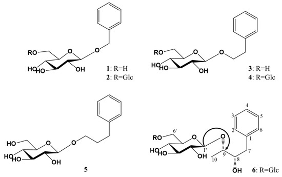
Figure 1.
Chemical structures of phenylalkyl glucosides 1–6 isolated from the flowers of Brugmansia arborea. Glc: β-D-glucopyranosyl; the gHMBC key correlations are represented by single-headed arrows from H to C.
Compound 6 was obtained as a white amorphous powder and became black on the TLC plate after spraying with 10% H2SO4 and then heating. FT−IR data exhibited the absorption (cm−1) of hydroxy (3352) and aromatic C=C (1651, 1528). The molecular weight and molecular formula were, respectively, determined to be 490 and C22H34O12 from the molecular ion peak [M + Na]+ m/z 513.1943 in the HRFABMS spectrum (calcd. for C22H34O12Na, 513.1948). In the 1H-NMR spectrum, the proton signals of five olefin methines δH 7.26 (m, 1H, H-4), δH 7.32 (dd, J = 8.4, 8.4 Hz, 2H, H-3, 5), δH 7.41 (br. d, J = 8.4 Hz, 2H, H-2, 6) owing to a phenyl moiety, two oxy-methines δH 3.42 (m, 1H, H-8), δH 3.94 (m, 1H, H-9), one methylene δH 2.76 and 2.93 (both dd, both 1H, J = 11.0, 3.0 Hz, H-7a; J = 11.0, 5.0 Hz, H-7b), and one methyl δH 1.20 (d, J = 5.7 Hz, 3H, H-10) were observed as the signals of an aglycon moiety. The proton signals of two hemiacetal δH 4.30 (d, J = 7.8 Hz, 1H, H-1″), δH 4.17 (d, J = 7.7 Hz, 1H, H-1′), eight oxy-methines δH 3.17 (overlapped, 2H, H-2′, 2″), δH 3.31 (overlapped, 2H, H-5′, 5″), δH 3.35 (overlapped, 2H, H-3′, 3″), δH 3.57 (dd, J = 7.7, 7.7 Hz, 1H, H-4″), δH 3.72 (overlapped, 1H, H-4′), and two oxy-methylenes δH 3.76 and 4.05 (both dd, both 1H, J = 11.6, 5.2 Hz, H-6′a; J = 11.6, 2.0 Hz, H-6′b), δH 3.65 and 3.86 (both 1H, br. d, J = 11.6 Hz, H-6″a; dd, J = 11.6, 5.0 Hz, H-6″b) due to two hexose moieties were detected. The above-mentioned 1H-NMR spectrum indicated that compound 6 was expected to be a phenylbutyl diglycoside. The 13C-NMR spectrum showed 22 carbon signals, confirming compound 6 to be composed of a phenylbutandiol and two hexoses. One olefin quaternary carbon signal (δC 140.2), three aromatic methine carbon signals (δC 127.2, 129.4, 130.0), two oxygenated methine carbon signals (δC 76.6, 88.2), one methylene carbon signal (δC 39.3), and one methyl carbon signal (δC 17.4) were observed as the signals of an aglycon, which was identified to be a phenylbutandiol. The chemical shift of the two sugar moieties, including two hemiacetal carbon signals (δC 104.5, 104.8), eight oxygenated methine carbon signals (δC 71.6, 71.9, 75.1, 75.2, 77.8, 78.0), and two oxygenated methylene carbon signals (δC 62.8, 69.9), revealed the sugar to be a β-glucopyranosyl-(1→6)-β-glucopyranose, and the coupling constant of the anomer proton signal (J = 8.0 Hz and J = 8.0 Hz) confirmed the two anomer hydroxyls to have β-configurations. In the gHMBC spectrum, the two anomer proton signals [δH 4.30 (H-1′′) and 4.17 (H-1′)] showed a cross-peak with the one oxygenated methine carbon signal [δC 88.2 (C-9)] and the one oxygenated methylene carbon signal [δC 69.9 (C-6′)], respectively, suggesting the β-D-glucopyranose to be at the C-9 and C-6′ positions. The oxygenated methine and oxygenated methylene carbon signals (C-9 and C-6′) were down-shifted owing to the glycosidation effect from their usual detection at δC 71.4 and δC 62.3, respectively, in β-D-glucopyranose [17]. The absolute configuration of C-8 and C-9 was proposed as S and S, respectively, by comparing the optical rotation value ( −23.0°) with that of (2S,3S)-1-phenyl-2,3-dihydroxybuthyl-3-O-β-D-glucopyranoside ( −21.9°) [18]. Taken together, these observations identified compound 6 as (2S,3S)-1-phenyl-2,3-dihydroxybuthyl-O-β-D-glucopyranosyl-(1→6)-β-D-glucopyranoside (Figure 1), which was revealed to be a new compound and named brugmansioside C.
2.2. Radical Scavenging Capacity
The antioxidant capacities of extract, solvent fractions, and phenylalkyl glycosides 1–6 of B. arborea flowers by the ABTS and DPPH assays are shown in Table 1. The experimental information with a description of the measurements of radical scavenging assay was described in a previous study [17]. EtOAc fraction (BAFE) showed the highest antioxidant capacities in both ABTS and DPPH assays. It was thought that the ethyl acetate fraction mainly contained compounds that contribute more to antioxidant capacities. The isolated compounds 1–6 showed ABTS radical scavenging capacities in order 1 > 3 > 2 ≥ 5 > 4 > 6. The DPPH radical scavenging activity was similar to ABTS radical scavenging activity (Table 1). BAFE showed the highest capacity, whereas compounds 2, 4, and 6 did not show DPPH scavenging capacity. Because the DPPH assay (80% methanol) measures the radical scavenging ability in the non-polar solvent system, compounds 2, 4, and 6, including two sugars in the structure, showed lower DPPH radical scavenging activity than ABTS [19,20]. Monoglycoside compounds 1 and 3 exhibited higher activity than the diglycoside compounds 2 and 4, respectively.

Table 1.
Radical scavenging capacity of extract, solvent fractions, and isolated compounds from Brugmansia arborea L. flowers using ABTS and DPPH radicals.
2.3. Protective Effects on Pancreatic Islets in Zebrafish Treated by Alloxan
Extracts, solvent fractions (EtOAc, n-BuOH), and isolated phenylalkyl glucosides 1–6 from B. arborea flowers were evaluated for protective activity against the pancreatic islets of zebrafish larvae damaged by alloxan. The larvae treated with alloxan were used to model type 1 diabetes due to their physiological similarities to mammals [21,22]. Alloxan is a diabetogenic chemical that has been reported to decrease β-cell mass in pancreatic islets [23]. To assess pancreatic islets treated with alloxan, the size changes of the pancreatic islets and fluorescence intensities of the NBDG-stained pancreatic islets under a fluorescence microscope were analyzed. When the zebrafish larvae were exposed to alloxan, pancreatic islet size decreased significantly by 51.8% (p = 0.0003) compared to the normal group (Figure 2a). Zebrafish larvae treated with glimepiride, a positive control, showed a pancreatic islet increase of 89.5% (p = 0.0047) compared to the alloxan group. The pancreatic islet sizes in the groups treated with EtOAc (BAFE) and n-BuOH (BAFB) fractions significantly increased up to 64.0 and 69.4% (p 0.0091 and 0.0065) compared with alloxan treatment (Figure 2). All of the phenylalkyl glucosides from B. arborea flowers also resulted in increases in pancreatic islet size. Compounds 1–6 (BAP 1–6) increased the injured pancreatic islets up to 82.0, 89.8, 80.0, 97.8, 103.1, and 99.6% (p 0.0011, 0.0037, 0.0012, 0.0002, 0.0011, 0.0011, and 0.0011), respectively, compared with alloxan treatment (Figure 2). All phenylalkyl glucosides isolated from B. arborea flowers in this study increased the sizes of pancreatic islets damaged by alloxan treatment in zebrafish larvae with high levels of significance. In particular, phenylpropyl glucoside 5 and the new phenylbutyl glucoside 6 displayed recovery effects greater than glimepiride. These diverse rates also showed structure–activity relationships. Monoglycoside compounds 1 and 3 exhibited lower activity than diglycoside compounds 2 and 4, respectively. And, compared to compounds 1, 3, and 5, the propyl group attached to phenyl was more effective than the methyl and ethyl groups.
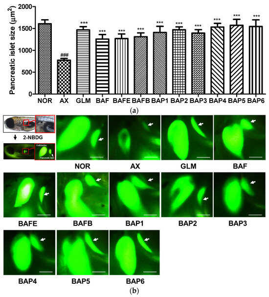
Figure 2.
Protective effect of EtOAc and n-BuOH fractions and compounds 1–6 from Brugmansia arborea flowers on alloxan-induced pancreatic islets in zebrafish. (a) Size of the pancreatic islets. (b) Pancreatic islet image: NOR: normal group, AX: alloxan group, GLM: glimepiride+AX group, BAF: Extract+AX, BAFE: EtOAc fraction+AX, BAFB: n-BuOH fraction+AX, BAP1-6: compound 1–6+AX. (### p < 0.001; compared to the normal group), (*** p < 0.001 compared to the alloxan-treated group). Scale bar = 100 μm.
2.4. Action of Diazoxide (DZ) on Alloxan-Induced Pancreatic Islets in Zebrafish
The metabolism of glucose in pancreatic cells is the key step in glucose-stimulated insulin secretion [24]. To study the involvement of the pancreatic β-Cell KATP channel stimulation activity, diazoxide (DZ), a KATP channel opener, was used. The size of the pancreatic islets in the DZ-treated normal group was significantly smaller (43.4%, p = 0.0052) relative to the normal group without DZ treatment. Furthermore, the alloxan (AX) group showed no significant difference compared to the DZ-treated group. Pancreatic islet size in the 10 μg/mL glimepiride (GLM), AX, and DZ co-treatment groups was significantly lower (48.9%, p = 0.0202) compared to the 10 μg/mL GLM and AX co-treated groups without DZ. Groups co-treated with compounds 1, 2, 4, or 6 and AX were not significantly different after treatment with DZ, indicating no relationship with KATP channels. Compounds 3 and 5, in addition to AX, yielded significantly smaller pancreatic islet sizes after treatment with DZ (3: 57.6%, p = 0.0358; 5: 69.3%, p = 0.0358) compared to the compound+AX groups (Figure 3). These results suggest that compounds 3 and 5 might stimulate insulin secretion by Ca2+ influx via the closure of the KATP channels in β-cells.
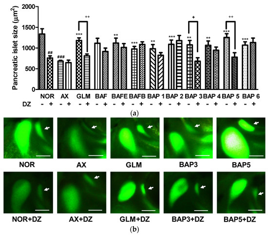
Figure 3.
Action of diazoxide (DZ) for pancreatic islets damage by alloxan (AX) in zebrafish fractions and compounds. (a) Size of the pancreatic islets. (b) Pancreatic islet image. Normal (NOR), NOR+alloxan-treatment (AX), AX+glimepiride, a positive control (GLM), AX+Extract, EtOAc, and n-BuOH fractions of B. arborea flowers (BAF, BAFE, BAFB), AX + phenylalkyl glucosides 1–6 (BAP1~BAP6), treated with and without diazoxide (DZ). (## p < 0.01, ### p < 0.001; compared to NOR), (** p < 0.01, *** p < 0.001; compared to AX), (+ p < 0.05, ++ p < 0.01). Scale bar = 100 μm.
3. Materials and Methods
3.1. Plant Materials
The dried flowers of B. arborea L. were supplied by Herb Island, Pocheon, Korea, in June 2014 and were identified by Professor Dae-Keun Kim, Woosuk University, Jeonju, Korea. A voucher specimen (KHU2014-0623) is reserved at the Laboratory of Natural Products Chemistry, Kyung Hee University, Yongin, Korea.
3.2. General Experimental Procedures
The silica gel (SiO2) and octadecyl SiO2 (ODS); Kiesel gel 60 (Merck, Darmstadt, Germany), Lichroprep RP-18 (40~60 m, Merck), and Sephadex LH-20 (Amersham Biosciences, Uppsala, Sweden). Thin layer chromatography (TLC); Kiesel gel 60 F254 and RP-18 F254S (Merck) TLC plates. The spots were detected using a UV lamp Spectroline Model ENF-240 C/F (Spectronics Corporation, Westbury, NY, USA) and a 10% H2SO4 solution. Nuclear magnetic resonance (NMR) spectra; 400 MHz FT-NMR spectrometer (Varian Inova AS-400, Palo Alto, CA, USA). Deuterium solvents; Merck Co. Ltd. and Sigma Aldrich Co. Ltd. (St. Louis, MO, USA). IR spectra; Perkin Elmer Spectrum One FT-IR spectrometer (Buckinghamshire, England). Electronic ionization mass spectrometry (EI/MS) and fast atom bombardment mass spectrometry (FABMS) spectra; JEOL JMS-700 (Tokyo, Japan). Melting points; Fisher-John’s melting point apparatus (Fisher Scientific, Miami, FL, USA) without correction.
3.3. Extraction and Isolation
B. arborea flowers (900 g, in drying) were extracted with 80% aqueous methanol (27 L × 3) at room temperature for 24 h. Concentrated methanol extracts (160 g) were suspended in water (1 L) and then successively partitioned with ethyl acetate (EtOAc, 1 L × 3), n-butanol (n-BuOH, 0.6 L × 3), and water. The extracts were concentrated to produce the residues of the EtOAc fraction (BAFE, 12 g), the n-BuOH fraction (BAFB, 16 g), and the H2O fraction (BAFW, 132 g), respectively. The EtOAc fraction was applied to SiO2 CC (7.0 × 15.0 cm) and eluted with n-hexane-EtOAc (4:1 → 2:1 → 1:1, 2.0 L of each) and CHCl3-MeOH-H2O (50:3:1 → 36:3:1 → 25:3:1 → 18:3:1 → 12:3:1 → 9:3:1 → 7:3:1 → 6:4:1, 1.5 L of each). The eluting solutions were monitored by TLC to produce 16 fractions (BAFE-1 to BAFE-16). BAFE-10 (966.2 mg, elution volume/total volume (Ve/Vt) 0.649–0.718) was subjected to ODS CC (4.5 × 10 cm) and eluted with acetone-H2O (1:2 → 1:1, 3.7 L of both) to yield 16 fractions (BAFE-10-1 to BAFE-10-16). BAFE-10-5 (64.2 mg, Ve/Vt 0.020–0.034) was subjected to SiO2 CC (2.5 × 7 cm) and eluted with CHCl3-MeOH-H2O (30:3:1, 1.2 L) to ultimately produce six fractions (BAFE-10-5-1 to BAFE-10-5-6) along with purified compound 5 [3-phenylpropyl glucoside, BAFE-10-5-4, 13.9 mg, Ve/Vt 0.458–0.733, TLC (SiO2 F254) Rf 0.54, CHCl3-MeOH-H2O = 9:3:1]. The n-BuOH fraction (BAFB) was implemented by SiO2 CC (7.0 × 13.0 cm) and eluted with CHCl3-MeOH-H2O (12:3:1 → 9:3:1 → 6:4:1, 12.0 L of each) yielding 14 parts (BAFB-1 to BAFB-14). BAFB-5 (447.5 mg, Ve/Vt 0.063–0.075) was subjected to Sephadex-LH-20 CC (2.5 × 50 cm) and eluted with MeOH-H2O (4:1, 296 mL) to yield six fractions (BAFB-5-1 to BAFB-5-6). BAFB-5-3 (151.9 mg, Ve/Vt 0.446–0.500) was subjected to ODS CC (2.0 × 17 cm) and eluted with MeOH-H2O (1:2, 485 mL) yielding nine parts (BAFB-5-3-1 to BAFB-5-3-9) as well as 1 [benzyl glucoside, BAFB-5-3-3, 88.0 mg, Ve/Vt 0.155–0.196, TLC (SiO2 F254) Rf 0.44, CHCl3-MeOH-H2O = 12:3:1] and 3 [2-phenylethyl glucoside, BAFB-5-3-6, 20.5 mg, Ve/Vt 0.289–0.330, TLC (SiO2 F254) Rf 0.44, CHCl3-MeOH-H2O = 12:3:1]. BAFB-9 (1.2 g, Ve/Vt 0.163–0.225) was subjected to SiO2 CC (4.0 × 15.0 cm) and eluted with CHCl3-MeOH-H2O (10:3:1, 3.1 L) to yield 14 fractions (BAFB-9-1 to BAFB-9-14). BAFB-9-8 (487.7 mg, Ve/Vt 0.308–0.607) was subjected to SiO2 CC (3.5 × 15.0 cm) and eluted with EtOAc-n-BuOH-H2O (22:3:1, 2.7 L) to yield 15 fractions (BAFB-9-8-1 to BAFB-9-8-15). BAFB-9-8-15 (33.7 mg, Ve/Vt 1.000) was implemented by ODS CC (2.0 × 5.0 cm) and eluted with MeOH-H2O (1:3, 430 mL) to ultimately produce seven fractions (BAFB-9-8-15-1 to BAFB-9-8-15-7) along with purified 6 [(2S, 3S)-1-phenyl-2,3-dihydroxybutyl glucosyl-(1→6)-glucoside, BAFB-9-8-15-3, 12.0 mg, Ve/Vt 0.070–0.140, TLC (SiO2 F254) Rf 0.50, EtOAc-n-BuOH-H2O = 4:5:1]. BAFB-12 (899.3 mg, Ve/Vt 0.392–0.495) was subjected to ODS CC (3.5 × 5.0 cm) and eluted with MeOH-H2O (1:3, 2.9 L) to yield 12 fractions (BAFB-12-1 to BAFB-12-12). BAFB-12-5 (182.2 mg, Ve/Vt 0.082–0.149) was subjected to SiO2 CC (2.5 × 16.0 cm) and eluted with CHCl3-MeOH-H2O (9:3:1, 1.1 L) to ultimately produce eight fractions (BAFB-12-5-1 to BAFB-12-5-8) along with purified 4 [2-phenylethyl glucosyl-(1→6)-glucoside, BAFB-12-5-3, 104.3 mg, Ve/Vt 0.407–0.433, TLC (SiO2 F254) Rf 0.54, CHCl3-MeOH-H2O = 6:4:1]. BAFB-13 (1.8 g, Ve/Vt 0.495–0.575) was subjected to ODS CC (4.0 × 9.0 cm) and eluted with MeOH-H2O (1:2, 2.9 L) to yield 13 fractions (BAFB-13-1 to BAFB-13-13). BAFB-13-5 (128.7 mg, Ve/Vt 0.075–0.097) was subjected to ODS CC (2.5 × 7.0 cm) and eluted with MeOH-H2O (1:2, 300 mL) to ultimately produce eight parts (BAFB-13-5-1 to BAFB-13-5-8) as well as 2 [benzyl glucosyl-(1→6)-glucoside, BAFB-13-5-2, 10.3 mg, Ve/Vt 0.120–0.140, TLC (SiO2 F254) Rf 0.57, CHCl3-MeOH-H2O = 6:4:1].
Benzyl O-β-D-glucopyranoside (1): White amorphous powder (CH3OH); −43° (c = 0.10, CH3OH); positive FABMS m/z 293 [M + Na]+; IR (KBr, v) 3502, 1697, 1530 cm−1; 1H- and 13C-NMR (400 MHz and 100 MHz, CD3OD, δH and δC); Table 2 and Table 3.
Table 2.
1H-NMR spectral data (CD3OD; 400 MHz) of compounds 1–6. δ in ppm, J in Hz.
Table 3.
13C-NMR spectral data (CD3OD; 100 MHz) of compounds 1–6. δ in ppm.
Benzyl O-β-D-glucopyranosyl-(1→6)-β-D-glucopyranoside (2): White amorphous powder (CH3OH); −90° (c = 0.20, CH3OH); positive FABMS m/z 433 [M + H]+; IR(KBr, ν) 3510, 1671, 1600 cm−1; 1H- and 13C-NMR (400 MHz and 100 MHz, CD3OD, δH and δC); Table 2 and Table 3.
2-phenylethyl O-β-D-glucopyranoside (3): White amorphous powder (CH3OH); −35.3° (c = 0.50, CH3OH); positive FABMS m/z 285 [M + H]+; IR(KBr, ν) 3368, 1683, 1573 cm−1; 1H- and 13C-NMR (400 MHz and 100 MHz, CD3OD, δH and δC); Table 2 and Table 3.
2-phenylethyl O-β-D-glucopyranosyl-(1→6)-β-D-glucopyranoside (4): White amorphous powder (CH3OH); −82° (c = 0.20, CH3OH); positive FABMS m/z 447 [M + H]+; IR(KBr, ν) 3370, 1685, 1586 cm−1; 1H- and 13C-NMR (400 MHz and 100 MHz, CD3OD, δH and δC); Table 2 and Table 3.
3-phenylpropyl O-β-D-glucopyranoside (5): White amorphous powder (CH3OH); −29.3° (c = 0.75, CH3OH); positive FABMS m/z 299 [M + H]+; IR(KBr, ν) 3364, 1687, 1574 cm−1; 1H- and 13C-NMR (400 MHz and 100 MHz, CD3OD, δH and δC); Table 2 and Table 3.
(2S,3S)-1-phenyl-2,3-dihydroxybutyl-O-β-D-glucopyranosyl-(1→6)-β-D-glucopyranoside (6): White amorphous powder (CH3OH); −23.0° (c = 0.10, CH3OH); HRFABMS m/z 513.1943 [M + Na]+ (calcd for C22H34O12Na, 513.1948); IR(KBr, ν) 3352, 1651, 1528 cm−1; 1H-NMR (600 MHz, CD3OD, δH) 7.41 (2H, br. d, J = 8.4 Hz, H-2, 6), 7.32 (2H, dd, J = 8.4, 8.4 Hz, H-3, 5), 7.26 (1H, m, H-4), 4.30 (1H, d, J = 7.8 Hz, H-1″), 4.17 (1H, d, J = 7.8 Hz, H-1′), 4.05 (1H, dd, J = 12.0, 5.4 Hz, H-6′a), 3.94 (1H, m, H-9), 3.86 (1H, br. d, J = 12.0 Hz, H-6″a), 3.76 (1H, dd, J = 12.0, 1.8 Hz, H-6′b), 3.72 (1H, overlapped, H-4′), 3.65 (1H, dd, J = 12.0, 5.4 Hz, H-6″b), 3.57 (1H, dd, J = 7.8, 7.8 Hz, H-4″), 3.42 (1H, m, H-8), 3.35 (2H, overlapped, H-3′, 3″), 3.31 (2H, overlapped, H-5′, 5″), 3.17 (2H, overlapped, H-2′, 2″), 2.93 (1H, dd, J = 12.0, 2.4 Hz, H-7a), 2.76 (1H, dd, J = 12.0, 6.0 Hz, H-7b), 1.20 (3H, d, J = 6.0 Hz, H-10); 13C-NMR (150 MHz, CD3OD, δC) 140.2 (C-1), 130.0 (C-3, 5), 129.4 (C-2, 6), 127.2 (C-4), 104.8 (C-1″), 104.5 (C-1′), 88.2 (C-9), 78.0 (C-3′), 78.0 (C-3″), 77.8 (C-5′), 77.8 (C-5″), 76.6 (C-8), 75.2 (C-2′), 75.1 (C-2″), 71.9 (C-4′), 71.6 (C-4″), 69.9 (C-6′), 62.8 (C-6″), 39.3 (C-7), 17.4 (C-10); Table 2 and Table 3.
3.4. Free Radical Scavenging Activity
The materials, equipment, and methods used for the free radical scavenging assay of extract, solvent fractions, and compounds 1–6 from B. arborea flowers were described in a previous study [17].
3.5. Antidiabetic Activity
3.5.1. Chemicals and Animals
The chemical materials and animal preparation used for the antidiabetic activity are described in a previous study [25].
3.5.2. Ethics Statement
All zebrafish experimental procedures were carried out in accordance with standard zebrafish protocols and were approved by the Animal Care and Use Committee of Kyung Hee University [KHUASP(SE)-15-10].
3.5.3. Evaluation of Recovery Efficacy on Pancreatic Islet Damaged by Alloxan in Zebrafish
The materials, equipment, and methods used for evaluation of recovery efficacy of extract (BAF), solvent fractions (BAFE and BAFB), and compounds 1–6 from B. arborea flowers on pancreatic islet damaged by alloxan in zebrafish are described in a previous study [25].
3.5.4. Action of Diazoxide on Alloxan-Induced Diabetic Zebrafish
The materials, equipment, and methods used for the action of diazoxide on alloxan-induced diabetic zebrafish are described in a previous study [25].
4. Conclusions
This study endeavored to find new active compounds of B. arborea flowers. Six phenylalkyl glucosides, including one new phenylbutyl diglucoside, were isolated through repeated column chromatography using SiO2, ODS, and Sephadex LH-20 resins and identified by the analysis of NMR, IR, UV, and FABMS data. Extracts, solvent fractions, and some compounds from B. arborea flowers were found to show scavenging activity in ABTS radicals with levels of significance. The EtOAc fraction (BAFE) showed the highest scavenging capacity, whereas compounds 2, 4, and 6 did not display a capacity to use the DPPH radical. Compounds 2, 4, and 6 were isolated from the BuOH fraction (BAFB). Therefore, the low antioxidant capacity of BAFB compared to the BAFE is expected to be due to the lower antioxidant capacity of these compounds. And the significant antioxidant activity of compound 5 is expected to be one of the reasons for the high antioxidant activity of the BAFE. All compounds and solvent fractions were also found to show protective activity against alloxan-induced pancreatic islet damage in zebrafish larvae with high levels of significance. In particular, compounds 3 and 5 stimulated insulin secretion by Ca2+ influx via closure of KATP channels in β-cells. These results indicate that B. arborea flowers and their isolated compounds are used as potential antioxidant and antidiabetic agents.
Supplementary Materials
The following supporting information can be downloaded at: https://www.mdpi.com/article/10.3390/plants12244075/s1, Figure S1: 1H NMR spectrum of brugmansioside C (6) in CD3OD (600 MHz); Figure S2: 13C NMR spectrum of brugmansioside C (6) in CD3OD (150 MHz); Figure S3: gHMBC spectrum of brugmansioside C (6) in CD3OD; Figure S4: gHSQC spectrum of brugmansioside C (6) in CD3OD; Figure S5: HRFABMS spectrum of brugmansioside C (6); Figure S6: IR spectrum of brugmansioside C (6).
Author Contributions
Conceptualization, H.-G.K., N.-I.B. and D.Y.L.; methodology, H.-G.K. and N.-I.B.; software, Y.H.N. and T.H.K.; validation, M.-H.L. and D.Y.L.; formal analysis, H.-G.K. and Y.H.N.; investigation, H.-G.K.; resources, M.-H.L.; data curation, H.-G.K., Y.H.N., T.H.K. and D.Y.L.; writing—original draft preparation, H.-G.K.; writing—review and editing, D.Y.L.; visualization, H.-G.K. and Y.H.N.; supervision, N.-I.B. and D.Y.L.; project administration, D.Y.L.; funding acquisition, M.-H.L. and D.Y.L. All authors have read and agreed to the published version of the manuscript.
Funding
This work was supported by the “Cooperative Research Program for Agri-culture Science & Technology Development” (Project no. PJ01420403), Rural Development Administration, Republic of Korea.
Data Availability Statement
The 1H, 13C, gHMBC, gHSQC, HRFABMS, and IR spectra of compound 6 are available from the supplementary materials.
Conflicts of Interest
The authors declare no conflict of interest.
References
- American Diabetes Association. Diagnosis and classification of diabetes mellitus. Diabetes Care 2009, 32, 62–67. [Google Scholar] [CrossRef] [PubMed]
- Piero, M.N.; Nzaro, G.M.; Njagi, J.M. Diabetes mellitus—A devastating metabolic disorder. Asian J. Biomed. Pharm. Sci. 2014, 4, 1–7. [Google Scholar]
- Bastaki, S. Diabetes mellitus and its treatment. Int. J. Diabetes Metab. 2005, 13, 111–134. [Google Scholar] [CrossRef]
- Olokoba, A.B.; Obateru, O.A.; Olokoba, L.B. Type 2 Diabetes mellitus: A review of current trends. Oman Med. J. 2012, 27, 269–273. [Google Scholar] [CrossRef] [PubMed]
- Romero Aroca, P.; Mendez Marin, I.; Baqet Bermaldiz, M.; Fernendez Ballart, J.; Santos Blanco, E.I. Review of the relationship between renal and retinal microangiopathy in diabetes mellitus patients. Curr. Diabetes Rev. 2010, 6, 88–101. [Google Scholar] [CrossRef] [PubMed]
- Parng, C.; Seng, W.L.; Semino, C.; McGrath, P.I. Zebrafish: A preclinical model for drug screening. Assay Drug Dev. Technol. 2002, 1, 41–48. [Google Scholar] [CrossRef]
- Spence, R.; Gerlach, G.; Lawrence, C.; Smith, C. The behaviour and ecology of the zebrafish, Danio rerio. Biol. Rev. 2008, 83, 13–34. [Google Scholar] [CrossRef]
- De Feo, V. The ritual use of Brugmansia species in traditional Andean medicine in Northern Peru. Econ. Bot. 2004, 58, S221–S229. [Google Scholar] [CrossRef]
- Pratt, C. An Encyclopedia of Shamanism; The Rosen Publishing Group: New York City, NY, USA, 2007; pp. 68–70. [Google Scholar]
- Fuller, T.C.; McClintock, E. Poisonous Plants of California; University of California Press: Berkeley, CA, USA, 1988; pp. 233–235. [Google Scholar]
- Capasso, A.; De Feo, V.; De Simone, F.; Sorrentino, L. Activity-directed isolation of spasmolytic (anti-cholinergic) alkaloids from Brugmansia arborea (L.) Lagerheim. Int. J. Pharmacogn. 1997, 35, 43–48. [Google Scholar] [CrossRef]
- Ghani, A. Cuscohygrine from some solanaceous plants. Indian J. Pharm. Sci. 1985, 47, 127–129. [Google Scholar]
- Zhang, C.; Ma, Y.; Gao, H.; Liu, X.; Chen, L.; Zhang, Q.; Wang, Z.; Li, A. Non-alkaloid components from Sophora flavescens. Zhongguo Zhongyao Zazhi 2013, 38, 3520–3524. [Google Scholar]
- Lin, S.; Zhang, Z.; Shen, Y.; Li, H.; Shan, L.; Liu, R.; Xu, X.; Zhang, W. New lignan glycoside from Senecio chrysanthemoides. Zhongguo Zhongyao Zazhi 2011, 36, 1755–1762. [Google Scholar]
- Luyen, B.T.T.; Tai, B.H.; Nguyen, P.T.; Cha, J.Y.; Lee, H.Y.; Lee, Y.M.; Kim, Y.H. Anti-inflammatory components of Chrysanthemum indicum flowers. Bioorg. Med. Chem. Lett. 2015, 25, 266–269. [Google Scholar] [CrossRef]
- Kurashima, K.; Fujii, M.; Ida, Y.; Akita, H. Simple synthesis of β-D-glycopyranosides using β-glycosidase from almonds. Chem. Pharm. Bull. 2004, 52, 270–275. [Google Scholar] [CrossRef]
- Kim, H.G.; Jung, Y.S.; Oh, S.M.; Oh, H.J.; Ko, J.H.; Kim, D.O.; Kang, S.C.; Lee, Y.G.; Lee, D.Y.; Baek, N.I. Coreolanceolins A–E, new flavanones from the flowers of Coreopsis lanceolate, and their antioxidant and anti-inflammatory effects. Antioxidants 2020, 9, 539–554. [Google Scholar] [CrossRef]
- Matsuda, H.; Morikawa, T.; Toguchida, I.; Harima, S.; Yoshikawa, M. Medicinal flowers. VI. Absolute stereostructures of two new flavanone glycosides and a phenylbutanoid glycoside from the flowers of Chrysanthemum indicum L.: Their inhibitory activities for rat lens aldose reductase. Chem. Pharm. Bull. 2002, 50, 972–975. [Google Scholar] [CrossRef]
- Kim, D.O.; Lee, K.W.; Lee, H.J.; Lee, C.Y. Vitamin C Equivalent Antioxidant Capacity (VCEAC) of Phenolic Phytochemicals. J. Agric. Food Chem. 2002, 50, 3713–3717. [Google Scholar] [CrossRef]
- Yokozawa, T.; Chen, C.P.; Dong, E.; Tanaka, T.; Nonaka, G.I.; Nishioka, I. Study on the Inhibitory Effect of Tannins and Flavonoids against the 1,1-Diphenyl-2-picrylhydrazyl Radical. Biochem. Pharmacol. 1998, 56, 213–222. [Google Scholar] [CrossRef]
- Elo, B.; Villano, C.M.; Govorko, D.; White, L.A. Larval zebrafish as a model for glucose metabolism: Expression of phosphoenolpyruvate carboxykinase as a marker for exposure to anti-diabetic compounds. J. Mol. Endocrinol. 2007, 38, 433–440. [Google Scholar] [CrossRef]
- Kim, H.G.; Nam, Y.H.; Jung, Y.S.; Oh, S.M.; Trong Nguyen, N.; Lee, M.H.; Kim, D.O.; Kang, D.H.; Lee, D.Y.; Baek, N.I. Aurones and flavonols from Coreopsis lanceolata L. flowers and their anti-oxidant, pro-inflammatory inhibition effects, and recovery effects on alloxan-induced pancreatic islets in zebrafish. Molecules 2021, 26, 6098. [Google Scholar] [CrossRef]
- Desgraz, R.; Bonal, C.; Herrera, P.L. β-Cell regeneration: The pancreatic intrinsic faculty. Trends Endocrinol. Metab. 2011, 22, 34–43. [Google Scholar] [CrossRef] [PubMed]
- Miki, T.; Nagashima, K.; Seino, S. The structure and function of the ATP-sensitive K+ channel in insulin-secreting pancreatic β-cells. J. Mol. Endocrinol. 1999, 22, 113–123. [Google Scholar] [CrossRef]
- Nam, Y.H.; Hong, B.N.; Rodriguez, I.; Park, M.S.; Jeong, S.Y.; Lee, Y.G.; Shim, J.H.; Yasmin, T.; Kim, N.W.; Koo, Y.T.; et al. Steamed ginger may enhance insulin secretion through KATP channel closure in pancreatic β-cells potentially by increasing 1-dehydro-6-gingerdione content. Nutrients 2020, 12, 324–338. [Google Scholar] [CrossRef]
Disclaimer/Publisher’s Note: The statements, opinions and data contained in all publications are solely those of the individual author(s) and contributor(s) and not of MDPI and/or the editor(s). MDPI and/or the editor(s) disclaim responsibility for any injury to people or property resulting from any ideas, methods, instructions or products referred to in the content. |
© 2023 by the authors. Licensee MDPI, Basel, Switzerland. This article is an open access article distributed under the terms and conditions of the Creative Commons Attribution (CC BY) license (https://creativecommons.org/licenses/by/4.0/).