Communication

Effect of In Vitro Culture of Long Shoot Tip on Variant Structure and Titer of Grapevine Viruses

Research Institute of Pomology, Chinese Academy of Agricultural Sciences, Xingcheng 125100, China
*
Author to whom correspondence should be addressed.
Plants 2022, 11(15), 1907; https://doi.org/10.3390/plants11151907
Submission received: 24 June 2022 / Revised: 18 July 2022 / Accepted: 20 July 2022 / Published: 23 July 2022
(This article belongs to the Section Plant Protection and Biotic Interactions)

Abstract

:
Shoot tip culture is a very effective approach for studying plant viruses. In this study, we evaluated the numbers, diversity, and titer of grapevine viruses in in vitro grapevine plants after long shoot tip culture. Six virus-infected grapevine cultivars (Cabernet Franc, Cabernet Gernischt, Cabernet Sauvignon, Wink, Victoria, and Merlot) collected from six regions of China were used as the research materials. Approximately 1.5 cm long shoot tips were used for meristem culture. The average survival rate of the six grapevine cultivars was 45.7%. Merlot collected from Beijing showed the highest survival rate (80.0%). Regeneration was not achieved in Cabernet Gernischt collected from Liaoning province and Cabernet Sauvignon from Tianjin due to bacterial and fungal contamination. Virus detection conducted in the surviving regenerated plants showed that the virus infection status, including the viral numbers and the species present in plants grown in vitro, was the same as that in corresponding in vivo plants. Moreover, the analysis of sequence diversity and the mutation frequency in grapevine viruses in vitro indicated that the structure of grapevine viruses was stable in long shoot tip culture after four sub-culture passages. Further, the relative viral titer of in vitro grapevine plants was much higher than that of in vivo plants. These results aid in the investigation of viruses in woody plants.

1. Introduction

Plant tissue culture is indispensable for the rapid multiplication of rare plants, plant genetic transformation, and production of plant-derived metabolites of important commercial value [1]. Modern plant tissue culture techniques increase the variety of cultures and provide a new platform for in vitro studies of plant cells. Compared to plants grown in the field, in vitro plants can acquire a better environmental containment. Generally, the production times are shorter for in vitro plants, the system is easily regulated, and proteins secreted from the cells, downstream processing and product purification are simpler and cheaper [2].
Tissue culture, such as meristem culture of shoot tips, is a very effective approach to obtain virus-free plants from a wide range of hosts. It can maximize the propagation of the sterile stocks suitable for certification schemes [3]. Some research has shown that shoot tip culture alone remained insufficient for the elimination of some plant viruses [4,5]. Therefore, shoot tip culture is usually combined with thermotherapy, chemotherapy, or cryotherapy to eliminate viruses [6,7,8].
Plant tissue culture also contributes substantially to exploring viral replication and virus–host interaction, and provides the basic methodology to generate virus resistant plants through genetic engineering [9,10,11]. Many woody plant viruses cannot be mechanically transmitted to herbaceous hosts, which is hampered by gene expression strategies and replication. Plant tissue culture techniques can be helpful to overcome these limitations [12]. The successful viral elimination of meristem tip culture depends on the size of the explant, and a smaller tip (<1 mm) could lead to a higher death rate [13,14].
In fact, large tissues can also be cultured, if only to obtain in vitro plants, and it is much easier to obtain a big tip than a small one. Studies have found that smaller shoot tip culture can remove viruses from infected plants [15,16,17], but the effect of longer shoot tip culture on viruses is unclear. Grapevine is a fruit crop with major relevance worldwide and is also among the most well-studied deciduous woody perennials. Viral diseases are major constraints to grape production worldwide. Nearly 90 virus species infect grapevine, and most of them cannot be inoculated to herbs by mechanical transmission from sap, which has seriously influenced the pathogenicity research of grapevine viruses [18,19,20,21]. Moreover, small grapevine shoot tips are commonly used as material for elimination treatment to obtain virus-free grapevine plants [22,23,24]. Meanwhile, as deciduous fruit trees, in vitro virus-infected plants are of great significance in the study of grapevine viruses. In vitro culture of long shoot tip is an effective way to achieve the above mentioned goal. However, the effect of long shoot tip culture on viruses is unclear. In this study, the numbers, diversity, and titer of viruses in in vitro grapevine plants were assessed after long shoot tip culture.

2. Results

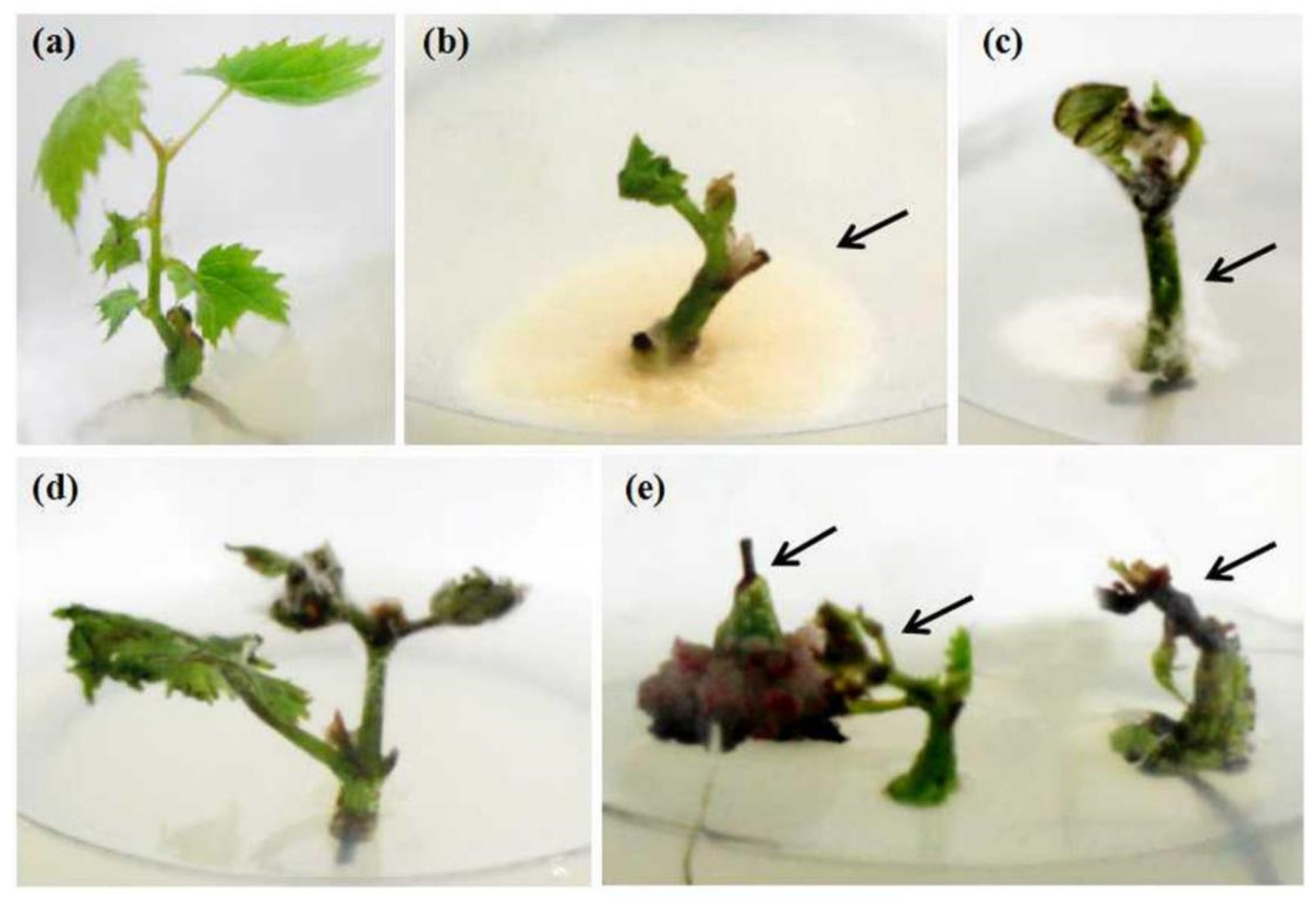
2.1. Survival of Shoot Tips

The survival rate was calculated after the shoot tip regenerated into a whole plant (Figure 1a). The average survival rate of six grapevine cultivars from six regions was 45.7%. Merlot collected from Beijing showed the highest survival rate of 80.0%. Regeneration was not observed in Cabernet Gernischt from Liaoning province and Cabernet Sauvignon from Tianjin. Other grapevine cultivars showed 50.0–70.0% survival rates (Table 1). There were two reasons related to shoot tip death: bacterial and fungal contamination owing to inadequate sterilization time (Figure 1b–d) and the highly differentiated callus on the base of the shoot tip, which caused the gradual browning, blackening, and wilting of meristem on the top (Figure 1e).

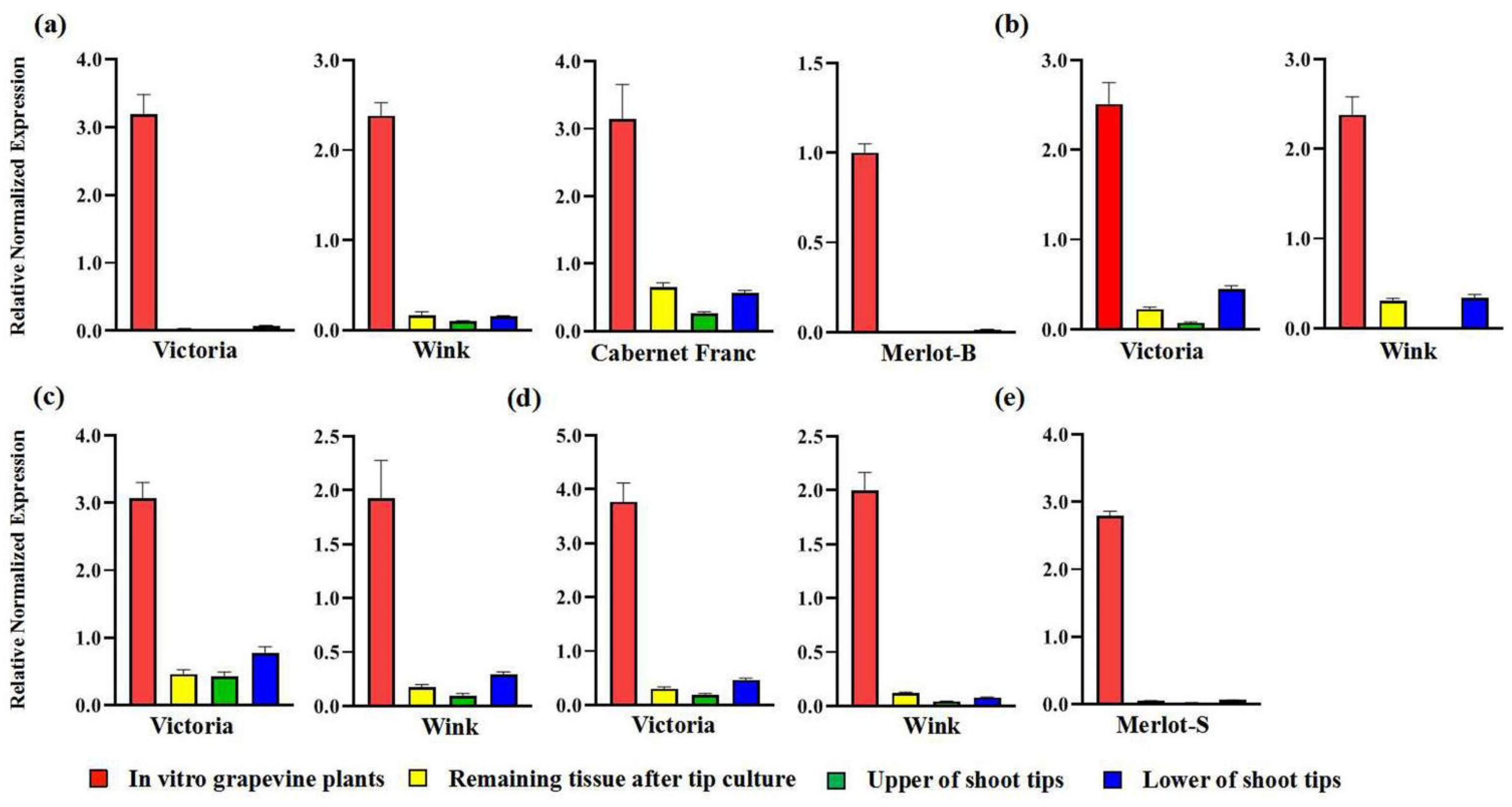
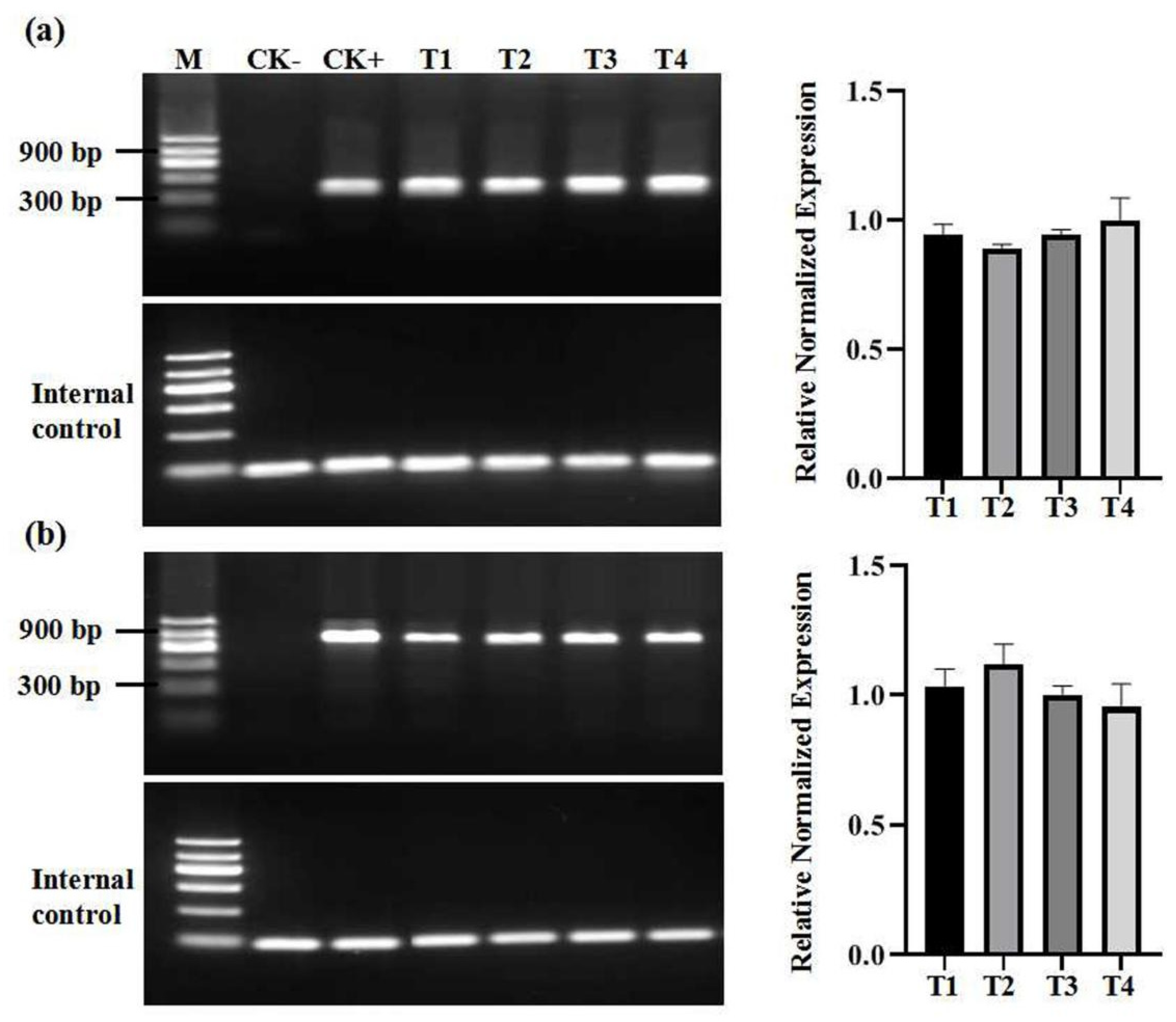
2.2. Virus Detection of In Vitro Grapevine Plantlets

Both Wink and Victoria from Guangxi province were infected by grapevine rupestris stem pitting-associated virus (GRSPaV), grapevine leafroll-associated virus 2 (GLRaV-2), grapevine leafroll-associated virus 3 (GLRaV-3), and grapevine fleck virus. Cabernet Franc from Ningxia and Merlot from Sichuan province were infected by two viruses (Table 1). Only one virus (GRSPaV) was detected in Merlot of Beijing. These viruses (GRSPaV, GLRaV-1, GLRaV-2, GLRaV-3, GFkV, and GVA) have been found to be present and common grapevine viruses in China [25,26]. The virus infection status, including the viral numbers and the species of in vitro plants, was the same as that of the corresponding in vivo plants (Table 1). For long shoot tips, the virus titer in the upper part of shoot tips was relatively lower than that in the lower part of shoot tips, while the relative viral titer of plants regenerated from the upper part of shoot tips was substantially higher than that of the remaining tissue after tip culture (Figure 2). Moreover, we also analyzed the viral titer in four sub-cultures of Cabernet Franc from Ningxia and observed that the titer of grapevine viruses was relatively stable and that no significant change was found during sub-culture (Figure 3).

2.3. Diversity of In Vitro Grapevine Plantlets

The diversity of GRLaV-2, GRLaV-3, and GRSPaV in grapevine cultivars was analyzed before and after tissue culture. The products of the viruses amplified by regular PCR were used to clone and sequence. The nucleotide identities of GRLaV-2 sequence variants isolated from Wink, Victoria, and Cabernet Franc shoot tips were 99.7–100.0%, 99.5–100.0%, and 98.9–100.0%, respectively. After tissue culture, the identities of sequence variants from the three GRLaV-2 isolates were 99.4–100.0%, 99.2–100.0%, and 99.5–100.0%, respectively. The two groups of the three cultivars showed 99.4–100.0%, 99.2–100.0%, and 98.9–100.0% sequence identities and 99.97%, 99.90%, and 99.73% consistencies, respectively. The GRLaV-3 sequence variants isolated from Wink and Victoria, and the GRSPaV sequence variants isolated from Wink and Merlot (Beijing, Merlot-B) also showed similar results (Table 2). Based on the taxonomic system related to the genera Closterovirus, Ampelovirus, and Foveavirus, these results indicated that the sequence diversity of grapevine viruses hardly changed during shoot tip culture.
To further demonstrate the stability of grapevine virus population composition during shoot tip culture, we also computed the mutation frequency. Table 2 lists the mutation frequency/nt of GRLaV-2, GRLaV-3, and GRSPaV in different cultivars before and after shoot tip culture. The nucleotide mutation frequencies of the GRLaV-2 sequence isolated from Cabernet Franc, the GRLaV-3 sequence isolated from Wink, and the GRSPaV sequence isolated from Merlot-B were the same for in vivo and in vitro plants. The nucleotide mutation frequency of the GRLaV-2 sequence isolated from in vitro Victoria was lower than that detected from in vivo plants, which demonstrated that the diversity of the population structure of GRLaV-2 scarcely changed after shoot tip culture. The nucleotide mutation frequencies of the GRLaV-2 and GRSPaV sequences isolated from in vitro Wink were 1.8 × 10⁻⁴ and 0.8 × 10⁻⁴ more than those from in vivo plants, respectively, but it was found that the two mutations were not produced during virus replication after they were revised by error rate (Table 2). These results also indicated that the structure of grapevine viruses was stable after long shoot tip culture.

3. Discussion

In the current study, the infection status and genetic diversity of viruses were the same in in vitro and in vivo grapevine plants after long shoot tip culture. Meanwhile, the viral titer markedly increased after shoot tip culture.
Most research has demonstrated that the size of shoot tips is directly related to the survival rate of cultures, and that smaller sizes could led to a higher death rate [13,14]. Han et al. [22] demonstrated that the survival rate of small shoot tips (<0.5 mm) was less than 65%. Moreover, previously we reported that the survival rate was related to the cultivars of grapevine [23,24]. Here, we observed that the extension of shoot tips (≈1.5 cm) did not significantly improve the culture survival rate. Abundant parasitic or infective fungi and bacteria existed on the surface and inside the new and tender long shoot tips. The timing of ethanol and mercury bichloride treatments should be optimized in order to select the suitable treatment for long shoot tips specifically.
Plant tissue culture has a wide range of uses in modern biotechnology. The regeneration abilities of different parts of explants are different [10,27,28]. We found that a large number of calli formed at the base of long shoot tips, but these calli were unable to regenerate. Moreover, the production of calli blocked the supply of nutrition and water, resulting in the death of newly differentiated leaves or apical tips. The callus development was closely correlated with the activity of accumulated auxins at the basal cut ends, which stimulates cell proliferation [29,30,31]. Therefore, regulating the concentration of the hormone could be helpful to increase the survival rate of the long shoot tips of grapevine.
RNA viruses have high mutation rates owing to the mismatch recognition of RNA-dependent RNA polymerase, which has been found to lead to increases in genetic diversity and evolutionary change [32]. Genetic variation is generated at the first replication cycle of a viral clone and unique viral lineages, and separate evolutionary trajectories might be created during the consecutive infection cycles [33]. It has been reported that the high levels of genetic diversity in plant viruses are linked to their ability to adapt to changing environments [34]. Our results demonstrated that even in the changed infection environment in shoot tip culture, the genetic diversity of grapevine viruses was unchanged. In addition, previous research proved that viral diversity was stable during sub-culture [35]. The accumulation of differences may be a slow process, and individual isolates of many plant viruses have shown low levels of variation [36]. Long-term monitoring can confirm this fact.
Different tissue culture techniques are of great significance for research on plant viruses. The viral replication and virus–host interaction of tobacco mosaic virus, potato spindle tuber viroid, and turnip yellow mosaic virus have been explored through callus culture, cell culture, and cell suspension culture of herbaceous host plants [9,37,38]. Svensson et al. [12] studied the genome replication and gene expression of European mountain ash ringspot-associated virus (EMARaV) in Sorbus aucuparia and found that the EMARaV infection of calli and cell suspension cultures was detectable even 18 months after callus induction. In this study, we demonstrated that long shoot tip culture could increase the titers of grapevine viruses. Viruses in woody plants often have low titers, which seriously affects research into their detection, replication and interaction with hosts. Our result is helpful for the above mentioned research. AlKhazindar et al. [28] found that virus elimination using small shoot tips (<0.5 mm) was related to the action of growth regulators, cell injury during excision, poor development of vascular tissue, and the descending virus concentration from the base of the plant towards the meristems. Bradamante [39] suggested that apical meristems possess effective antiviral barriers that prevent many pathogenic viruses from entering and/or establishing infection. Therefore, we speculate that increases in viral concentration after long shoot tip culture may have an explanation contrary to the above mentioned reasons. Moreover, plant viruses only move among adjacent cells that share plasmodesmata connections or over long distances via the phloem. These spatial restrictions are potential bottlenecks for plant viruses in an infected plant, while long shoot tip culture breaks these restrictions [40]. This study used a novel strategy to study grapevine viruses, which should support future studies on plant viruses, especially those infecting woody plants, e.g., in vitro grafting experiments during evaluation of grapevine resistance to viruses, or environmentally secured long term maintenance of grapevine virus collections in vitro.

4. Materials and Methods

4.1. Plant Materials

New shoot tips (length about 3.0 cm) of six grapevine cultivars (Cabernet Franc, Cabernet Gernischt, Cabernet Sauvignon, Wink, Victoria, and Merlot) were collected from six regions of China in August 2021 (Table 1).

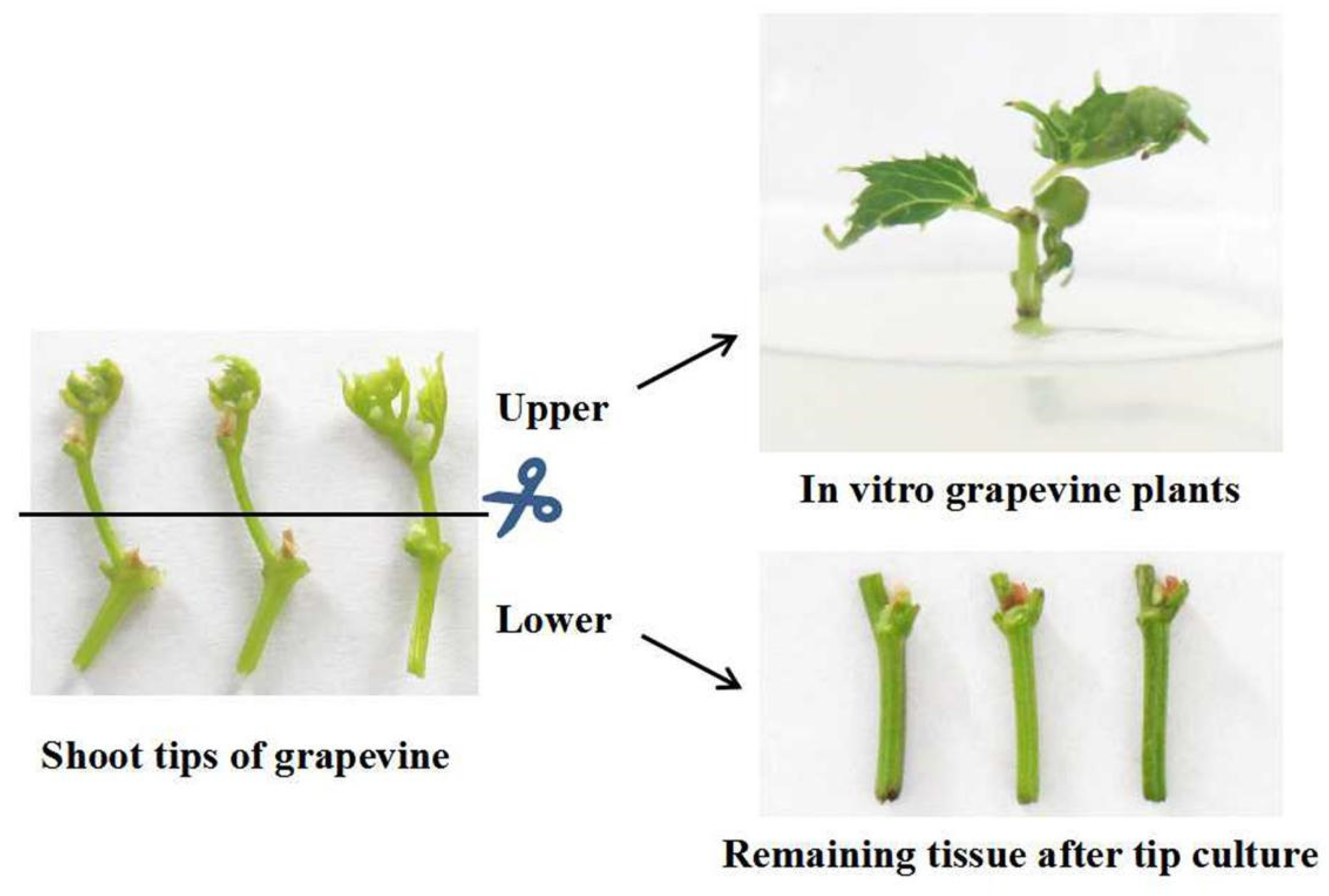
4.2. Establishment of In Vitro Cultures

Firstly, the shoot tips were cleaned under tap water, placed in sterilized glasses, and then sterilized by treating them with 75% ethanol and 0.1% mercury bichloride for 30 s and 15 min, respectively. After washing them with sterilized distilled water, the shoot tips were pruned to 1.5 cm in size and were inserted in modified 1/2MS solid medium [41]. These explants were incubated under a controlled environment (24 °C, 16 h photoperiod, and 2000 lx light intensity). The survival rates of the explants were investigated after 60 days, when the whole plants were regenerated. The presence and diversity of viruses in six grapevine cultivars was assessed before and after shoot tip culture; the virus titers in the lower and upper part of shoot tips and the residual tissue after collecting explants (the same tissue as the lower part of shoot tips) and in vitro grapevine plants were also analyzed (Figure 4). The in vitro grown plants were transferred to fresh MS medium at 45-day intervals.

4.3. RNA Extraction, cDNA Synthesis and Regular PCR Amplification

Total RNA was extracted from different materials of grapevine plants following the protocol described by Hu et al. [41]. First-strand cDNA was synthesized with M-MLV reverse transcriptase (Promega, Madison, WI, USA). A 25 μL PCR reaction was set up following the protocol described by Hu et al. [38] with specific virus primer pairs (Table 3). All these primer pairs have previously proven to be effective and polyvalent against grapevine virus variants present in China [25].

4.4. Cloning and Sequencing

The PCR products of grapevine viruses were purified using a PCR purification kit (Axygen, Hangzhou, China). Then, the purified products were inserted into the pTOPO-TA vector (Aidlab, Beijing, China) and transformed into Escherichia coli DH5α. For each grape cultivar, 13-17 positive clones selected from in vitro and in vivo materials were sequenced in both orientations. Multiple sequence alignment was conducted using the DNAMAN 5.2.2 program (Lynnon BioSoft, Vaudreuil, QC, Canada). To determine the variations during shoot tip culture, the mutation frequency/nucleotide (nt) of viral sequences was computed using the RT-PCR mutation frequency/nt subtracted from the uncorrected difference to yield total corrected mutation frequency/nt [35].

4.5. Quantitative Real-Time PCR (qPCR)

The RT Reagent Kit with gDNA Eraser (TaKaRa, Dalian, China) and SYBR® Premix Ex Taq™ Tli RNaseH Plus (TaKaRa) were used in qPCR. The ΔCt method was used to calculate the relative virus titers in different plant samples. Each sample was analyzed in triplicate.

Author Contributions

G.H. and Y.D. conceived and designed the experiments. Z.Z. and X.F. collected the samples. F.R. conducted the experiments and analyzed the data. G.H. and Y.D. discussed the results, and drafted and revised the manuscript. All authors approved the final draft of the manuscript. All authors have read and agreed to the published version of the manuscript.

Funding

This research was funded by China Agriculture Research System of MOF and MARA and the Agricultural Science and Technology Innovation Program of the Chinese Academy of Agricultural Sciences.

Institutional Review Board Statement

Not applicable.

Informed Consent Statement

Not applicable.

Data Availability Statement

All data were presented in the manuscript, so the study did not report other data.

Conflicts of Interest

The authors declare no conflict of interest.

References

  1. Espinosa-Leal, C.A.; Puente-Garza, C.A.; García-Lara, S. In vitro plant tissue culture: Means for production of biological active compounds. Planta 2018, 248, 1–18. [Google Scholar] [CrossRef] [PubMed]
  2. Doran, P.M. Foreign protein degradation and instability in plants and plant tissue cultures. Trends Biotechnol. 2006, 24, 426–432. [Google Scholar] [CrossRef] [PubMed]
  3. Mourad, A. Elimination of grapevine bois noir phytoplasma by tissue culture coupled or not with heat therapy or hot water treatment. Adv. Crop Sci. Technol. 2013, 1, 1–4. [Google Scholar] [CrossRef]
  4. Wang, M.R.; Li, B.Q.; Feng, C.H.; Wang, Q.C. Culture of shoot tips from adventitious shoots can eradicate apple stem pitting virus but fails in apple stem grooving virus. Plant Cell Tiss. Org. Cult. 2016, 125, 283–291. [Google Scholar] [CrossRef]
  5. 5. Sim, S.T.; Khuu, N.; Shoulders, J.R.; Pudlo, W.; Hoang, N.H.; Golino, D.A. Elimination of rose viruses using microshoot tip tissue culture. Acta Hortic. 2019, 1232, 241–246. [Google Scholar] [CrossRef]
  6. Kang, H.C.; Shin, J.; Kim, D.H.; Park, S.J.; Han, C.L. Elimination of apple stem grooving virus from ‘mansoo’ pear (Pyrus pyrifolia L.) by an antiviral agent combined with shoot tip culture. J. Plant Biotechnol. 2016, 43, 391–396. [Google Scholar]
  7. Hu, G.J.; Dong, Y.F.; Zhang, Z.P.; Fan, X.D.; Ren, F.; Li, Z.N. Efficacy of virus elimination from apple by thermotherapy coupled with in vivo shoot-tip grafting and in vitro meristem culture. J. Phytopathol. 2017, 165, 701–706. [Google Scholar] [CrossRef]
  8. Bettoni, J.C.; Fazio, G.; Carvalho Costa, L.; Hurtado-Gonzales, O.P.; Rwahnih, M.A.; Nedrow, A.; Volk, G.M. Thermotherapy followed by shoot tip cryotherapy eradicates latent viruses and apple hammerhead viroid from in vitro apple rootstocks. Plants 2022, 11, 582. [Google Scholar] [CrossRef]
  9. Camborde, L.; Tournier, V.; Noizet, M.; Jupin, I. A Turnip yellow mosaic virus infection system in Arabidopsis suspension cell culture. FEBS Lett. 2007, 581, 337–341. [Google Scholar] [CrossRef] [Green Version]
  10. Shih, S.M.; Doran, P.M. In vitro propagation of plant virus using different forms of plant tissue culture and modes of culture operation. J. Biotechnol. 2009, 143, 198–206. [Google Scholar] [CrossRef]
  11. Scorza, R.; Callahan, A.; Dardick, C.; Ravelandro, M.; Polak, J.; Malinowski, T.; Zagrai, I.; Cambra, M.; Kamenova, I. Genetic engineering of plum pox virus resistance: ‘Honey Sweet’ plum-from concept to product. Plant Cell, Tiss. Org. Cult. 2013, 115, 1–12. [Google Scholar] [CrossRef]
  12. Svensson, D.; Perveen, R.; Hirschfeld, C.; Robel, J.; Mühlbach, H.P. In vitro cultures of Sorbus aucuparia sustain replication of European mountain ash ringspot-associated virus (EMARaV). Plant Cell, Tiss. Org. Cult. 2014, 119, 441–445. [Google Scholar] [CrossRef]
  13. Ramgareeb, S.; Snyman, S.J.; Antwerpen, T.V.; Rutherford, R.S. Elimination of virus and rapid propagation of disease-free sugarcane (Saccharum spp. cultivar NCo376) using apical meristem culture. Plant Cell, Tiss. Org. Cult. 2010, 100, 175–181. [Google Scholar] [CrossRef]
  14. Špak, J.; Pavingerova, D.; Přibylová, J.; Špaková, V.; Sedlak, J. Blueberry red ringspot virus eliminated from highbush blueberry by shoot tip culture. Plant Protect. Sci. 2014, 50, 174–178. [Google Scholar] [CrossRef] [Green Version]
  15. Azad, M.; Hossen, M.I.; Khatun, Z.; Eaton, E.J.; Soren, E.B. Generation of virus free potato plantlets through meristem culture and their field evaluation. Am. J. Plant Sci. 2020, 11, 1827–1846. [Google Scholar] [CrossRef]
  16. Guerra, P.A.; Souza, E.H.; Andrade, E.C.; Max, D.A.S.; Oliveira, R.S.; Souza, F.V.D. Comparison of shoot tip culture and cryotherapy for eradication of ampeloviruses associated with Pineapple mealybug wilt in wild varieties. Vitro Cell Dev. Biol.-Plant 2020, 56, 903–910. [Google Scholar] [CrossRef]
  17. Yan, K.; Du, X.; Mao, B. Production of virus-Free chrysanthemum (Chrysanthemum morifolium Ramat) by tissue culture techniques. Methods in Molecular Biology 2022, 2400, 171–186. [Google Scholar]
  18. Tello, J.; Roux, C.; Chouiki, H.; Laucou, V.; Sarah, G.; Weber, A.; Santoni, S.; Flutre, T.; Pons, T.; This, P.; et al. A novel high-density grapevine (Vitis vinifera L.) integrated linkage map using GBS in a half-diallel population. Theor. Appl. Genet. 2019, 132, 2237–2252. [Google Scholar] [CrossRef]
  19. Blaisdell, G.K.; Zhang, S.; Rowhani, A.; Klaassen, V.; Almeida, R. Trends in vector-borne transmission efficiency from coinfected hosts: Grapevine leafroll-associated virus-3 and grapevine virus A. In Eur. J. Plant Pathol.; 2020; Volume 156, pp. 1163–1167. [Google Scholar]
  20. Fuchs, M. Grapevine viruses: A multitude of diverse species with simple but overall poorly adopted management solutions in the vineyard. J. Phytopathol. 2020, 102, 643–653. [Google Scholar] [CrossRef]
  21. Messmer, N.; Bohnert, P.; Schumacher, S. Studies on the occurrence of viruses in planting material of grapevines in southwestern Germany. Viruses 2021, 13, 248. [Google Scholar] [CrossRef]
  22. Han, Y.; Shi, X.; Yang, G.; Wang, X.; Liu, K.; Feng, X.; Ni, J. Shoot tip tissue virus-free culture and RT-PCR detection of GLRaV-3 in red globe grape. Chin. Agric. Sci. Bull. 2011, 27, 198–202. [Google Scholar]
  23. Hu, G.J.; Dong, Y.F.; Zhang, Z.P.; Fan, X.D.; Ren, F. Efficiency of chemotherapy combined with thermotherapy for eliminating grapevine leafroll-associated virus 3 (GLRaV-3). Sci. Hortic. 2020, 271, 109462. [Google Scholar] [CrossRef]
  24. Hu, G.J.; Dong, Y.F.; Zhang, Z.P.; Fan, X.D.; Ren, F. Elimination of grapevine fleck virus and grapevine rupestris stem pitting-associated virus from vitis vinifera ‘87-1′ by ribavirin combined with thermotherapy. J. Integr. Agr. 2021, 20, 2463–2470. [Google Scholar] [CrossRef]
  25. Li, C.; Fan, X.D.; Zhang, Z.P.; Hu, G.J.; Ren, F.; Zhang, B.D.; Dong, Y.F. Analysis of virus infection status based on RT-PCR detection of 6 flavoring grape varieties. China Fruits 2021, 2, 66–69. [Google Scholar]
  26. Wu, B.; Xu, D.K.; Jiang, S.S.; Zhang, M.; Wang, S.J.; Ma, Y.L.; Xin, Z.M. Distribution and pathogen identification of grape main virus diseases in Shandong province. Shandong Agric. Sci. 2019, 51, 73–77. [Google Scholar]
  27. Asakura, I.; Hoshino, Y. Evaluation of plant regeneration ability of different explants and establishment of an efficient regeneration system using immature seeds in actinidia kolomikta, a cold-hardy kiwifruit relative. Sci. Hortic. 2017, 220, 275–282. [Google Scholar] [CrossRef]
  28. Amano, R.; Momoi, R.; Omata, E.; Nakahara, T.; Kimura, S. Molecular and biochemical differences in leaf explants and the implication for regeneration ability in Rorippa Aquatica (Brassicaceae). Plants 2020, 9, 1372. [Google Scholar] [CrossRef] [PubMed]
  29. Okamoto, A.; Kishine, S.; Hirosawa, T. Efficient plant regeneration by smashing callus in rice (Oriza Sativa L.) cell culture. Tissue Cult. Lett. 2010, 13, 291–295. [Google Scholar] [CrossRef] [Green Version]
  30. Alkhazindar, M. Elimination of tomato ppotted wilt virus (genus Tospovirus) from infected tomato plants by meristem tip culture. Egypt. J. Bot. 2015, 55, 149–159. [Google Scholar]
  31. Klimek-Chodacka, M.; Kadluczka, D.; Lukasiewicz, A.; Malec-Pala, A.; Grzebelus, E. Effective callus induction and plant regeneration in callus and protoplast cultures of Nigella Damascena L. Plant Cell Tiss. Org. Cult. 2020, 143, 693–707. [Google Scholar] [CrossRef]
  32. Moya, A.; Elena, S.F.; Bracho, A.; Miralles, R.; Barrio, E. The evolution of RNA viruses: A population genetics view. Proc. Natl. Acad. Sci. USA 2000, 97, 6967–6973. [Google Scholar] [CrossRef] [PubMed] [Green Version]
  33. Cuevas, J.M.; Moya, A.; Sanjuán, R. Following the very initial growth of biological RNA viral clones. J. Gen. Virol. 2005, 86, 435–443. [Google Scholar] [CrossRef] [PubMed]
  34. Simmons, H.E.; Holmes, E.C.; Stephenson, A.G. Rapid turnover of intra-host genetic diversity in zucchini yellow mosaic virus. Virus Res. 2011, 155, 389–396. [Google Scholar] [CrossRef] [PubMed] [Green Version]
  35. Hu, G.J.; Wang, G.P.; Hong, N. Effect of in vitro sub-culture of sandy pear on variant structure of apple stem grooving virus. Trop. Plant Pathol. 2019, 44, 302–307. [Google Scholar] [CrossRef]
  36. Schneider, W.L.; Roossinck, M.J. Evolutionarily related Sindbis-like plant viruses maintain different levels of population diversity in a common host. J. Virol. 2000, 74, 3130–3134. [Google Scholar] [CrossRef] [Green Version]
  37. Murakishi, H.H.; Hartmann, J.X.; Pelcher, L.E.; Beachy, R.N. Improved inoculation of cultured plant cells resulting in high virus titer and crystal formation. Virology 1970, 41, 365–367. [Google Scholar] [CrossRef]
  38. Zelcer, A.; van Adelsberg, J.; Leonard, D.A.; Zaitlin, M. Plant cell suspension cultures sustain long-term replication of potato spindle tuber viroid. Virology 1981, 109, 314–322. [Google Scholar] [CrossRef]
  39. Bradamante, G.; Scheid, O.M.; Incarbone, M. Under siege: Virus control in plant meristems and progeny. Plant Cell 2021, 33, 2523–2537. [Google Scholar] [CrossRef]
  40. Waigmann, E.; Ueki, S.; Trutnyeva, K.; Citovsky, V. The ins and outs of nondestructive cell-to-cell and systemic movement of plant viruses. Crit. Rev. Plant Sci. 2004, 23, 195–250. [Google Scholar] [CrossRef]
  41. Hu, G.J.; Dong, Y.F.; Zhang, Z.P.; Fan, X.D.; Ren, F.; Li, Z.N.; Zhang, S.N. Elimination of Grapevine rupestris stem pitting-associated virus from Vitis vinifera ‘Kyoho’ by an antiviral agent combined with shoot tip culture. Sci. Hortic. 2018, 229, 99–106. [Google Scholar] [CrossRef]
  42. Osman, F.; Leutenegger, C.; Golino, D.; Rowhani, A. Comparison of low-density arrays, RT-PCR and real-time taqman RT-PCR in detection of grapevine viruses. J. Virol. Methods 2008, 149, 292–299. [Google Scholar] [CrossRef] [PubMed]
  43. Minafra, A.; Saldarelli, P.; Grieco, F.; Martelli, G.P. Nucleotide sequence of the 3′ terminal region of the RNA of two filamentous grapevine viruses. Arch. Virol. 1994, 137, 249–261. [Google Scholar] [CrossRef] [PubMed]
  44. Nolasco, G.; Mansinho, A.; Santos, M.T.; Soares, C.; Sequeira, Z.; Sequeira, C.; Sequeira, O.A. Large scale evaluation of primers for diagnosis of rupestris stem pitting associated virus-1. Eur. J. Plant Pathol. 2000, 106, 311–318. [Google Scholar] [CrossRef]
  45. Gambino, G.; Gribaudo, I. Simultaneous detection of nine grapevine viruses by multiplex reverse transcription-polymerase chain reaction with coamplification of a plant RNA as internal control. Phytopathology 2006, 96, 1223–1229. [Google Scholar] [CrossRef] [PubMed] [Green Version]
  46. Fan, X.D.; Dong, Y.F.; Zhang, Z.P.; Ren, F.; Hu, G.J.; Zhou, J. Detection and sequence analysis of grapevine leafroll-associated virus 2 isolates from China. J. Phytopathol. 2015, 163, 978–986. [Google Scholar] [CrossRef]
  47. Turturo, C.; Saldarelli, P.; Dong, Y.; Digiaro, M.; Martelli, G.P. Genetic variability and population structure of grapevine leafroll-associated virus 3 isolates. J. Gen. Virol. 2005, 86, 217–224. [Google Scholar] [CrossRef]
  48. Poojari, S.; Alabi, O.J.; Okubara, P.A.; Naidu, R.A. SYBR® Green-based real-time quantitative reverse-transcription PCR for detection and discrimination of grapevine viruses. J. Virol. Methods 2016, 235, 112–118. [Google Scholar] [CrossRef] [Green Version]
  49. Beuve, M.; Sempé, L.; Lemaire, O. A sensitive one-step real-time RT-PCR method for detecting grapevine leafroll-associated virus 2 variants in grapevine. J. Virol. Methods 2007, 141, 117–124. [Google Scholar] [CrossRef]
  50. Bester, R.; Pepler, P.T.; Burger, J.T.; Maree, H.J. Relative quantitation goes viral: An RT-qPCR assay for a grapevine virus. J. Virol. Methods 2014, 210, 67–75. [Google Scholar] [CrossRef]
  51. Wang, J.; Xi, D.; Liu, J.; Chen, K.; Li, H.; Liu, X.; Yan, S.; Ercisli, S.; Lin, H. Genetic variability in grapevine virus A from Vitis vinifera L.× Vitis labrusca L. in Sichuan, China. Turk. J.Biol. 2012, 36, 542–551. [Google Scholar] [CrossRef]
Figure 1. Factors affecting the survival of long shoot tip culture. (a) Survival regenerated grapevine plants; (b) Contamination of bacteria; (c,d) Contamination of fungi; (e) Callus of shoot tip base and browning of shoot tip top.
Plants 11 01907 g001
Figure 2. The relative concentrations of (a) GRSPaV, (b) GRLaV-3, (c) GFkV, (d) GRLaV-2, and (e) GRLaV-1 in different materials of different grapevine cultivars.
Plants 11 01907 g002
Figure 3. Assays of (a) GRLaV-2 and (b) GRSPaV in in vitro sub-culture of Cabernet Franc. M: maker II (Tiangen, China); CK-: negative control; CK+: positive control; T1-T4: passage number of in vitro sub-culture.
Plants 11 01907 g003
Figure 4. Sampling of long shoot tip culture.
Plants 11 01907 g004
Table 1. The survival and virus infection status of regenerated grapevine plants.
CultivarsOriginsIn Vivo In Vitro
Infected VirusNo. of Dissected TipsNo. of Survival Tips (%)Infected Virus
Cabernet FrancNingxia, ChinaGRSPaV, GRLaV-22010 (50.0)GRSPaV, GRLaV-2
Cabernet GernischtLiaoning, ChinaGRSPaV, GRLaV-3, GFkV200/
Cabernet SauvignonTianjin, ChinaGRSPaV, GRLaV-2, GFkV200/
WinkGuangxi, ChinaGRSPaV, GRLaV-2, GRLaV-3, GFkV2012 (60.0)GRSPaV, GRLaV-2, GRLaV-3, GFkV
VictoriaGuangxi, ChinaGRSPaV, GRLaV-2, GRLaV-3, GFkV2014 (70.0)GRSPaV, GRLaV-2, GRLaV-3, GFkV
MerlotBeijing, ChinaGRSPaV2016 (80.0)GRSPaV
MerlotSichuan, ChinaGRLaV-1, GVA2012 (60.0)GRLaV-1, GVA
GRSPaV: grapevine rupestris stem pitting-associated virus; GLRaV-1: grapevine leafroll-associated virus 1; GLRaV-2: grapevine leafroll-associated virus 2; GLRaV-3: grapevine leafroll-associated virus 3; GFkV: grapevine fleck virus; GVA: grapevine virus A.
Table 2. Comparison of nucleotide identities and differences of grapevine viruses before and after shoot tip culture.
Infected VirusCultivars No. of Clones SequencedIdentities (%)Consistency (%)Mutation Frequency/nt
In VivoIn VitroBetween Group In VivoIn VitroBetween Group In VivoIn VitroUncorrected DifferenceCorrected Difference a
GRLaV-2Wink1599.7–100.099.4–100.099.4–100.099.9499.9699.971.7 × 10−43.5 × 10−41.8 × 10−40
Victoria1599.5–100.099.2–100.099.2–100.099.8999.9199.908.7 × 10−48.7 × 10−400
Cabernet Franc1398.9–100.099.5–100.098.9–100.099.7999.8599.738.1 × 10−48.1 × 10−400
GRLaV-3Wink1599.7–100.099.5–100.099.5–100.099.9399.9399.937.1 × 10−47.1 × 10−400
Victoria1799.5–100.099.2–100.099.2–100.099.8799.8799.878.1 × 10−46.3 × 10−400
GRSPaVWink1699.2–100.099.2–100.099.1–100.099.8999.8899.836.2 × 10−47.0 × 10−40.8 × 10−40
Merlot-B1699.8–100.099.6–100.099.7–100.099.9499.9399.939.7 × 10−49.7 × 10−400
a Sporadic changes attributed to combined error (5.4 × 10⁻⁴/nt) were subtracted to yield the corrected mutation frequency/nucleotide.
Table 3. Primers used for grapevine virus detection.
VirusesPrimersSequences (5′-3′)Size (bp)Reference
Regular PCR
GFkVC1/RTGGTCCTCGGCCCAGTGAAAGTA344[42]
V1/RGGCCAGGTTGTAGTCGGTGTTGTC
GVAH587GACAAATGGCACACTACG429[43]
C995AAGCCTGACCTAGTCATCTTGG
GRSPaVRSP52TGAAGGCTTTAGGGGTTAG 905[44]
RSP53CTTAACCCAGCCTTGAAAT
GRLaV-1L1ATCTTTACCAACCCCGAGATGAA232[45]
L1BGTGTCTGGTGACGTGCTAAACG
GRLaV-2L2HSPLCARAAYAATTCGGCGTACAT386[46]
L2HSPRTAATTGGCRGGYACYGAACA
GRLaV-3LR3PUCGCTCATGGTGAAAGCAGACG653[47]
LR3PDCTTAGAACAAAAATATGGAGCAG
Quantitative real-time PCR
GFkVF1TCAAGGACTCCGTCACCTACA110This study
R1AGGATGGAGCCGCAGAT
GRSPaVY-cpf1GCACGTCACTGCTCTGATGTTGG170[41]
Y-cpr1GTCTCCAGATGGATGTTCCACACGAT
GRLaV-1GLRaV-1FGTGGAGAGTATGATTCCGTGGTCAC267[48]
GLRaV-1RCACTGGCACGTTAACTTGAGGTCG
GRLaV-2RL2 P19CTAACAATTTCTTCTTTGGATCGCAT155[49]
RL2 P24AGAATGTCTTCAGCTTCATAAGGAG
GRLaV-3LR3-F1GGGRACGGARAAGTGTTACC143[50]
LR3-R1 TCCAAYTGGGTCATRCACAA
Internal control Vivi-18Sf AAGCCCGATCCAGCAATA176[51]
Vivi-18SrGCCCTTTACGCCCAGTCA
Publisher’s Note: MDPI stays neutral with regard to jurisdictional claims in published maps and institutional affiliations.

