Review

Massive Activation of GABAA Receptors: Rundown, Ionic and Neurodegenerative Consequences

by
Sergey A. Menzikov
*,
Danila M. Zaichenko
,
Aleksey A. Moskovtsev
,
Sergey G. Morozov
and
Aslan A. Kubatiev
Institute of General Pathology and Pathophysiology, 8, Baltiyskaya st., Moscow 125315, Russia
*
Author to whom correspondence should be addressed.
Biomolecules 2025, 15(7), 1003; https://doi.org/10.3390/biom15071003
Submission received: 9 June 2025 / Revised: 9 July 2025 / Accepted: 11 July 2025 / Published: 13 July 2025

Abstract

The GABAA receptors, through a short-term interaction with a mediator, induce hyperpolarization of the membrane potential (Vm) via the passive influx of chloride ions (Cl) into neurons. The massive (or intense) activation of the GABAARs by the agonist could potentially lead to depolarization/excitation of the Vm. Although the ionic mechanisms of GABAA-mediated depolarization remain incompletely understood, a combination of the outward chloride current and the inward bicarbonate current and the resulting pH shift are the main reasons for this event. The GABAA responses are determined by the ionic gradients—neuronal pH/bicarbonate homeostasis is maintained by carbonic anhydrase and electroneutral/electrogenic bicarbonate transporters and the chloride level is maintained by secondary active cation–chloride cotransporters. Massive activation can also induce the rundown effect of the receptor function. This rundown effect partly involves phosphorylation, Ca2+ and the processes of receptor desensitization. In addition, by various methods (including fluorescence and optical genetic methods), it has been shown that massive activation of GABAARs during pathophysiological activity is also associated with an increase in [Cl]i and a decline in the pH and ATP levels in neurons. Although the relationship between the neuronal changes induced by massive activation of GABAergic signaling and the risk of developing neurodegenerative disease has been extensively studied, the molecular determinants of this process remain somewhat mysterious. The aim of this review is to summarize the data on the relationship between the massive activation of inhibitory signaling and the ionic changes in neurons. The potential role of receptor dysfunction during massive activation and the resulting ionic and metabolic disruption in neurons during the manifestation of network/seizure activity will be considered.

1. Introduction

Pentameric ligand-gated ion channels (pLGICs) are classic mediators of electrochemical signaling in the central nervous system of most kingdoms of life [1,2]. The cys-loop receptors in this family directly transduce the binding of an agonist into the opening of an intrinsic ion channel pore, enabling the passive conduction of cations or anions. These processes induce decremental fluctuations in the transmembrane potential (Vm) that dynamically control/modify the neuronal excitation/inhibition balance in the brain [3]. pLGICs have similar cylindrical structures, consisting of five subunits arranged around a central axis [4,5]. The general features of each subunit include a large hydrophilic extracellular domain (ED), four transmembrane domains (TMDs), where TMD2 forms the ionic pore, and a small extracellular C-terminal region [2]. pLGICs are the object of extensive research because of their role in regulating synaptic signaling in the healthy brain and their involvement in various neurological disorders [6].
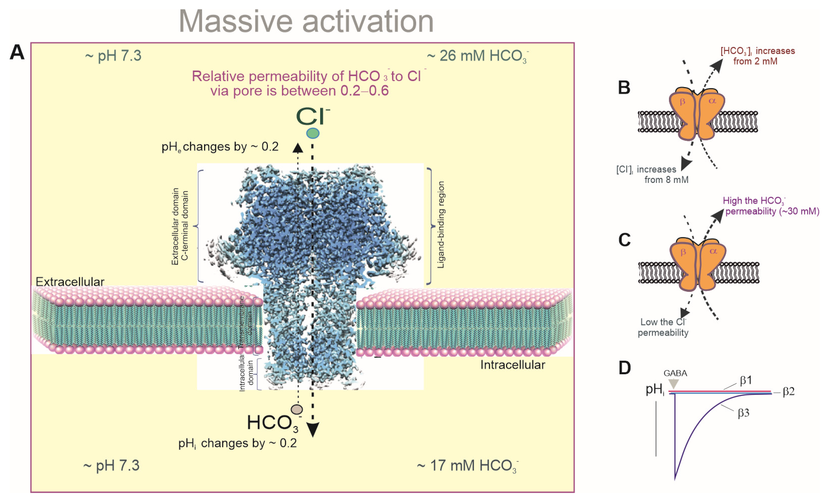
As members of the pLGIC family, γ-aminobutyric acid type A (GABAAR) receptors are the primary inhibitory receptors in the adult brain [7]. Nineteen different receptor subunits (α1–6, β1–3, γ1–3, δ, ρ1–3, ε, θ, and π) enable a variety of pentameric ensembles. The most prevalent receptor subtypes observed in the human brain have a 2α:2β:1γ composition [8,9,10]. After agonist binding with the ED of different GABAAR types [11], the receptors undergo conformational changes, causing pore opening followed by a rapid increase in the chloride ion (Cl) permeability (Figure 1A). In mature neurons, this change leads to hyperpolarization/inhibition of the Vm (Figure 1B) [12]. In addition to the hyperpolarization responses, GABAARs can induce the depolarization/excitation of the Vm, not only during embryonic and early post-embryonic development [13,14] but also in adult brains (Figure 1C) [15,16,17]. In immature neurons, GABA-mediated depolarization is associated with a distinctive electrochemical gradient for Cl, which is controlled by chloride transport systems (mainly secondary active cation–chloride cotransporters (CCCs)) [15,16]. This affects the direction and magnitude of the receptor’s effect on the neuronal excitability [17]. The depolarizing effects in mature neurons are associated with the formation of a high intracellular chloride concentration ([Cl]i), which is observed during the massive (or intense) activation of the receptors by an agonist [18] or in neurodegenerative disorders, such as seizures [19,20]. During massive activation, the receptor pores become permeable not only to Cl but also to bicarbonate (HCO3), which always flows out of neurons (Figure 1C) [21]. Such GABAA-mediated depolarization is associated with both a decrease in the intracellular HCO3 concentrations ([HCO3]i) and a fundamental shift in the intra- and extracellular pH, as shown in Figure 2A [22,23]. High HCO3 (EHCO3−) equilibrium potential is a direct consequence of lower neuronal HCO3 than extracellular HCO3 levels [24] and is sustained through secondary active HCO3 uptake via electroneutral and electrogenic Na+/HCO3 symporters (NBCN1/NBCN2), Na+/H+ exchangers (NHEs), and Na+-dependent/independent Cl/HCO3 exchanges (NCBE/NDCBE) [25,26] (Figure 1A). However, the maintenance of the neuronal HCO3 levels is directly correlated with the intracellular pH (pHi) through cytosolic carbonic anhydrase (CA) activity (Figure 2D), which rapidly converts CO2 into HCO3 under a constant intracellular pH (Figure 1A) [27,28]. Thus, the opposing transport of Cl and HCO3 through the receptor pores (Figure 2B,C) and the resulting decrease in the neuronal pH are essential factors that regulate GABAergic signaling during the massive activation of receptors in mature neurons (Figure 2A) [29]. In addition, the massive activation of GABAARs [30] can directly modulate the receptor states by altering desensitization and/or resensitization [31,32]. Indeed, over time, inhibitory receptors can become less responsive (desensitized) to GABA, but factors that prevent or delay this process can result in prolonged activation [31]. Studies conducted in recent decades have shown that prolonged receptor activation is coupled with phosphorylation mechanisms [33], the presence of Ca2+ and the state of receptor resensitization [34]. Specifically, the reduction in the neuronal ATP during massive GABAAR activation causes a decline in their functional activity, resulting in neuronal excitation. Different receptor subtypes have varying sensitivity to agonists (including GABA), and their subunit composition can influence their overall function [35]. Understanding these mechanisms is crucial for comprehending the role of various GABAAR subtypes in both normal brain function and neurodegenerative disorders.
The objective of this work is to analyze information on the relationship between the massive activation of inhibitory signaling and the ionic changes in neurons. The role of the massive activation of receptors in GABAergic depolarization and theoretical considerations regarding the mechanisms of chloride and bicarbonate ion permeability through ion channel pores are also discussed. This review summarizes the role of the transport systems involved in inhibitory synaptic plasticity and facilitating the restoration of the [Cl]i and [HCO3]i/pHi following GABAAR activity. Finally, the potential role of receptor dysfunction during massive activation and the resulting ionic and metabolic disruption in neurons during the manifestation of network/seizure activity are considered.

2. The “GABA Switch” from Inhibition to Excitation During Massive Activation

The GABAA-mediated single inhibitory postsynaptic potentials (IPSPs) can be hyperpolarizing, shunting or depolarizing under resting conditions [36,37]. Brief activation of dendritic GABAAR leads to hyperpolarization of the Vm (Figure 1B), whereas the massive activation of dendritic GABAARs induces Vm depolarization (Figure 1C) [38]. The massive activation of GABAA receptors is a physiological event that initially leads to significant inhibition, followed by excitation of the nervous system. There are several factors that can cause this event. High concentrations of GABA in the extracellular space may be the main cause of massive activation. This may be due to increased synthesis, reduced reuptake, or disrupted degradation of GABA [35]. Furthermore, the use of GABAergic modulators such as barbiturates, benzodiazepines or neurosteroids can contribute to intensive receptor activation [31]. These drugs enhance the effects of low concentrations of GABA by binding to specific sites on the receptor structure and increasing the frequency with which the channel opens [12]. However, the effect of these ligands on depolarization depends on the concentration and the specific neural context: they can both induce and inhibit it. Specifically, benzodiazepines and pentobarbital can potentiate GABAA-mediated depolarization at low concentrations but mimic GABA’s depolarizing effect at higher concentrations [39]. Thus, GABAergic ligands have the capacity to modulate substantial receptor activation by GABA. Some studies have shown that the massive activation of dendritic GABAARs induces depolarization [15,40,41]. The dual nature of the GABA response involves a fast, initial hyperpolarizing component followed by a depolarizing phase that can last several seconds. This phase outlasts the action of any possible synaptically released mediator and is followed by prolonged epileptic afterdischarges that resemble seizures [35]. It is important to note that the depolarization is strong enough to trigger spike bursts in CA1 pyramids [42,43]. This demonstrates that the signaling role of GABA can change qualitatively from inhibitory to excitatory during intense interneuronal network activity [43]. For example, the dual GABAAR responses in hippocampal pyramidal neurons can be induced by the intensive activation of hippocampal interneurons [44]. Bonnet and Bingmann showed that the postsynaptic GABAA-mediated responses of cortical neurons consisted of early somatic hyperpolarization and dendritic depolarization [45]. The depolarizing responses can be seen in structures with low volume-to-receptor-density ratios and are easily elicited in dendrites but not in somas [46,47].

2.1. Changes in pHi During Massive Activation

Early studies demonstrated that sustained GABAAR activation could lead to changes in the distribution of Cl and HCO3 and a decrease in the pHi from 7.3 to 7.1 (Figure 2A) [48,49,50,51,52]. In pyramidal neurons in adult neocortical slices with a more hyperpolarized resting membrane potential and low [Cl]i, the HCO3-mediated current component can exceed the GABA-mediated Cl current, resulting in HCO3-dependent depolarizing IPSPs, as shown in some studies [41,53,54]. The authors showed that HCO3 fluxes contributed to GABA-induced depolarization rather than to hyperpolarization, which was mainly driven by Cl flux. In a solution containing 30 mM HCO3, the GABA-mediated current increase was associated with a depolarization of approximately 15 mV, an increase in the [Cl]i, and a decrease in the [HCO3]i. During the intensive activation of GABAARs, Cl inflow was shown to overwhelm KCC2-induced Cl extrusion, resulting in the collapse of homeostasis in neurons. In such situations, the HCO3 current would predominate and drive the Vm toward the action potential threshold, which would result in GABAergic depolarization and excitation [16,24]. Further research confirmed that in pyramidal neurons in adult neocortical slices with a more hyperpolarized resting Vm and low [Cl]i, the HCO3-mediated current component could exceed the GABA-mediated Cl current and result in HCO3-dependent depolarization [55,56]. GABA-evoked dendritic depolarization has a much slower rise time than either the hyperpolarizing response or the depolarizing response seen in neurons that maintain a depolarized resting Cl reversal potential (ECl) [21].

2.2. Maintaining pHi/Bicarbonate Homeostasis

The maintenance of the acid–base balance is ensured by homeostatic ionic mechanisms, which are of critical importance for all cells, including neurons and glia [57]. The typical resting or steady-state pHi of a hippocampal neuron in CO2/HCO3-containing media is ~7.03–7.46, depending on the preparation, while the pHe is ~7.35 (Figure 1A) [58,59]. The transmembrane transporters implicated in bicarbonate/pH regulation include Na+/K+ (NKE) and Na+/H+ exchangers (NHE), Cl/HCO3 exchangers, and Na+/HCO3cotransporters (Figure 1A) [60,61]. The SLC4 family of Cl/HCO3 exchangers is one of the most well-known families of HCO3 transporters and is widely expressed in cells [62,63]. The family comprises ten members (SLC4A1–5 and A7–11). Some of these members mediate Na+-independent Cl/HCO3 exchange (AE1-AE3), while others facilitate Na+-dependent Cl/HCO3 exchange (NCBE and NDCBE) [64]. AE transporters facilitate the extrusion of HCO3 while simultaneously transporting Cl within the neuronal compartment. In contrast, NCBE and NDCBE facilitate the transport of HCO3 into the cell and Cl out of the cell (Figure 1A). Cl/HCO3 exchangers have been demonstrated to play a pivotal role in maintaining the baseline pHi levels and facilitating recovery following pH modifications [25]. Specifically, the process of acid extrusion, which typically increases the pHi, is achieved in neurons by the action of the SLC4 and SLC9 families of Na+-coupled transport proteins. The inwardly directed Na+ gradient established by Na+/K+-ATPase is utilized by both transporter families to either extrude H+ from the cell (in the case of Na+/H+ exchangers (NHEs)) or accumulate a weak base, such as HCO3−. [HCO3]i values of approximately 17 mM have been estimated [22]. Excessive alkalosis, which is probably a rather exceptional event under physiological conditions, leads to the activation of the Na+-independent anion exchanger (AE3), which extrudes base equivalents in the form of HCO3 in exchange for Cl [65,66]. AE3 mediates the countertransport of one Cl with one HCO3, leading to the depletion of [Cl]i and Cl accumulation in the extracellular medium [25]. The role of Na+-dependent Cl/HCO3 antiporters in regulating Cl extrusion to maintain neuronal [Cl]i homeostasis is not fully understood [67]. Neuronal acid extrusion is mainly achieved by Na+-driven Cl/HCO3 exchange (NDCBE) and Na+/H+ exchange [64] (Figure 1A). Both types of transporters are activated by a decrease in the pHi, which can be caused by GABAergic transmission; for example, leading to a channel-mediated net efflux of HCO3.
Buffering by CO2/HCO3 is one of the most important mechanisms of tissue pH control. The presence of a dynamic equilibrium between CO2 and H2O and the presence of H+ and HCO3 is a key feature of this system [68]. This equilibrium is quickly reached through the action of enzymes from the CAs family (see Figure 1A). Carbonic anhydrases catalyze the reversible hydration of CO2 to H2O and HCO3. Twelve catalytically active CA isoenzymes are known, five of which show cytosolic localization [69]. Bicarbonate buffering and carbonic anhydrase activity protect brain tissue from acidification by converting H+ and HCO3 into H2O and CO2. The resulting CO2 is then removed by the cerebral circulation and transported to the lungs for exhalation. Therefore, to maintain the stability of the brain tissue pH, there is a need for an effective mechanism that can supply HCO3 to the extracellular space in a way that is responsive to neuronal activity. The rapid replenishment of [HCO3]i by the activity of cytosolic CA isoforms occurs during a large GABAA-mediated net efflux of HCO3 [70]. Thus, the net extrusion of acid from neurons and glial cells is achieved by secondary active transport in which the efflux of H+ or influx of HCO3 is coupled with Na+ influx using energy stored in the transmembrane Na+ gradient. Several acid–base-coupled carriers are also involved in the transport of metabolites, including lactate and amino acids. Lactate transport via monocarboxylate transporters has been suggested to play an important role in the energy supply of neurons, leading to the “astrocyte-to-neuron lactate shuttle hypothesis” [71]. The major acid–base transporters identified in astrocytes include the electrogenic sodium–bicarbonate cotransporter 1 (NBCe1) encoded by the SLC4A4 gene, the sodium–hydrogen exchanger (NHE1) encoded by the SLC9A1 gene, the sodium-dependent chloride-bicarbonate exchanger (NCBE) encoded by the SLC4A10 gene, the chloride–bicarbonate exchanger (AE) encoded by the SLC4A1 gene, a proton pump, and vacuolar V-ATPase (Figure 1) [72]. Moreover, astrocytes express both intra- and extracellular CA isoforms, with carbonic anhydrase II (CAII) being particularly abundant. Of the various cells involved in the synaptic neuropil, astrocytes appear to be well suited to actively controlling the local brain pHe microenvironment.

2.3. Changes in [Cl]i During Massive Activation

A detailed analysis of the ionic basis of the GABAAR-mediated biphasic responses indicated that the initial part of the depolarization was caused by anionic redistribution in which the inward HCO3 current drove a depolarization that promoted Cl uptake and hence caused a positive shift in the EGABA [21,23,43]. Such studies demonstrated that neuronal transmembrane Cl transport is rate-limited. Significant changes in the dendritic [Cl]i and ECl occur because of intense GABAAR activation, and the transmembrane gradients for Cl in somas and HCO3 in dendrites are significantly less affected by large GABAAR-mediated Cl currents. Several theoretical models have been proposed to explain the ionic basis of slow post-tetanic GABAAR depolarization. Specifically, the GABAAR-induced depolarization potential has been shown to be dependent on the preservation of HCO3 and the collapse of the Cl gradients (Figure 2B) [21]. The key argument of the gradient collapse hypothesis is that influx of chloride ions through the receptor pore would lead to an increase in the [Cl]i, thereby shifting the EGABA in a positive direction. Experiments using the massive activation of dendritic GABAARs demonstrated that synaptic Cl influx through the ion channel pore could exceed the maximum transport rate. This hypothesis is reasonable, given that pH alterations would accompany dissipation of the HCO3 gradient at a constant pCO2, while the intracellular pH would be 6.3 if the EHCO3 was driven to the Vm, a process that would cause protein denaturation and neuronal damage. To avoid this type of damage, the neuronal HCO3- concentration is stabilized via the diffusion and hydration of CO2, while the pHi is supported by intracellular buffers and H+ efflux. Opposing HCO3 and Cl flux through ionic channel pores resulted in a reduction in the electrochemical gradient that drove the hyperpolarizing Cl inflow, thereby allowing the depolarizing HCO3 outflow to prevail. These studies provided estimates of the maximum transport rates for Cl of 5 to 7 mM/s and indicated that the Vmax could be exceeded by GABAAR-evoked Cl inflow into neurons. The authors reported that during depolarization, the [Cl]i increased from 8 mM, whereas the [HCO3]e increased from 2 mM (Figure 2B) [15]. The Cl accumulation model is attractive because it could explain why the enhanced synaptic release of GABA is required to elicit depolarizing responses. However, an argument against the Cl gradient collapse hypothesis is that the stability of the HCO3 gradient appears to require both extra- and intracellular CA function. However, benzolamide, an enzyme inhibitor that is confined to the extracellular space, does not block the depolarizing GABA responses [21].
An alternative model contradicts the Cl accumulation hypothesis and suggests that distinct GABAAR subtypes with special permeability for anions are present, namely lower Cl and higher HCO3 permeability through ion channel pores and slower kinetics. However, the authors were unable to identify these receptor subtypes [40,41]. In addition, when the intracellular anion concentrations were manipulated and the polarity of the giant biphasic GABA-mediated postsynaptic current was reversed, neuronal Cl accumulation did not occur during the early inward current. Thus, this result does not explain why an inward Cl/HCO3 current would switch to an outward one. The authors suggested that for neurons with a high [Cl]i, the Cl in the cell would be depleted during the early (inward current) part of the response, which would have to occur at a rate comparable to that of the Cl accumulation, and the polarity of the late response would be determined by the HCO3 gradient. Recent molecular studies support the existence of GABAAR subtypes with high HCO3 permeability, which could be responsible for the depolarizing GABAA responses. Only the homopentameric GABAAR β3 isoform, in contrast to the β1 and β2 isoforms, was shown to participate in GABA-mediated HCO3 outflow from HEK 293FT cells (Figure 2D) [73].

2.4. Maintaining [Cl]i Homeostasis

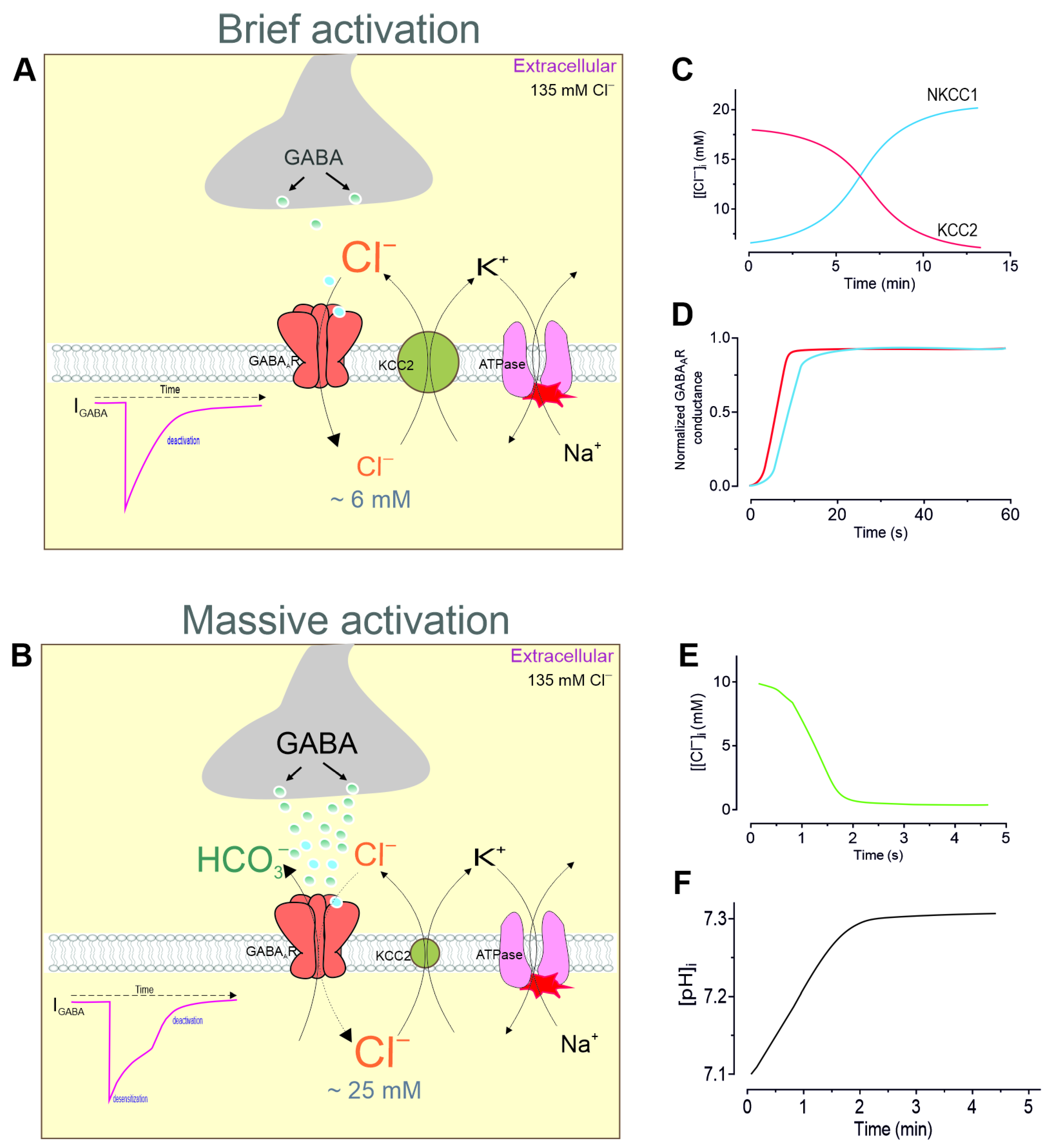
The neuronal anion concentrations are key parameters controlling the EGABA; their effect depends on the activity of the neuronal network and the stage of neuronal development [73,74]. Although neuronal chloride homeostasis is determined by several Cl-conducting systems [75], secondary active cation–chloride cotransporters (CCCs) play a key role in maintaining the [Cl]i (Figure 3A) [76]. CCCs are members of the SLC12A nine-member gene family, which have a common evolutionary origin and include four K+/Cl cotransporters (KCCs), two Na+/K+/2Cl cotransporters (NKCCs), an Na+/Cl cotransporter (NCC), and two cotransporters, CCC9 and CIP [77]. CCCs are large glycoproteins (∼110–130 kDa) with 12 transmembrane segments and intracellular amino- and carboxy-terminal domains [78]. Two members of the SLC12A family, NKCC1 and KCC2, which are expressed in the vertebrate brain and whose activity determines the switching of GABAergic signaling from excitation to inhibition, are of particular interest [17]. CCCs do not consume ATP directly but use the energy stored in the ionic gradients established by Na+/K+-ATPase to transport other ions, including Cl, against their electrochemical gradients. NKCC1 mediates Cl uptake driven by the intracellular Na+/K+ gradients (Figure 3A), whereas KCC2 extrudes Cl under physiological conditions using the K+ gradient (Figure 3B) [79,80]. NKCCs are activated by phosphorylation processes and KCCs are activated by dephosphorylation [81]. The phosphorylation pathway that targets NKCC is repressed by intracellular [Cl], leading to an inhibitory negative feedback loop when the intracellular [Cl]i reaches a sufficiently high level.
Progress in understanding the contribution of specific CCCs to GABAergic signaling has been made possible using blockers of these chloride transport systems. Specifically, the Cl gradient was shown to be reduced by the loop diuretic furosemide and to depend on the extracellular K+ [82], which together revealed a distinct neuronal K+/Cl transport process. The net transport activity of KCC2 depends on the chemical gradients of K+ and Cl and is inhibited by the diuretic furosemide [68]. When adults are treated with furosemide, there is a greater increase in the [Cl]i than when treated with P10 cells. This indicates that KCC2 activity rises with age (see Figure 1A) [83]. Although furosemide is often used to block KCC2, this drug also effectively blocks NKCC1 [84,85]. Selective KCC2 blockers (e.g., VU0463271) currently exist, while another diuretic, bumetanide, selectively blocks depolarizing GABAergic responses in immature pyramidal neurons and is considered to be a specific NKCC1 blocker [86,87]. However, despite many molecular biological studies demonstrating the involvement of CCCs [88,89,90], some studies have questioned their role in the rapid recovery of chloride homeostasis after receptor activity [22,91]. The time constant for neuronal chloride homeostasis recovery after a [Cl]i increase of about 10 mM was shown to be more than 5 min (Figure 3C) [92]. Another study showed that the KCC2-mediated extrusion of ~5 mM Cl required more than 5 min after furosemide-induced [Cl]i changes [83]. Bumetanide-sensitive NKCC1-mediated Cl uptake in immature rat neocortical neurons and human neuroblastoma cells was observed over a time interval of 5–15 min (Figure 3D) [93,94]. Gonzalez-Islas et al. demonstrated that Cl recovery following depletion in chick embryonic motoneurons exhibited both NKCC1-independent (1–10 min) and NKCC1-dependent (10–50 min) properties [95]. In contrast, the recovery of GABAAR conductance following desensitization exhibited a time constant of a few seconds (Figure 3D). Specifically, the recovery of GABA-evoked conductance after desensitization in different neurons had a time constant of approximately 13 s [96,97], while the time course of [Cl]i recovery (Figure 3E) inferred from the IGABA series was several seconds and the restoration of the pHi took about 1 min (Figure 3F). In addition, in vivo studies of mouse pyramidal neurons revealed that an increase in the [Cl]i of 12 mM, which occurred following epileptic activity, was recovered in under 30 s [98].

3. Conformational Changes During Massive Activation

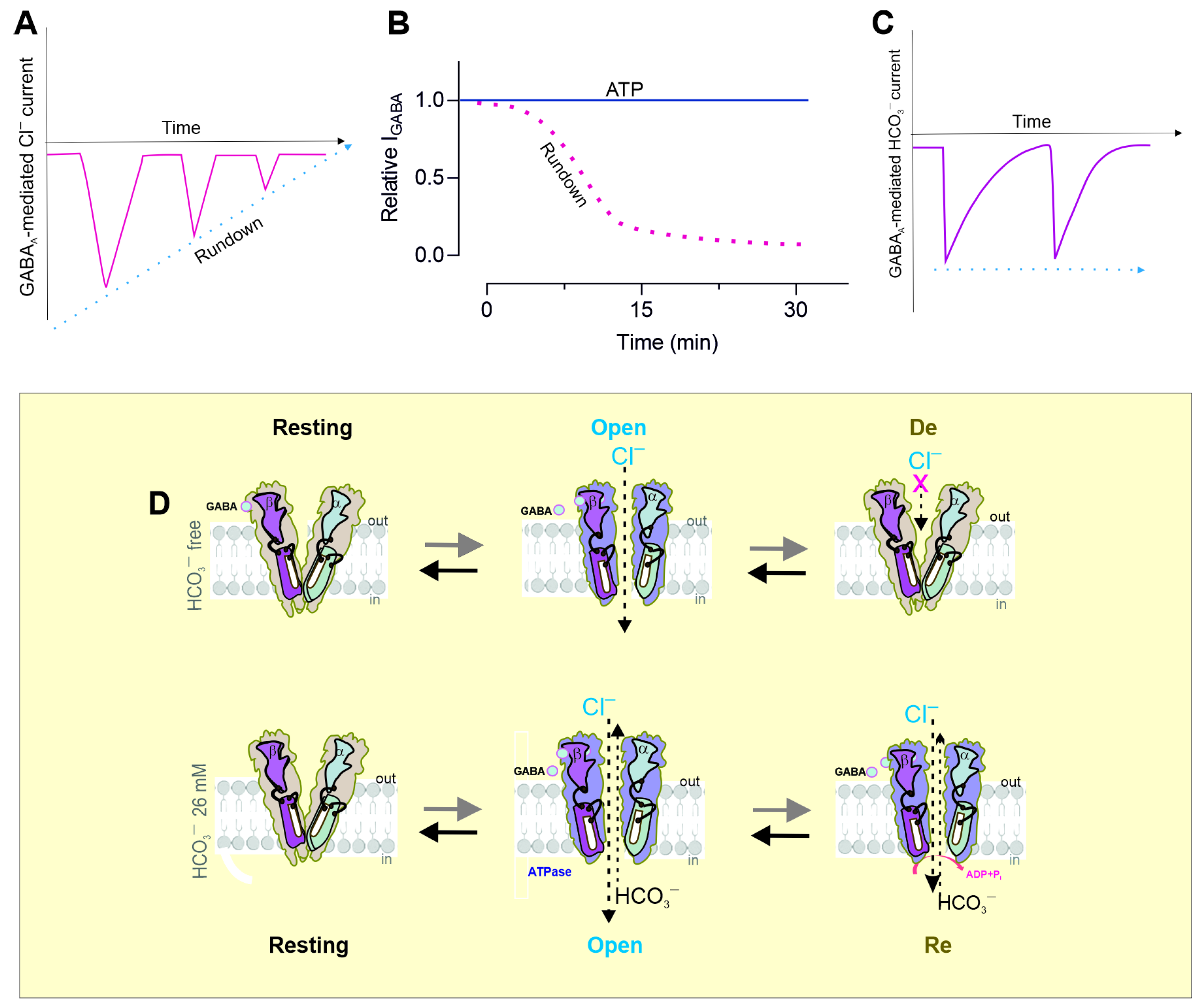
After GABA is released from vesicles, synapse receptors are activated by the brief or intensive action of an agonist. As a result, GABAARs undergo conformational changes that result in channel opening and GABAergic mIPSC induction [99]. As the GABA concentration changes are transient and short, the IPSC decay reflects the closure of channels following ligand removal, namely deactivation [34,100]. Some studies demonstrated that exposure to micromolar GABA concentrations could lead to receptor activation, the generation of sustained low-amplitude current, and the conversion of the receptor into a desensitized state [31]. Currently, four basic types of receptor function states during sustained or repeated agonist binding have been demonstrated: resting, open, desensitized, and resensitized (Figure 4D) [101,102,103,104]. GABAARs’ function is to pass through the series of states described above. The time spent in each state and, thus, the time course of the postsynaptic current are determined by the receptor’s properties and the agonist exposure profile [105,106,107]. Specifically, the expression of different receptor subtypes contributes to differences in the IPSC decay observed at various stages of development [108] and in different cell types [109]. Typically, IPSCs last considerably longer in immature neurons than in mature neurons. For example, currents activated by brief pulses of (>1 µM) GABA to outside-out patches from cultured hippocampal neurons were found to mimic GABA-mediated IPSCs, during which channels enter lengthy closed states and then subsequently reopen. Movement through these states was shown to result in paired-pulse desensitization [110]. This high-affinity state was found to disappear upon agonist removal [106]. However, [3H]GABA dissociates four-fold faster than functional recovery, indicating that desensitized receptors need not be bound by an agonist. Following the application of 1 µM GABA, approximately 5% of the α1β3γ2L subtype was found to have been functionally eliminated due to desensitization [111].
Many receptor desensitization studies have focused on the changes to receptors following massive activation with a neuromediator or allosteric drug that affects the occupancy of this state during synaptic inhibition [112,113]. Such changes may include the phosphorylation of receptor subunits by kinase activity [31] and changes in expression, clustering [114], and pharmacological selectivity. Some authors have shown that the cross-desensitization of GABAAR-mediated Cl currents is evoked by changes in the [Cl]i [96]. In contrast, other studies demonstrated that the extent of the desensitization, as assessed by the steady-state-to-peak ratio, was significantly greater at a high intracellular ([Ca2+]i) [115]. Recovery from desensitization was measured at 30 s intervals using a double-pulse protocol and found to be significantly slower at a high [Ca2+]i than at a low [Ca2+]i [116]. Receptor mutants and allosteric modulators were used to demonstrate that the desensitization of GABAARs facilitated their phosphorylation by protein kinase C (PKC), as well as increases in the number of receptors at inhibitory synapses. These observations provide physiological relevance for the desensitized state, which may act as a signal regulating the efficacy of inhibitory synapses during prolonged periods [31].
Structural studies have demonstrated that differences between the agonist-bound open state and the desensitized state are conformational changes that occur at the inner face of the receptor. However, recent functional and structural studies provide compelling evidence for a dual-gate model in which the TMD of pLGICs contains both an activation gate, which is in the upper half of the channel, and a de-gate, located on the intracellular side. Some authors have reported a very diverse and labile conformational landscape during desensitization after introducing mutations in different subunits [3]. Kinetic modeling indicated that the subunits could move independently, with the desensitization of two subunits being necessary to occlude the pores. These findings are consistent with the results of other studies. For example, differences in the conformational properties of the activation and desensitization states of GABAARs were identified using the sulfhydryl alkylating reagent n-ethylmaleimide (NEM) [104]. In particular, the important role of the conformational rearrangements and the resulting changes in the phosphorylation and dephosphorylation processes were confirmed using a mutant receptor that produced a significant alteration in the receptor’s capability to access the transition from a desensitized to a resensitized state. Receptor desensitization strongly correlates with the slowing of its deactivation, namely, resensitization [115]. The correlation between the desensitization and resensitization of GABAAR-mediated currents can be seen more clearly in response to prolonged or repeated mediator application, which allows more desensitization rather than a short pulse (Figure 4A) [113]. In particular, the reapplication of 100 μM GABA did not result in the desensitization of GABAAR-mediated HCO3 (Figure 4C). Recently, fluorescence studies also demonstrated that GABAAR-mediated HCO3 outflow in the presence of HCO3 did not occur after a second application of the agonist; instead, only resensitization occurred [103]. Furthermore, the recovery of the [HCO3]i after receptor activity, which appears to occur at ~30–40 s, was consistent with data on the recovery of GABAAR-mediated conductance after desensitization at ~40 s [22,96,104]. Thus, although the regulation of GABAAR desensitization and resensitization is an important mechanism controlled at the receptor level and by its signaling pathway [31], such regulation may be important in understanding the role of HCO3 in receptor activity. It is more likely that the change in the [HCO3]i is likely the major determinant of the onset of receptor resensitization, whereas the change in the [Cl]i in bicarbonate-free medium plays a critical role in desensitization.

4. Modulation of Desensitization by pH

Recent works have shown that pH alterations and the membrane potential affect GABAAR gating, mainly via modulation in terms of agonist binding and receptor desensitization. Their combined actions were investigated to test whether the effects of the membrane potential and pH are additive [117]. An examination of the existing responses to rapid GABA applications revealed that the current–voltage relationship was almost straight at an acidic pH. Nevertheless, an increase in the pH induced inward rectification. The process of desensitization was found to be enhanced at depolarizing potentials, although this depolarization was observed to be pH-dependent, with a weaker effect observed at an acidic pH and a stronger effect at a basic pH. A comparable pattern was identified with regard to the onset of responses to saturating GABA concentrations. Additionally, extracellular H+ was demonstrated to allosterically modulate GABAARs. Specifically, the modulation of GABAARs by H+ was shown to generate a synaptic GABA transient, highlighting the importance of the desensitization process [118]. In the case of the α1β2γ2 and α1β2 receptor subtypes, the application of H+ notably affected the current time course characterized by a reduction in the binding and desensitization rates [119]. The amplitude of the α1β2 receptor-mediated currents was found to be significantly affected by protons, while the effect on their kinetics was observed to be relatively limited. This finding indicates that the primary mechanism is likely to be direct, noncompetitive inhibition, with a lesser contribution from allosteric regulation. The evidence demonstrates that extracellular H+ has a significant impact on GABAARs.

5. Metabolic Changes During Massive Activation

Early studies of the GABAAR properties in hippocampal neurons [33], cultured chick spinal cord neurons [120], dissociated nucleus tractus solitarii [121], isolated bullfrog dorsal root ganglion neurons [122], cerebellar granule cells [123], rat pyramidal neurons [124], and hypothalamic hamartomas [125] demonstrated that the GABA-mediated Cl conductance decreased progressively with the time (maximum effect at 12 min) (Figure 4B). The inactivation of GABAARs could be prevented by the concomitant inclusion of ATP and Mg2+ at concentrations of ≥2 mM, indicating that these chemicals are necessary to prevent the inactivation of the GABA response [33]. The GABAA Cl current was also maintained by the addition of 2 mM ADP or ATPγS (adenosine 5′-O-3-thiotriphosphate) to an internal medium containing Mg2+ [124]. The nonhydrolyzable ATP analog β,γ-imidoadenosine-5′-triphosphate did not produce any effects [125]. It appears that the maintenance of GABAR function is linked to an ATP-dependent process, which is likely to be a phosphorylation event. The magnitude of the rundown was comparable in both recombinant α1β2γ2 and β2γ2 receptors [126]. Neither the stimulation nor the inhibition of protein kinase A or C significantly affected the rundown. However, the addition of sodium vanadate, an inhibitor of protein tyrosine phosphatase, led to a significant reduction in the rundown. In addition, inhibiting protein tyrosine kinase activity by either genistein or lavendustin A induced the rundown of the GABA response. The authors presented the hypothesis that an ATP-sensitive binding site exists on the cytoplasmic side of the cell membrane and that receptor activity may entail not only ATP binding but also ATP hydrolysis mechanisms [33,121,127]. Additionally, hypoxia was shown to cause the depression of GABA-mediated IPSCs in the CA1 neurons of hippocampal slices [124,128].

6. Ionic Changes During Network/Seizure Activity

Dynamic functional brain connectivity plays a pivotal role in enabling adaptive cognition and behavior. Abnormal changes in this connectivity can lead to dysfunction, which is common in a range of neurological conditions. Epilepsy is one of the most common neurological disorders, defined by the seemingly random occurrence of spontaneous seizures [129,130,131]. The epileptic brain network is made up of a set of cortical and subcortical brain regions that are structurally and functionally connected. These regions are distributed across lobes and hemispheres, and their connections and dynamics evolve over time [132]. A range of epileptic manifestations, including focal and generalized seizures, were proposed to originate from, propagate through and terminate at network nodes and edges, which also generate and maintain normal physiological brain dynamics. Network-based approaches can help us to gain a deeper understanding of the mechanisms that underlie the emergence of seizures in evolving epileptic brain networks [133]. Considering the above, concerted experimental and theoretical efforts have been made to elucidate the underlying mechanisms of seizure onset and propagation [134,135,136]. Understanding more about these mechanisms may help in developing more effective therapeutic interventions [137]. However, the current technical problems make it challenging to study their specific contribution in an experimental setting [22].
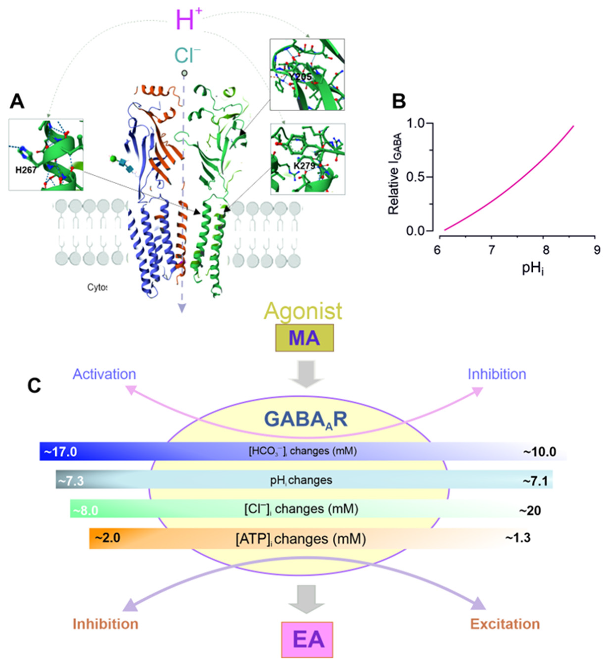
The synchronization of neuronal activity is said to be fundamentally dependent on GABAergic neurotransmission, according to experimental and theoretical evidence [138]. The role of GABAARs in epileptiform activity (EA) has received increasing attention, in part because GABA-induced Cl currents can be depolarizing and, thus, excitatory under certain circumstances [139]. Furthermore, as described in previous sections, during prolonged GABAergic stimulation (e.g., epileptic seizures), the stable depolarizing drive of GABAAR-mediated HCO3 currents will enhance the activity-dependent Cl inflow into neurons and, thus, directly contribute to the generation of excitation under these conditions [18,140,141]. For example, depolarizing shifts in the EGABA of the pyramidal neurons in the hippocampus are caused by intense receptor activation during an epileptiform discharge [142,143,144], which can switch their action such that they begin to promote rather than oppose EA [145]. However, such [Cl]i dynamics coupled with EA coincide with other changes, including increases in intracellular H+ (i.e., acidification) and reductions in extracellular H+ (Figure 5A) [146,147].
Specifically, whole-cell recordings from stratum interneurons and CA1 pyramidal cells demonstrated that some GABA-mediated currents were depolarizing during EA and were suppressed by CA blockers or activators [148,149,150]. Fluorescence and optical genetic methods have demonstrated a clear increase in the dynamic [Cl]i and pHi changes during neurological disorders [151,152,153] and increased network activity (Figure 5B). For example, during epileptiform discharge, all the GABAergic interneuron subtypes converge on elevated [Cl]i [154,155,156]. In parallel with these dynamics, EA leads to varying degrees of intracellular acidification, reflecting the baseline pH [146,157]. Călin and coauthors demonstrated that similar to pyramidal neurons, SST and VIP interneurons tended to accumulate [Cl]i during EA, whereas the neuronal chloride levels remained unchanged in the PV interneuron populations during EA [158]. Chloride imaging data allowed the authors to suggest that the GABAergic synaptic input to the different neuronal subtypes exhibits different chloride-dependent changes during EA [159]. These differences are unlikely to reflect EA-associated damage, as the ion concentrations recovered in the neuronal populations following each EA. Rather, the degree of loading across the subtypes appeared to be inversely related to the baseline chloride levels, suggesting that the initial driving force of GABAARs affects the degree of the EA-associated chloride shift. This is consistent with the observation that all the neuronal subtypes converged toward a common, high [Cl]i of approximately 20 mM during intense periods of EA [158]. Moreover, intracellular acidification was observed during EA, whereas the intracellular pH recovered between seizures.

7. Role of pH in GABAAR Modulation

The neuronal pH is determined by the equilibrium between the rate of acid influx/generation or alkali outflow/consumption and the rate of acid influx/consumption or alkali influx/generation [160]. The maintenance of a steady-state pH is the result of a balance between these processes [25,61]. The concentration of free H+ in neurons is found to be in the nanomolar range. The high buffering capacity of the cells provides a reservoir of acid equivalents in the millimolar range. The changes in the extracellular pH (pHe) accompanying physiological and pathological processes are sufficient to affect a number of important structures, such as ion channels, transporters and receptors. However, although several reports indicate that inhibitory signaling is strongly modulated by increasing or decreasing the pH [161,162,163] (Figure 5B), the physiological significance of the pH in the modulation of GABAAR activity is not fully understood. The authors hypothesized that the presence of H+ would affect the apparent binding affinity for receptors. Indeed, a significant shift in the EC50 for GABA was observed when the pH was adjusted to an acidic state, without any substantial impact on the maximal currents or the Hill coefficient [164]. Furthermore, an increase in the pH to an alkaline level was found to increase the frequency of channel opening and reduce the duration of the long-closed state, thereby raising the open probability [161]. Conversely, when the channels were exposed to an acidic pH, the outcome was the opposite: a decrease in the open probability. Other electrophysiological studies have shown that a pH (6.4) decreases and a pH (8.4) increases GABA-mediated chloride current in rat hypothalamic neurons and in α3β2γ2 GABAAR isoform stably expressed in HEK 293 cells [161,165,166]. The inhibitory effect of an acidic pH on the agonist response is reversible and concentration-dependent, resulting in a notable increase in the EC50 without a substantial change in the slope or maximal currents induced by GABA in the α1β2γL and α1β2 receptors [166]. In addition, H+ competitively inhibits the bicuculline-induced antagonism on mediator effects in the α1β2γL receptors. Other authors have also demonstrated that, within the pH range of 5.0–9.0, an increase in the extracellular pH enhances, while a decrease suppresses, the GABA-mediated Cl current in rat primary sensory neurons [167], as well as in freshly isolated rat dorsal root ganglia neurons, in a reversible and concentration-dependent manner (Figure 5B) [168]. The IC50 for these neurons was 7.1 pH. By contrast, several studies have demonstrated that the GABA-mediated chloride currents in rat cerebellar granule cells (CGCs) increase at an acidic pH of 6.4 and decrease at a basic pH of 8.4 [169,170]. The rectification of chloride channel conductance is associated with the protonation of base amino acids at an acidic pH. For example, in 6–12-day-old rat hippocampal CA3 neurons, it was demonstrated that increasing the extracellular pH to 8.5 or reducing it to 6.0 markedly enhanced or diminished the frequency and amplitude of spontaneous GABAAR-mediated postsynaptic currents [171].

8. Role of β3 Subunit in pHi Changes and Seizure Activity

Several studies have shown that the impact of H+ on the functional activity of the GABAARs depends on the subunit composition. Although it is accepted that protons can control the probability of ion channels opening, the specific amino acid residues and receptor subunits that are responsible for H+-mediated modification are not well understood (see Figure 5A). To illustrate this, an elevation in the pH resulted in a reduction in the amplitudes of the currents evoked by saturating [GABA] for the α1β2γ2 and α1β2 subtypes. However, this effect was more pronounced for the α1β2 isoforms [119]. At pH 6.4, GABA activation of the αβγ receptors was enhanced at low GABA concentrations. A further decrease in the pH to 5.4 inhibited the GABAAR-mediated responses via αβγ receptors, while the mediator responses via αβ isoforms were enhanced. Inserting homologous β-subunit amino acid residues into the γ2 subunit in order to recreate the H+ modulatory profile of the αβ and αβγ receptor isoforms revealed that, in the presence of β2H267, the γ2T294K mutation is necessary in order to enhance the mediator response at pH 5.4 (Figure 5A). This residue, T294, is structurally similar to K279 in the β subunit. The authors suggested that the presence of a lysine at this position plays a key role in mediating the allosteric effects of changes in the pH, rather than forming a direct site for protonation within the GABAAR [29]. Replacing all the conserved external histidine residues with alanine on the β subunits demonstrated that H267, which is located in the TM2 domain, plays a key role in regulating H+ passage activity. The contrasting functional impacts of Zn2+ and H+ on this single histidine residue are probably due to differences in the charge delocalization on the imidazole rings within the receptor structure. Additional replacement of H267 in the β subunit with charged side-chain residues (such as glutamate and lysine) shows that this region of the ion channel significantly affects the functional properties of receptors [172]. A comparable report found that site-specific mutations of βY205 to F or α1F64 to A—both of which affect agonist binding—resulted in a notable decline in the pH sensitivity of the GABA response [164]. Other research found that the mediator response was reversibly and concentration-dependently inhibited by an acidic pH. A significant increase in the EC50 was observed without any notable change in the slope or maximal currents induced by GABA at the α1β2γ2L and α1β2 isoforms [166]. It is worth noting that H+ was effective in counteracting the bicuculline-induced antagonism of the agonist in the α1β2γ2L isoforms. The homomeric β3 subunit has recently been identified as an H+-gated anion channel [173]. Specifically, it has been demonstrated that a single H267A mutation in the β3 subunit completely eliminates channel activation by protons. Molecular dynamics simulations of the β3 crystal structure revealed that protonation of H267 enhances the number of hydrogen bonds formed between the H267 and E270 of the neighboring subunit. This resulted in the formation of a pore-stabilizing ring and Cl accumulation within the transmembrane pore. The activation of GABA-mediated Cl/HCO3 currents at physiological pH changes (pH50 is in the range 6.0–6.3) and kinetic studies suggest a physiological role in neuronal and non-neuronal tissues that express the β3 subunit [173].
Numerous works have emphasized the pivotal role of the β3 subunit in shaping the functional characteristics of GABAARs [174]. Specifically, β3 is the only subunit that determines the ionic selectivity of the recombinant receptor and can form a functional homomeric ion channel pore [175]. Furthermore, only the β3 subunit can fully maintain or restore receptor activity to control levels in the hippocampus when the β1–β2 subunit is knocked out [176]. The β3 subunit is also widely expressed in the immature and adult brain in circuits involved in seizure generation, such as the cortex, hippocampus, and thalamic reticular nucleus [177,178]. Genetic variants associated with developmental and epileptic encephalopathies have been identified in the GABRB3 gene, which is responsible for encoding the β3 subunit. The absence of the β3 subunit results in a lower production of functional receptors in null-β3 mice. Pharmacological evidence indicates that this reduction is not compensated for by other β subunits. Recent research has revealed that gain-of-function GABRB3 variants can increase or decrease the properties of receptor desensitization and that there is a correlation with the degree of disease severity [179]. Variants that reduced desensitization at equilibrium were found to be clustered within the transmembrane regions that constitute the channel pore, and they were found to be associated with greater disease severity. Conversely, variants that accelerated current decay were found to be clustered within the coupling loops responsible for receptor activation, and they were also found to be associated with lower severity.

9. Conclusions and Future Directions

Although receptor pores were shown to be permeable not only to chloride but also to bicarbonate ions over 30 years ago, the role and mechanisms of ionic changes during GABAergic depolarization remain somewhat mysterious. Bicarbonate is involved in the GABAA-mediated current only in the adult brain and under certain conditions. Here, we showed that the massive activation of GABAARs and the resulting ionic and metabolic changes in neurons can be considered to be a single pool of interrelated events (Figure 5C). Furthermore, the analysis of the data presented in this review suggests that if bicarbonate is involved in GABAergic depolarization/excitation, it may be the primary cause of these changes in neuronal metabolism. These changes are likely to occur because of the dysfunction of the β3-containing receptor subtypes. Furthermore, protons can directly regulate receptor function by modulating desensitization or resensitization. These results are consistent with other studies. For example, HCO3 may modulate neuronal activity in a potentially pH-independent manner through the allosteric modulation of intracellular messengers. Nevertheless, it is crucial to recognize that no current, objective analytical compartment can be used to study and anticipate the heterogeneity associated with the propagation dynamics of seizure activity. Here, we demonstrate the β subunits to be important targets for the proton modulation of receptor function and drivers of a cascade of molecular events leading to the manifestation of neurodegenerative disorders. The analysis of the relationship between these processes could be useful in understanding the physiological and pathological processes in the brain. However, clear insight into the mechanisms of network or seizure activity requires the detailed structural analysis of the conformational rearrangements.

Author Contributions

S.A.M.: Conceptualization, writing—original draft. D.M.Z.: Formal analysis, validation, writing—review and editing. A.A.M.: Formal analysis, validation, writing—review and editing. S.G.M.: Project administration, resources, writing—review and editing. A.A.K.: Project administration, resources, writing—review and editing. All authors have read and agreed to the published version of the manuscript.

Funding

This research received Institutional funding at the expense of the State Assignment (topic ”Pathophysiological mechanisms of development of chronic pain, epileptogenesis, emotional and cognitive disorders and new possibilities of their pathogenetic therapy”, FGFU-2025-0012).

Acknowledgments

We thank Olga Menzikova for the help with the preparation of the figures.

Conflicts of Interest

The authors declare that they have no known competing financial interests or personal relationships that could have appeared to influence the work reported in this paper.

References

  1. Hu, H.; Howard, R.J.; Bastolla, U.; Lindahl, E.; Delarue, M. Structural basis for allosteric transitions of a multidomain pentameric ligand-gated ion channel. Proc. Natl. Acad. Sci. USA 2020, 117, 13437–13446. [Google Scholar] [CrossRef] [PubMed]
  2. Petroff, J.T.; Dietzen, N.M.; Santiago-McRae, E.; Deng, B.; Washington, M.S.; Chen, L.J.; Moreland, K.T.; Deng, Z.; Rau, M.; Fitzpatricket, J.A.J.; et al. Open-channel structure of a pentameric ligand-gated ion channel reveals a mechanism of leaflet-specific phospholipid modulation. Nat. Commun. 2022, 13, 7017. [Google Scholar] [CrossRef] [PubMed]
  3. Gielen, M.; Corringer, P.J. The dual-gate model for pentameric ligand-gated ion channels activation and desensitization. J. Physiol. 2018, 596, 1873–1902. [Google Scholar] [CrossRef]
  4. Howard, R.J. Elephants in the dark: Insights and incongruities in pentameric ligand-gated ion channel models. J. Mol. Biol. 2021, 433, 167128. [Google Scholar] [CrossRef] [PubMed]
  5. Pless, S.A.; Sivilotti, L.G. A tale of ligands big and small: An update on how pentameric ligand-gated ion channels interact with agonists and proteins. Curr. Opin. Physiol. 2019, 2, 19–26. [Google Scholar] [CrossRef]
  6. Lara, C.O.; Burgos, C.F.; Moraga-Cid, G.; Carrasco, M.A.; Yévenes, G.E. Pentameric ligand-gated ion channels as pharmacological targets against chronic pain. Front. Pharmacol. 2020, 11, 167. [Google Scholar] [CrossRef]
  7. Goetz, T.; Arslan, A.; Wisden, W.; Wulff, P. GABA(A) receptors: Structure and function in the basal ganglia. Prog. Brain Res. 2007, 160, 21–41. [Google Scholar] [CrossRef]
  8. Sigel, E.; Steinmann, M.E. Structure, function, and modulation of GABA(A) receptors. J. Biol. Chem. 2012, 287, 40224–40231. [Google Scholar] [CrossRef]
  9. Zhu, S.; Noviello, C.M.; Teng, J.; Walsh, R.M., Jr.; Kim, J.J.; Hibbs, R.E. Structure of a human synaptic GABAA receptor. Nature 2018, 559, 67–72. [Google Scholar] [CrossRef]
  10. Borghese, C.M.; Wang, H.L.; McHardy, S.F.; Messing, R.O.; Trudell, J.R.; Harris, R.A.; Bertaccini, E.J. Modulation of α1β3γ2 GABAA receptors expressed in X. laevis oocytes using a propofol photoswitch tethered to the transmembrane helix. Proc. Natl. Acad. Sci. USA 2021, 118, e2008178118. [Google Scholar] [CrossRef]
  11. Iorio, M.T.; Vogel, F.D.; Koniuszewski, F.; Scholze, P.; Rehman, S.; Simeone, X.; Schnürch, M.; Mihovilovic, M.D.; Ernst, M. GABAA Receptor Ligands Often Interact with Binding Sites in the Transmembrane Domain and in the Extracellular Domain-Can the Promiscuity Code Be Cracked? Int. J. Mol. Sci. 2020, 21, 334. [Google Scholar] [CrossRef] [PubMed]
  12. Ghit, A.; Assal, D.; Al-Shami, A.S.; Hussein, D.E.E. GABAA receptors: Structure, function, pharmacology, and related disorders. J. Genet. Eng. Biotechnol. 2021, 19, 123. [Google Scholar] [CrossRef]
  13. Herbison, A.E.; Moenter, S.M. Depolarising and hyperpolarising actions of GABA(A) receptor activation on gonadotrophin-releasing hormone neurones: Towards an emerging consensus. J. Neuroendocrinol. 2011, 23, 557–569. [Google Scholar] [CrossRef] [PubMed]
  14. Raimondo, J.V.; Richards, B.A.; Woodin, M.A. Neuronal chloride and excitability—The big impact of small changes. Curr. Opin. Neurobiol. 2017, 43, 35–42. [Google Scholar] [CrossRef]
  15. Staley, K.J.; Soldo, B.L.; Proctor, W.R. Ionic mechanisms of neuronal excitation by inhibitory GABAA receptors. Science 1995, 269, 977–981. [Google Scholar] [CrossRef]
  16. Kilb, W. When Are Depolarizing GABAergic Responses Excitatory? Front. Mol. Neurosci. 2021, 14, 747835. [Google Scholar] [CrossRef] [PubMed]
  17. Pressey, J.C.; de Saint-Rome, M.; Raveendran, V.A.; Woodin, M.A. Chloride transporters controlling neuronal excitability. Physiol. Rev. 2023, 103, 1095–1135. [Google Scholar] [CrossRef]
  18. Isomura, Y.; Sugimoto, M.; Fujiwara-Tsukamoto, Y.; Yamamoto-Muraki, S.; Yamada, J.; Fukuda, A. Synaptically activated Cl accumulation responsible for depolarizing GABAergic responses in mature hippocampal neurons. J. Neurophysiol. 2003, 90, 2752–2756. [Google Scholar] [CrossRef]
  19. Jin, X.; Huguenard, J.R.; Prince, D.A. Impaired Cl extrusion in layer V pyramidal neurons of chronically injured epileptogenic neocortex. J. Neurophysiol. 2005, 93, 2117–2126. [Google Scholar] [CrossRef]
  20. Kan, A.S.H.; Kusay, A.S.; Mohammadi, N.A.; Lin, S.X.N.; Liao, V.W.Y.; Lesca, G.; Souci, S.; Milh, M.; Christophersen, P.; Chebib, M.; et al. Understanding paralogous epilepsy-associated GABAA receptor variants: Clinical implications, mechanisms, and potential pitfalls. Proc. Natl. Acad. Sci. USA 2024, 121, e2413011121. [Google Scholar] [CrossRef]
  21. Staley, K.J.; Proctor, W.R. Modulation of mammalian dendritic GABA(A) receptor function by the kinetics of Cl and HCO3 transport. J. Physiol. 1999, 519, 693–712. [Google Scholar] [CrossRef]
  22. Lombardi, A.; Jedlicka, P.; Luhmann, H.J.; Kilb, W. Interactions between Membrane Resistance, GABA-A Receptor Properties, Bicarbonate Dynamics and Cl-Transport Shape Activity-Dependent Changes of Intracellular Cl Concentration. Int. J. Mol. Sci. 2019, 20, 1416. [Google Scholar] [CrossRef]
  23. Sulis Sato, S.; Artoni, P.; Landi, S.; Cozzolino, O.; Parra, R.; Pracucci, E.; Trovato, F.; Szczurkowska, J.; Luin, S.; Arosio, D.; et al. Simultaneous two-photon imaging of intracellular chloride concentration and pH in mouse pyramidal neurons in vivo. Proc. Natl. Acad. Sci. USA 2017, 114, 8770–8779. [Google Scholar] [CrossRef]
  24. Kim, D.Y.; Fenoglio, K.A.; Kerrigan, J.F.; Rho, J.M. Bicarbonate contributes to GABAA receptor-mediated neuronal excitation in surgically resected human hypothalamic hamartomas. Epilepsy Res. 2009, 83, 89–93. [Google Scholar] [CrossRef] [PubMed]
  25. Ruffin, V.A.; Salameh, A.I.; Boron, W.F.; Parker, M.D. Intracellular pH regulation by acid-base transporters in mammalian neurons. Front. Physiol. 2014, 5, 43. [Google Scholar] [CrossRef] [PubMed]
  26. Lee, H.J.; Park, H.J.; Lee, S.; Kim, Y.H.; Choi, I. The sodium-driven chloride/bicarbonate exchanger NDCBE in rat brain is upregulated by chronic metabolic acidosis. Brain Res. 2011, 1377, 13–20. [Google Scholar] [CrossRef] [PubMed]
  27. Sinning, A.; Hübner, C.A. Minireview: pH and synaptic transmission. FEBS Lett. 2013, 587, 1923–1928. [Google Scholar] [CrossRef]
  28. Ruusuvuori, E.; Huebner, A.K.; Kirilkin, I.; Yukin, A.Y.; Blaesse, P.; Helmy, M.; Kang, H.J.; El Muayed, M.; Hennings, J.C.; Voipio, J.; et al. Neuronal carbonic anhydrase VII provides GABAergic excitatory drive to exacerbate febrile seizures. EMBO J. 2013, 32, 2275–2286. [Google Scholar] [CrossRef]
  29. Wilkins, M.E.; Hosie, A.M.; Smart, T.G. Proton modulation of recombinant GABA(A) receptors: Influence of GABA concentration and the beta subunit TM2-TM3 domain. J. Physiol. 2005, 567, 365–377. [Google Scholar] [CrossRef]
  30. Halbhuber, L.; Achtner, C.; Luhmann, H.J.; Sinning, A.; Kilb, W. Coincident Activation of Glutamate Receptors Enhances GABAA Receptor-Induced Ionic Plasticity of the Intracellular Cl-Concentration in Dissociated Neuronal Cultures. Front. Cell. Neurosci. 2019, 13, 497. [Google Scholar] [CrossRef]
  31. Field, M.; Dorovykh, V.; Thomas, P.; Smart, T.G. Physiological role for GABAA receptor desensitization in the induction of long-term potentiation at inhibitory synapses. Nat. Commun. 2021, 12, 2112. [Google Scholar] [CrossRef]
  32. Tia, S.; Wang, J.F.; Kotchabhakdi, N.; Vicini, S. Distinct deactivation and desensitization kinetics of recombinant GABAA receptors. Neuropharmacology 1996, 35, 1375–1382. [Google Scholar] [CrossRef]
  33. Stelzer, A.; Kay, A.R.; Wong, R.K. GABAA-receptor function in hippocampal cells is maintained by phosphorylation factors. Science 1988, 241, 339–341. [Google Scholar] [CrossRef] [PubMed]
  34. Menzikov, S.A.; Zaichenko, D.M.; Moskovtsev, A.A.; Morozov, S.G.; Kubatiev, A.A. Physiological Role of ATPase for GABAA Receptor Resensitization. Int. J. Mol. Sci. 2022, 23, 5320. [Google Scholar] [CrossRef] [PubMed]
  35. Glykys, J.; Mody, I. Activation of GABAA receptors: Views from outside the synaptic cleft. Neuron 2007, 56, 763–770. [Google Scholar] [CrossRef] [PubMed]
  36. Lambert, N.; Grover, L. The mechanism of biphasic GABA responses. Science 1995, 269, 928–929. [Google Scholar] [CrossRef]
  37. Peerboom, C.; Wierenga, C.J. The postnatal GABA shift: A developmental perspective. Neurosci. Biobehav. Rev. 2021, 124, 179–192. [Google Scholar] [CrossRef]
  38. Chavas, J.; Marty, A. Coexistence of excitatory and inhibitory GABA synapses in the cerebellar interneuron network. J. Neurosci. 2003, 23, 2019–2031. [Google Scholar] [CrossRef]
  39. Backus, K.H.; Kettenmann, H.; Schachner, M. Effect of benzodiazepines and pentobarbital on the GABA-induced depolarization in cultured astrocytes. Glia 1988, 1, 132–140. [Google Scholar] [CrossRef]
  40. Perkins, K.L.; Wong, R.K. The depolarizing GABA response. Can. J. Physiol. Pharmacol. 1997, 75, 516–519. [Google Scholar] [CrossRef]
  41. Perkins, K.L.; Wong, R.K. Ionic basis of the postsynaptic depolarizing GABA response in hippocampal pyramidal cells. J. Neurophysiol. 1996, 76, 3886–3894. [Google Scholar] [CrossRef] [PubMed]
  42. Dallwig, R.; Deitmer, J.W.; Backus, K.H. On the mechanism of GABA-induced currents in cultured rat cortical neurons. Pflugers Arch. 1999, 437, 289–297. [Google Scholar] [CrossRef]
  43. Zeng, X.J.; Tietz, E.I. Role of bicarbonate ion in mediating decreased synaptic conductance in benzodiazepine tolerant hippocampal CA1 pyramidal neurons. Brain Res. 2000, 868, 202–214. [Google Scholar] [CrossRef]
  44. Smirnov, S.; Paalasmaa, P.; Uusisaari, M.; Voipio, J.; Kaila, K. Pharmacological isolation of the synaptic and nonsynaptic components of the GABA-mediated biphasic response in rat CA1 hippocampal pyramidal cells. J. Neurosci. 1999, 19, 9252–9260. [Google Scholar] [CrossRef]
  45. Bonnet, U.; Bingmann, D. GABAA-responses of CA3 neurones: Contribution of bicarbonate and of Cl-extrusion mechanisms. Neuroreport 1995, 6, 700–704. [Google Scholar] [CrossRef] [PubMed]
  46. Grover, L.M.; Lambert, N.A.; Schwartzkroin, P.A.; Teyler, T.J. Role of HCO3 ions in depolarizing GABAA receptor-mediated responses in pyramidal cells of rat hippocampus. J. Neurophysiol. 1993, 69, 1541–1555. [Google Scholar] [CrossRef]
  47. Branchereau, P.; Cattaert, D.; Delpy, A.; Allain, A.E.; Martin, E.; Meyrand, P. Depolarizing GABA/glycine synaptic events switch from excitation to inhibition during frequency increases. Sci. Rep. 2016, 6, 21753. [Google Scholar] [CrossRef] [PubMed]
  48. Mason, M.J.; Mattsson, K.; Pasternack, M.; Voipio, J.; Kaila, K. Postsynaptic fall in intracellular pH and increase in surface pH caused by efflux of formate and acetate anions through GABA-gated channels in crayfish muscle fibres. Neuroscience 1990, 34, 359–368. [Google Scholar] [CrossRef]
  49. Kulik, A.; Nishimaru, H.; Ballanyi, K. Role of bicarbonate and chloride in GABA- and glycine-induced depolarization and [Ca2+]i rise in fetal rat motoneurons in situ. J. Neurosci. 2000, 20, 7905–7913. [Google Scholar] [CrossRef]
  50. Chen, J.C.; Chesler, M.A. Bicarbonate-dependent increase in extracellular pH mediated by GABAA receptors in turtle cerebellum. Neurosci. Lett. 1990, 116, 130–135. [Google Scholar] [CrossRef]
  51. Chesler, M.; Chen, J.C. Alkaline extracellular pH shifts generated by two transmitter-dependent mechanisms. Can. J. Physiol. Pharmacol. 1992, 70, 286–292. [Google Scholar] [CrossRef]
  52. Lückermann, M.; Trapp, S.; Ballanyi, K. GABA- and glycine-mediated fall of intracellular pH in rat medullary neurons in situ. J. Neurophysiol. 1997, 77, 1844–1852. [Google Scholar] [CrossRef]
  53. Gulledge, A.T.; Stuart, G.J. Excitatory actions of GABA in the cortex. Neuron 2003, 37, 299–309. [Google Scholar] [CrossRef] [PubMed]
  54. Ma, B.F.; Xie, M.J.; Zhou, M. Bicarbonate efflux via GABA(A) receptors depolarizes membrane potential and inhibits two-pore domain potassium channels of astrocytes in rat hippocampal slices. Glia 2012, 60, 1761–1772. [Google Scholar] [CrossRef] [PubMed]
  55. Kaila, K.; Voipio, J.; Paalasmaa, P.; Pasternack, M.; Deisz, R.A. The role of bicarbonate in GABAA receptor-mediated IPSPs of rat neocortical neurones. J. Physiol. 1993, 464, 273–289. [Google Scholar] [CrossRef] [PubMed]
  56. Zhang, Q.; Jian, L.; Yao, D.; Rao, B.; Xia, Y.; Hu, K.; Li, S.; Shen, Y.; Cao, M.; Qin, A.; et al. The structural basis of the pH-homeostasis mediated by the Cl/HCO3 exchanger, AE2. Nat. Commun. 2023, 14, 1812. [Google Scholar] [CrossRef]
  57. Schwiening, C.J.; Boron, W.F. Regulation of intracellular pH in pyramidal neurones from the rat hippocampus by Na+-dependent Cl-HCO3 exchange. J. Physiol. 1994, 475, 59–67. [Google Scholar] [CrossRef]
  58. Bouyer, P.; Bradley, S.R.; Zhao, J.; Wang, W.; Richerson, G.B.; Boron, W.F. Effect of extracellular acid-base disturbances on the intracellular pH of neurones cultured from rat medullary raphe or hippocampus. J. Physiol. 2004, 559, 85–101. [Google Scholar] [CrossRef]
  59. Bouyer, P.G.; Salameh, A.I.; Zhou, Y.; Kolba, T.N.; Boron, W.F. Effects of extracellular metabolic acidosis and out-of-equilibrium CO2/HCO3 solutions on intracellular pH in cultured rat hippocampal neurons. Front. Physiol. 2024, 15, 1434359. [Google Scholar] [CrossRef]
  60. Hamm, L.L.; Nakhoul, N.; Hering-Smith, K.S. Acid-Base Homeostasis. Clin. J. Am. Soc. Nephrol. 2015, 10, 2232–2242. [Google Scholar] [CrossRef]
  61. Li, Y.; Zhou, X.; Sun, S.X. Hydrogen, Bicarbonate, and Their Associated Exchangers in Cell Volume Regulation. Front. Cell Dev. Biol. 2021, 9, 683686. [Google Scholar] [CrossRef]
  62. Casey, J.R.; Sly, W.S.; Shah, G.N.; Alvarez, B.V. Bicarbonate homeostasis in excitable tissues: Role of AE3 Cl/HCO3 exchanger and carbonic anhydrase XIV interaction. Am. J. Physiol. Cell Physiol. 2009, 297, 1091–1102. [Google Scholar] [CrossRef]
  63. Liu, Y.; Yang, J.; Chen, L.M. Structure and Function of SLC4 Family Transporters. Front. Physiol. 2015, 6, 355. [Google Scholar] [CrossRef] [PubMed]
  64. Wang, C.Z.; Yano, H.; Nagashima, K.; Seino, S. The Na+-driven Cl/HCO3 exchanger. Cloning, tissue distribution, and functional characterization. J. Biol. Chem. 2000, 275, 35486–35490. [Google Scholar] [CrossRef] [PubMed]
  65. Deitmer, J.W.; Theparambil, S.M.; Ruminot, I.; Becker, H.M. The role of membrane acid/base transporters and carbonic anhydrases for cellular pH and metabolic processes. Front. Neurosci. 2015, 8, 430. [Google Scholar] [CrossRef] [PubMed]
  66. Shcheynikov, N.; Son, A.; Hong, J.H.; Yamazaki, O.; Ohana, E.; Kurtz, I.; Shin, D.M.; Muallem, S. Intracellular Cl as a signaling ion that potently regulates Na+/HCO3 transporters. Proc. Natl. Acad. Sci. USA 2015, 112, 329–337. [Google Scholar] [CrossRef]
  67. Hübner, C.A.; Holthoff, K. Anion transport and GABA signaling. Front. Cell. Neurosci. 2013, 7, 177. [Google Scholar] [CrossRef]
  68. Roos, A.; Boron, W.F. Intracellular pH. Physiol. Rev. 1981, 61, 296–434. [Google Scholar] [CrossRef]
  69. Ruusuvuori, E.; Li, H.; Huttu, K.; Palva, J.M.; Smirnov, S.; Rivera, C.; Kaila, K.; Voipio, J. Carbonic anhydrase isoform VII acts as a molecular switch in the development of synchronous gamma-frequency firing of hippocampal CA1 pyramidal cells. J. Neurosci. 2004, 24, 2699–2707. [Google Scholar] [CrossRef]
  70. Pasternack, M.; Smirnov, S.; Kaila, K. Proton modulation of functionally distinct GABAA receptors in acutely isolated pyramidal neurons of rat hippocampus. Neuropharmacology 1996, 35, 1279–1288. [Google Scholar] [CrossRef]
  71. Deitmer, J.W.; Theparambil, S.M.; Ruminot, I.; Noor, S.I.; Becker, H.M. Energy Dynamics in the Brain: Contributions of Astrocytes to Metabolism and pH Homeostasis. Front. Neurosci. 2019, 13, 1301. [Google Scholar] [CrossRef] [PubMed]
  72. Blumenfeld, H. What is a seizure network? Long-range network consequences of focal seizures. Adv. Exp. Med. Biol. 2014, 813, 63–70. [Google Scholar] [CrossRef] [PubMed]
  73. Menzikov, S.A.; Zaichenko, D.M.; Moskovtsev, A.A.; Morozov, S.G.; Kubatiev, A.A. Zinc Inhibits the GABAAR/ATPase during Postnatal Rat Development: The Role of Cysteine Residue. Int. J. Mol. Sci. 2023, 24, 2764. [Google Scholar] [CrossRef] [PubMed]
  74. Halverson, H.E.; Kim, J.; Freeman, J.H. Dynamic Changes in Local Activity and Network Interactions among the Anterior Cingulate, Amygdala, and Cerebellum during Associative Learning. J. Neurosci. 2023, 43, 8385–8402. [Google Scholar] [CrossRef]
  75. Ferrini, F.; Perez-Sanchez, J.; Ferland, S.; Lorenzo, L.E.; Godin, A.G.; Plasencia-Fernandez, I.; Cottet, M.; Castonguay, A.; Wang, F.; Salio, C.; et al. Differential chloride homeostasis in the spinal dorsal horn locally shapes synaptic metaplasticity and modality-specific sensitization. Nat. Commun. 2020, 11, 3935. [Google Scholar] [CrossRef]
  76. Schulte, J.T.; Wierenga, C.J.; Bruining, H. Chloride transporters and GABA polarity in developmental, neurological and psychiatric conditions. Neurosci. Biobehav. Rev. 2018, 90, 260–271. [Google Scholar] [CrossRef]
  77. Schmidt, T.; Ghaffarian, N.; Philippot, C.; Seifert, G.; Steinhäuser, C.; Pape, H.C.; Blaesse, P. Differential regulation of chloride homeostasis and GABAergic transmission in the thalamus. Sci. Rep. 2018, 8, 13929. [Google Scholar] [CrossRef]
  78. Hartmann, A.M.; Tesch, D.; Nothwang, H.G.; Bininda-Emonds, O.R. Evolution of the cation chloride cotransporter family: Ancient origins, gene losses, and subfunctionalization through duplication. Mol. Biol. Evol. 2014, 31, 434–447. [Google Scholar] [CrossRef]
  79. Hartmann, A.M.; Nothwang, H.G. Molecular and evolutionary insights into the structural organization of cation chloride cotransporters. Front. Cell. Neurosci. 2015, 8, 470. [Google Scholar] [CrossRef]
  80. Chamma, I.; Chevy, Q.; Poncer, J.C.; Lévi, S. Role of the neuronal K-Cl co-transporter KCC2 in inhibitory and excitatory neurotransmission. Front. Cell. Neurosci. 2012, 21, 5. [Google Scholar] [CrossRef]
  81. Khirug, S.; Huttu, K.; Ludwig, A.; Smirnov, S.; Voipio, J.; Rivera, C.; Kaila, K.; Khiroug, L. Distinct properties of functional KCC2 expression in immature mouse hippocampal neurons in culture and in acute slices. Eur. J. Neurosci. 2005, 21, 899–904. [Google Scholar] [CrossRef]
  82. He, Y.; Ji, W.; Huang, H. Effect of the cation-chloride cotransporter inhibitor furosemide in a rat model of postoperative pain. Pain Med. 2011, 12, 1427–1434. [Google Scholar] [CrossRef] [PubMed]
  83. Kakazu, Y.; Uchida, S.; Nakagawa, T.; Akaike, N.; Nabekura, J. Reversibility and cation selectivity of the K+-Cl cotransport in rat central neurons. J. Neurophysiol. 2000, 84, 281–288. [Google Scholar] [CrossRef]
  84. Yelhekar, T.D.; Druzin, M.; Johansson, S. Contribution of Resting Conductance, GABAA-Receptor Mediated Miniature Synaptic Currents and Neurosteroid to Chloride Homeostasis in Central Neurons. eNeuro 2017, 4, ENEURO.0019-17.2017. [Google Scholar] [CrossRef] [PubMed]
  85. McMoneagle, E.; Zhou, J.; Zhang, S.; Huang, W.; Josiah, S.S.; Ding, K.; Wang, Y.; Zhang, J. Neuronal K+-Cl cotransporter KCC2 as a promising drug target for epilepsy treatment. Acta Pharmacol. Sin. 2024, 45, 1–22. [Google Scholar] [CrossRef]
  86. Prael, F.J., III; Kim, K.; Du, Y.; Spitznagel, B.D.; Sulikowski, G.A.; Delpire, E.; Weaver, C.D. Discovery of Small Molecule KCC2 Potentiators Which Attenuate In Vitro Seizure-Like Activity in Cultured Neurons. Front. Cell Dev. Biol. 2022, 10, 912812. [Google Scholar] [CrossRef] [PubMed]
  87. Xu, J.C.; Lytle, C.; Zhu, T.T.; Payne, J.A.; Benz, E., Jr.; Forbush, B. Molecular cloning and functional expression of the bumetanide-sensitive Na-K-Cl cotransporter. Proc. Natl. Acad. Sci. USA 1994, 91, 2201–2205. [Google Scholar] [CrossRef]
  88. Wang, C.; Shimizu-Okabe, C.; Watanabe, K.; Okabe, A.; Matsuzaki, H.; Ogawa, T.; Mori, N.; Fukuda, A.; Sato, K. Developmental changes in KCC1, KCC2, and NKCC1 mRNA expressions in the rat brain. Brain Res. Dev. Brain Res. 2002, 139, 59–66. [Google Scholar] [CrossRef]
  89. Williams, J.R.; Payne, J.A. Cation transport by the neuronal K+-Cl cotransporter KCC2: Thermodynamics and kinetics of alternate transport modes. Am. J. Physiol. Cell Physiol. 2004, 287, C919–C931. [Google Scholar] [CrossRef]
  90. Zhu, L.; Lovinger, D.; Delpire, E. Cortical neurons lacking KCC2 expression show impaired regulation of intracellular chloride. J. Neurophysiol. 2005, 93, 1557–1568. [Google Scholar] [CrossRef]
  91. Doyon, N.; Vinay, L.; Prescott, S.A.; De Koninck, Y. Chloride Regulation: A Dynamic Equilibrium Crucial for Synaptic Inhibition. Neuron 2016, 89, 1157–1172. [Google Scholar] [CrossRef]
  92. DeFazio, R.A.; Keros, S.; Quick, M.W.; Hablitz, J.J. Potassium-coupled chloride cotransport controls intracellular chloride in rat neocortical pyramidal neurons. J. Neurosci. 2000, 20, 8069–8076. [Google Scholar] [CrossRef]
  93. Sun, D.; Murali, S.G. Stimulation of Na+-K+-2Cl cotransporter in neuronal cells by excitatory neurotransmitter glutamate. Am. J. Physiol. 1998, 275, 772–779. [Google Scholar] [CrossRef] [PubMed]
  94. Achilles, K.; Okabe, A.; Ikeda, M.; Shimizu-Okabe, C.; Yamada, J.; Fukuda, A.; Luhmann, H.J.; Kilb, W. Kinetic properties of Cl uptake mediated by Na+-dependent K+-2Cl cotransport in immature rat neocortical neurons. J. Neurosci. 2007, 27, 8616–8627. [Google Scholar] [CrossRef] [PubMed]
  95. Gonzalez-Islas, C.; Chub, N.; Wenner, P. NKCC1 and AE3 appear to accumulate chloride in embryonic motoneurons. J. Neurophysiol. 2009, 101, 507–518. [Google Scholar] [CrossRef] [PubMed]
  96. Karlsson, U.; Druzin, M.; Johansson, S. Cl concentration changes and desensitization of GABA(A) and glycine receptors. J. Gen. Physiol. 2011, 138, 609–626. [Google Scholar] [CrossRef]
  97. Chen, M.; Wang, J.; Jiang, J.; Zheng, X.; Justice, N.J.; Wang, K.; Ran, X.; Li, Y.; Huo, Q.; Zhang, J.; et al. APP modulates KCC2 expression and function in hippocampal GABAergic inhibition. eLife 2017, 6, e20142. [Google Scholar] [CrossRef]
  98. Deisz, R.A.; Lehmann, T.N.; Horn, P.; Dehnicke, C.; Nitsch, R. Components of neuronal chloride transport in rat and human neocortex. J. Physiol. 2011, 589, 1317–1347. [Google Scholar] [CrossRef]
  99. Brickley, S.G.; Cull-Candy, S.G.; Farrant, M. Single-channel properties of synaptic and extrasynaptic GABAA receptors suggest differential targeting of receptor subtypes. J. Neurosci. 1999, 19, 2960–2973. [Google Scholar] [CrossRef]
  100. Sallard, E.; Letourneur, D.; Legendre, P. Electrophysiology of ionotropic GABA receptors. Cell. Mol. Life Sci. 2021, 78, 5341–5370. [Google Scholar] [CrossRef]
  101. Bianchi, M.T.; Macdonald, R.L. Slow phases of GABA(A) receptor desensitization: Structural determinants and possible relevance for synaptic function. J. Physiol. 2002, 544, 3–18. [Google Scholar] [CrossRef] [PubMed]
  102. Gielen, M.; Barilone, N.; Corringer, P.J. The desensitization pathway of GABAA receptors, one subunit at a time. Nat. Commun. 2020, 11, 5369. [Google Scholar] [CrossRef]
  103. Böhm, S.K.; Khitin, L.M.; Grady, E.F.; Aponte, G.; Payan, D.G.; Bunnett, N.W. Mechanisms of desensitization and resensitization of proteinase-activated receptor-2. J. Biol. Chem. 1996, 271, 22003–22016. [Google Scholar] [CrossRef]
  104. Kaila, K.; Saarikoski, J.; Voipio, J. Mechanism of action of GABA on intracellular pH and on surface pH in crayfish muscle fibres. J. Physiol. 1990, 427, 241–260. [Google Scholar] [CrossRef] [PubMed]
  105. Barberis, A.; Mozrzymas, J.W.; Ortinski, P.I.; Vicini, S. Desensitization and binding properties determine distinct alpha1beta2gamma2 and alpha3beta2gamma2 GABA(A) receptor-channel kinetic behavior. Eur. J. Neurosci. 2007, 5, 2726–2740. [Google Scholar] [CrossRef] [PubMed]
  106. Chang, Y.; Ghansah, E.; Chen, Y.; Ye, J.; Weiss, D.S. Desensitization mechanism of GABA receptors revealed by single oocyte binding and receptor function. J. Neurosci. 2002, 22, 7982–7990. [Google Scholar] [CrossRef]
  107. Terejko, K.; Michałowski, M.A.; Iżykowska, I.; Dominik, A.; Brzóstowicz, A.; Mozrzymas, J.W. Mutations at the M2 and M3 Transmembrane Helices of the GABAARs α1 and β2 Subunits Affect Primarily Late Gating Transitions Including Opening/Closing and Desensitization. ACS Chem. Neurosci. 2021, 12, 2421–2436. [Google Scholar] [CrossRef]
  108. Huntsman, M.M.; Huguenard, J.R. Fast IPSCs in rat thalamic reticular nucleus require the GABAA receptor beta1 subunit. J. Physiol. 2006, 572, 459–475. [Google Scholar] [CrossRef]
  109. Rotaru, D.C.; Olezene, C.; Miyamae, T.; Povysheva, N.V.; Zaitsev, A.V.; Lewis, D.A.; Gonzalez-Burgos, G. Functional properties of GABA synaptic inputs onto GABA neurons in monkey prefrontal cortex. J. Neurophysiol. 2015, 113, 1850–1861. [Google Scholar] [CrossRef]
  110. Jones, M.V.; Westbrook, G.L. The impact of receptor desensitization on fast synaptic transmission. Trends Neurosci. 1996, 19, 96–101. [Google Scholar] [CrossRef]
  111. Pierce, S.R.; Germann, A.L.; Evers, A.S.; Steinbach, J.H.; Akk, G. Reduced Activation of the Synaptic-Type GABAA Receptor Following Prolonged Exposure to Low Concentrations of Agonists: Relationship between Tonic Activity and Desensitization. Mol. Pharmacol. 2020, 98, 762–769. [Google Scholar] [CrossRef] [PubMed]
  112. Gravielle, M.C.C. Regulation of GABAA receptors by prolonged exposure to endogenous and exogenous ligands. Neurochem. Int. 2018, 118, 96–104. [Google Scholar] [CrossRef]
  113. Kang, Y.; Saito, M.; Toyoda, H. Molecular and Regulatory Mechanisms of Desensitization and Resensitization of GABAA Receptors with a Special Reference to Propofol/Barbiturate. Int. J. Mol. Sci. 2020, 21, 563. [Google Scholar] [CrossRef]
  114. Chen, L.; Wang, H.; Vicini, S.; Olsen, R.W. The γ-aminobutyric acid type A (GABAA) receptor-associated protein (GABARAP) promotes GABAA receptor clustering and modulates the channel kinetics. Proc. Natl. Acad. Sci. USA 2000, 97, 11557–11562. [Google Scholar] [CrossRef] [PubMed]
  115. Toyoda, H.; Saito, M.; Sato, H.; Tanaka, T.; Ogawa, T.; Yatani, H.; Kawano, T.; Kanematsu, T.; Hirata, M.; Kang, Y. Enhanced desensitization followed by unusual resensitization in GABAA receptors in phospholipase C-related catalytically inactive protein-1/2 double-knockout mice. Pflugers Arch. 2015, 467, 267–284. [Google Scholar] [CrossRef]
  116. Mozrzymas, J.W.; Cherubini, E. Changes in intracellular calcium concentration affect desensitization of GABAA receptors in acutely dissociated P2-P6 rat hippocampal neurons. J. Neurophysiol. 1998, 79, 1321–1328. [Google Scholar] [CrossRef] [PubMed]
  117. Pytel, M.; Mercik, K.; Mozrzymas, J.W. The voltage dependence of GABAA receptor gating depends on extracellular pH. Neuroreport 2005, 16, 1951–1954. [Google Scholar] [CrossRef]
  118. Mozrzymas, J.W.; Zarnowska, E.D.; Pytel, M.; Mercik, K. Modulation of GABA(A) receptors by hydrogen ions reveals synaptic GABA transient and a crucial role of the desensitization process. J. Neurosci. 2003, 23, 7981–7992. [Google Scholar] [CrossRef]
  119. Mercik, K.; Pytel, M.; Cherubini, E.; Mozrzymas, J.W. Effect of extracellular pH on recombinant alpha1beta2gamma2 and alpha1beta2 GABAA receptors. Neuropharmacology 2006, 51, 305–314. [Google Scholar] [CrossRef]
  120. Gyenes, M.; Farrant, M.; Farb, D.H. “Run-down” of gamma-aminobutyric acid A receptor function during whole-cell recording: A possible role for phosphorylation. Mol. Pharmacol. 1988, 34, 719–723. [Google Scholar] [CrossRef]
  121. Shirasaki, T.; Aibara, K.; Akaike, N. Direct modulation of GABAA receptor by intracellular ATP in dissociated nucleus tractus solitarii neurones of rat. J. Physiol. 1992, 449, 551–572. [Google Scholar] [CrossRef] [PubMed]
  122. Bradshaw, D.J.; Simmons, M.A. gamma-Aminobutyric acid A receptor function is modulated by cyclic GMP. Brain Res. Bull. 1995, 37, 67–72. [Google Scholar] [CrossRef] [PubMed]
  123. Amico, C.; Cupello, A.; Fossati, C.; Robello, M. Involvement of phosphatase activities in the run-down of GABA(A) receptor function in rat cerebellar granule cells in culture. Neuroscience 1998, 84, 529–535. [Google Scholar] [CrossRef]
  124. Harata, N.; Wu, J.; Ishibashi, H.; Ono, K.; Akaike, N. Run-down of the GABAA response under experimental ischaemia in acutely dissociated CA1 pyramidal neurones of the rat. J. Physiol. 1997, 500, 673–688. [Google Scholar] [CrossRef] [PubMed]
  125. Li, G.; Yang, K.; Zheng, C.; Liu, Q.; Chang, Y.; Kerrigan, J.F.; Wu, J. Functional rundown of gamma-aminobutyric acid(A) receptors in human hypothalamic hamartomas. Ann. Neurol. 2011, 69, 664–672. [Google Scholar] [CrossRef]
  126. Huang, R.Q.; Dillon, G.H. Maintenance of recombinant type A gamma-aminobutyric acid receptor function: Role of protein tyrosine phosphorylation and calcineurin. J. Pharmacol. Exp. Ther. 1998, 286, 243–255. [Google Scholar] [CrossRef]
  127. Rapallino, M.V.; Cupello, A.; Hydén, H. An electrogenic ionic pump derived from an ionotropic receptor: Assessment of a candidate. Cell. Mol. Neurobiol. 1999, 19, 681–690. [Google Scholar] [CrossRef]
  128. Katchman, A.N.; Vicini, S.; Hershkowitz, N. Mechanism of early anoxia-induced suppression of the GABAA-mediated inhibitory postsynaptic current. J. Neurophysiol. 1994, 71, 1128–1138. [Google Scholar] [CrossRef]
  129. Lehnertz, K.; Bröhl, T.; Wrede, R.V. Epileptic-network-based prediction and control of seizures in humans. Neurobiol. Dis. 2023, 181, 106098. [Google Scholar] [CrossRef]
  130. Khambhati, A.N.; Chang, E.F.; Baud, M.O.; Rao, V.R. Hippocampal network activity forecasts epileptic seizures. Nat. Med. 2024, 30, 2787–2790. [Google Scholar] [CrossRef]
  131. Pedersen, M.; Pardoe, H.; Mito, R.; Sethi, M.; Vaughan, D.N.; Carney, P.W.; Jackson, G.D. Brain network changes after the first seizure: An insight into medication response? Brain Commun. 2024, 6, fcae328. [Google Scholar] [CrossRef] [PubMed]
  132. Steinmetz, P.N.; Wait, S.D.; Lekovic, G.P.; Rekate, H.L.; Kerrigan, J.F. Firing behavior and network activity of single neurons in human epileptic hypothalamic hamartoma. Front. Neurol. 2013, 4, 210. [Google Scholar] [CrossRef] [PubMed]
  133. Fujiwara-Tsukamoto, Y.; Isomura, Y.; Takada, M. Comparable GABAergic mechanisms of hippocampal seizurelike activity in posttetanic and low-Mg2+ conditions. J. Neurophysiol. 2006, 95, 2013–2019. [Google Scholar] [CrossRef] [PubMed]
  134. Wu, J.; Gao, M.; Shen, J.X.; Qiu, S.F.; Kerrigan, J.F. Mechanisms of intrinsic epileptogenesis in human gelastic seizures with hypothalamic hamartoma. CNS Neurosci. Ther. 2015, 21, 104–111. [Google Scholar] [CrossRef]
  135. Giacopelli, G.; Tegolo, D.; Migliore, M. The role of network connectivity on epileptiform activity. Sci. Rep. 2021, 11, 20792. [Google Scholar] [CrossRef]
  136. Sinha, N.; Joshi, R.B.; Sandhu, M.R.S.; Netoff, T.I.; Zaveri, H.P.; Lehnertz, K. Perspectives on Understanding Aberrant Brain Networks in Epilepsy. Front. Netw. Physiol. 2022, 2, 868092. [Google Scholar] [CrossRef]
  137. Cifelli, P.; Palma, E.; Roseti, C.; Verlengia, G.; Simonato, M. Changes in the sensitivity of GABAA current rundown to drug treatments in a model of temporal lobe epilepsy. Front. Cell. Neurosci. 2013, 7, 108. [Google Scholar] [CrossRef]
  138. Sakimoto, Y.; Oo, P.M.; Goshima, M.; Kanehisa, I.; Tsukada, Y.; Mitsushima, D. Significance of GABAA Receptor for Cognitive Function and Hippocampal Pathology. Int. J. Mol. Sci. 2021, 22, 12456. [Google Scholar] [CrossRef] [PubMed]
  139. Mazzuferi, M.; Palma, E.; Martinello, K.; Maiolino, F.; Roseti, C.; Fucile, S.; Fabene, P.F.; Schio, F.; Pellitteri, M.; Sperk, G.; et al. Enhancement of GABA(A)-current run-down in the hippocampus occurs at the first spontaneous seizure in a model of temporal lobe epilepsy. Proc. Natl. Acad. Sci. USA 2010, 107, 3180–3185. [Google Scholar] [CrossRef]
  140. Galanopoulou, A.S. GABA(A) receptors in normal development and seizures: Friends or foes? Curr. Neuropharmacol. 2008, 6, 1–20. [Google Scholar] [CrossRef]
  141. Kim, Y.S.; Yoon, B.E. Altered GABAergic Signaling in Brain Disease at Various Stages of Life. Exp. Neurobiol. 2017, 26, 122–131. [Google Scholar] [CrossRef]
  142. Fujiwara-Tsukamoto, Y.; Isomura, Y.; Imanishi, M.; Fukai, T.; Takada, M. Distinct types of ionic modulation of GABA actions in pyramidal cells and interneurons during electrical induction of hippocampal seizure-like network activity. Eur. J. Neurosci. 2007, 25, 2713–2725. [Google Scholar] [CrossRef]
  143. Lillis, K.P.; Kramer, M.A.; Mertz, J.; Staley, K.J.; White, J.A. Pyramidal cells accumulate chloride at seizure onset. Neurobiol. Dis. 2012, 47, 358–366. [Google Scholar] [CrossRef] [PubMed]
  144. Ellender, T.J.; Raimondo, J.V.; Irkle, A.; Lamsa, K.P.; Akerman, C.J. Excitatory effects of parvalbumin-expressing interneurons maintain hippocampal epileptiform activity via synchronous afterdischarges. J. Neurosci. 2014, 34, 15208–15222. [Google Scholar] [CrossRef] [PubMed]
  145. D’Antuono, M.; Louvel, J.; Köhling, R.; Mattia, D.; Bernasconi, A.; Olivier, A.; Turak, B.; Devaux, A.; Pumain, R.; Avoli, M. GABAA receptor-dependent synchronization leads to ictogenesis in the human dysplastic cortex. Brain 2004, 127, 1626–1640. [Google Scholar] [CrossRef] [PubMed]
  146. Xiong, Z.Q.; Saggau, P.; Stringer, J.L. Activity-dependent intracellular acidification correlates with the duration of seizure activity. J. Neurosci. 2000, 20, 1290–1296. [Google Scholar] [CrossRef]
  147. Raimondo, J.V.; Joyce, B.; Kay, L.; Schlagheck, T.; Newey, S.E.; Srinivas, S.; Akerman, C.J. A genetically-encoded chloride and pH sensor for dissociating ion dynamics in the nervous system. Front. Cell. Neurosci. 2013, 7, 202. [Google Scholar] [CrossRef]
  148. Perez Velazquez, J.L. Bicarbonate-dependent depolarizing potentials in pyramidal cells and interneurons during epileptiform activity. Eur. J. Neurosci. 2003, 18, 1337–1342. [Google Scholar] [CrossRef]
  149. Magheru, C.; Magheru, S.; Coltau, M.; Hoza, A.; Moldovan, C.; Sachelarie, L.; Gradinaru, I.; Hurjui, L.L.; Marc, F.; Farcas, D.M. Antiepileptic Drugs and Their Dual Mechanism of Action on Carbonic Anhydrase. J. Clin. Med. 2022, 11, 2614. [Google Scholar] [CrossRef]
  150. Poggetti, V.; Salerno, S.; Baglini, E.; Barresi, E.; Da Settimo, F.; Taliani, S. Carbonic Anhydrase Activators for Neurodegeneration: An Overview. Molecules 2022, 27, 2544. [Google Scholar] [CrossRef]
  151. Liu, R.; Wang, J.; Liang, S.; Zhang, G.; Yang, X. Role of NKCC1 and KCC2 in Epilepsy: From Expression to Function. Front. Neurol. 2020, 10, 1407. [Google Scholar] [CrossRef] [PubMed]
  152. Ciccone, L.; Cerri, C.; Nencetti, S.; Orlandini, E. Carbonic Anhydrase Inhibitors and Epilepsy: State of the Art and Future Perspectives. Molecules 2021, 26, 6380. [Google Scholar] [CrossRef] [PubMed]
  153. Pracucci, E.; Graham, R.T.; Alberio, L.; Nardi, G.; Cozzolino, O.; Pillai, V.; Pasquini, G.; Saieva, L.; Walsh, D.; Landi, S.; et al. Daily rhythm in cortical chloride homeostasis underpins functional changes in visual cortex excitability. Nat. Commun. 2023, 14, 7108. [Google Scholar] [CrossRef]
  154. Huberfeld, G.; Wittner, L.; Clemenceau, S.; Baulac, M.; Kaila, K.; Miles, R.; Rivera, C. Perturbed chloride homeostasis and GABAergic signaling in human temporal lobe epilepsy. J. Neurosci. 2007, 27, 9866–9873. [Google Scholar] [CrossRef] [PubMed]
  155. Briggs, S.W.; Galanopoulou, A.S. Altered GABA signaling in early life epilepsies. Neural. Plast. 2011, 2011, 527605. [Google Scholar] [CrossRef]
  156. Wang, Y.; Wang, Y.; Chen, Z. Double-edged GABAergic synaptic transmission in seizures: The importance of chloride plasticity. Brain Res. 2018, 1701, 126–136. [Google Scholar] [CrossRef]
  157. Elverson, K.; Freeman, S.; Manson, F.; Warwicker, J. Computational Investigation of Mechanisms for pH Modulation of Human Chloride Channels. Molecules 2023, 28, 5753. [Google Scholar] [CrossRef]
  158. Călin, A.; Waseem, T.; Raimondo, J.V.; Newey, S.E.; Akerman, C.J. A genetically targeted ion sensor reveals distinct seizure-related chloride and pH dynamics in GABAergic interneuron populations. iScience 2023, 26, 106363. [Google Scholar] [CrossRef]
  159. Benarroch, E.E. GABAA receptor heterogeneity, function, and implications for epilepsy. Neurology 2007, 20, 612–614. [Google Scholar] [CrossRef]
  160. Boron, W.F. Intracellular pH Regulation. In Membrane Transport Processes in Organized Systems; Andreoli, T.E., Hoffman, J.F., Fanestil, D.D., Schultz, S.G., Eds.; Springer: Boston, MA, USA, 1987. [Google Scholar] [CrossRef]
  161. Huang, R.Q.; Dillon, G.H. Effect of extracellular pH on GABA-activated current in rat recombinant receptors and thin hypothalamic slices. J. Neurophysiol. 1999, 82, 1233–1243. [Google Scholar] [CrossRef]
  162. Pasternack, M.; Bountra, C.; Voipio, J.; Kaila, K. Influence of extracellular and intracellular pH on GABA-gated chloride conductance in crayfish muscle fibres. Neuroscience 1992, 47, 921–929. [Google Scholar] [CrossRef] [PubMed]
  163. Limon, A.; Delbruck, E.; Yassine, A.; Pandya, D.; Myers, R.M.; Barchas, J.D.; Lee, F.; Schatzberg; Watson, S.J.; Akil, H.; et al. Electrophysiological evaluation of extracellular spermine and alkaline pH on synaptic human GABAA receptors. Transl. Psychiatry 2019, 9, 218. [Google Scholar] [CrossRef] [PubMed]
  164. Huang, R.Q.; Chen, Z.; Dillon, G.H. Molecular basis for modulation of recombinant alpha1beta2gamma2 GABAA receptors by protons. J. Neurophysiol. 2004, 92, 883–894. [Google Scholar] [CrossRef]
  165. Chen, Z.L.; Huang, R.Q. Extracellular pH modulates GABAergic neurotransmission in rat hypothalamus. Neuroscience 2014, 271, 64–76. [Google Scholar] [CrossRef]
  166. Wang, M.D.; Rahman, M.; Zhu, D. Protons inhibit Cl conductance by direct or allosteric interaction with the GABA-binding site in the rat recombinant alpha1beta2gamma2L and alpha1beta2 GABAA receptor. Eur. J. Pharmacol. 2005, 528, 1–6. [Google Scholar] [CrossRef]
  167. Zhai, I.J.; Peoples, R.W.; Li, C. Proton inhibition of GABA-activated current in rat primary sensory neurons. Pflugers Arch. 1998, 435, 539–545. [Google Scholar] [CrossRef]
  168. Kisiel, M.; Jatczak-Śliwa, M.; Mozrzymas, J.W. Protons modulate gating of recombinant α1β2γ2 GABAA receptor by affecting desensitization and opening transitions. Neuropharmacology 2019, 146, 300–315. [Google Scholar] [CrossRef]
  169. Krishek, B.J.; Smart, T.G. Proton sensitivity of rat cerebellar granule cell GABAA receptors: Dependence on neuronal development. J. Physiol. 2001, 530, 219–233. [Google Scholar] [CrossRef] [PubMed]
  170. Robello, M.; Baldelli, P.; Cupello, A. Modulation by extracellular pH of the activty of GABAA receptors on rat cerebellum granule cells. Neuroscience 1994, 61, 833–837. [Google Scholar] [CrossRef]
  171. Zhou, C.; Xiao, C.; Deng, C.; Ye, J.H. Extracellular proton modulates GABAergic synaptic transmission in rat hippocampal CA3 neurons. Brain Res. 2007, 1145, 213–220. [Google Scholar] [CrossRef]
  172. Wilkins, M.E.; Hosie, A.M.; Smart, T.G. Identification of a beta subunit TM2 residue mediating proton modulation of GABA type A receptors. J. Neurosci. 2002, 22, 5328–5333. [Google Scholar] [CrossRef] [PubMed]
  173. Garifulina, A.; Friesacher, T.; Stadler, M.; Zangerl-Plessl, E.M.; Ernst, M.; Stary-Weinzinger, A.; Willam, A.; Hering, S. β subunits of GABAA receptors form proton-gated chloride channels: Insights into the molecular basis. Commun. Biol. 2022, 5, 784. [Google Scholar] [CrossRef] [PubMed]
  174. Connolly, C.N.; Wooltorton, J.R.; Smart, T.G.; Moss, S.J. Subcellular localization of gamma-aminobutyric acid type A receptors is determined by receptor beta subunits. Proc. Natl. Acad. Sci. USA 1996, 93, 9899–9904. [Google Scholar] [CrossRef] [PubMed]
  175. Jensen, M.L.; Timmermann, D.B.; Johansen, T.H.; Schousboe, A.; Varming, T.; Ahring, P.K. The beta subunit determines the ion selectivity of the GABAA receptor. J. Biol. Chem. 2002, 277, 41438–41447. [Google Scholar] [CrossRef]
  176. Nguyen, Q.A.; Nicoll, R.A. The GABAA Receptor β Subunit is Required for Inhibitory Transmission. Neuron 2018, 98, 718–725. [Google Scholar] [CrossRef]
  177. Hernandez, C.C.; Zhang, Y.; Hu, N.; Shen, D.; Shen, W.; Liu, X.; Kong, W.; Jiang, Y.; Macdonald, R.L. GABA A Receptor Coupling Junction and Pore GABRB3 Mutations are Linked to Early-Onset Epileptic Encephalopathy. Sci. Rep. 2017, 7, 15903. [Google Scholar] [CrossRef]
  178. Hörtnagl, H.; Tasan, R.O.; Wieselthaler, A.; Kirchmair, E.; Sieghart, W.; Sperk, G. Patterns of mRNA and protein expression for 12 GABAA receptor subunits in the mouse brain. Neuroscience 2013, 236, 345–372. [Google Scholar] [CrossRef]
  179. Lin, S.X.N.; Ahring, P.K.; Keramidas, A.; Liao, V.W.Y.; Møller, R.S.; Chebib, M.; Absalom, N.L. Correlations of receptor desensitization of gain-of-function GABRB3 variants with clinical severity. Brain 2024, 147, 224–239. [Google Scholar] [CrossRef]
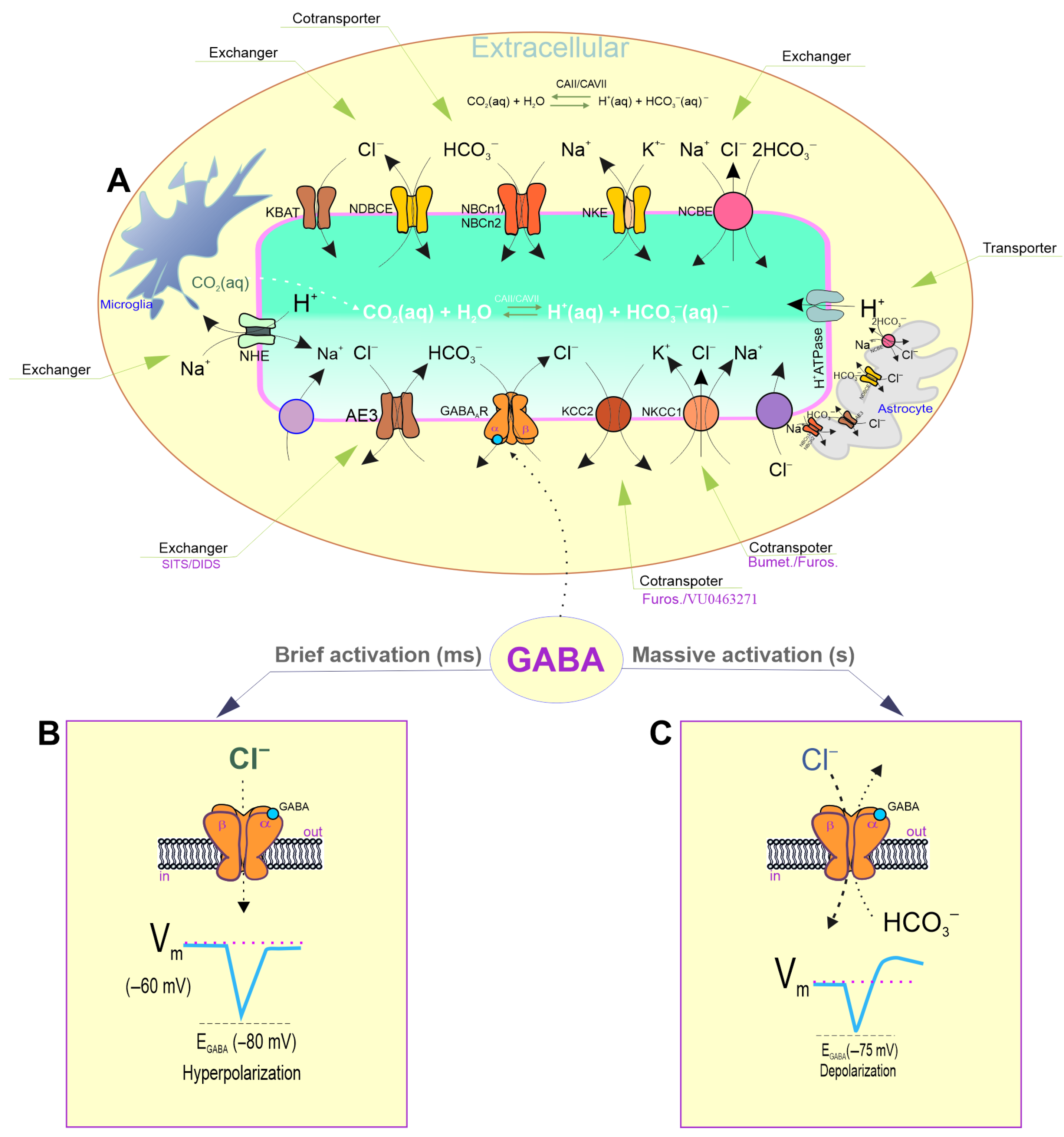
Figure 1. General diagram of the role of chloride and bicarbonate in GABAergic signaling. (A) Bicarbonate/pH and chloride homeostasis in neurons is maintained by membrane transporters and channels. Neuronal bicarbonate/pH levels are maintained by secondary active HCO3 uptake via electro-neutral and electrogenic Na+/HCO3 symporters (NBCN1/NBCN2), Na+/H+ exchangers (NHEs), and Na+-dependent/independent Cl/HCO3 exchanges (NCBE/NDCBE). The function of secondary active cation–chloride cotransporters (CCCs) and the activity of bicarbonate transporters are closely connected, and they are responsible for maintaining the neuronal Cl concentration ([Cl]i) through K+ or Na+/K+-coupled cation transport. The correlation between the [HCO3]i and intracellular pH is directly linked to carbonic anhydrase activity, which quickly replenishes their intraneuronal level after changes. (B) After brief activation by agonist binding to the extracellular site in the structure, the receptors undergo conformational changes, resulting in pore opening and rapid Cl permeability, leading to hyperpolarization/inhibition of the Vm in mature neurons. (C) During intense activation, GABAA receptors can be permeable to both Cl and bicarbonate ions. Bicarbonate ions, unlike Cl, always flow out of neurons in the open state during such activation, resulting in depolarization/excitation of the Vm.
Biomolecules 15 01003 g001
Figure 2. Neuronal bicarbonate/pH changes occur when GABAA receptors are massively activated. (A) GABAA-mediated depolarization is connected to both a decrease in the intracellular bicarbonate concentration and a fundamental shift in the intra- and extracellular pH. (B,C) Models that hypothetically explain the ionic basis of GABAAR-mediated depolarization. (D) The homopentameric GABAAR β3 isoform, unlike the β1 and β2 isoforms, is capable of participating in GABA-mediated HCO3 outflow from HEK 293 cells.
Biomolecules 15 01003 g002
Figure 3. Inhibitory synaptic plasticity during brain development. (A) A diagram showing the Cl influx through the ion channel pore during the brief activation of GABAARs and the main structures that hyperpolarize the Vm in mature neurons. (B) A diagram illustrating the Cl influx and HCO3 outflow through the channel pore during GABAARs’ massive activation, and the primary structures responsible for the Vm depolarization in mature neurons. (C) Time course of the [Cl]i recovery through NKCC1 and KCC2 activity. (D) The recovery of GABAAR conductance following massive activation. (E) The time required for the recovery of the [Cl]i after receptor activity. (F) The time required for pH recovery following receptor activity.
Biomolecules 15 01003 g003
Figure 4. Bicarbonate and ATP/ATPase in GABAAR function. (A,B) The rundown effect of the receptor function during massive activation or long-term potentiation, respectively. The rundown effect of GABAAR activity is not observed when Mg2+ATP (2 mM) is present. (C) The rundown effect of the receptor function is not significantly manifested in the presence of HCO3 (~26 mM). (D) A model illustrating the four basic conformational states that represent GABAAR function, which include a resting state, an open state, a desensitization state, and a resensitization state. In an HCO3-free experimental medium, the first application of GABA shifted the equilibrium from the resting state to the high-affinity open state, which then entered a state of desensitization. In contrast, in the presence of HCO3 (~26 mM) in an experimental medium, GABA application induced changes in receptor conformation and shifted the equilibrium from the resting state to the high-affinity open state; here, agonist application resulted in receptor resensitization. Bicarbonate preferentially stabilized the receptor structure.
Biomolecules 15 01003 g004
Figure 5. Massive GABAAR activation and network/seizure activity. (A) Sites of H+ binding with the GABAAR β subunit (Protein Data Bank, 4COF). (B) Relative IGABA as a function of the pHi. (C) The diagram illustrates how receptor massive activation (MA) affects the [HCO3]i, [Cl]i, pHi, and [ATP]i, leading to the appearance of epileptiform activity (EA).
Biomolecules 15 01003 g005
Disclaimer/Publisher’s Note: The statements, opinions and data contained in all publications are solely those of the individual author(s) and contributor(s) and not of MDPI and/or the editor(s). MDPI and/or the editor(s) disclaim responsibility for any injury to people or property resulting from any ideas, methods, instructions or products referred to in the content.