You are currently viewing a new version of our website. To view the old version click .
Biomolecules
  • Review
  • Open Access

15 December 2021

Amyloid Fragmentation and Disaggregation in Yeast and Animals

,
and
Bach Institute of Biochemistry, Federal Research Center “Fundamentals of Biotechnology” of the Russian Academy of Sciences, 119071 Moscow, Russia
*
Authors to whom correspondence should be addressed.
This article belongs to the Special Issue α-Synuclein Amyloids & Parkinson’s Disease: On Growth, Spread, and Neurodegeneration

Abstract

Amyloids are filamentous protein aggregates that are associated with a number of incurable diseases, termed amyloidoses. Amyloids can also manifest as infectious or heritable particles, known as prions. While just one prion is known in humans and animals, more than ten prion amyloids have been discovered in fungi. The propagation of fungal prion amyloids requires the chaperone Hsp104, though in excess it can eliminate some prions. Even though Hsp104 acts to disassemble prion fibrils, at normal levels it fragments them into multiple smaller pieces, which ensures prion propagation and accelerates prion conversion. Animals lack Hsp104, but disaggregation is performed by the same complement of chaperones that assist Hsp104 in yeast—Hsp40, Hsp70, and Hsp110. Exogenous Hsp104 can efficiently cooperate with these chaperones in animals and promotes disaggregation, especially of large amyloid aggregates, which indicates its potential as a treatment for amyloid diseases. However, despite the significant effects, Hsp104 and its potentiated variants may be insufficient to fully dissolve amyloid. In this review, we consider chaperone mechanisms acting to disassemble heritable protein aggregates in yeast and animals, and their potential use in the therapy of human amyloid diseases.

1. Introduction

Amyloids are filamentous protein aggregates with regular cross-beta type structure. They are generated from the soluble form of a single protein through self-catalytic structural conversion. Amyloid formation is regarded to be the cause of more than forty incurable age-associated diseases, including Alzheimer’s disease and Parkinson’s disease (PD) []. Due to their self-catalytic nature, some amyloids can be infectious, and, in this case, they are called prions, i.e., proteinaceous infectious agents. Only one protein in humans and animals, PrP, is known to form a bona fide prion that is able to infect other organisms naturally []. However, many amyloids are able to spread within an organism, or in certain tissues, thus showing prion-like properties [,], and there is emerging evidence that some can also be infectious via direct injection []. Not all amyloids discovered in animals are associated with pathologies. Some of them serve useful functions, such as amyloids of the proteins CPEB of Aplysia or Orb2 of Drosophila, which are involved in the formation of long-term memory [,].
Several amyloids with prion properties were found in yeast []. The first yeast prion, [PSI+], was discovered by Brian Cox in 1965 as an unusual nonsense-suppressor phenotype with non-Mendelian genetic properties []. This phenotype segregated 4:0 in crosses of [PSI+] with non-prion [psi] cells; it could be spontaneously lost or cured with certain chemicals, as well as appearing de novo []. Another genetic element with similar behavior, [URE3], which allows the utilization of poor nitrogen sources in the presence of rich ones, was discovered by Francois Lacroute in 1971 []. However, their prion nature was explained only in 1994 by Reed Wickner, who related them to the prion-like conversion of the Sup35 and Ure2 proteins []. Further studies revealed several other yeast proteins that formed amyloid prions: Rnq1, Swi1, Cyc8, Mod5, Mot3, and Nup100 [,,,,,]. Amyloidogenic domains of more than ten other proteins form prions when fused to the Sup35 functional domain, though the propagation in a prion mode was not shown for these proteins on their own []. Transient prion-like amyloids can be induced by heat stress [,], and non-heritable functional amyloids called mnemons have been shown to mediate yeast cellular memory [].
Of note, there are many prions that are apparently not based on the amyloid structure, like the [GAR+] prion, which allows overriding glucose-associated repression of alternative carbon source utilization []. A recent groundbreaking systematic search revealed about 46 novel yeast prions, most of which relied on some type of protein aggregation of a poorly characterized, seemingly non-amyloid, type [].
Yeast prions are usually not highly detrimental to cells and can even increase the ability of a yeast population to adapt to changing environmental conditions [,], though some prion-associated phenotypes are not necessarily beneficial []. Yeast amyloid prions proved to be a very efficient model for understanding the basic principles of the formation and propagation of amyloids and prions. Though many specific manifestations of mammalian amyloids and prions cannot be reproduced in yeast, this organism is very convenient for the study of their basic properties due to the ease of all kinds of biological manipulations. Moreover, yeast is used as a model to study the aggregation and toxicity of many human and animal amyloidogenic proteins [,,].
Yeast studies were central in the formulation of the concept of amyloid fragmentation, the process by which amyloid fibrils are split into smaller pieces through interactions with chaperones. Fragmentation is strictly required for amyloid propagation as a prion []. Fragmentation also strongly affects intraorganismal propagation and properties of non-prion amyloids, since smaller amyloids are more mobile and may be more toxic. Here, we review existing data on how chaperones work to disassemble amyloids in yeast and animals, resulting in either amyloid dissolution or prion-like propagation, and the potential of chaperone-based mechanisms in therapies for human amyloid diseases and PD in particular.

2. The Sup35 Protein and Its Prion Structures

2.1. Sup35 Protein Function and Architecture

The majority of yeast studies described below involve [PSI+], the most studied yeast prion, which relates to the prion state of the Sup35 protein. Sup35, also known as translation termination factor eRF3, terminates protein synthesis in cooperation with the Sup45 protein (eRF1) [,]. The reduction of Sup35 function due to mutations or prion formation results in an increased readthrough of nonsense codons. This may be conveniently detected as a phenotypic reversion of nonsense mutations. Either one of the two nonsense mutations in the adenine synthesis pathway, ade1-14 or ade2-1, are usually used for the observation of nonsense codon readthrough. Such mutants are unable to grow in the absence of adenine, but they re-acquire this ability when Sup35 converts to its prion state and only a small fraction of it remains soluble and functional. Moreover, ade nonsense mutants produce red colonies due to the accumulation of a red intermediate of adenine biosynthesis, while wild-type colonies are white. Depending on the levels of Sup35 that remain soluble/functional, the restoration of adenine biosynthesis can be nearly complete or partial, which results in white or pink colonies.
Sup35 is composed of three distinct domains. The C-terminal domain (C, residues 254–685) is homologous to the translation elongation factor eEF-1A and is essential for translation termination and cell viability. The N-terminal (N, 1–123) and middle (M, 124–253) domains are non-essential, non-conservative in sequence, and are natively unfolded []. The N domain is required for prion properties and forms prion structures. It is rich in glutamine, asparagine, and tyrosine, as is typical of many yeast prion domains. The M domain has no clear function, though it was reported to interact with Hsp104 [] and to modulate the formation of Sup35 liquid droplets under stress conditions []. Lack of the M domain strongly reduces Sup35 levels ([] and our unpublished observations) via an unknown mechanism. The M domain is rich in the charged residues of lysine and glutamic acid, which is atypical of yeast prion domains.

2.2. [PSI+] Prion Variants

Similar to many other amyloids and prions [,], Sup35 can form many different amyloid structures (variants), which manifest as different phenotypes in vivo [,]. [PSI+] variants are usually distinguished by the level of the nonsense codon readthrough and the stability of inheritance. These phenotypes usually correlate with each other, where higher readthrough corresponds to higher stability. However, such phenotypes do not allow one to reliably distinguish between more than two or three [PSI+] variants, since these phenotypes can vary slightly depending on the cellular background and so cannot be defined with sufficient precision. In particular, two other prions, [SWI+] and [PIN+], can significantly affect the nonsense readthrough by decreasing levels of the eRF1 (Sup45) translation termination factor and can even produce a detectable nonsense-suppressor phenotype in the absence of [PSI+] []. To overcome this restriction, King et al. proposed to distinguish [PSI+] variants based on their interaction with a panel of Sup35 N domain mutants and GFP fusions []. This allowed the authors to confidently distinguish 23 [PSI+] variants []. Three more variants were able to propagate in the presence of the weakened T160M mutant of the Hsp104 chaperone, but not wild-type Hsp104 []. In addition, some Sup35 amyloid structures can be obtained in vitro, but cannot stably propagate in vivo [,]. All this gives us an idea of how many different folds a single amyloidogenic protein can acquire.
Notably, only five of the mentioned 23 [PSI+] variants can be obtained by the standard procedure used in the majority of publications, i.e., Sup35 overproduction in the wild-type background. The additional [PSI+] variants were obtained by passing these “basic” [PSI+] variants to various Sup35 mutants and back []. The basic [PSI+] variants were usually roughly divided into two categories. “Strong” [PSI+] variants showed a high level of nonsense codon readthrough due to very low levels of soluble non-prion Sup35. “Weak” variants exhibited moderate readthrough related to observable levels of soluble Sup35. According to Huang and King, only one of the basic [PSI+] variants is strong, while the four others are weak []. Compared to weak prions, strong ones have smaller fibrils due to better fragmentation [].
Interestingly, some tests reveal a sharp (rather than gradual) difference between the strong and weak [PSI+] variants. The strong variant is not cured by the multicopy HSP104 gene and is lethal in the presence of multicopy SUP35, while weak isolates are readily curable and non-toxic under these conditions [].

2.3. Sup35 Prion Structures

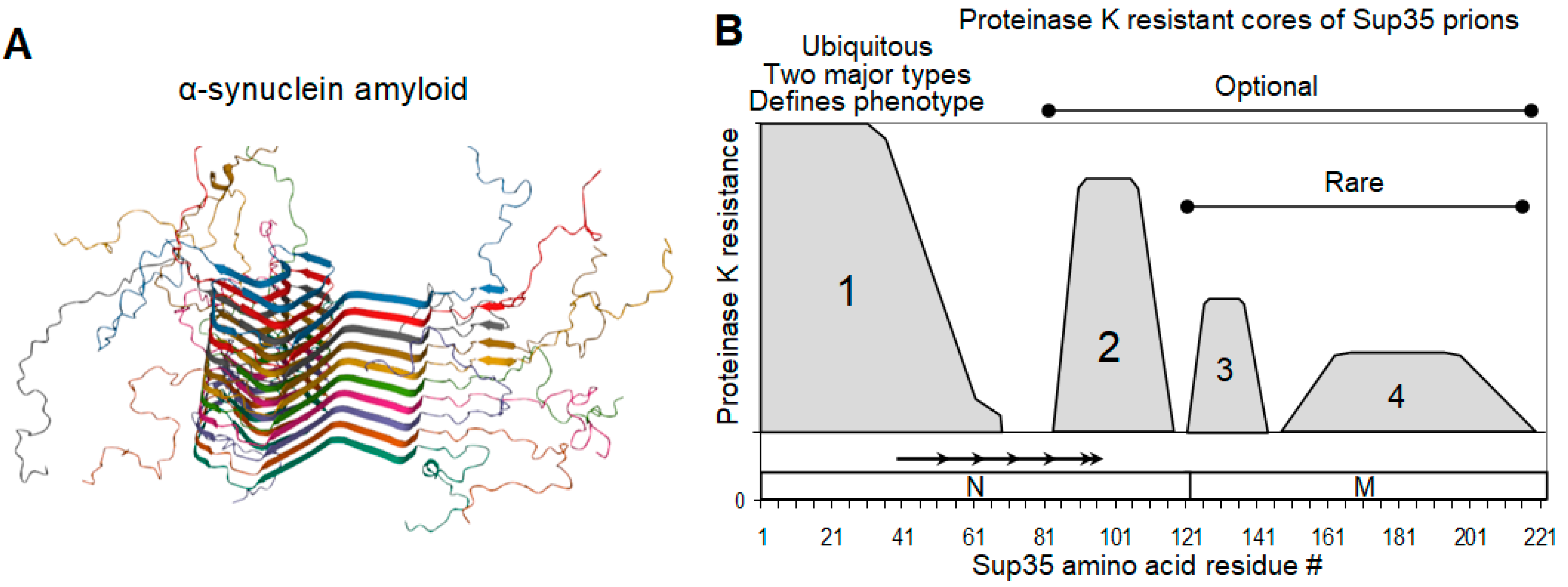
Most [PSI+] phenotypic variants are likely to have a structural basis in the manner in which monomers are folded in the amyloid fibril. While a full spatial structure of the Sup35 prion has not yet been obtained, it is known that it is a parallel in-register cross-beta structure based on intermolecular beta sheets [,]. In this structure, each Sup35 molecule (protomer) occupies a layer of 0.48 nm along the fibril axis, which corresponds to the standard interstrand distance in a beta sheet. Such layers have a uniform thickness, though they are not necessarily perfectly flat, and they may be regarded as a one strand-thick two-dimensional structure. A good example of such a structure is α-synuclein (α-syn) amyloid [] (Figure 1A). The strain variations in such structures may be due to two partly dependent parameters: (a) the difference in the exact regions involved in the beta structure and (b) the different folding within a layer. The former parameter can be defined by several different methods, including fluorescent labels [], hydrogen/deuterium exchange (HD) [], and the resistance to proteinase K (PK) [,]. These data largely agree with each other, though with some reservations. Fluorescent labeling with pyrene maleimide (297 Da) and acrylodan (225 Da) indicated that region 1–20 is loosely structured and does not form intermolecular contacts []. This contradicts other works and is likely to be an artifact, since [PSI+] is highly sensitive to single amino acid changes in this region [,], while the addition of fluorescent labels with considerable masses may cause an even greater structural perturbation. Therefore, we will describe the Sup35NM prion structure based on our PK mapping data, since they (1) were obtained with ex vivo Sup35 prions, rather than in vitro-generated Sup35 amyloids used in other works, and (2) were made for 26 [PSI+] isolates differing in their phenotypes and origins [].
Figure 1. α-synuclein amyloid and Sup35 prion structure. (A) A typical parallel in-register amyloid structure exemplified by that of α-synuclein (PDB 2N0A) []. (B). Map of proteinase K-resistant cores of Sup35 prions. The picture summarizes data for 26 [PSI+] isolates differing in their origin and phenotype []. The location of Sup35 N and M domains, as well as of the imperfect oligopeptide repeats, is shown.
The Sup35NM region can contain up to four separate protease-resistant structures, called Cores 1 to 4, located strictly within four regions of residues 2–72, 73–124, 125–153, and 154–221 (Regions 1 to 4, Figure 1B). As the only exception, the structure spanning residues 90 to 143 was observed infrequently. However, only the N-terminal of these structures (Core 1) was present in all [PSI+] isolates. The majority of weak [PSI+] variants have a Core 2 structure at residues 91-121, while a few strong [PSI+] variants have a Core 2 at residues 82–100. Structures in the regions 125–153 and 154–221 belonging to the M domain were present in a small proportion of [PSI+] isolates. All four structures together were observed only once in the W8 [PSI+] variant []. Of note, some [PSI+] variants were likely heterogenous for Cores 2 to 4. This implies the simultaneous presence of Core 2 at locations 82–100 and 91–119, and/or, presumably, the presence of any one of the Cores 2 to 4 in only a part of the prion population. This agrees with electron microscopic observations of the structural heterogeneity of Sup35 fibrils seeded in vitro by [PSI+] lysates [], and can complicate future detailed structural studies.
To our knowledge, Sup35 is the only amyloid for which a structure composed of more than one part was reported. However, a multipart structure could be present in Rnq1 (our unpublished data) and other yeast prions with large glutamine- and asparagine-rich prion domains.
Surprisingly, the [PSI+] nonsense-suppressor phenotype seems to be defined solely by apparently small differences in Core 1 structure, rather than by the presence or absence of the other Cores. Core 1 can be further divided into two parts, of which part A (2–32) is fully resistant to PK, while part B (33–72) is only partially resistant and shows lower resistance in weak [PSI+] [].
The Sup35 N domain is known to contain five imperfect oligopeptide repeats with the sequence PQGGYQ(Q)QYN. Despite being rich in glutamine, tyrosine, and glycine residues typical of yeast prions, the repeats tend to be unstructured or acquire partial PK resistance as a lateral part of Core 1 or Core 2, like Core 1B (33–72) []. Thus, the oligopeptide repeats are a likely target for recognition by Hsp104 partner chaperones, which facilitates subsequent prion fragmentation. This agrees with earlier observations that the deletion of some of these repeats decreases fragmentation and turns a strong [PSI+] into a phenotypically weak one, despite preserving strong [PSI+] structure [,].
The finding that some prion structures of Sup35 can be phenotypically silent [] adds uncertainty to the definition of prion variants. The assay for the discrimination of [PSI+] variants [] relies on mutations in Region 1 and thus can only discriminate the variation of Core 1. It is not clear whether presence of Cores 2 to 4, which seem to be nearly phenotypically silent, should be regarded as additional [PSI+] strain variation.

2.4. On the Equivalence of Sup35 In Vitro Fibrils and In Vivo Prions

Structural studies of the Sup35 amyloid are often performed with Sup35 fibrils that are spontaneously generated in vitro. Fibrils obtained at 4 °C induce mainly strong [PSI+] variants when introduced to yeast cells and those generated at 25–37 °C induce predominantly weak [PSI+] variants []. Due to this, such fibrils are usually regarded as structurally equivalent to the strong and weak Sup35 prions. Although such equivalence is likely to be correct as a rough approximation, there are some significant concerns as to its complete validity. The first is that some level of structural heterogeneity of the in vitro fibrils and the existence of four different weak [PSI+] variants potentially correspond to the 25–37 °C fibrils []. Secondly, the amyloid fold obtained in vitro can be altered upon propagation in yeast. For example, Ohhashi et al. obtained in vitro Sup35 amyloids with protease-resistant cores at residues 81–148 and 62–144 []. When introduced to yeast cells, these amyloids induce strong and weak [PSI+] variants, VH and VK [], both of which, as we observed, have an entirely different structure, including the N-terminal protease-resistant Core 1 (2–72), but they lack the original in vitro-generated core []. The 4 °C “strong” fibrils show significantly higher fragility than the 25–37 °C “weak” fibrils [], which agrees with their lower thermal stability []. However, the 4 °C fibrils also show an almost five-fold polymerization reduction [], which, according to the quantitative prion conversion model presented in the same work, poorly agrees with the much lower level of soluble Sup35 in strong [PSI+] cells.

3. Mechanisms for the Fragmentation and Disassembly of Amyloids in Yeast and Animals

3.1. Replication of Yeast Prions

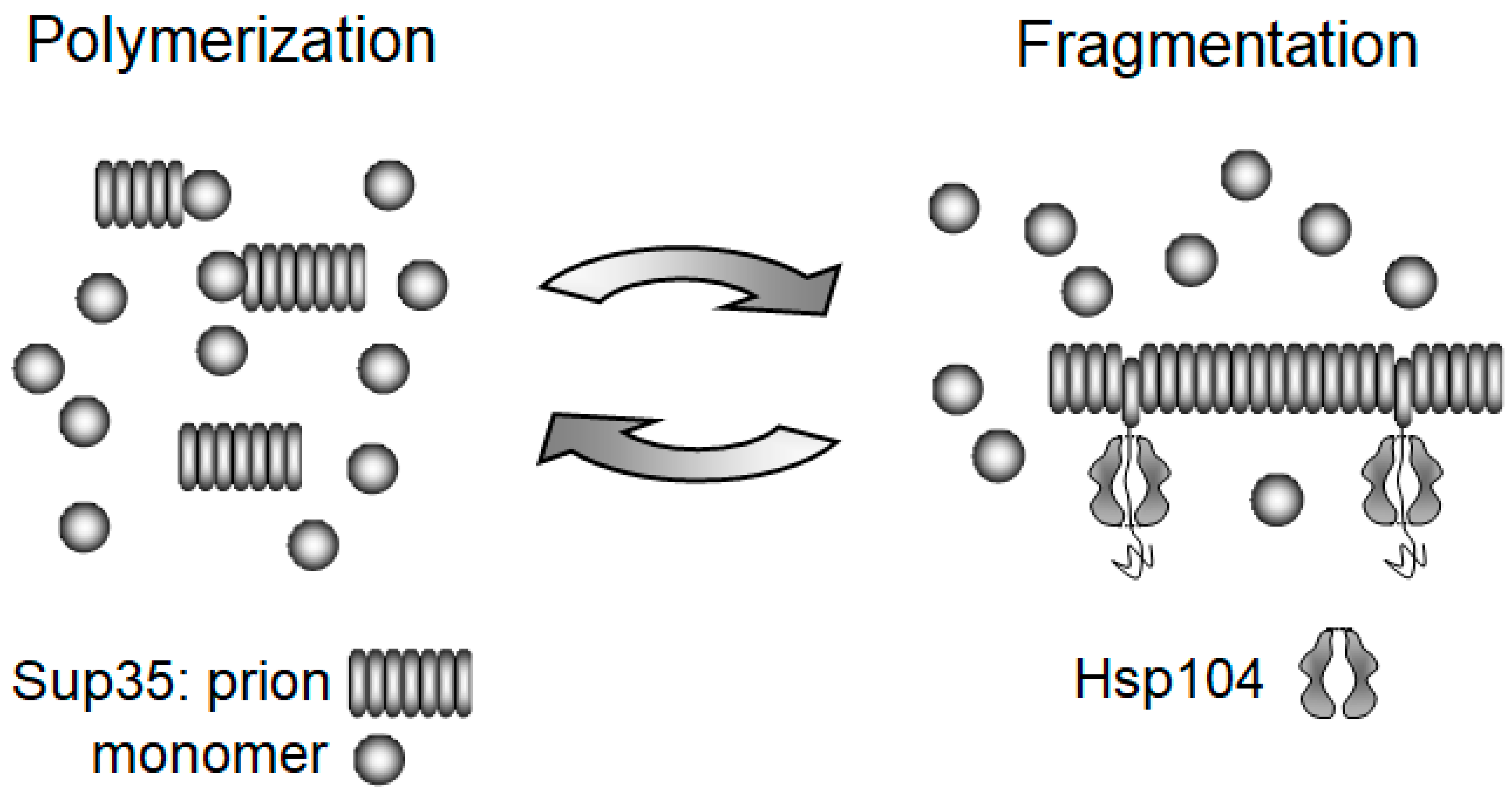
The study of yeast prions played a crucial role in understanding how prions and amyloids multiply. It was found that the mild overproduction of the Hsp104 chaperone can cure some variants of the [PSI+] prion. Paradoxically, the lack of Hsp104 has an even stronger effect, eliminating all [PSI+] variants []. Our group offered an explanation for this paradox and the role of Hsp104 [,], relying on three earlier observations. Firstly, Hsp104 is able to extract single molecules from the large aggregates of heat-denatured proteins []. Secondly, Sup35, in agreement with the amyloid model, forms amyloid fibrils in vitro [,]. Thirdly, prion conversion activity correlates with the aggregated, rather than the soluble, form of Sup35 [,,]. We reasoned that if Hsp104 would extract a molecule from the middle of a fibril the same way it extracts them from heat-denatured aggregates, the fibril would split, producing two prion particles from one. This, in fact, completes the prion replication cycle, the other part of which is the growth of Sup35 fibrils by their joining, as well as the conversion of soluble Sup35 molecules (Figure 2). This simple mechanism ensures prion propagation in three ways. Firstly, the inheritance of a prion requires that the prion particles should multiply, not just grow in size, since they are constantly diluted, i.e. reduced in number during cell division. Secondly, the distribution of prion particles to daughter cells is nearly random for smaller particles, while larger ones tend to be retained in the mother cells []. Therefore, reliable inheritance requires tens or hundreds of prion particles per cell. Thirdly, since prion conversion occurs at the fibril ends, fragmentation creates new ends and thus accelerates the conversion of soluble proteins to their prion form. We presumed initially that overproduced Hsp104 cures [PSI+] by dissolving prion fibrils, but, as discussed in Section 4.2, the curing mechanism is likely to be different.
Figure 2. Yeast prion replication cycle []. Prion fibrils are multiplied through the action of Hsp104, which randomly extracts protomers from amyloid, thus fragmenting it into smaller pieces. The size of amyloid fragments is restored through growth by accretion of soluble Sup35 molecules. The action of Hsp104 is preceded by, and depends on, binding of the Sis1 and Ssa chaperones [], which are not shown here.
The paradoxical beauty of this mechanism is that none of its parts are likely to have evolved for prion replication. The fibrillar shape is a universal property of amyloids, while the Hsp104 mechanism was most likely developed to cope with amorphous stress-derived aggregates, rather than prions. It is also noteworthy that the replication of yeast prions relies on the Hsp104 action aimed at their destruction.
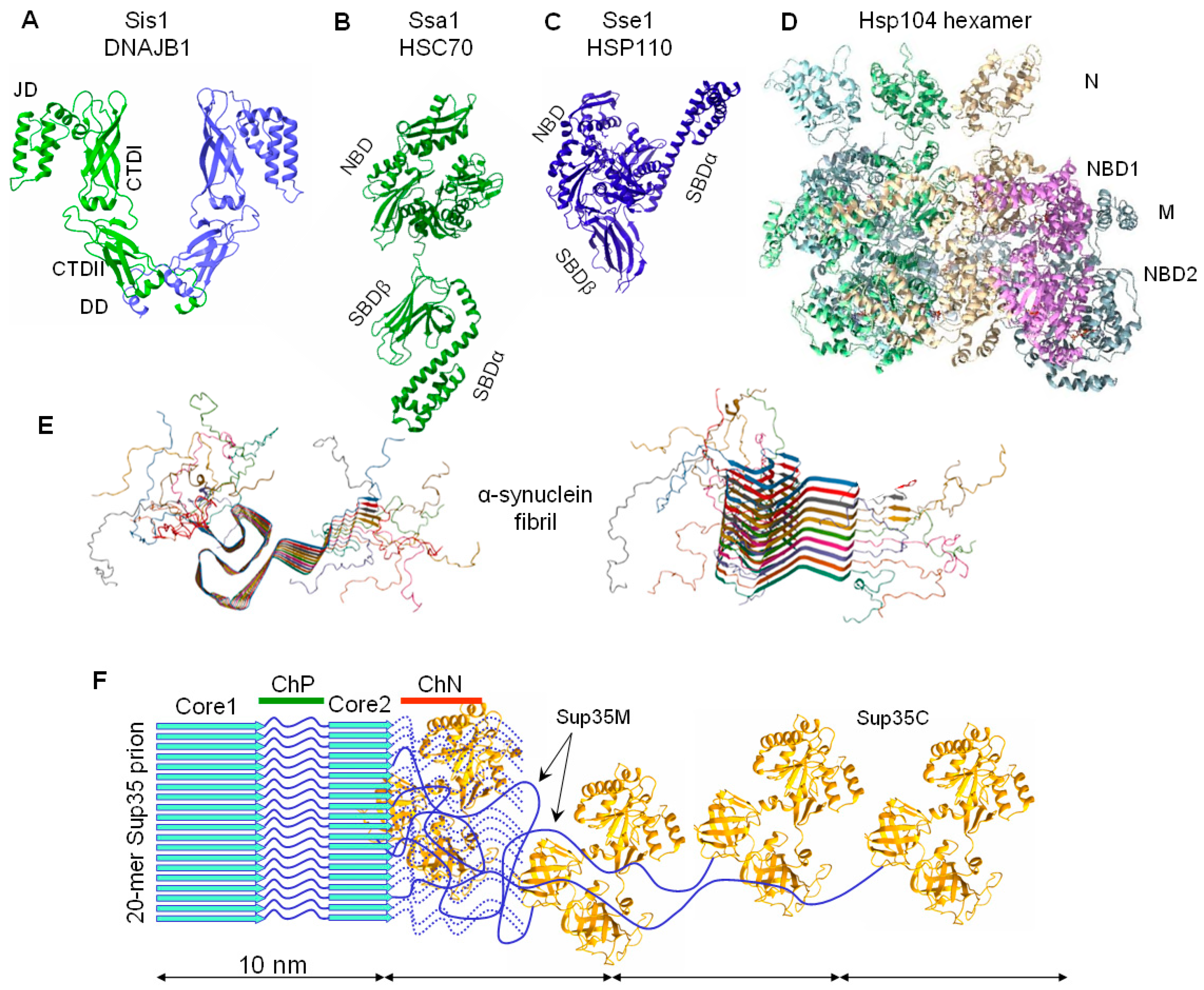
The proposed mechanism of prion fragmentation by Hsp104 was later confirmed by many studies. In particular, we developed an electrophoretic method to determine the size of amyloid fibrils, and showed that Sup35 prion particles are relatively small and include 10 to 50 Sup35 molecules []. Hsp104 is inhibited by a low concentration of guanidinium hydrochloride (GuHCl, 3–5 mM) in the growth medium []. Such inhibition increases the size of Sup35 prion particles two-fold per generation, exactly as expected in the case of the complete block of fragmentation []. Notably, with such a size, Sup35 fibrils should look quite different from the long fibrils obtained in vitro, since the length of a 20-mer fibril (~10 nm) is about the same as its width (Figure 3).
Figure 3. Relative sizes of a α-synuclein fibril, Sup35 prion, and the chaperones involved in their fragmentation. The sizes are drawn to scale. Structures of yeast chaperones are presented, while their mentioned human orthologs are of similar size and structure, such as (A) Sis1/DNAJB1 dimer (Sis1: PDB 6D6X+1CG3). JD: J domain; CTDI, CTDII: carboxy-terminal domains I and II; DD: dimerization domain. (B,C) Ssa1/Hsc70 and Sse1/HSP110 (Ssa1: PDB 2KHO, Sse1: PDB 2QXL). NBD: nucleotide binding domain; SBDα and SBDβ: substrate-binding domain. (D) Hsp104 hexamer (PDB 5KNE). N, M: domains N and M; NBD1, NBD2: nucleotide binding domains 1 and 2. (E) α-syn fibril (PDB 2N0A). (F) Sup35 prion (Sup35C: PDB 1R5B). Sup35 prion structure with two protease-resistant cores is depicted, though in some [PSI+] variants Core 2 is absent and/or additional structures in the M domain are present. For clarity, only four Sup35 C domains and M domains (blue line) are shown; other copies of the M domain are indicated by dotted lines. ChP—a non-structured protease-sensitive region of the Sup35N domain, presumably used for productive binding of Hsp104 via Sis1 and Ssa chaperones. ChN—the region 129–148 of direct but non-productive Hsp104 binding. The diameter of the amyloid core, presumably corresponding to the Sup35 N domain, is about 8–10 nm [,]. The unfolded M domain allows the C domain to be widely offset from the fibril core, thus they form a loose halo with a diameter of 60–65 nm []. Notably, the M domain can acquire a folded [] protease-resistant state, at least in a part of the prion population [,].
It may appear that the prion fragmentation concept is too obvious, but it was not so clear at the time when it was proposed. The discovery of yeast prions was inspired by the human prion concept of Stanley Prusiner [], but this concept included the heterodimer model for prion replication [], which considered prions as monomers. In this model, a prion molecule PrPSc forms a heterodimer with its normal conformer PrPC, converts it into PrPSc, and then two PrPSc molecules dissociate. We concluded that such a process is unlikely, because (1) it cannot occur without an external source of energy and (2) it poorly accommodates the existence of prion strains or multiple alternative conformations of PrP, capable of self-catalytic conversion. In contrast, amyloids are known to form alternative self-catalytic structures (reviewed in []). Therefore, we switched to the amyloid model of the prion, which was free from these problems, though initially it did not focus on the multiplication of amyloid particles, which is especially important in the case of prions. We proposed that the multiplication is performed through fragmentation by the Hsp104 chaperone, which simultaneously provides the required source of external energy [].

3.2. The Chaperone-Mediated Fragmentation of Yeast Prions

While Hsp104 is a key element of the yeast fragmentation mechanism, it does not act alone. For its function, Hsp104 requires the assistance of chaperones belonging to the Hsp40 (called J-proteins, due to their homology to bacterial DnaJ) and Hsp70 (similar to DnaK) families. In particular, Ydj1 (Hsp40) and Ssa-family (Hsp70) proteins are required to solubilize and reactivate denatured luciferase []. There are four functionally interchangeable Ssa proteins in yeast, Ssa1 to Ssa4, which we call here, collectively, Ssa. Ssa1 and Ssa2 are constitutively expressed and associate with prions [], as well as with non-prion Sup35 complexes []. Yeast has 13 proteins in the Hsp40 family, which differ in their substrate specificity and their participation in the maintenance of yeast prions. Among them, the key role in prion propagation belongs to Sis1, which is required for the propagation of at least four yeast prions, [PSI+], [PIN+], [URE3], and [SWI+] [,,,,]. Three other J-proteins are also involved in prion propagation: Ydj1, Apj1, and Swa2 [,,,]. Current models assume that Sis1 and Ssa proteins act together to recognize prions and present them to Hsp104 for unfolding, which is required for Hsp104 disaggregating activity [,].
Hsp104 functions in the form of a ring-like hexamer with a central pore. Hsp104 extracts proteins from amyloid or amorphous aggregates by threading their polypeptide chain through the pore and allowing it to fold properly at the distal end. Hsp104 is a large protein containing two ATPase units, nucleotide binding domains (NBD) 1 and 2. The movement of the polypeptide chain through the Hsp104 pore is achieved by the sequential contraction of Hsp104 protomers at the expense of ATP hydrolysis by NBD1, while NBD2 appears to be required for the assembly of the hexamer []. For each ATP molecule spent by NBD1, Hsp104 advances along the peptide chain by two residues [,,]. A frequent scenario is that Hsp104 starts unfolding from a loop in the middle of a protein. In this case, Hsp104 threads two polypeptide strands simultaneously [], thus doubling its energy efficiency.
The mechanism of Hsp104 inhibition by GuHCl has also been elucidated. It was shown that in the bacterial Hsp104 homolog ClpB, GuHCl binds specifically to the active site of NBD1 []. GuHCl strengthens the Hsp104 M domain/NBD1 interaction, stabilizing Hsp104 in a repressed conformation, abrogating Hsp70 cooperation and inhibiting continuous ATP turnover by NBD1 [].
Hsp104 is a very powerful, but also costly, molecular machine: under non-stressing conditions, cells lacking Hsp104 grow noticeably faster. When wild-type and hsp104Δ cells were grown together in a competition assay, the proportion of wild-type cells decreased from 50% to 20% during two days of growth. However, this proportion remained constant for the next two days [], which suggests that the cells remaining after two days were not inhibited in growth. It is possible that the starting wild-type cells were heterogeneous for the Hsp104 level and there was a fraction of cells with low Hsp104 not inhibited in growth.
Different Hsp104 substrates may vary in their mechanical strength and, therefore, may require different mechanical forces to unfold them. By mixing functional and nonfunctional Hsp104 subunits, DeSantis et al. showed that “easy” substrates like disordered heat-denatured aggregates of luciferase do not require the cooperation of Hsp104 subunits and can tolerate a high proportion of mutant Hsp104. A stronger substrate, like Sup35’s “strong” in vitro formed fibrils, requires the cooperation of three sequential subunits. The disassembly of the most mechanically stable “weak” Sup35 fibrils requires the cooperation of all six Hsp104 subunits and is the least tolerant to the presence of nonfunctional Hsp104 [].

3.3. Amyloid Fragmentation in Animals

Hsp104 orthologs are present in all kingdoms of life, in prokaryotes (ClpB) and eukaryotes, except for animals (metazoa). Hsp104 was present in the last common ancestor of animals and choanoflagellates, but was lost in the last common ancestor of animals. The mitochondrial Hsp104 analog, Hsp78, was lost simultaneously [], and so the loss does not appear accidental. The authors relate the loss to the simultaneous disappearance of some Hsp104 clients among metabolic enzymes [], but one can also suggest that mobility gives animals more opportunities to avoid stressful conditions requiring Hsp104, the costly machine with 12 ATPase units that may be disadvantageous in the absence of stress.
Searches for disaggregase activity in animal cells revealed that even without Hsp104, the Hsp40-Hsp70-mediated mechanism can result in moderately efficient disaggregase activity. It shows a strong dependence on another participant of this mechanism, the Hsp110 (APG-2) chaperone, which is a nucleotide exchange factor for Hsp70 [,,]. In yeast, the role of analogous factors like Sse1/2 is also important, but it becomes more evident in animals, which lack Hsp104 [].
This mechanism was studied in fine detail by Wentink et al. using α-syn amyloids as a target []. In humans, this mechanism includes the J protein DNAJB1, an ortholog of yeast Sis1, the constitutive human HSP70 (HSC70, also known as HSPA8) and HSP110 (APG-2, also known as HSPH2 and HSPA4). First, these authors showed that the DNAJB1 binding affinity to α-syn fibrils is much higher than to the monomer α-syn and that binding is strongly required for DNAJB1 dimerization. The maximal binding density proved to be about one DNAJB1 dimer to ten α-syn protomers. To envision the interaction of chaperones with amyloids, it is helpful to keep in mind a characteristic property of amyloids: the very close spacing of protomers, equal to 0.48 nm. The low proportion of DNAJB1 is then not surprising, since its dimer size is about 10nm, which corresponds to the length of a 20-protomer fibril (Figure 3). DNAJB1 stimulates HSP70 binding to fibrils up to ten-fold, at a proportion of up to five HSP70 molecules per one DNAJB1. Thus, the maximal binding proportion of HSP70-to-α-syn is one for every two protomers. This appears to be a very high density of HSP70, considering the size of HSP70 molecule (Figure 3). Such dense packing of HSP70 molecules should cause steric pushing and create forces that break the amyloid fibrils. This mechanism of amyloid disassembly is termed entropic pulling.
The way HSP110 helps this mechanism proved to be very unexpected. Due to its large size of 94 kDa, HSP110 can reach and release only those HSP70 molecules, which are not clustered. In this way, HSP110 locally concentrates HSP70, thus increasing the efficiency of entropic pulling. BAG1 is another nucleotide exchange factor for HSP70, but it is a small molecule with a size of 31 kDa. BAG1 does not stimulate amyloid disassembly, but, remarkably, it starts doing so when its size is artificially increased [].

3.4. Yeast Prions Based on Putative “Soft” Amyloids or Non-Amyloid Structures

Two basic properties of amyloids seem to be essential for them to exhibit prion properties: the ability for the indefinite autocatalytic propagation of altered protein folds, and the filamentous shape that is important for prion replication by fragmentation. Accordingly, the existence of a prion related to a heritable structural change in a protein, which is not of an amyloid type, seemed unlikely. Nevertheless, such prions were discovered in yeast. It is worth noting that one prion formally matching such a definition is known: it is an artificial prion [β], which is related to the proteolytic self-activation of protease Prb1 []. However, in this review, we consider only prions that are based on the alterations of spatial structure that do not modify the primary structure of the protein.
Study of the [GAR+] prion revealed that it is related to some non-amyloid structural alterations of the Pma1 and/or Std1 proteins. [GAR+] shows some properties typical of amyloid prions: it appears with a greatly increased frequency upon the overproduction of Pma1 or Std1, it is transmissible by cytoduction (the transfer of cytoplasmic genetic elements between yeast cells through an abortive mating procedure, in which nuclear fusion is prevented by a mutation blocking karyogamy, usually kar1 []), and it shows a barrier in prion transmission between closely related species. However, no change in the aggregation state of either Pma1 or Std1 was detected [].
In 2016, a groundbreaking study revealed a large swath of 46 yeast prions, most of which are not based on SDS-resistant aggregates [] (one of the main hallmarks of amyloids [,]). These prions were discovered by monitoring heritable alteration of yeast phenotypes (sensitivity to various salts and antibiotics) after transient overproduction of the respective proteins. Their prion state is readily transmitted through spheroplast “protein” transformation with [PRION+] lysates treated with DNase and RNase. In some, but not all, cases, a change of the aggregation state is evident by the microscopic observation of fluorescently labeled prion molecules. Most of the new prion proteins have large intrinsically disordered regions, but these are not rich in glutamine and asparagine as is typical of amyloid-based yeast prions. The new prions show a different dependence on chaperones.
Eleven of these 46 prions are curable by GuHCl and therefore require Hsp104 activity like the amyloid-based yeast prions. However, these 11 prions did not form detergent-resistant polymers, which allowed the authors to conclude that they do not form amyloids. However, such a conclusion could be incorrect. While all known yeast prion amyloids are insoluble in SDS at room temperature [,], this does not necessitate that all amyloids must show such a property. For instance, yeast Pub1 forms both SDS-insoluble and soluble amyloids in vivo, though the latter are insoluble in a milder ionic detergent, sarcosyl []. SDS-soluble amyloids have also been observed for several proteins related to human amyloidosis, such as FUS and hnRNPA2 [,], as well as for yeast Pbp1, which is an ortholog of human ataxin-2 and a binding partner of Pbp2, one of the newly identified “non-amyloid” prions [].
The remaining 35 Hsp104-independent prions were tested for their requirements in other chaperones. Nineteen prions proved to be curable by the transient expression of the Ssa1-K69M mutant protein, which dominantly inhibits Ssa function []. This subset includes the three best-studied members of the non-amyloid prion cohort: [GAR+], [BIG+], and [SMAUG+]. [BIG+], the prion form of pseudouridine synthase (Pus4), accelerates cell growth and reduces the replicative lifespan. [SMAUG+], the prion form of the Vts1 protein, orchestrates gene expression by regulating mRNA decay. [SMAUG+] was proposed to have a transmissible agent in the form of a hydrogel that can seed the conversion of monomeric Vts1 molecules into a gel-like state in vitro. This hydrogel is soluble in ionic detergents even at their low concentrations and shows no signs of filamentous structures [].
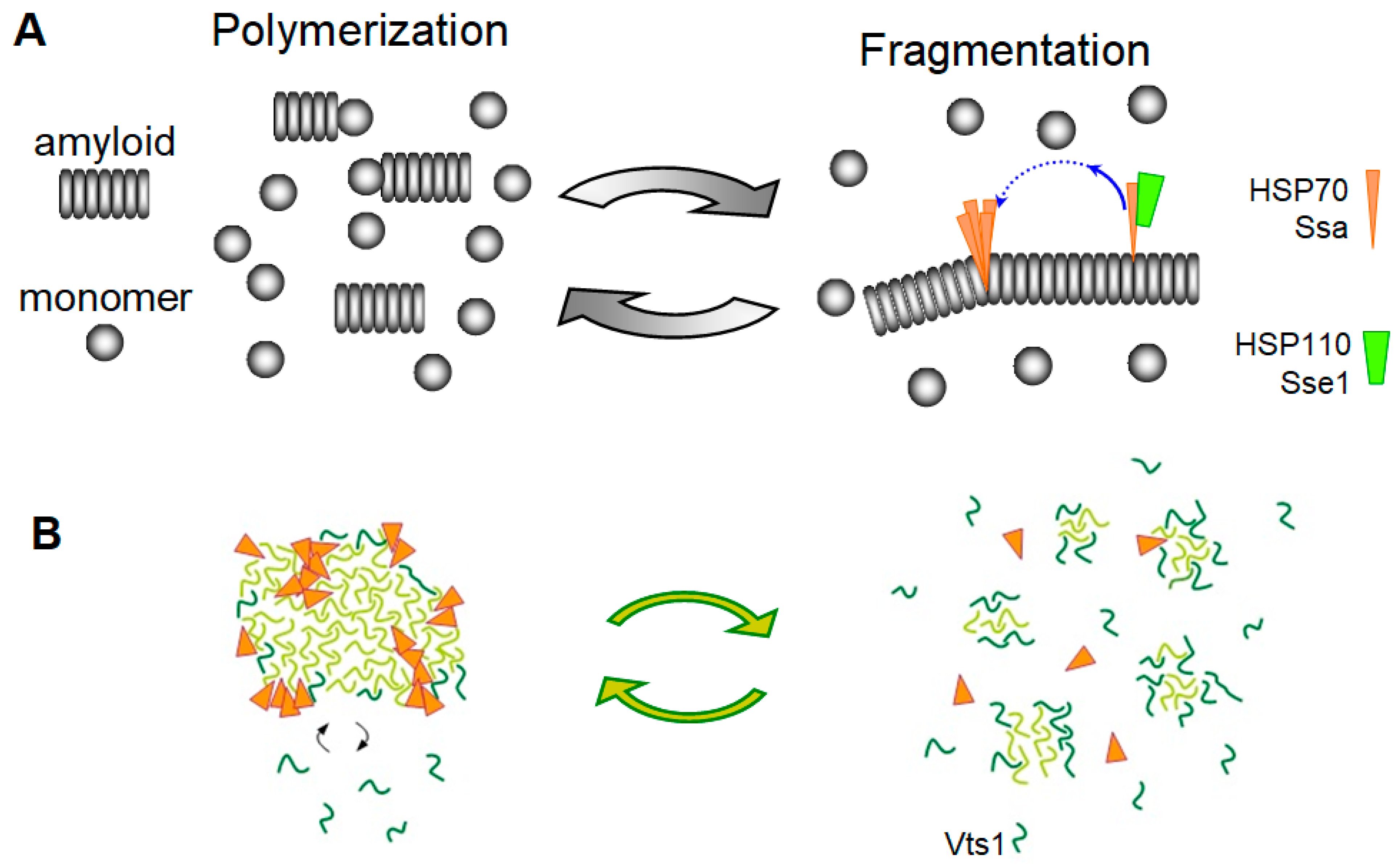
Thus, it may be proposed that the yeast Hsp70 Ssa proteins can fragment and can thus replicate prions made of either amyloids with a soft SDS-soluble structure like Pbp1, or hydrogels like Vts1, using the same “entropic pulling” mechanism described above for the animal Hsp70 and its co-chaperones []. Amyloids are one-dimensional crystals (i.e., their basic unit is repeated in just one dimension), and such topology was important for formulating the fragmentation model []. Though hydrogels are three-dimensional, this model could be extended to 3-D structures (Figure 4).
Figure 4. Amyloid and prion fragmentation in the absence of Hsp104. (A) “Entropic pulling” amyloid fragmentation by HSP70 and HSP110. Hsp110 preferentially removes non-clustered HSP70 molecules from the amyloid, thus promoting formation of HSP70 clusters, where entropic pulling can create sufficient force to fragment amyloid. Hsp40 that initiates Hsp70 binding is not shown. Originally described for α-syn [], this model could also work for yeast prions based on soft amyloid structure and independent of Hsp104, but dependent on Ssa. (B) Proposed fragmentation by Ssa of yeast prions based on hydrogels like [SMAUG+]. Vts1 monomers joining hydrogel are shown in dark green.
Importantly, the intrinsically disordered regions present in the novel yeast prions are also found in homologous human proteins. Some of these human proteins were expressed in yeast as GFP fusions and exhibited a heritable aggregation induced by the overproduction of these proteins. These included Ipo11 (a homolog of yeast Kap120), Pold3 (a homolog of Pol32), [] and hSMAUG (a homolog of Vts1) []. This suggests that the corresponding epigenetic elements may also act in humans and animals.

Author Contributions

Conceptualization, V.V.K.; investigation (literature review), text writing and editing—all authors. All authors have read and agreed to the published version of the manuscript.

Funding

This work was supported by the Ministry of Science and Higher Education of the Russian Federation.

Institutional Review Board Statement

Not applicable.

Data Availability Statement

Not applicable.

Conflicts of Interest

The authors declare no conflict of interest.

References

  1. Chiti, F.; Dobson, C.M. Protein Misfolding, Amyloid Formation, and Human Disease: A Summary of Progress over the Last Decade. Annu. Rev. Biochem. 2017, 86, 27–68. [Google Scholar] [CrossRef]
  2. Prusiner, S.B. Biology and genetics of prions causing neurodegeneration. Annu. Rev. Genet. 2013, 47, 601–623. [Google Scholar] [CrossRef] [PubMed]
  3. Brundin, P.; Melki, R.; Kopito, R. Prion-like transmission of protein aggregates in neurodegenerative diseases. Nat. Rev. Mol. Cell Biol. 2010, 11, 301–307. [Google Scholar] [CrossRef]
  4. Goedert, M.; Masuda-Suzukake, M.; Falcon, B. Like prions: The propagation of aggregated tau and α-synuclein in neurodegeneration. Brain 2017, 140, 266–278. [Google Scholar] [CrossRef]
  5. Lam, S.; Petit, F.; Hérard, A.-S.; Boluda, S.; Eddarkaoui, S.; Guillermier, M.; Letournel, F.; Martin-Négrier, M.-L.; Faisant, M.; Godfraind, C.; et al. Transmission of amyloid-beta and tau pathologies is associated with cognitive impairments in a primate. Acta Neuropathol. Commun. 2021, 9, 165. [Google Scholar] [CrossRef]
  6. Keleman, K.; Krüttner, S.; Alenius, M.; Dickson, B.J. Function of the Drosophila CPEB protein Orb2 in long-term courtship memory. Nat. Neurosci. 2007, 10, 1587–1593. [Google Scholar] [CrossRef]
  7. Mastushita-Sakai, T.; White-Grindley, E.; Samuelson, J.; Seidel, C.; Si, K. Drosophila Orb2 targets genes involved in neuronal growth, synapse formation, and protein turnover. Proc. Natl. Acad. Sci. USA 2010, 107, 11987–11992. [Google Scholar] [CrossRef]
  8. Wickner, R.B. Yeast and Fungal Prions. Cold Spring Harb. Perspect. Biol. 2016, 8, a023531. [Google Scholar] [CrossRef]
  9. Cox, B.S. Ψ, A cytoplasmic suppressor of super-suppressor in yeast. Heredity 1965, 20, 505–521. [Google Scholar] [CrossRef]
  10. Cox, B.; Tuite, M. The life of [PSI]. Curr. Genet. 2018, 64, 1–8. [Google Scholar] [CrossRef]
  11. Lacroute, F. Non-Mendelian mutation allowing ureidosuccinic acid uptake in yeast. J. Bacteriol. 1971, 106, 519–522. [Google Scholar] [CrossRef]
  12. Wickner, R.B. [URE3] as an altered URE2 protein: Evidence for a prion analog in Saccharomyces cerevisiae. Science 1994, 264, 566–569. [Google Scholar] [CrossRef]
  13. Derkatch, I.L.; Bradley, M.E.; Hong, J.Y.; Liebman, S.W. Prions affect the appearance of other prions: The story of [PIN+]. Cell 2001, 106, 171–182. [Google Scholar] [CrossRef]
  14. Du, Z.; Park, K.-W.; Yu, H.; Fan, Q.; Li, L. Newly identified prion linked to the chromatin-remodeling factor Swi1 in Saccharomyces cerevisiae. Nat. Genet. 2008, 40, 460–465. [Google Scholar] [CrossRef] [PubMed]
  15. Patel, B.K.; Gavin-Smyth, J.; Liebman, S.W. The yeast global transcriptional co-repressor protein Cyc8 can propagate as a prion. Nat. Cell Biol. 2009, 11, 344–349. [Google Scholar] [CrossRef]
  16. Suzuki, G.; Shimazu, N.; Tanaka, M. A yeast prion, Mod5, promotes acquired drug resistance and cell survival under environmental stress. Science 2012, 336, 355–359. [Google Scholar] [CrossRef]
  17. Alberti, S.; Halfmann, R.; King, O.; Kapila, A.; Lindquist, S. A systematic survey identifies prions and illuminates sequence features of prionogenic proteins. Cell 2009, 137, 146–158. [Google Scholar] [CrossRef]
  18. Halfmann, R.; Wright, J.R.; Alberti, S.; Lindquist, S.; Rexach, M. Prion formation by a yeast GLFG nucleoporin. Prion 2012, 6, 391–399. [Google Scholar] [CrossRef]
  19. Chernova, T.A.; Kiktev, D.A.; Romanyuk, A.V.; Shanks, J.R.; Laur, O.; Ali, M.; Ghosh, A.; Kim, D.; Yang, Z.; Mang, M.; et al. Yeast Short-Lived Actin-Associated Protein Forms a Metastable Prion in Response to Thermal Stress. Cell Rep. 2017, 18, 751–761. [Google Scholar] [CrossRef]
  20. Cereghetti, G.; Wilson-Zbinden, C.; Kissling, V.M.; Diether, M.; Arm, A.; Yoo, H.; Piazza, I.; Saad, S.; Picotti, P.; Drummond, D.A.; et al. Reversible amyloids of pyruvate kinase couple cell metabolism and stress granule disassembly. Nat. Cell Biol. 2021, 23, 1085–1094. [Google Scholar] [CrossRef]
  21. Reichert, P.; Caudron, F. Mnemons and the memorization of past signaling events. Curr. Opin. Cell Biol. 2021, 69, 127–135. [Google Scholar] [CrossRef] [PubMed]
  22. Brown, J.C.S.; Lindquist, S. A heritable switch in carbon source utilization driven by an unusual yeast prion. Genes Dev. 2009, 23, 2320–2332. [Google Scholar] [CrossRef] [PubMed]
  23. Chakrabortee, S.; Byers, J.S.; Jones, S.; Garcia, D.M.; Bhullar, B.; Chang, A.; She, R.; Lee, L.; Fremin, B.; Lindquist, S.; et al. Intrinsically Disordered Proteins Drive Emergence and Inheritance of Biological Traits. Cell 2016, 167, 369–381.e12. [Google Scholar] [CrossRef] [PubMed]
  24. True, H.L.; Lindquist, S.L. A yeast prion provides a mechanism for genetic variation and phenotypic diversity. Nature 2000, 407, 477–483. [Google Scholar] [CrossRef]
  25. Namy, O.; Galopier, A.; Martini, C.; Matsufuji, S.; Fabret, C.; Rousset, J.-P. Epigenetic control of polyamines by the prion [PSI+]. Nat. Cell Biol. 2008, 10, 1069–1075. [Google Scholar] [CrossRef] [PubMed]
  26. Tuite, M.F. Yeast models of neurodegenerative diseases. Prog. Mol. Biol. Transl. Sci. 2019, 168, 351–379. [Google Scholar] [CrossRef]
  27. Chernoff, Y.O.; Grizel, A.V.; Rubel, A.A.; Zelinsky, A.A.; Chandramowlishwaran, P.; Chernova, T.A. Application of yeast to studying amyloid and prion diseases. Adv. Genet. 2020, 105, 293–380. [Google Scholar] [CrossRef]
  28. Serpionov, G.V.; Alexandrov, A.I.; Antonenko, Y.N.; Ter-Avanesyan, M.D. A protein polymerization cascade mediates toxicity of non-pathological human huntingtin in yeast. Sci. Rep. 2015, 5, 18407. [Google Scholar] [CrossRef]
  29. Kushnirov, V.V.; Ter-Avanesyan, M.D. Structure and replication of yeast prions. Cell 1998, 94, 13–16. [Google Scholar] [CrossRef]
  30. Stansfield, I.; Jones, K.M.; Kushnirov, V.V.; Dagkesamanskaya, A.R.; Poznyakovski, A.I.; Paushkin, S.V.; Nierras, C.R.; Cox, B.S.; Ter-Avanesyan, M.D.; Tuite, M.F. The products of the SUP45 (eRF1) and SUP35 genes interact to mediate translation termination in Saccharomyces cerevisiae. EMBO J. 1995, 14, 4365–4373. [Google Scholar] [CrossRef] [PubMed]
  31. Zhouravleva, G.; Frolova, L.; Le Goff, X.; Le Guellec, R.; Inge-Vechtomov, S.; Kisselev, L.; Philippe, M. Termination of translation in eukaryotes is governed by two interacting polypeptide chain release factors, eRF1 and eRF3. EMBO J. 1995, 14, 4065–4072. [Google Scholar] [CrossRef]
  32. Ter-Avanesyan, M.D.; Kushnirov, V.V.; Dagkesamanskaya, A.R.; Didichenko, S.A.; Chernoff, Y.O.; Inge-Vechtomov, S.G.; Smirnov, V.N. Deletion analysis of the SUP35 gene of the yeast Saccharomyces cerevisiae reveals two non-overlapping functional regions in the encoded protein. Mol. Microbiol. 1993, 7, 683–692. [Google Scholar] [CrossRef]
  33. Helsen, C.W.; Glover, J.R. Insight into molecular basis of curing of [PSI+] prion by overexpression of 104-kDa heat shock protein Hsp104. J. Biol. Chem. 2012, 287, 542–556. [Google Scholar] [CrossRef]
  34. Franzmann, T.M.; Jahnel, M.; Pozniakovsky, A.; Mahamid, J.; Holehouse, A.S.; Nüske, E.; Richter, D.; Baumeister, W.; Grill, S.W.; Pappu, R.V.; et al. Phase separation of a yeast prion protein promotes cellular fitness. Science 2018, 359, 6371. [Google Scholar] [CrossRef]
  35. Mehra, S.; Gadhe, L.; Bera, R.; Sawner, A.S.; Maji, S.K. Structural and Functional Insights into α-Synuclein Fibril Polymorphism. Biomolecules 2021, 11, 1419. [Google Scholar] [CrossRef]
  36. Carta, M.; Aguzzi, A. Molecular foundations of prion strain diversity. Curr. Opin. Neurobiol. 2021, 72, 22–31. [Google Scholar] [CrossRef]
  37. Derkatch, I.L.; Chernoff, Y.O.; Kushnirov, V.V.; Inge-Vechtomov, S.G.; Liebman, S.W. Genesis and variability of [PSI+] prion factors in Saccharomyces cerevisiae. Genetics 1996, 144, 1375–1386. [Google Scholar] [CrossRef]
  38. Wickner, R.B.; Son, M.; Edskes, H.K. Prion Variants of Yeast are Numerous, Mutable, and Segregate on Growth, Affecting Prion Pathogenesis, Transmission Barriers, and Sensitivity to Anti-Prion Systems. Viruses 2019, 11, 238. [Google Scholar] [CrossRef]
  39. Nizhnikov, A.A.; Ryzhova, T.A.; Volkov, K.V.; Zadorsky, S.P.; Sopova, J.V.; Inge-Vechtomov, S.G.; Galkin, A.P. Interaction of Prions Causes Heritable Traits in Saccharomyces cerevisiae. PLoS Genet. 2016, 12, e1006504. [Google Scholar] [CrossRef]
  40. Chang, H.-Y.; Lin, J.-Y.; Lee, H.-C.; Wang, H.-L.; King, C.-Y. Strain-specific sequences required for yeast [PSI+] prion propagation. Proc. Natl. Acad. Sci. USA 2008, 105, 13345–13350. [Google Scholar] [CrossRef]
  41. Huang, Y.-W.; King, C.-Y. A complete catalog of wild-type Sup35 prion variants and their protein-only propagation. Curr. Genet. 2020, 66, 97–122. [Google Scholar] [CrossRef]
  42. Huang, Y.-W.; Kushnirov, V.V.; King, C.-Y. Mutable yeast prion variants are stabilized by a defective Hsp104 chaperone. Mol. Microbiol. 2021, 115, 774–788. [Google Scholar] [CrossRef]
  43. Ohhashi, Y.; Yamaguchi, Y.; Kurahashi, H.; Kamatari, Y.O.; Sugiyama, S.; Uluca, B.; Piechatzek, T.; Komi, Y.; Shida, T.; Müller, H.; et al. Molecular basis for diversification of yeast prion strain conformation. Proc. Natl. Acad. Sci. USA 2018, 115, 2389–2394. [Google Scholar] [CrossRef]
  44. Kryndushkin, D.S.; Alexandrov, I.M.; Ter-Avanesyan, M.D.; Kushnirov, V. V Yeast [PSI+] prion aggregates are formed by small Sup35 polymers fragmented by Hsp104. J. Biol. Chem. 2003, 278, 49636–49643. [Google Scholar] [CrossRef] [PubMed]
  45. Dergalev, A.; Alexandrov, A.; Ivannikov, R.; Ter-Avanesyan, M.; Kushnirov, V. Yeast Sup35 Prion Structure: Two Types, Four Parts, Many Variants. Int. J. Mol. Sci. 2019, 20, 2633. [Google Scholar] [CrossRef]
  46. Shewmaker, F.; Wickner, R.B.; Tycko, R. Amyloid of the prion domain of Sup35p has an in-register parallel beta-sheet structure. Proc. Natl. Acad. Sci. USA 2006, 103, 19754–19759. [Google Scholar] [CrossRef] [PubMed]
  47. Shewmaker, F.; Kryndushkin, D.; Chen, B.; Tycko, R.; Wickner, R.B. Two prion variants of Sup35p have in-register parallel β-sheet structures, independent of hydration. Biochemistry 2009, 48, 5074–5082. [Google Scholar] [CrossRef]
  48. Tuttle, M.D.; Comellas, G.; Nieuwkoop, A.J.; Covell, D.J.; Berthold, D.A.; Kloepper, K.D.; Courtney, J.M.; Kim, J.K.; Barclay, A.M.; Kendall, A.; et al. Solid-state NMR structure of a pathogenic fibril of full-length human α-synuclein. Nat. Struct. Mol. Biol. 2016, 23, 409–415. [Google Scholar] [CrossRef]
  49. Krishnan, R.; Lindquist, S.L. Structural insights into a yeast prion illuminate nucleation and strain diversity. Nature 2005, 435, 765–772. [Google Scholar] [CrossRef]
  50. Toyama, B.H.; Kelly, M.J.S.; Gross, J.D.; Weissman, J.S. The structural basis of yeast prion strain variants. Nature 2007, 449, 233–237. [Google Scholar] [CrossRef]
  51. Depace, A.H.; Santoso, A.; Hillner, P.; Weissman, J.S. A Critical Role for Amino-Terminal Glutamine/Asparagine Repeats in the Formation and Propagation of a Yeast Prion. Cell 1998, 93, 1241–1252. [Google Scholar] [CrossRef]
  52. Huang, Y.W.; Chang, Y.C.; Diaz-Avalos, R.; King, C.Y. W8, a new Sup35 prion strain, transmits distinctive information with a conserved assembly scheme. Prion 2015, 9, 207–227. [Google Scholar] [CrossRef][Green Version]
  53. Ghosh, R.; Dong, J.; Wall, J.; Frederick, K.K. Amyloid fibrils embodying distinctive yeast prion phenotypes exhibit diverse morphologies. FEMS Yeast Res. 2018, 18, foy059. [Google Scholar] [CrossRef]
  54. Parham, S.N.; Resende, C.G.; Tuite, M.F. Oligopeptide repeats in the yeast protein Sup35p stabilize intermolecular prion interactions. EMBO J. 2001, 20, 2111–2119. [Google Scholar] [CrossRef]
  55. Shkundina, I.S.; Kushnirov, V.V.; Tuite, M.F.; Ter-Avanesyan, M.D. The role of the N-terminal oligopeptide repeats of the yeast Sup35 prion protein in propagation and transmission of prion variants. Genetics 2006, 172, 827–835. [Google Scholar] [CrossRef] [PubMed]
  56. Tanaka, M.; Collins, S.R.; Toyama, B.H.; Weissman, J.S. The physical basis of how prion conformations determine strain phenotypes. Nature 2006, 442, 585–589. [Google Scholar] [CrossRef] [PubMed]
  57. Alexandrov, A.I.; Polyanskaya, A.B.; Serpionov, G.V.; Ter-Avanesyan, M.D.; Kushnirov, V.V. The Effects of Amino Acid Composition of Glutamine-Rich Domains on Amyloid Formation and Fragmentation. PLoS ONE 2012, 7, e46458. [Google Scholar] [CrossRef] [PubMed]
  58. Chernoff, Y.O.; Lindquist, S.L.; Ono, B.; Inge-Vechtomov, S.G.; Liebman, S.W. Role of the chaperone protein Hsp104 in propagation of the yeast prion-like factor [PSI+]. Science 1995, 268, 880–884. [Google Scholar] [CrossRef]
  59. Paushkin, S.V.; Kushnirov, V.V.; Smirnov, V.N.; Ter-Avanesyan, M.D. Propagation of the yeast prion-like [PSI+] determinant is mediated by oligomerization of the SUP35-encoded polypeptide chain release factor. EMBO J. 1996, 15, 3127–3134. [Google Scholar] [CrossRef] [PubMed]
  60. Parsell, D.A.; Kowal, A.S.; Singer, M.A.; Lindquist, S. Protein disaggregation mediated by heat-shock protein Hsp104. Nature 1994, 372, 475–478. [Google Scholar] [CrossRef]
  61. King, C.-Y.; Tittmann, P.; Gross, H.; Gebert, R.; Aebi, M.; Wuthrich, K. Prion-inducing domain 2-114 of yeast Sup35 protein transforms in vitro into amyloid-like filaments. Proc. Natl. Acad. Sci. USA 1997, 94, 6618–6622. [Google Scholar] [CrossRef]
  62. Glover, J.R.; Kowal, A.S.; Schirmer, E.C.; Patino, M.M.; Liu, J.J.; Lindquist, S. Self-seeded fibers formed by Sup35, the protein determinant of [PSI+], a heritable prion-like factor of S. cerevisiae. Cell 1997, 89, 811–819. [Google Scholar] [CrossRef]
  63. Patino, M.M.; Liu, J.J.; Glover, J.R.; Lindquist, S. Support for the prion hypothesis for inheritance of a phenotypic trait in yeast. Science 1996, 273, 622–626. [Google Scholar] [CrossRef]
  64. Paushkin, S.V.; Kushnirov, V.V.; Smirnov, V.N.; Ter-Avanesyan, M.D. In vitro propagation of the prion-like state of yeast Sup35 protein. Science 1997, 277, 381–383. [Google Scholar] [CrossRef]
  65. Byrne, L.J.; Cole, D.J.; Cox, B.S.; Ridout, M.S.; Morgan, B.J.T.; Tuite, M.F. The number and transmission of [PSI+] prion seeds (Propagons) in the yeast Saccharomyces cerevisiae. PLoS ONE 2009, 4, e4670. [Google Scholar] [CrossRef]
  66. Winkler, J.; Tyedmers, J.; Bukau, B.; Mogk, A. Hsp70 targets Hsp100 chaperones to substrates for protein disaggregation and prion fragmentation. J. Cell Biol. 2012, 198, 387–404. [Google Scholar] [CrossRef]
  67. Tuite, M.F.; Mundy, C.R.; Cox, B.S. Agents that cause a high frequency of genetic change from [PSI+] to [psi-] in Saccharomyces cerevisiae. Genetics 1981, 98, 691–711. [Google Scholar] [CrossRef]
  68. Baxa, U.; Keller, P.W.; Cheng, N.; Wall, J.S.; Steven, A.C. In Sup35p filaments (the [PSI+] prion), the globular C-terminal domains are widely offset from the amyloid fibril backbone. Mol. Microbiol. 2011, 79, 523–532. [Google Scholar] [CrossRef]
  69. Prusiner, S.B. Prions. Proc. Natl. Acad. Sci. USA 1998, 95, 13363–13383. [Google Scholar] [CrossRef]
  70. Cohen, F.E.; Pan, K.M.; Huang, Z.; Baldwin, M.; Fletterick, R.J.; Prusiner, S.B. Structural clues to prion replication. Science 1994, 264, 530–531. [Google Scholar] [CrossRef] [PubMed]
  71. Walker, L.C. Proteopathic Strains and the Heterogeneity of Neurodegenerative Diseases. Annu. Rev. Genet. 2016, 50, 329–346. [Google Scholar] [CrossRef]
  72. Glover, J.R.; Lindquist, S. Hsp104, Hsp70, and Hsp40: A novel chaperone system that rescues previously aggregated proteins. Cell 1998, 94, 73–82. [Google Scholar] [CrossRef]
  73. Bagriantsev, S.N.; Gracheva, E.O.; Richmond, J.E.; Liebman, S.W. Variant-specific [PSI+] infection is transmitted by Sup35 polymers within [PSI+] aggregates with heterogeneous protein composition. Mol. Biol. Cell 2008, 19, 2433–2443. [Google Scholar] [CrossRef]
  74. Park, S.; Wang, X.; Xi, W.; Richardson, R.; Laue, T.M.; Denis, C.L. The non-prion SUP35 preexists in large chaperone-containing molecular complexes. Proteins 2021. [Google Scholar] [CrossRef]
  75. Sondheimer, N.; Lopez, N.; Craig, E.A.; Lindquist, S. The role of Sis1 in the maintenance of the [RNQ+] prion. EMBO J. 2001, 20, 2435–2442. [Google Scholar] [CrossRef]
  76. Higurashi, T.; Hines, J.K.; Sahi, C.; Aron, R.; Craig, E.A. Specificity of the J-protein Sis1 in the propagation of 3 yeast prions. Proc. Natl. Acad. Sci. USA 2008, 105, 16596–16601. [Google Scholar] [CrossRef] [PubMed]
  77. Tipton, K.A.; Verges, K.J.; Weissman, J.S. In vivo monitoring of the prion replication cycle reveals a critical role for Sis1 in delivering substrates to Hsp104. Mol. Cell 2008, 32, 584–591. [Google Scholar] [CrossRef]
  78. Hines, J.K.; Li, X.; Du, Z.; Higurashi, T.; Li, L.; Craig, E.A. [SWI], the prion formed by the chromatin remodeling factor Swi1, is highly sensitive to alterations in Hsp70 chaperone system activity. PLoS Genet. 2011, 7, e1001309. [Google Scholar] [CrossRef]
  79. Schilke, B.A.; Ciesielski, S.J.; Ziegelhoffer, T.; Kamiya, E.; Tonelli, M.; Lee, W.; Cornilescu, G.; Hines, J.K.; Markley, J.L.; Craig, E.A. Broadening the functionality of a J-protein/Hsp70 molecular chaperone system. PLoS Genet. 2017, 13, e1007084. [Google Scholar] [CrossRef] [PubMed]
  80. Bradley, M.E.; Edskes, H.K.; Hong, J.Y.; Wickner, R.B.; Liebman, S.W. Interactions among prions and prion “strains” in yeast. Proc. Natl. Acad. Sci. USA 2002, 99 (Suppl. 4), 16392–16399. [Google Scholar] [CrossRef]
  81. Kryndushkin, D.S.; Smirnov, V.N.; Ter-Avanesyan, M.D.; Kushnirov, V.V. Increased expression of Hsp40 chaperones, transcriptional factors, and ribosomal protein Rpp0 can cure yeast prions. J. Biol. Chem. 2002, 277, 23702–23708. [Google Scholar] [CrossRef]
  82. Troisi, E.M.; Rockman, M.E.; Nguyen, P.P.; Oliver, E.E.; Hines, J.K. Swa2, the yeast homolog of mammalian auxilin, is specifically required for the propagation of the prion variant [URE3-1]. Mol. Microbiol. 2015, 97, 926–941. [Google Scholar] [CrossRef]
  83. Parsell, D.A.; Kowal, A.S.; Lindquist, S. Saccharomyces cerevisiae Hsp104 protein. Purification and characterization of ATP-induced structural changes. J. Biol. Chem. 1994, 269, 4480–4487. [Google Scholar] [CrossRef]
  84. Shorter, J.; Southworth, D.R. Spiraling in Control: Structures and Mechanisms of the Hsp104 Disaggregase. Cold Spring Harb. Perspect. Biol. 2019, 11, a034033. [Google Scholar] [CrossRef]
  85. Gates, S.N.; Yokom, A.L.; Lin, J.; Jackrel, M.E.; Rizo, A.N.; Kendsersky, N.M.; Buell, C.E.; Sweeny, E.A.; Mack, K.L.; Chuang, E.; et al. Ratchet-like polypeptide translocation mechanism of the AAA+ disaggregase Hsp104. Science 2017, 357, 273–279. [Google Scholar] [CrossRef]
  86. Avellaneda, M.J.; Franke, K.B.; Sunderlikova, V.; Bukau, B.; Mogk, A.; Tans, S.J. Processive extrusion of polypeptide loops by a Hsp100 disaggregase. Nature 2020, 578, 317–320. [Google Scholar] [CrossRef]
  87. Zeymer, C.; Werbeck, N.D.; Schlichting, I.; Reinstein, J. The molecular mechanism of Hsp100 chaperone inhibition by the prion curing agent guanidinium chloride. J. Biol. Chem. 2013, 288, 7065–7076. [Google Scholar] [CrossRef]
  88. Kummer, E.; Oguchi, Y.; Seyffer, F.; Bukau, B.; Mogk, A. Mechanism of Hsp104/ClpB inhibition by prion curing Guanidinium hydrochloride. FEBS Lett. 2013, 587, 810–817. [Google Scholar] [CrossRef]
  89. Escusa-Toret, S.; Vonk, W.I.M.; Frydman, J. Spatial sequestration of misfolded proteins by a dynamic chaperone pathway enhances cellular fitness during stress. Nat. Cell Biol. 2013, 15, 1231–1243. [Google Scholar] [CrossRef] [PubMed]
  90. Desantis, M.E.; Leung, E.H.; Sweeny, E.A.; Jackrel, M.E.; Cushman-Nick, M.; Neuhaus-Follini, A.; Vashist, S.; Sochor, M.A.; Knight, M.N.; Shorter, J. Operational plasticity enables Hsp104 to disaggregate diverse amyloid and nonamyloid clients. Cell 2012, 151, 778–793. [Google Scholar] [CrossRef]
  91. Erives, A.J.; Fassler, J.S. Metabolic and chaperone gene loss marks the origin of animals: Evidence for Hsp104 and Hsp78 chaperones sharing mitochondrial enzymes as clients. PLoS ONE 2015, 10, e0117192. [Google Scholar] [CrossRef]
  92. Rampelt, H.; Kirstein-Miles, J.; Nillegoda, N.B.; Chi, K.; Scholz, S.R.; Morimoto, R.I.; Bukau, B. Metazoan Hsp70 machines use Hsp110 to power protein disaggregation. EMBO J. 2012, 31, 4221–4235. [Google Scholar] [CrossRef]
  93. Gao, X.; Carroni, M.; Nussbaum-Krammer, C.; Mogk, A.; Nillegoda, N.B.; Szlachcic, A.; Guilbride, D.L.; Saibil, H.R.; Mayer, M.P.; Bukau, B. Human Hsp70 Disaggregase Reverses Parkinson’s-Linked α-Synuclein Amyloid Fibrils. Mol. Cell 2015, 59, 781–793. [Google Scholar] [CrossRef]
  94. Shorter, J. The mammalian disaggregase machinery: Hsp110 synergizes with Hsp70 and Hsp40 to catalyze protein disaggregation and reactivation in a cell-free system. PLoS ONE 2011, 6, e26319. [Google Scholar] [CrossRef]
  95. Wentink, A.S.; Nillegoda, N.B.; Feufel, J.; Ubartaitė, G.; Schneider, C.P.; de los Rios, P.; Hennig, J.; Barducci, A.; Bukau, B. Molecular dissection of amyloid disaggregation by human HSP70. Nature 2020, 587, 483–488. [Google Scholar] [CrossRef]
  96. Roberts, B.T.; Wickner, R.B. Heritable activity: A prion that propagates by covalent autoactivation. Genes Dev. 2003, 17, 2083–2087. [Google Scholar] [CrossRef]
  97. Zakharov, I.A.; Yarovoy, B.P. Cytoduction as a new tool in studying the cytoplasmic heredity in yeast. Mol. Cell. Biochem. 1977, 14, 15–18. [Google Scholar] [CrossRef]
  98. Urakov, V.N.; Vishnevskaya, A.B.; Alexandrov, I.M.; Kushnirov, V.V.; Smirnov, V.N.; Ter-Avanesyan, M.D. Interdependence of amyloid formation in yeast: Implications for polyglutamine disorders and biological functions. Prion 2010, 4, 45–52. [Google Scholar] [CrossRef]
  99. Kato, M.; Han, T.W.; Xie, S.; Shi, K.; Du, X.; Wu, L.C.; Mirzaei, H.; Goldsmith, E.J.; Longgood, J.; Pei, J.; et al. Cell-free Formation of RNA Granules: Low Complexity Sequence Domains Form Dynamic Fibers within Hydrogels. Cell 2012, 149, 753–767. [Google Scholar] [CrossRef]
  100. Murakami, T.; Qamar, S.; Lin, J.Q.; Schierle, G.S.K.; Rees, E.; Miyashita, A.; Costa, A.R.; Dodd, R.B.; Chan, F.T.S.; Michel, C.H.; et al. ALS/FTD Mutation-Induced Phase Transition of FUS Liquid Droplets and Reversible Hydrogels into Irreversible Hydrogels Impairs RNP Granule Function. Neuron 2015, 88, 678–690. [Google Scholar] [CrossRef]
  101. Yang, Y.-S.; Kato, M.; Wu, X.; Litsios, A.; Sutter, B.M.; Wang, Y.; Hsu, C.-H.; Wood, N.E.; Lemoff, A.; Mirzaei, H.; et al. Yeast Ataxin-2 Forms an Intracellular Condensate Required for the Inhibition of TORC1 Signaling during Respiratory Growth. Cell 2019, 177, 697–710.e17. [Google Scholar] [CrossRef]
  102. Lagaudrière-Gesbert, C.; Newmyer, S.L.; Gregers, T.F.; Bakke, O.; Ploegh, H.L. Uncoating ATPase Hsc70 is recruited by invariant chain and controls the size of endocytic compartments. Proc. Natl. Acad. Sci. USA 2002, 99, 1515–1520. [Google Scholar] [CrossRef]
  103. Chakravarty, A.K.; Smejkal, T.; Itakura, A.K.; Garcia, D.M.; Jarosz, D.F. A Non-amyloid Prion Particle that Activates a Heritable Gene Expression Program. Mol. Cell 2020, 77, 251–265.e9. [Google Scholar] [CrossRef] [PubMed]
  104. Cox, B.; Ness, F.; Tuite, M. Analysis of the generation and segregation of propagons: Entities that propagate the [PSI+] prion in yeast. Genetics 2003, 165, 23–33. [Google Scholar] [CrossRef]
  105. Ghaemmaghami, S.; Huh, W.-K.; Bower, K.; Howson, R.W.; Belle, A.; Dephoure, N.; O’Shea, E.K.; Weissman, J.S. Global analysis of protein expression in yeast. Nature 2003, 425, 737–741. [Google Scholar] [CrossRef]
  106. Ness, F.; Cox, B.S.; Wongwigkarn, J.; Naeimi, W.R.; Tuite, M.F. Over-expression of the molecular chaperone Hsp104 in Saccharomyces cerevisiae results in the malpartition of [PSI+] propagons. Mol. Microbiol. 2017, 104, 125–143. [Google Scholar] [CrossRef]
  107. Kushnirov, V.V.; Kochneva-Pervukhova, N.V.; Chechenova, M.B.; Frolova, N.S.; Ter-Avanesyan, M.D. Prion properties of the Sup35 protein of yeast Pichia methanolica. EMBO J. 2000, 19, 324–331. [Google Scholar] [CrossRef] [PubMed]
  108. Gorkovskiy, A.; Reidy, M.; Masison, D.C.; Wickner, R.B. Hsp104 disaggregase at normal levels cures many [PSI +] prion variants in a process promoted by Sti1p, Hsp90, and Sis1p. Proc. Natl. Acad. Sci. USA 2017, 114, E4193–E4202. [Google Scholar] [CrossRef]
  109. Moriyama, H.; Edskes, H.K.; Wickner, R.B. [URE3] prion propagation in Saccharomyces cerevisiae: Requirement for chaperone Hsp104 and curing by overexpressed chaperone Ydj1p. Mol. Cell. Biol. 2000, 20, 8916–8922. [Google Scholar] [CrossRef]
  110. Derkatch, I.L.; Bradley, M.E.; Masse, S.V.; Zadorsky, S.P.; Polozkov, G.V.; Inge-Vechtomov, S.G.; Liebman, S.W. Dependence and independence of [PSI+] and [PIN+]: A two-prion system in yeast? EMBO J. 2000, 19, 1942–1952. [Google Scholar] [CrossRef]
  111. Matveenko, A.G.; Barbitoff, Y.A.; Jay-Garcia, L.M.; Chernoff, Y.O.; Zhouravleva, G.A. Differential effects of chaperones on yeast prions: CURrent view. Curr. Genet. 2018, 64, 317–325. [Google Scholar] [CrossRef]
  112. Kushnirov, V.V.; Kryndushkin, D.S.; Boguta, M.; Smirnov, V.N.; Ter-Avanesyan, M.D. Chaperones that cure yeast artificial [PSI+] and their prion-specific effects. Curr. Biol. 2000, 10, 1443–1446. [Google Scholar] [CrossRef]
  113. Greene, L.E.; Saba, F.; Silberman, R.E.; Zhao, X. Mechanisms for curing yeast prions. Int. J. Mol. Sci. 2020, 21, 6536. [Google Scholar] [CrossRef]
  114. Villali, J.; Dark, J.; Brechtel, T.M.; Pei, F.; Sindi, S.S.; Serio, T.R. Nucleation seed size determines amyloid clearance and establishes a barrier to prion appearance in yeast. Nat. Struct. Mol. Biol. 2020, 27, 540–549. [Google Scholar] [CrossRef]
  115. Frederick, K.K.; Debelouchina, G.T.; Kayatekin, C.; Dorminy, T.; Jacavone, A.C.; Griffin, R.G.; Lindquist, S. Distinct prion strains are defined by amyloid core structure and chaperone binding site dynamics. Chem. Biol. 2014, 21, 295–305. [Google Scholar] [CrossRef]
  116. Newnam, G.P.; Birchmore, J.L.; Chernoff, Y.O. Destabilization and recovery of a yeast prion after mild heat shock. J. Mol. Biol. 2011, 408, 432–448. [Google Scholar] [CrossRef]
  117. Howie, R.L.; Jay-Garcia, L.M.; Kiktev, D.A.; Faber, Q.L.; Murphy, M.; Rees, K.A.; Sachwani, N.; Chernoff, Y.O. Role of the Cell Asymmetry Apparatus and Ribosome-Associated Chaperones in the Destabilization of a Saccharomyces cerevisiae Prion by Heat Shock. Genetics 2019, 212, 757–771. [Google Scholar] [CrossRef]
  118. Alexandrov, I.M.; Vishnevskaya, A.B.; Ter-Avanesyan, M.D.; Kushnirov, V.V. Appearance and propagation of polyglutamine-based amyloids in yeast: Tyrosine residues enable polymer fragmentation. J. Biol. Chem. 2008, 283, 15185–15192. [Google Scholar] [CrossRef]
  119. Franco, A.; Gracia, P.; Colom, A.; Camino, J.D.; Fernández-Higuero, J.Á.; Orozco, N.; Dulebo, A.; Saiz, L.; Cremades, N.; Vilar, J.M.G.; et al. All-or-none amyloid disassembly via chaperone-triggered fibril unzipping favors clearance of α-synuclein toxic species. Proc. Natl. Acad. Sci. USA 2021, 118, e2105548118. [Google Scholar] [CrossRef]
  120. Shorter, J. Hsp104: A weapon to combat diverse neurodegenerative disorders. NeuroSignals 2007, 16, 63–74. [Google Scholar] [CrossRef]
  121. Mosser, D.D.; Ho, S.; Glover, J.R. Saccharomyces cerevisiae Hsp104 enhances the chaperone capacity of human cells and inhibits heat stress-induced proapoptotic signaling. Biochemistry 2004, 43, 8107–8115. [Google Scholar] [CrossRef]
  122. Lo Bianco, C.; Shorter, J.; Régulier, E.; Lashuel, H.; Iwatsubo, T.; Lindquist, S.; Aebischer, P. Hsp104 antagonizes alpha-synuclein aggregation and reduces dopaminergic degeneration in a rat model of Parkinson disease. J. Clin. Invest. 2008, 118, 3087–3097. [Google Scholar] [CrossRef]
  123. Liu, Y.-H.; Han, Y.-L.; Song, J.; Wang, Y.; Jing, Y.-Y.; Shi, Q.; Tian, C.; Wang, Z.-Y.; Li, C.-P.; Han, J.; et al. Heat shock protein 104 inhibited the fibrillization of prion peptide 106-126 and disassembled prion peptide 106-126 fibrils in vitro. Int. J. Biochem. Cell Biol. 2011, 43, 768–774. [Google Scholar] [CrossRef]
  124. Cushman-Nick, M.; Bonini, N.M.; Shorter, J. Hsp104 suppresses polyglutamine-induced degeneration post onset in a drosophila MJD/SCA3 model. PLoS Genet. 2013, 9, e1003781. [Google Scholar] [CrossRef]
  125. Vacher, C.; Garcia-Oroz, L.; Rubinsztein, D.C. Overexpression of yeast hsp104 reduces polyglutamine aggregation and prolongs survival of a transgenic mouse model of Huntington’s disease. Hum. Mol. Genet. 2005, 14, 3425–3433. [Google Scholar] [CrossRef]
  126. Satyal, S.H.; Schmidt, E.; Kitagawa, K.; Sondheimer, N.; Lindquist, S.; Kramer, J.M.; Morimoto, R.I. Polyglutamine aggregates alter protein folding homeostasis in Caenorhabditis elegans. Proc. Natl. Acad. Sci. USA 2000, 97, 5750–5755. [Google Scholar] [CrossRef]
  127. Perrin, V.; Régulier, E.; Abbas-Terki, T.; Hassig, R.; Brouillet, E.; Aebischer, P.; Luthi-Carter, R.; Déglon, N. Neuroprotection by Hsp104 and Hsp27 in lentiviral-based rat models of Huntington’s disease. Mol. Ther. 2007, 15, 903–911. [Google Scholar] [CrossRef] [PubMed]
  128. Mack, K.L.; Shorter, J. Engineering and Evolution of Molecular Chaperones and Protein Disaggregases with Enhanced Activity. Front. Mol. Biosci. 2016, 3, 8. [Google Scholar] [CrossRef]
  129. Jackrel, M.E.; Yee, K.; Tariq, A.; Chen, A.I.; Shorter, J. Disparate Mutations Confer Therapeutic Gain of Hsp104 Function. ACS Chem. Biol. 2015, 10, 2672–2679. [Google Scholar] [CrossRef]
  130. Jackrel, M.E.; Desantis, M.E.; Martinez, B.A.; Castellano, L.M.; Stewart, R.M.; Caldwell, K.A.; Caldwell, G.A.; Shorter, J. Potentiated Hsp104 variants antagonize diverse proteotoxic misfolding events. Cell 2014, 156, 170–182. [Google Scholar] [CrossRef]
  131. Sweeny, E.A.; Jackrel, M.E.; Go, M.S.; Sochor, M.A.; Razzo, B.M.; DeSantis, M.E.; Gupta, K.; Shorter, J. The Hsp104 N-terminal domain enables disaggregase plasticity and potentiation. Mol. Cell 2015, 57, 836–849. [Google Scholar] [CrossRef]
  132. Tariq, A.; Lin, J.; Noll, M.M.; Torrente, M.P.; Mack, K.L.; Murillo, O.H.; Jackrel, M.E.; Shorter, J. Potentiating Hsp104 activity via phosphomimetic mutations in the middle domain. FEMS Yeast Res. 2018, 18, foy042. [Google Scholar] [CrossRef]
  133. Tariq, A.; Lin, J.; Jackrel, M.E.; Hesketh, C.D.; Carman, P.J.; Mack, K.L.; Weitzman, R.; Gambogi, C.; Hernandez Murillo, O.A.; Sweeny, E.A.; et al. Mining Disaggregase Sequence Space to Safely Counter TDP-43, FUS, and α-Synuclein Proteotoxicity. Cell Rep. 2019, 28, 2080–2095.e6. [Google Scholar] [CrossRef]
  134. Ryan, J.J.; Bao, A.; Bell, B.; Ling, C.; Jackrel, M.E. Drivers of Hsp104 potentiation revealed by scanning mutagenesis of the middle domain. Protein Sci. 2021, 30, 1667–1685. [Google Scholar] [CrossRef]
  135. Reidy, M.; Miot, M.; Masison, D.C. Prokaryotic Chaperones Support Yeast Prions and Thermoto lerance and Define Disaggregation Machinery Interactions. Genetics 2012, 192, 185–193. [Google Scholar] [CrossRef]
  136. March, Z.M.; Sweeney, K.; Kim, H.; Yan, X.; Castellano, L.M.; Jackrel, M.E.; Lin, J.; Chuang, E.; Gomes, E.; Willicott, C.W.; et al. Therapeutic genetic variation revealed in diverse Hsp104 homologs. Elife 2020, 9, e57457. [Google Scholar] [CrossRef] [PubMed]
  137. Michalska, K.; Zhang, K.; March, Z.M.; Hatzos-Skintges, C.; Pintilie, G.; Bigelow, L.; Castellano, L.M.; Miles, L.J.; Jackrel, M.E.; Chuang, E.; et al. Structure of Calcarisporiella thermophila Hsp104 Disaggregase that Antagonizes Diverse Proteotoxic Misfolding Events. Structure 2019, 27, 449–463.e7. [Google Scholar] [CrossRef]
  138. Cupo, R.R.; Shorter, J. Skd3 (human ClpB) is a potent mitochondrial protein disaggregase that is inactivated by 3-methylglutaconic aciduria-linked mutations. Elife 2020, 9, e55279. [Google Scholar] [CrossRef]
  139. Smith, D.M.; Benaroudj, N.; Goldberg, A. Proteasomes and their associated ATPases: A destructive combination. J. Struct. Biol. 2006, 156, 72–83. [Google Scholar] [CrossRef] [PubMed]
  140. Brooks, C.; Snoberger, A.; Belcastro, M.; Murphy, J.; Kisselev, O.G.; Smith, D.M.; Sokolov, M. Archaeal Unfoldase Counteracts Protein Misfolding Retinopathy in Mice. J. Neurosci. 2018, 38, 7248–7254. [Google Scholar] [CrossRef]
  141. Tittelmeier, J.; Sandhof, C.A.; Ries, H.M.; Druffel-Augustin, S.; Mogk, A.; Bukau, B.; Nussbaum-Krammer, C. The HSP110/HSP70 disaggregation system generates spreading-competent toxic α-synuclein species. EMBO J. 2020, 39, e103954. [Google Scholar] [CrossRef] [PubMed]
  142. Demaegd, K.; Schymkowitz, J.; Rousseau, F. Transcellular Spreading of Tau in Tauopathies. Chembiochem 2018, 19, 2424–2432. [Google Scholar] [CrossRef] [PubMed]
Publisher’s Note: MDPI stays neutral with regard to jurisdictional claims in published maps and institutional affiliations.

Article Metrics

Citations

Article Access Statistics

Multiple requests from the same IP address are counted as one view.