
Theoretical and Experimental Study on Assessment of Flow-Mediated Dilatation Using the Cuff Method in Brachial Arteries


Abstract
:1. Introduction
2. Materials and Methods
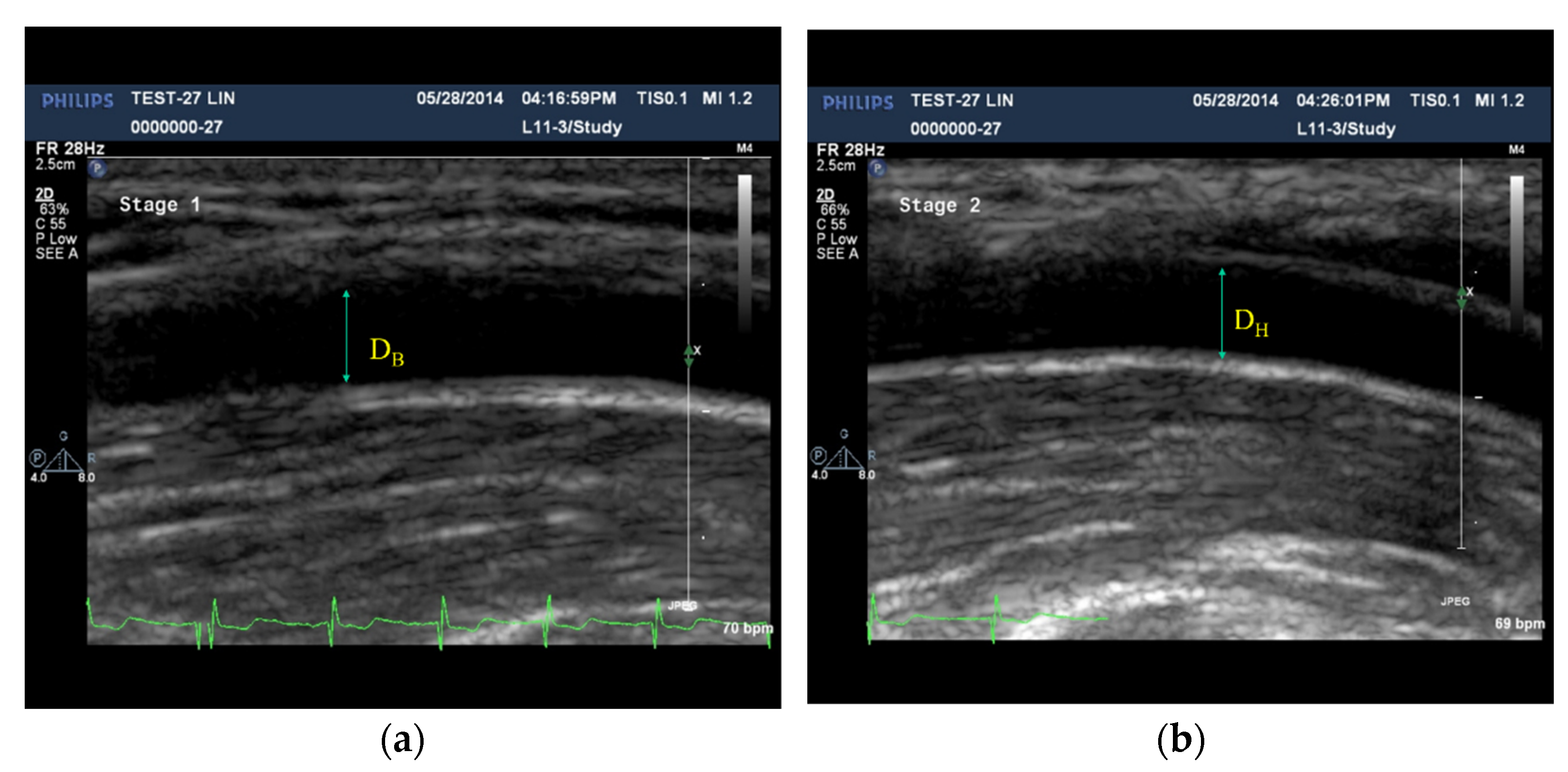
2.1. Assessment of FMD Using the Ultrasonography
2.2. Assessment of FMD Using Sphygmomanometer Cuff
2.3. Assessment of FMD Using the Photoplethysmography
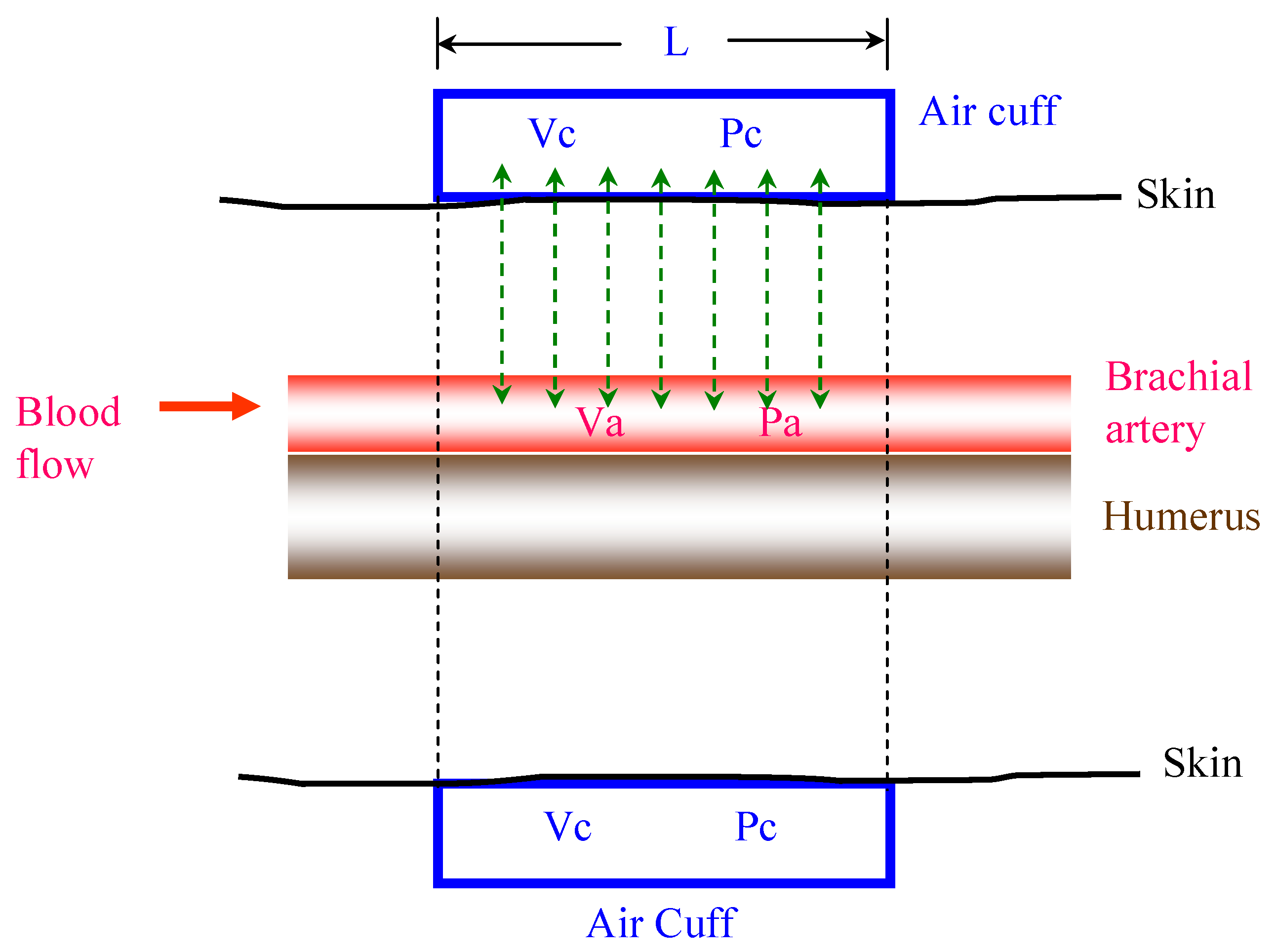
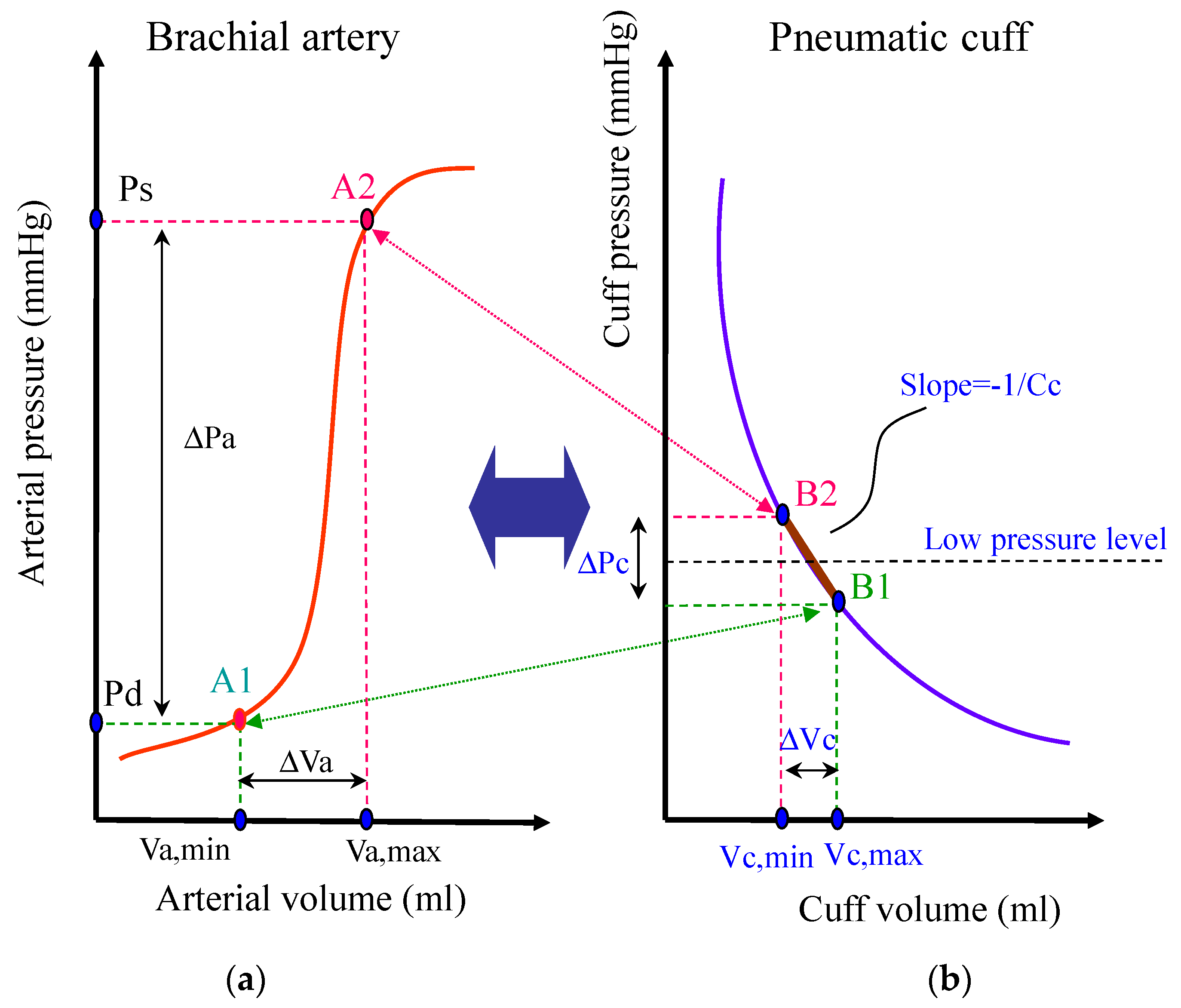
2.4. Cuff Model
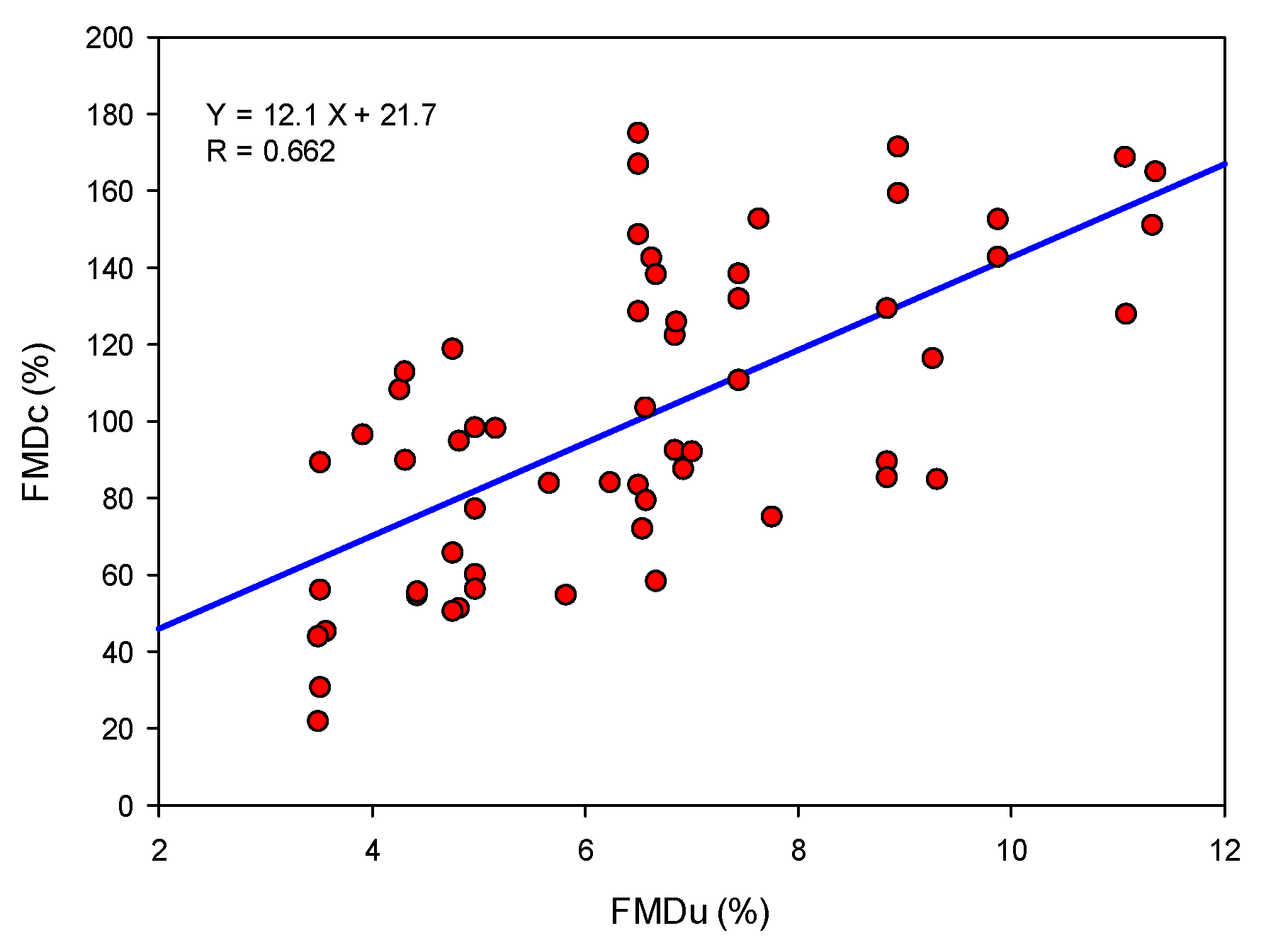
2.5. Relation between FMDc and FMDu
2.6. Experimental Protocol and Measurement
2.7. Data Measurement and Processing
3. Experimental Results
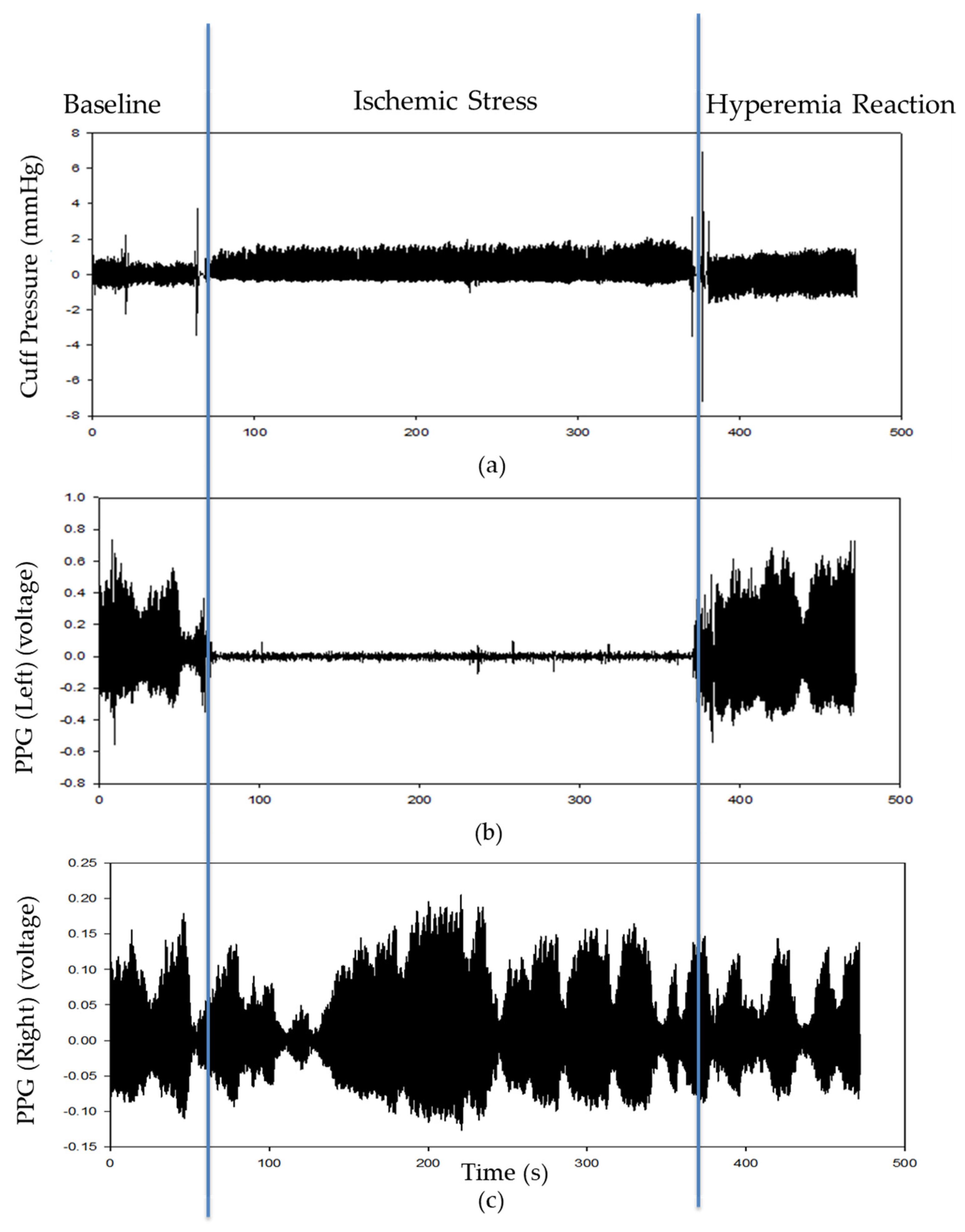
3.1. Typical Recording
3.2. FMD Ratios Measurement
4. Discussion
5. Conclusions
Author Contributions
Funding
Institutional Review Board Statement
Informed Consent Statement
Acknowledgments
Conflicts of Interest
References
- Little, P.J.; Askew, C.D.; Xu, S.; Kamato, D. Endothelial dysfunction and cardiovascular disease: History and analysis of the clinical utility of the relationship. Biomedicines 2021, 9, 699. [Google Scholar] [CrossRef] [PubMed]
- Costa, J.; Borges, M.; Oliveira, E.; Gouveia, M.; Carneiro, A.V. Incidence and prevalence of hypercholesterolemia in Portugal: A systematic review. Part 3. Rev. Port. Cardiol. 2003, 22, 829–836. [Google Scholar]
- Sakakura, K.; Nakano, M.; Otsuka, F.; Ladich, E.; Kolodgie, F.D.; Virmani, R. Pathophysiology of atherosclerosis plaque progression. Heart Lung Circ. 2013, 22, 399–411. [Google Scholar] [CrossRef] [PubMed] [Green Version]
- Bergheanu, S.C.; Bodde, M.C.; Jukema, J.W. Pathophysiology and treatment of atherosclerosis current view and future perspective on lipoprotein modification treatment. Neth. Heart J. 2017, 25, 231–242. [Google Scholar] [CrossRef] [PubMed] [Green Version]
- Singh, R.B.; Mengi, S.A.; Xu, Y.-J.; Arneja, A.S.; Dhalla, N.S. Pathogenesis of atherosclerosis: A multifactorial process. Exp. Clin. Cardiol. 2002, 7, 40–53. [Google Scholar] [PubMed]
- Smedby, O.; Johansson, J.; Molgaard, J.; Olsson, A.G.; Walldius, G.; Erikson, U. Predilection of atherosclerosis for the inner curvature in the femoral artery: A digitized angiography study. Arterioscler. Thromb. Vasc. Biol. 1995, 15, 912–917. [Google Scholar] [CrossRef]
- Furchgott, R.F.; Zawadzki, J.V. The obligatory role of endothelial cells in the relaxation of arterial smooth muscle by acetylcholine. Nature 1980, 288, 373–376. [Google Scholar] [CrossRef]
- Rassaf, T.; Feelisch, M.; Kelm, M. Circulating NO pool: Assessment of nitrite and nitroso species in blood and tissues. Free Radic. Biol. Med. 2004, 36, 413–422. [Google Scholar] [CrossRef]
- Wu, H.D.; Katz, S.D.; Beniaminovitz, A.; Khan, T.; DiTullio, M.R.; Homma, S. Assessment of endothelium-mediated vasodilation of the peripheral circulation by transcutaneous ultrasonography and venous occlusion plethysmography. Heart Vessels 1999, 14, 143–148. [Google Scholar] [CrossRef]
- Charakida, M.; Masi, S.; Luscher, T.F.; Kastelein, J.J.P.; Deanfield, J.E. Assessment of atherosclerosis: The role of flow-mediated dilatation. Eur. Heart J. 2010, 31, 2854–2861. [Google Scholar] [CrossRef]
- Corretti, M.C.; Anderson, T.J.; Benjamin, E.J. Guidelines for the ultrasound assessment of endothelial-dependent flow-mediated vasodilation of the brachial artery: A report of the international brachial artery reactivity task force. J. Am. Coll. Cardiol. 2002, 39, 257–265. [Google Scholar] [CrossRef] [Green Version]
- Ghiadoni, L.; Versari, D.; Giannarelli, C.; Faita, F.; Taddei, S. Non-invasive diagnostic tools for investigating endothelial dysfunction. Curr. Pharm. Des. 2008, 14, 3715–3722. [Google Scholar] [CrossRef] [PubMed]
- Stadler, R.W.; Karl, W.C.; Lees, R.S. New methods for arterial diameter measurement from B-mode images. Ultrasound Med. Biol. 1996, 22, 25–34. [Google Scholar] [CrossRef]
- Rubinshte, R.; Kuvin, J.T.; Soffler, M.; Lennon, R.J.; Lavi, S.; Nelson, R.E.; Pumper, G.M.; Lerman, L.O.; Lerman, A. Assessment of endothelial function by non-invasive peripheral arterial tonometry predicts late cardiovascular adverse events. Eur. Heart J. 2010, 31, 1142–1148. [Google Scholar] [CrossRef] [PubMed] [Green Version]
- Hamburg, N.M.; Benjamin, E.J. Assessment of endothelial function using digital pulse amplitude tonometry. Trends Cardiovasc. Med. 2009, 19, 6–11. [Google Scholar] [CrossRef] [Green Version]
- Kuvin, J.T.; Patel, A.R.; Sliney, K.A.; Pandian, N.G.; Sheffy, J.; Schnall, R.P.; Udelson, J.E. Assessment of peripheral vascular endothelial function with finger arterial pulse wave amplitude. Am. Heart J. 2003, 146, 168–174. [Google Scholar] [CrossRef]
- Hashimoto, M.; Akishita, M.; Eto, M.; Ishikawa, M.; Kozaki, K.; Toba, K.; Sagara, Y.; Taketani, Y.; Orimo, H.; Ouchi, Y. Modulation of endothelium-dependent flow-mediated dilatation of the brachial artery by sex and menstrual cycle. Circulation 1995, 92, 3431–3435. [Google Scholar] [CrossRef]
- Liu, S.-H.; Wang, J.-J.; Cheng, D.-C.; Su, C.-H.; Lin, T.-H. Assessment of the endothelial function with changed volume of brachial artery by menstrual cycle. Biomed. Eng. 2016, 15, 106. [Google Scholar] [CrossRef] [Green Version]
- Liu, S.-H.; Wang, J.-J.; Huang, K.-S. A New Oscillometry-Based Method for Estimating the Dynamic Brachial Artery Compliance under loaded conditions. IEEE Trans. Biomed. Eng. 2008, 55, 2463–2470. [Google Scholar] [CrossRef]
- Liu, S.-H.; Wang, J.-J.; Cheng, D.-C. Non-invasive determination of the instantaneous brachial blood flow using the oscillometric method. Biomed. Tech. 2009, 54, 171–177. [Google Scholar] [CrossRef]
- Liu, S.-H.; Lin, T.-H.; Cheng, D.-C.; Wang, J.-J. Assessment of stroke volume from brachial blood pressure using arterial characteristics. IEEE Trans. Biomed. Eng. 2015, 62, 2151–2157. [Google Scholar] [CrossRef] [PubMed]
- Wu, H.-T.; Lee, C.-H.; Liu, A.-B.; Chung, W.-S.; Tang, C.-J.; Sun, C.-K.; Yip, H.-K. Arterial stiffness using radial arterial waveforms measured at the wrist as an indicator of diabetic control in the elderly. IEEE Trans. Biomed. Eng. 2011, 58, 243–252. [Google Scholar] [PubMed]
- Wu, H.-T.; Lee, C.-H.; Sun, C.-K.; Hsu, J.-T.; Huang, R.-M.; Tang, C.-J. Arterial waveforms measured at the wrist as indicators of diabetic endothelial dysfunction in the elderly. IEEE Trans. Instrum. Meas. 2012, 61, 162–169. [Google Scholar] [CrossRef]
- Maltz, J.S.; Tison, G.H.; Alley, H.F.; Budinger, T.F.; Owens, C.D.; Olgin, J. Measurement of brachial artery endothelial function using a standard blood pressure cuff. Physiol. Meas. 2015, 36, 2247–2268. [Google Scholar] [CrossRef] [PubMed] [Green Version]
- Chowienczyk, P.J.; Kelly, R.P.; MacCallum, H.; Millasseau, S.C.; Andersson, T.L.; Gosling, R.G.; Änggård, E.E. Photoplethysmographic assessment of pulse wave reflection: Blunted response to endothelium-dependent beta2-adrenergic vasodilation in type II diabetes mellitus. J. Am. Coll. Cardiol. 1999, 34, 2007–2014. [Google Scholar] [CrossRef] [Green Version]
- Bonetti, P.O.; Pumper, G.M.; Higano, S.T.; Holmes, D.R., Jr.; Kuvin, J.T.; Lerman, A. Noninvasive identification of patients witj early coronary atherosclerosis by assessment of digital reaction hyperemia. J. Am. Coll. Cardiol. 2004, 44, 2137–2141. [Google Scholar] [CrossRef] [Green Version]
- Lekakis, J.; Abraham, P.; Balbarini, A.; Blann, A.; Boulanger, C.M.; Cockcroft, J.; Vlachopoulos, C. Methods for evaluating endothelial function: A position statement from the European society of cardiology working group on peripheral circulation. Eur. J. Cardiovasc. Prev. Rehabil. 2011, 18, 775–789. [Google Scholar] [CrossRef]


| Subjects (N = 51) | Control Group (N = 38) | Patient Group (N = 13) | p-Value | |
|---|---|---|---|---|
| Weight (kg) | 71.6 ± 20.5 | 72.3 ± 23.3 | 69.5 ± 7.9 | 0.526 |
| Height (cm) | 165.5 ± 18.3 | 166.5 ± 20.9 | 162.6 ± 5.3 | 0.295 |
| Age (years) | 32.7 ± 19.4 | 22.5 ± 4.8 | 62.5 ± 14.1 | <0.001 |
| SBP (mmHg) | 127.7 ± 16.6 | 122.3 ± 13.4 | 143.5 ± 15.2 | <0.001 |
| DIA (mmHg) | 77.5 ± 10.9 | 75.3 ± 10.4 | 83.9 ± 10.2 | 0.016 |
| BMI (kg/m2) | 24.5 ± 4 | 23.8 ± 4.2. | 26.3 ± 2.7 | 0.021 |
| FMDu% | FMDp% | FMDc% | ||||
|---|---|---|---|---|---|---|
| Control (N = 38) | Patient (N = 13) | Control (N = 38) | Patient (N = 13) | Control (N = 38) | Patient (N = 13) | |
| M ± SD | (8.1 ± 4.3)% | (4.5 ± 1.1)% | (93.4 ± 34.4)% | (84.4 ± 44.4)% | (121.6 ± 48.6)% | (55.2 ± 22.8)% |
| p-value | 0.040 | 0.06 | <0.001 | |||
| DB,max (cm) | DH,max (cm) | p-Value | DB,min (cm) | DH,min (cm) | p-Value | |
|---|---|---|---|---|---|---|
| Control | 0.323 ± 0.057 | 0.348 ± 0.060 | 0.001 | 0.297 ± 0.055 | 0.321 ± 0.056 | 0.116 |
| Patient | 0.400 ± 0.057 | 0.422 ± 0.220 | 0.002 | 0.350 ± 0.052 | 0.380 ± 0.052 | 0.302 |
| p-value | 0.001 | 0.004 | 0.018 | 0.015 |
| ΔPc,B (mmHg) | ΔPc,H (mmHg) | |
|---|---|---|
| Control | 1.441 ± 0.75 | 3.0 ± 1.48 |
| Patient | 2.302 ± 1.83 | 3.58 ± 2.73 |
| p-value | 0.002 | 0.036 |
Publisher’s Note: MDPI stays neutral with regard to jurisdictional claims in published maps and institutional affiliations. |
© 2022 by the authors. Licensee MDPI, Basel, Switzerland. This article is an open access article distributed under the terms and conditions of the Creative Commons Attribution (CC BY) license (https://creativecommons.org/licenses/by/4.0/).
Share and Cite
Wang, J.-J.; Liu, S.-H.; Pan, Y.-H.; Tseng, W.-K.; Chen, W. Theoretical and Experimental Study on Assessment of Flow-Mediated Dilatation Using the Cuff Method in Brachial Arteries. Electronics 2022, 11, 351. https://doi.org/10.3390/electronics11030351
Wang J-J, Liu S-H, Pan Y-H, Tseng W-K, Chen W. Theoretical and Experimental Study on Assessment of Flow-Mediated Dilatation Using the Cuff Method in Brachial Arteries. Electronics. 2022; 11(3):351. https://doi.org/10.3390/electronics11030351
Chicago/Turabian StyleWang, Jia-Jung, Shing-Hong Liu, Yong-Hong Pan, Wei-Kung Tseng, and Wenxi Chen. 2022. "Theoretical and Experimental Study on Assessment of Flow-Mediated Dilatation Using the Cuff Method in Brachial Arteries" Electronics 11, no. 3: 351. https://doi.org/10.3390/electronics11030351
APA StyleWang, J.-J., Liu, S.-H., Pan, Y.-H., Tseng, W.-K., & Chen, W. (2022). Theoretical and Experimental Study on Assessment of Flow-Mediated Dilatation Using the Cuff Method in Brachial Arteries. Electronics, 11(3), 351. https://doi.org/10.3390/electronics11030351