
Characterizing the Mechanism of Action of Essential Oils on Skin Homeostasis—Data from Sonographic Imaging, Epidermal Water Dynamics, and Skin Biomechanics


 ,
,  ,
, 
Abstract
1. Introduction
2. Methods
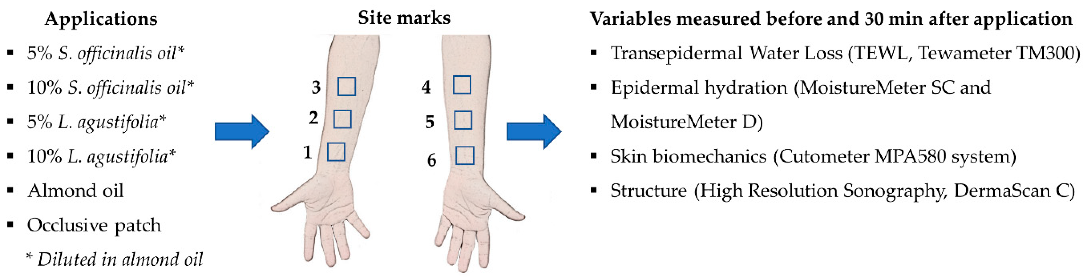
2.1. Participants
2.2. Essential Oils Obtention and Formulations
2.3. Procedure
2.4. Statistics
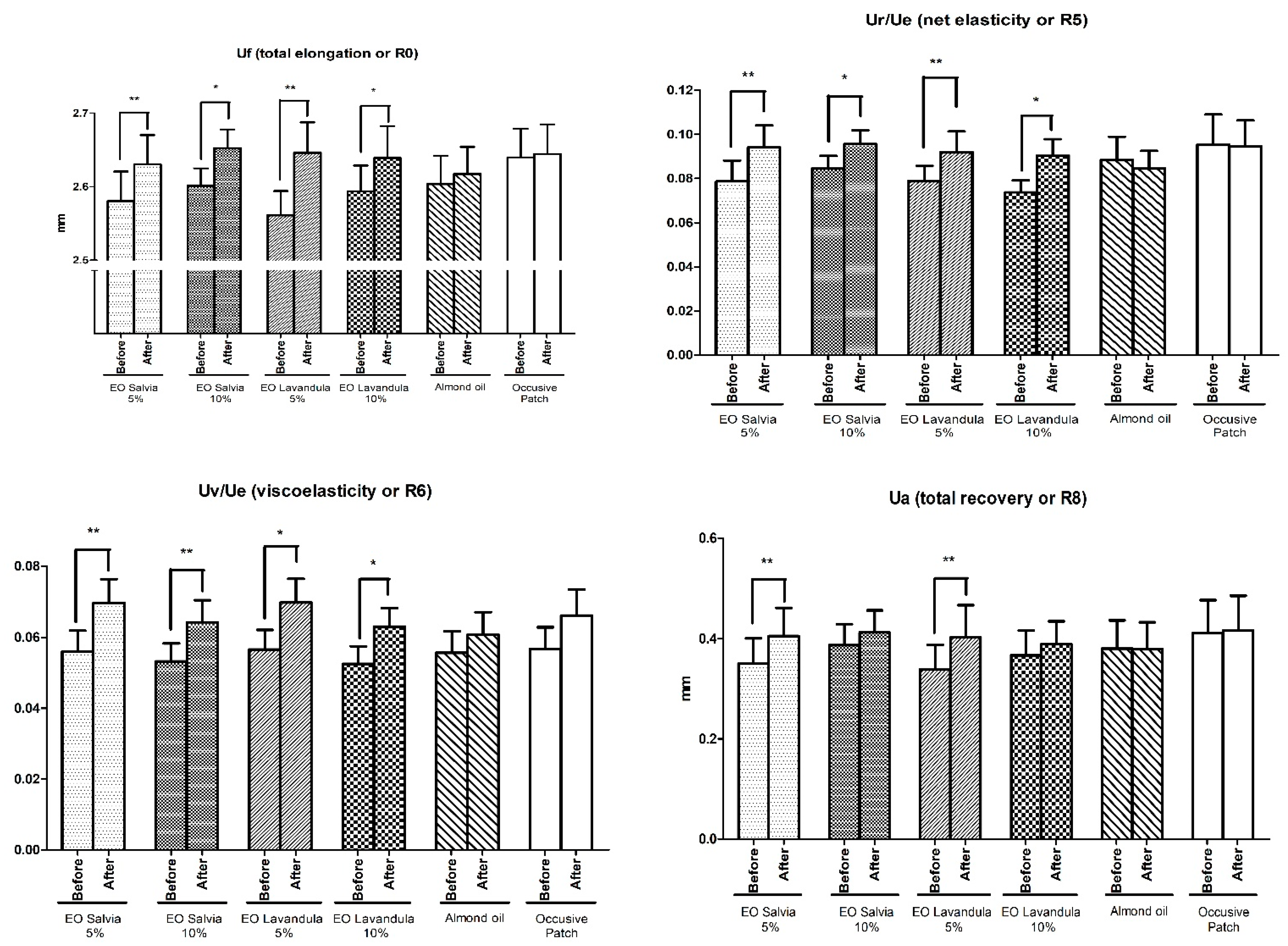
3. Results
4. Discussion
5. Conclusions
Author Contributions
Funding
Institutional Review Board Statement
Informed Consent Statement
Data Availability Statement
Acknowledgments
Conflicts of Interest
References
- Elshafie, H.; Camele, I. An Overview of The Biological Effects of Some Mediterranean Essential Oils on Human Health. BioMed Res. Intern. 2017, 2017, 9268468. [Google Scholar] [CrossRef]
- Vaughn, A.; Clark, A.; Sivamani, R.; Shi, V. Natural Oils for Skin-Barrier Repair: Ancient Compounds Now Backed by Modern Science. Am. J. Clin. Dermatol. 2017, 19, 103–117. [Google Scholar] [CrossRef]
- Pérez-Recalde, M.; Ruiz Arias, I.; Hermida, É. Could Essential Oils Enhance Biopolymers Performance for Wound Healing? A Systematic Review. Phytomedicine 2018, 38, 57–65. [Google Scholar] [CrossRef]
- Ahn, C.; Lee, J.; Park, M.; Kim, J.; Yang, J.; Yoo, Y.; Jeung, E. Cytostatic effects of plant essential oils on human skin and lung cells. Exper. Therap. Med. 2020, 19, 2008–2018. [Google Scholar] [CrossRef] [PubMed]
- Esposito, E.; Nastruzzi, C.; Sguizzato, M.; Cortesi, R. Nanomedicines to Treat Skin Pathologies with Natural Molecules. Curr. Pharm. Des. 2019, 25, 2323–2337. [Google Scholar] [CrossRef]
- de Groot, A.C.; Schmidt, E. Essential Oils, Part I: Introduction. Dermatitis 2016, 27, 39–42. [Google Scholar] [CrossRef]
- Firenzuoli, F.; Jaitak, V.; Horvath, G.; Bassolé, I.H.N.; Setzer, W.N.; Gori, L. Essential oils: New perspectives in human health and wellness. Evid. Based Complement. Alternat. Med. 2014. [Google Scholar] [CrossRef]
- Bakkali, F.; Averbeck, S.; Averbeck, D.; Idaomar, M. Biological effects of essential oils—A review. Food Chem. Toxicol. 2008, 46, 446–475. [Google Scholar] [CrossRef] [PubMed]
- Mekonnen, A.; Tesfaye, S.; Christos, S.; Dires, K.; Zenebe, T.; Zegeye, N.; Shiferaw, Y.; Lulekal, E. Evaluation of Skin Irritation and Acute and Subacute Oral Toxicity of Lavandula angustifolia Essential Oils in Rabbit and Mice. J. Toxicol. 2019, 2019, 5979546. [Google Scholar] [CrossRef]
- Aziz, Z.A.; Ahmad, A.; Setapar, S.H.M.; Karakucuk, A.; Azim, M.M.; Lokhat, D.; Rafatullah, M.; Ganash, M.; Kamal, M.A.; Ashraf, G.M. Essential Oils: Extraction Techniques, Pharmaceutical and Therapeutic Potential—A Review. Curr. Drug. Metab. 2018, 19, 1100–1110. [Google Scholar] [CrossRef] [PubMed]
- Orchard, A.; van Vuuren, S. Commercial Essential Oils as Potential Antimicrobials to Treat Skin Diseases. Evid.-Based Complement. Alternat. Med. 2017, 2017, 4517971. [Google Scholar] [CrossRef]
- Patzelt, A.; Lademann, J.; Richter, H.; Darvin, M.; Schanzer, S.; Thiede, G.; Sterry, W.; Vergou, T.; Hauser, M. In Vivo Investigations on The Penetration of Various Oils and Their Influence on The Skin Barrier. Skin Res. Technol. 2011, 18, 364–369. [Google Scholar] [CrossRef]
- Jiang, Q.; Wu, Y.; Zhang, H.; Liu, P.; Yao, J.; Yao, P.; Chen, J.; Duan, J. Development of Essential Oils as Skin Permeation Enhancers: Penetration Enhancement Effect and Mechanism of Action. Pharmaceut. Biol. 2017, 55, 1592–1600. [Google Scholar] [CrossRef] [PubMed]
- De Groot, A.; Schmidt, E. Essential Oils, Part V: Peppermint Oil, Lavender Oil, and Lemongrass Oil. Dermatitis 2016, 27, 325–332. [Google Scholar] [CrossRef] [PubMed]
- Smigielski, K.; Prusinowska, R.; Raj, A.; Sikora, M.; Woliñska, K.; Gruska, R. Effect of Drying on The Composition of Essential Oil from Lavandula angustifolia. J. Essent. Oil Bear. Plant. 2011, 14, 532–542. [Google Scholar] [CrossRef]
- Raal, A.; Orav, A.; Arak, E. Composition of the Essential Oil of Salvia officinalis L. from Various European Countries. Nat. Prod. Res. 2007, 21, 406–411. [Google Scholar] [PubMed]
- Tosun, A.; Khan, S.; Kim, Y.; Calín-Sánchez, A.; Hysenaj, X.; Carbonell-Barrachina, A. Essential Oil Composition and Anti-Inflammatory Activity of Salvia officinalis L (Lamiaceae) in Murin Macrophages. Trop. J. Pharm. Res. 2014, 13, 937. [Google Scholar] [CrossRef]
- World Medical Association. World Medical Association Declaration of Helsinki. JAMA 2013, 310, 2191. [Google Scholar] [CrossRef]
- Seidenari, S.; Nakijo, A.; Pepe, P.; Giannetti, A. Ultrasound B Scanning with Image Analysis for Assessment of Allergic Patch Test Reactions. Contact Derm. 1991, 24, 216–222. [Google Scholar] [CrossRef]
- Pinnagoda, J.; Tupkek, R.; Agner, T.; Serup, J. Guidelines for Transepidermal Water Loss (TEWL) Measurement. Contact Derm. 1990, 22, 164–178. [Google Scholar] [CrossRef]
- Berardesca, E.; Loden, M.; Serup, J.; Masson, P.; Rodrigues, L. The Revised EEMCO Guidance for The In Vivo Measurement of Water in The Skin. Skin Res. Technol. 2018, 24, 351–358. [Google Scholar] [CrossRef]
- Mayrovitz, H.; Luis, M. Spatial Variations in Forearm Skin Tissue Dielectric Constant. Skin Res. Technol. 2010, 16, 438–443. [Google Scholar] [CrossRef]
- Ryu, H.; Joo, Y.; Kim, S.; Park, K.; Youn, S. Influence of Age and Regional Differences on Skin Elasticity as Measured by The Cutometer. Skin Res. Technol. 2008, 14, 354–358. [Google Scholar] [CrossRef] [PubMed]
- Monteiro Rodrigues, L.; Fluhr, J. EEMCO Guidance for The In Vivo Assessment of Biomechanical Properties of The Human Skin and Its Annexes: Revisiting Instrumentation and Test Modes. Skin Pharm. Physiol. 2019, 33, 44–60. [Google Scholar] [CrossRef]
- Mazzarello, V.; Gavini, E.; Rassu, G.; Donadu, M.; Usai, D.; Piu, G.; Pomponi, V.; Sucato, F.; Zanetti, S.; Montesu, M.A. Clinical Assessment of New Topical Cream Containing Two Essential Oils Combined with Tretinoin in the Treatment of Acne. Clin. Cosm. Investig. Derm. 2020, 13, 233–239. [Google Scholar] [CrossRef] [PubMed]
- Wertz, P. Lipids and Barrier Function of The Skin. Acta Derm.-Venereol. 2000, 80, 7–11. [Google Scholar] [CrossRef]
- Sahle, F.; Gebre-Mariam, T.; Dobner, B.; Wohlrab, J.; Neubert, R. Skin Diseases Associated with the Depletion of Stratum Corneum Lipids and Stratum Corneum Lipid Substitution Therapy. Skin Pharm. Physiol. 2015, 28, 42–55. [Google Scholar] [CrossRef]
- Ernst, E. Adverse Effects of Herbal Drugs in Dermatology. Br. J. Dermat. 2000, 143, 923–929. [Google Scholar] [CrossRef]
- Nielsen, J. Natural Oils Affect the Human Skin Integrity and the Percutaneous Penetration of Benzoic Acid Dose-Dependently. Bas. Clin. Pharmacol. Toxicol. 2006, 98, 575–581. [Google Scholar] [CrossRef] [PubMed]
- Viciolle, E.; Castilho, P.; Rosado, C. In Vitroandin Vivoassessment of the Effect Oflaurus Novocanariensisoil and Essential Oil in Human Skin. Int. J. Cosm. Sci. 2012, 34, 546–550. [Google Scholar] [CrossRef] [PubMed]
- Lin, T.; Zhong, L.; Santiago, J. Anti-Inflammatory and Skin Barrier Repair Effects of Topical Application of Some Plant Oils. Int. J. Mol. Sci. 2017, 19, 70. [Google Scholar] [CrossRef]
- Martschick, A.; Teichmann, A.; Richter, H.; Schanzer, S.; Antoniou, C.; Sterry, W.; Lademann, J. Analysis of The Penetration Profiles of Topically Applied Substances by Laser Scanning Microscopy. Laser Phys. Lett. 2007, 4, 395–398. [Google Scholar] [CrossRef]
- Lademann, J.; Otberg, N.; Richter, H.; Meyer, L.; Audring, H.; Teichmann, A.; Thomas, S.; Knüttel, A.; Sterry, W. Application of Optical Non-Invasive Methods in Skin Physiology: A Comparison of Laser Scanning Microscopy and Optical Coherent Tomography with Histological Analysis. Skin Res. Technol. 2007, 13, 119–132. [Google Scholar] [CrossRef]
- Patzelt, A.; Sterry, W.; Lademann, J. In Vivo Measurements of Skin Barrier: Comparison of Different Methods and Advantages of Laser Scanning Microscopy. Laser Phys. Lett. 2010, 7, 843–852. [Google Scholar] [CrossRef]
- Capetti, F.; Sgorbini, B.; Cagliero, C.; Argenziano, M.; Cavalli, R.; Milano, L.; Bicchi, C.; Rubiolo, P. Melaleuca alternifolia Essential Oil: Evaluation of Skin Permeation and Distribution from Topical Formulations with a Solvent-Free Analytical Method. Planta Med. 2020, 86, 442–450. [Google Scholar] [CrossRef]
- Marzulli, F.; Maibach, H. Contact Allergy: Predictive Testing in Man. Contact Derm. 1976, 2, 1–17. [Google Scholar] [CrossRef] [PubMed]
- Alanen, E.; Nuutinen, J.; Nicklen, K.; Lahtinen, T.; Monkkonen, J. Measurement of Hydration in the Stratum Corneum with The Moisturemeter and Comparison with The Corneometer. Skin Res. Technol. 2004, 10, 32–37. [Google Scholar] [CrossRef] [PubMed]
- Rosado, C.; Barbosa, R.; Fernando, R.; Antunes, F.; Rodrigues, L. Study of the Effect of Epidermal Overhydration by Occlusion, on the Skin Biomechanical Behaviour Assessed In Vivo with the Systems Cutometer®, Reviscometer® and Cutiscan®. J. Biomed. Biopharm. Res. 2015, 12, 203–213. [Google Scholar] [CrossRef]
- Wolf, R.; Wolf, D.; Rudikoff, D.; Parish, L. Nutrition and Water: Drinking Eight Glasses of Water A Day Ensures Proper Skin Hydration—Myth or Reality? Clin. Dermatol. 2010, 28, 380–383. [Google Scholar] [CrossRef]
- Rodrigues, L.; Palma, L.; Tavares Marques, L.; Bujan Varela, J. Dietary Water Affects Human Skin Hydration and Biomechanics. Clin. Cosmet. Investig. Dermatol. 2015, 8, 413. [Google Scholar] [CrossRef]
- Monteiro Rodrigues, L.; Palma, L.; Santos, O.; Almeida, M.; Bujan, J.; Tavares, L. Excessive Weight Favours Skin Physiology—Up to A Point: Another Expression of the Obesity Paradox. Skin Pharm. Physiol. 2017, 30, 94–101. [Google Scholar] [CrossRef] [PubMed]


| Treatment | TEWL g/m2/h before (T0) after (T30) | Epidermal Superficial Hydration (AU’s) before (T0) after (T30) | Epidermal Deep Hydration (AU’s) before (T0) after (T30) | |||
|---|---|---|---|---|---|---|
| EO S. officinalis 5% | 6.36 ± 0.61 | 4.43 ± 0.34 ** | 32.80 ± 2.78 | 41.51 ± 3.08 ** | 17.38 ± 0.92 | 20.04 ± 0.63 ** |
| EO S. officinalis 10% | 6.91 ± 0.73 | 5.14 ± 0.59 * | 33.55 ± 1.93 | 42.35 ± 2.73 ** | 18.01 ± 0.95 | 19.81 ± 0.69 * |
| EO L. angustifolia 5% | 6.50 ± 0.63 | 4.69 ± 0.51 ** | 33.88 ± 2.37 | 44.41 ± 4.64 * | 17.29 ± 0.85 | 19.18 ± 0.86 ** |
| EO L. angustifolia 10% | 6.68 ± 1.81 | 4.84 ± 1.70 ** | 32.12 ± 2.33 | 46.25 ± 3.67 ** | 17.86 ± 0.95 | 20.11± 0.69 ** |
| Almond Oil | 5.99 ± 0.81 | 4.22 ± 0.55 * | 31.59 ± 3.16 | 40.61 ± 3.67 ** | 16.44 ± 0.81 | 18.48 ± 0.76 * |
| Occlusive Patch | 6.95 ± 0.80 | 10.62 ± 1.33 | 33.86 ± 2.22 | 42.50 ± 3.20 | 17.50 ± 0.62 | 18.69 ± 0.64 |
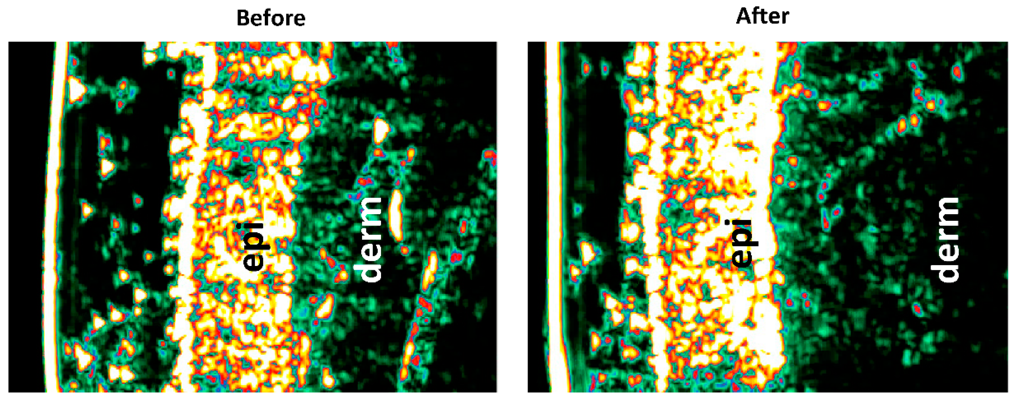
| Echogenic Changes/Area (%) | Echogenic Changes/Area (%) | |
|---|---|---|
| Epidermis | Dermis | |
| EO S. officinalis 5% | 20.10 ± 1.70 | −27.81 ± 4.88 ** |
| EO S. officinalis 10% | 14.62 ± 1.60 | −22.63 ± 1.52 * |
| EO L. angustifolia 5% | 19.02 ± 2.70 | −28.53 ± 4.46 ** |
| EO L. angustifolia 10% | 15.95 ± 2.68 | −25.62 ± 4.95 * |
| Almond Oil | 14.34 ± 3.52 | −17.88 ± 3.70 |
| Occlusive Patch | 15.84 ± 0.72 | −7.25 ± 2.06 |
Publisher’s Note: MDPI stays neutral with regard to jurisdictional claims in published maps and institutional affiliations. |
© 2021 by the authors. Licensee MDPI, Basel, Switzerland. This article is an open access article distributed under the terms and conditions of the Creative Commons Attribution (CC BY) license (https://creativecommons.org/licenses/by/4.0/).
Share and Cite
de Andrade, S.F.; Rijo, P.; Rocha, C.; Zhu, L.; Rodrigues, L.M. Characterizing the Mechanism of Action of Essential Oils on Skin Homeostasis—Data from Sonographic Imaging, Epidermal Water Dynamics, and Skin Biomechanics. Cosmetics 2021, 8, 36. https://doi.org/10.3390/cosmetics8020036
de Andrade SF, Rijo P, Rocha C, Zhu L, Rodrigues LM. Characterizing the Mechanism of Action of Essential Oils on Skin Homeostasis—Data from Sonographic Imaging, Epidermal Water Dynamics, and Skin Biomechanics. Cosmetics. 2021; 8(2):36. https://doi.org/10.3390/cosmetics8020036
Chicago/Turabian Stylede Andrade, Sérgio Faloni, Patricia Rijo, Clemente Rocha, Lin Zhu, and Luis Monteiro Rodrigues. 2021. "Characterizing the Mechanism of Action of Essential Oils on Skin Homeostasis—Data from Sonographic Imaging, Epidermal Water Dynamics, and Skin Biomechanics" Cosmetics 8, no. 2: 36. https://doi.org/10.3390/cosmetics8020036
APA Stylede Andrade, S. F., Rijo, P., Rocha, C., Zhu, L., & Rodrigues, L. M. (2021). Characterizing the Mechanism of Action of Essential Oils on Skin Homeostasis—Data from Sonographic Imaging, Epidermal Water Dynamics, and Skin Biomechanics. Cosmetics, 8(2), 36. https://doi.org/10.3390/cosmetics8020036