You are currently viewing a new version of our website. To view the old version click .
Cosmetics
  • Article
  • Open Access

7 November 2025

Fractional CO2 Laser 2-Mercaptonicotinoyl Glycine Drug Delivery for Melasma and Facial Hyperpigmentation: A Real-Observational World Experience

,
,
,
and
Dermatology Unit, Department of Clinical Internal, Anesthesiological and Cardiovascular Sciences, Sapienza University, Viale del Policlinico 155, 00185 Rome, Italy
*
Author to whom correspondence should be addressed.
These authors contributed equally to this work.
Cosmetics2025, 12(6), 251;https://doi.org/10.3390/cosmetics12060251 
(registering DOI)
This article belongs to the Section Cosmetic Dermatology

Abstract

Background: Melasma and facial hyperpigmentation are common dermatologic conditions that significantly impact patients’ quality of life. New approaches such as laser-assisted delivery of topical agents have been proposed to enhance treatment outcomes. Objective: To evaluate the comparative effectiveness of fractional CO2 laser-assisted delivery of 2-mercaptonicotinoyl glycine (2-MNG) versus topical 2-MNG alone in a real-world clinical setting. Methods: Fourteen female patients with mild-to-moderate melasma or hyperpigmentation were enrolled, and eleven completed the 16-week study. Participants were allocated to either the laser +2-MNG group (n = 6) or topical 2-MNG only group (n = 5). Assessments were performed at baseline and every four weeks using VISIA CR® imaging (visible, UV, and brown spots), Modified Melasma Area and Severity Index (mMASI), and Dermatology Life Quality Index (DLQI). Results: Both groups showed significant improvement in visible spots, brown spots, mMASI, and DLQI scores. The laser group showed earlier improvement, with significant changes starting at week 8, while the control group reached significance at week 12. No significant change in UV spot scores was observed. Conclusion: Both approaches were effective in reducing clinically visible pigmentation and improving quality of life. Fractional CO2 laser-assisted delivery offered a faster onset of efficacy.

1. Introduction

Hyperpigmentation refers to an increase in skin pigmentation, resulting in dark spots or areas that are visibly darker than the surrounding tissue. It includes a range of conditions, such as melasma, freckles, and post-inflammatory hyperpigmentation, each of which can affect the quality of the skin and the psychological well-being of those affected. Although hyperpigmentation is not typically dangerous, it can significantly impact quality of life, leading to emotional distress and social discomfort. This condition often arises from a combination of genetic, environmental, and hormonal factors. Effective management requires a comprehensive understanding of the underlying mechanisms involving melanin production, as well as a personalized therapeutic approach [,].
Biologically, hyperpigmentation develops when melanocytes, the skin’s melanin-producing cells, are stimulated to produce excessive amounts of pigment. Melanin has a role in protecting the skin against ultraviolet (UV) radiation, and its overproduction can lead to noticeable discoloration. The most common cause of hyperpigmentation is sun exposure. Ultraviolet radiation triggers melanocytes to produce melanin as a natural defense mechanism. However, chronic or unprotected sun exposure can lead to sunspots, which are signs of photoaging. Inflammatory processes are another significant factor. When the skin is subjected to injury, inflammation, or conditions like acne or eczema, it can respond with an overproduction of melanin, resulting in post-inflammatory hyperpigmentation that may persist long after the initial lesion has healed [,].
Hormonal factors also play a crucial role, particularly in cases of melasma, often called the “mask of pregnancy,” which arises due to elevated estrogen and progesterone levels during pregnancy or the use of oral contraceptives. Additionally, perimenopause and menopause have been associated with the onset or exacerbation of melasma, likely due to fluctuating estrogen levels and changes in skin melanocyte activity []. Certain medications, including tetracyclines, antimalarials, chemotherapeutic agents can induce photosensitivity, leading to hyperpigmentation. Genetic predisposition must also be considered, as individuals with darker skin phototypes are inherently more susceptible to pigmentary disorders. Finally, intrinsic aging contributes to the formation of age spots, particularly in individuals with significant cumulative sun exposure [].
Diagnosing hyperpigmentation typically relies on clinical examination conducted by dermatologists. A detailed patient history focusing on sun exposure, inflammatory skin events, medication use, and hormonal influences is essential. Diagnostic tools such as the Wood’s lamp can help differentiate between epidermal and dermal pigmentation, a distinction that significantly influences therapeutic decisions []. Dermatoscopy further enhances diagnostic accuracy by enabling a more precise evaluation of pigmented lesions, ensuring appropriate treatment and ruling out malignancies [].
Treatment of hyperpigmentation must be tailored to the underlying cause and the individual patient’s skin type. Daily application of sunscreen with an SPF of 50 or higher and a strong UVA protection is fundamental, as ongoing UV exposure do not help any therapeutic approach. Topical agents represent the first line of intervention []. Hydroquinone remains a gold standard for its strong depigmenting effects, although it must be used with caution to avoid adverse reactions []. Other topical options include glycolic acid, an alpha-hydroxy acid that promotes skin renewal; kojic acid, which inhibits melanin synthesis; and retinoids like tretinoin, which accelerate cell turnover and reduce the appearance of dark spots [].
In cases where topical treatments are insufficient, chemical peels using glycolic acid, mandelic acid, or trichloroacetic acid can effectively exfoliate the skin and reduce pigmentation []. More advanced technologies, such as fractional CO2 laser therapy, have shown great promise in managing difficult cases of hyperpigmentation. These lasers target pigment with precision while preserving surrounding healthy tissue, improving overall skin quality through collagen stimulation. However, potential side effects may include post-inflammatory hyperpigmentation (PIH), erythema, crusting, and a temporary worsening of pigmentation, particularly in darker phototypes [].
The fractional CO2 laser is an innovative tool in dermatologic aesthetics, representing a significant evolution from traditional ablative laser technologies. Unlike conventional CO2 lasers that ablate the entire skin surface, the fractional mode divides the laser beam into thousands of microscopic zones that create controlled micro-injuries while sparing surrounding tissues. This approach not only minimizes downtime but also reduces the risk of side effects such as scarring or post-inflammatory hyperpigmentation. Operating at a wavelength of 10,600 nm, the CO2 laser is selectively absorbed by water molecules within skin cells, allowing precise vaporization and thermal stimulation. The laser’s energy initiates a healing cascade, characterized by the production of new collagen and elastin, leading to skin rejuvenation and repair [].
In the treatment of hyperpigmentation, fractional CO2 lasers offer a dual benefit. They effectively target and vaporize melanocytes loaded with excess pigment, thereby reducing the appearance of dark spots. Simultaneously, they stimulate the production of new, evenly pigmented skin. This dual action makes the technology particularly effective for solar lentigo, melasma, and post-inflammatory hyperpigmentation. Compared to non-fractional ablative lasers, the fractional CO2 laser provides substantial clinical improvements with fewer side effects and shorter recovery periods, making it a preferred option for many patients and clinicians alike [].
Among emerging therapies for hyperpigmentation, a promising new agent inhibiting melanin production has been developed; 2-mercaptonicotinoyl glycine (2-MNG, MelasylTM, France). Unlike conventional depigmenting agents that inhibit tyrosinase, 2-MNG exerts its effect by selectively trapping melanin precursors such as DHICA, DHI, and dopaquinone. This action prevents their oxidation and subsequent polymerization into visible melanin pigments (eumelanin and pheomelanin). Through the formation of covalent bonds with these intermediates, 2-MNG disrupts the melanogenic cascade at an earlier step, preserving melanocyte viability and minimizing irritation. This innovative mechanism may explain its favorable safety profile and makes it a novel option in the treatment of hyperpigmentation [,,].
A depigmenting serum formulated with the novel anti-pigmentation molecule 2-MNG (2-MNG-serum), has been developed to specifically target hyperpigmentary disorders such as post-acne hyperpigmentation and melasma. The serum contains 0.5% 2-MNG (MelasylTM), was combined with 10% Niacinamide, Cystoseira tamariscifolia extract, LHA, Carnosine, Retinyl Palmitate and Dipotassium Glycyrrhizate (MelaB3 serum, MB3 La Roche-Posay Laboratoire Dermatologique, France). Clinical studies have already proven the efficacy of 2-MNG-serum in pigmentation disorders such as post-acne hyperpigmentation, with 32 participants in a study on PIH and 109 in a randomized melasma trial [,]. 2-MNG-serum is a comprehensive formula which exerts, through its different active ingredients, its effects through multiple biochemical mechanisms. Its antioxidant activity is primarily driven by its capacity to scavenge free radicals such as superoxide anions, peroxides, and hydroxyl radicals, all of which contribute to cellular aging and pigmentary disorders when produced in excess. Chronic inflammation is increasingly recognized as a central player in the pathogenesis of hyperpigmentation and other skin disorders, and by dampening these pathways, 2-MNG-serum may offer significant therapeutic benefits [].
Thanks to its multifaceted actions—neutralizing oxidative stress, modulating inflammation, strengthening skin barrier, and inhibiting melanin synthesis—2-MNG-serum represents a significant advancement in the dermatologic and cosmetic treatment of pigmentation disorders. Its integration into therapeutic regimens, either alone or in combination with physical modalities such as fractional CO2 laser therapy, holds great potential to improve outcomes for patients suffering from melasma, sunspots, and other pigmentary conditions. Continued research will help further define its role and optimize its use in clinical practice. Therefore, the present study aimed to evaluate the comparative efficacy and safety of fractional CO2 laser-assisted delivery of 2-mercaptonicotinoyl glycine (2-MNG)-containing serum versus topical 2-MNG-containing serum alone in patients with facial melasma and hyperpigmentation.

2. Materials and Methods

2.1. Study Design

This real-world observational study was conducted to evaluate the efficacy and safety of fractional CO2 laser-assisted delivery of 2-mercaptonicotinoyl glycine (2-MNG)-containing serum (2-MNG-serum) compared to topical 2-MNG-serum alone for the treatment of facial hyperpigmentation, including melasma.

2.2. Participants

Fourteen female patients, aged between 30 and 70 years, with mild to moderate facial melasma and/or other facial hyperpigmentation (Fitzpatrick skin types I to III), were enrolled between September 2024 and April 2025. Diagnosis was confirmed by a dermatologist, and baseline severity was assessed using the Modified Melasma Area and Severity Index (mMASI), with inclusion scores ranging from 4.5 to 9.0.

2.3. Informed Consent and Data Collection

All participants provided informed consent at their baseline visit. Detailed data, including age, Fitzpatrick skin phototype, medical history, chronic medication use, smoking and alcohol habits, weight, and height, were meticulously recorded. The study adhered to national legislation for non-interventional observational studies and complied with the ethical principles of the Declaration of Helsinki. Since this study did not involve any experimental intervention or change in clinical management, approval from the Ethics Committee was not required according to applicable regulations.

Treatment Groups and Clinical Procedures

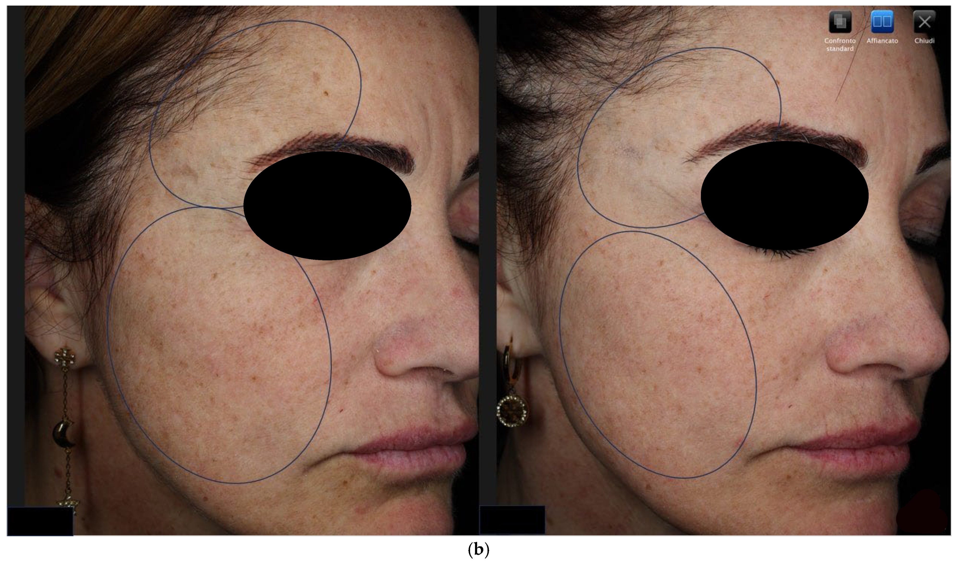
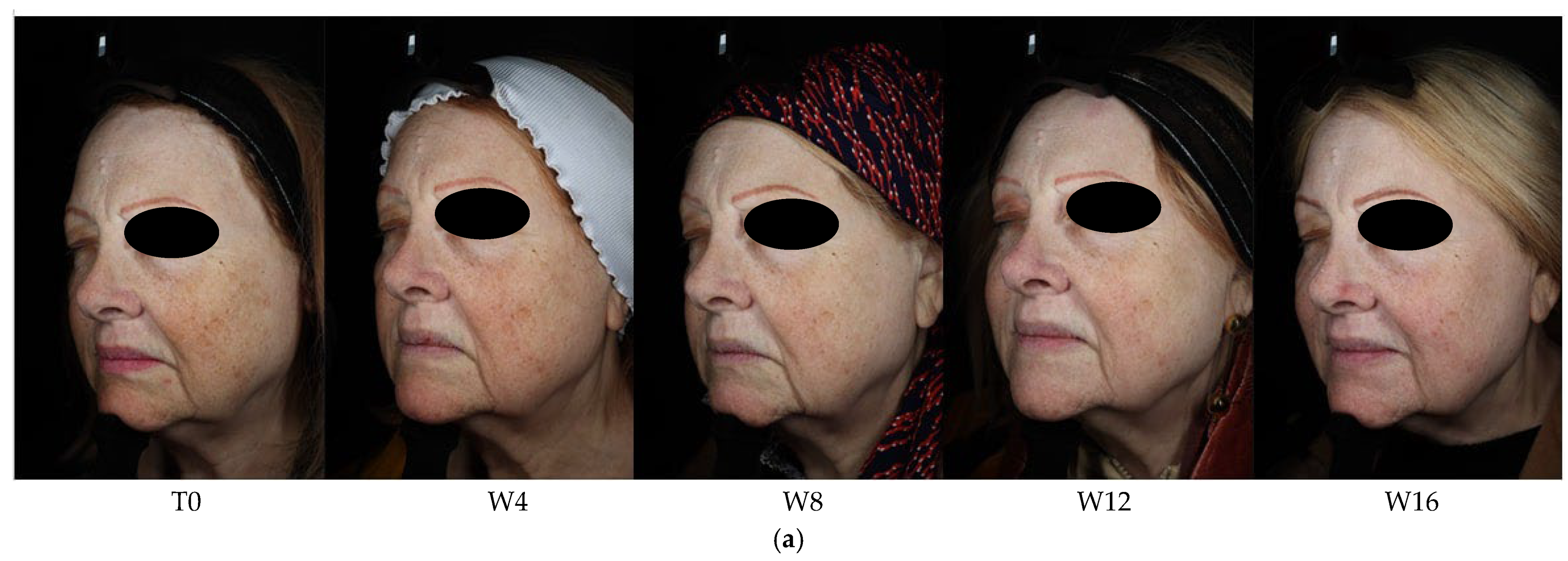
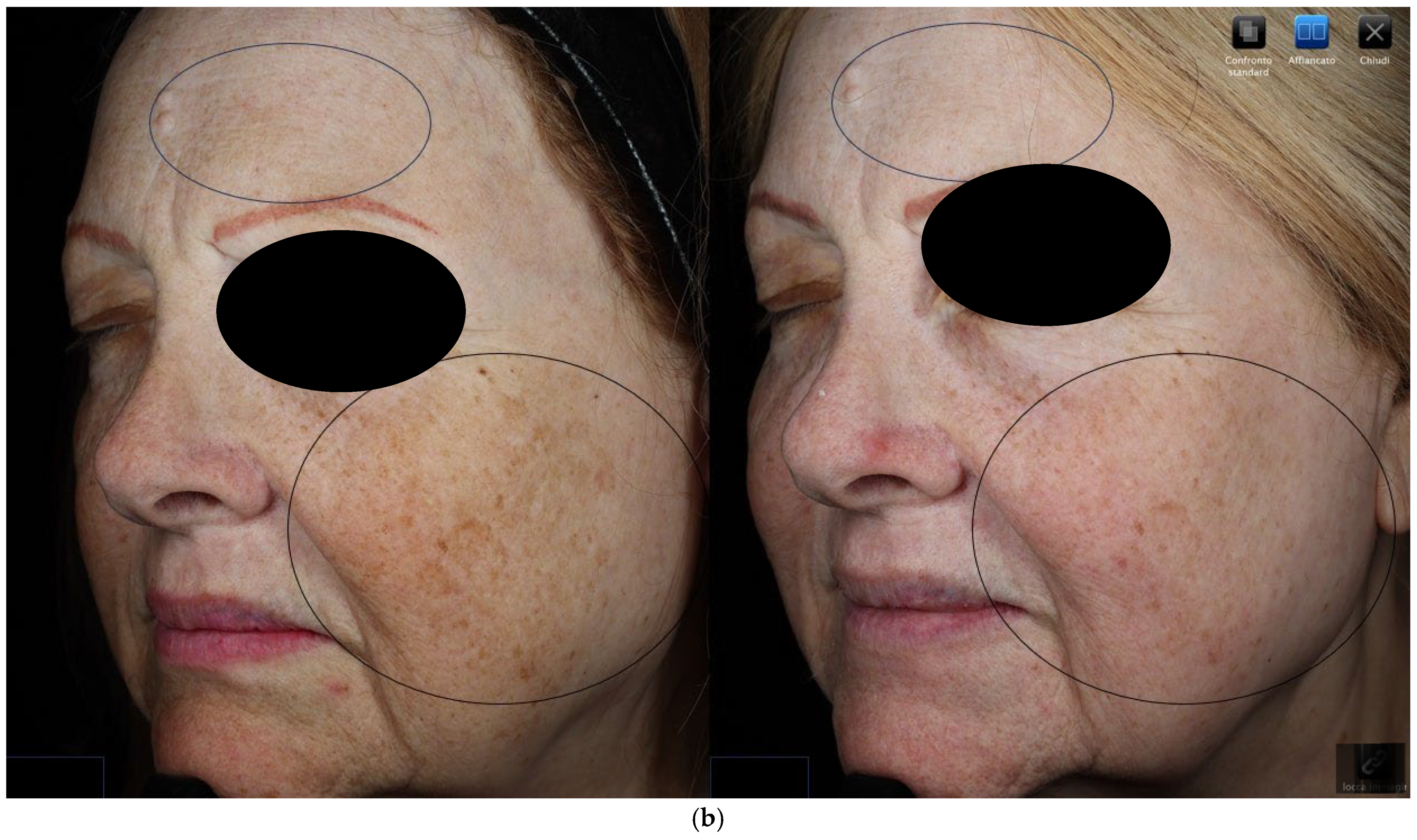
Participants were allocated into two groups based on the treatment regimen they were already receiving or had independently selected in consultation with their dermatologist, reflecting real-world clinical practice. Laser Group (n = 6): Patients received fractional CO2 laser treatment (SmartXide Punto, DEKA, Florence, Italy) in HighPeak mode to the affected facial areas every four weeks at Tw0 (baseline), Tw4, Tw8, and Tw12. Following each session, all patients applied a hyaluronic acid-based booster gel for three days, then twice per day 2-MNG-serum (1 mL per application). (Figure 1 and Figure 2).
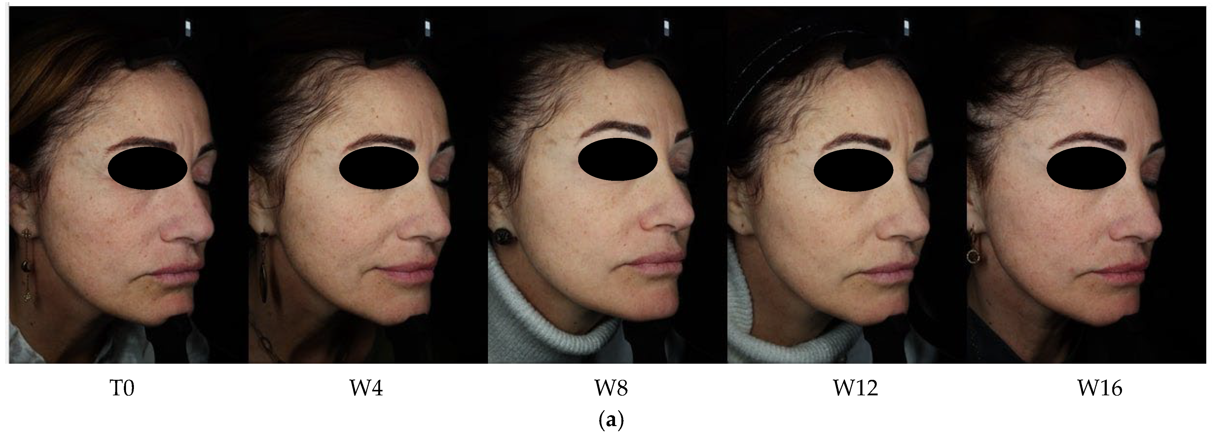
Figure 1. Patient 1 (laser group)—right projection—visible spot (cross polarized light). (a): sequential clinical images at baseline (T0), week 4 (W4), week 8 (W8), week 12 (W12), and week 16 (W16), illustrating progressive improvement over the study period. (b): side-by-side comparison of baseline (T0, left) and week 16 (W16, right) images.
Figure 2. Patient 2 (laser group)–left projection—visible spot (cross polarized light). (a): sequential clinical images at baseline (T0), week 4 (W4), week 8 (W8), week 12 (W12), and week 16 (W16), illustrating progressive improvement over the study period. (b): side-by-side comparison of baseline (T0, left) and week 16 (W16, right) images.
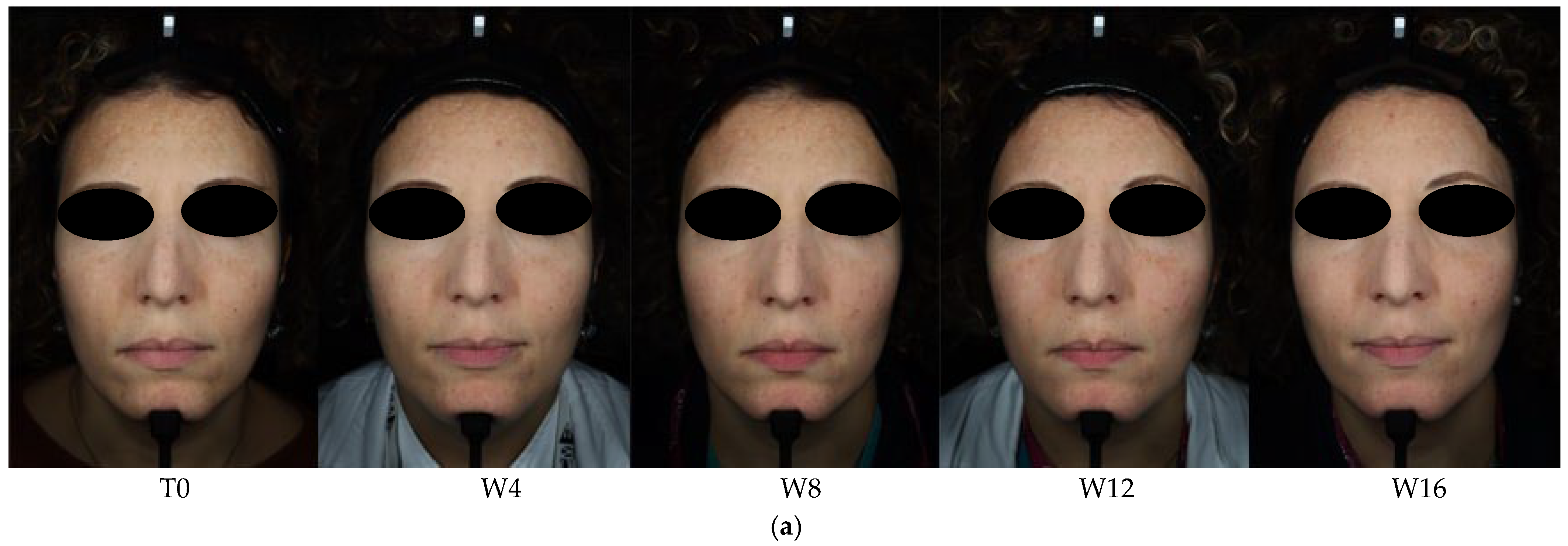
Control Group (n = 5): Patients applied the same twice per day 2-MNG-serum (1 mL per application) throughout the 16-week study. (Figure 3).
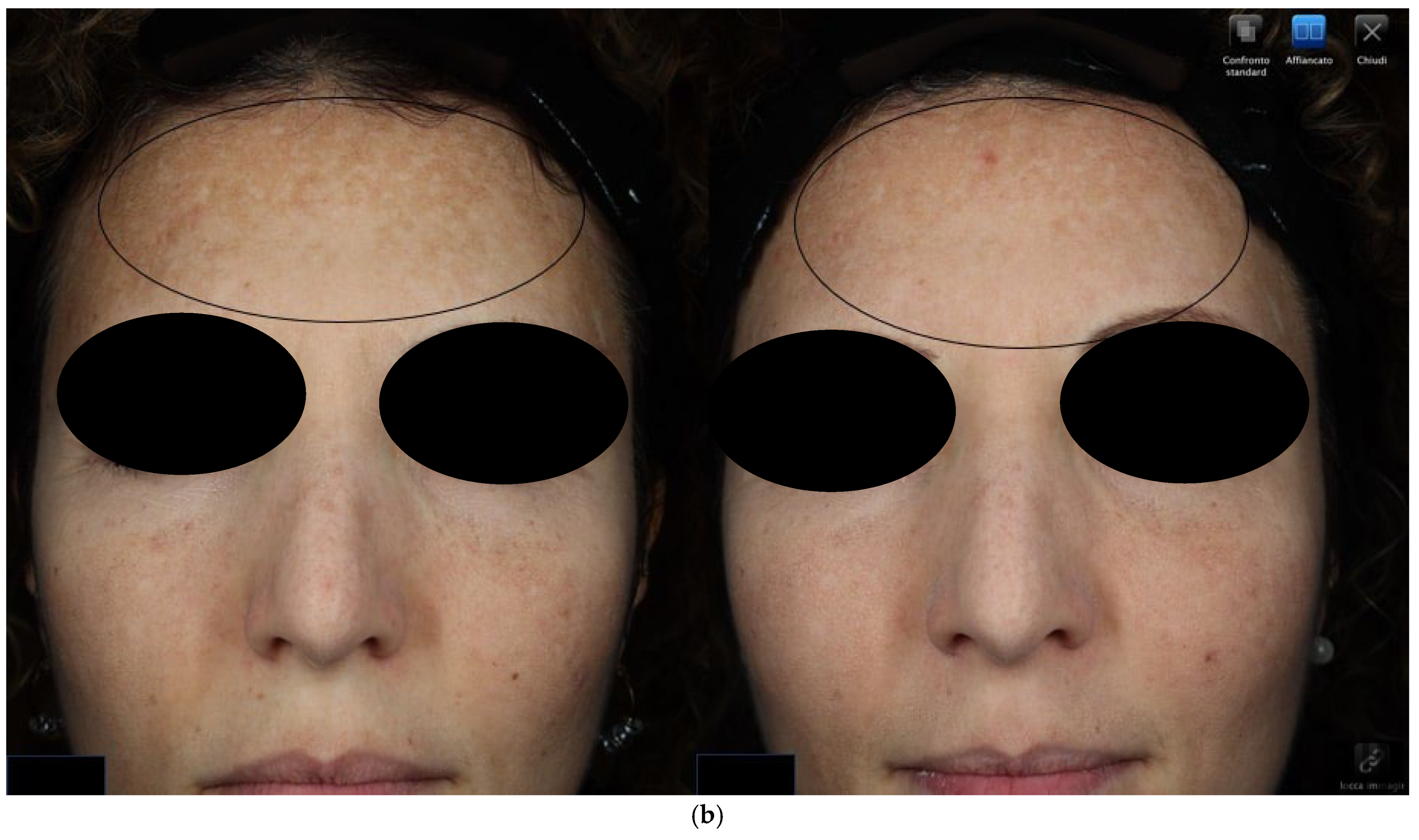
Figure 3. Patient 1 (control group)–frontal projection—visible spot (cross polarized light). (a): sequential clinical images at baseline (T0), week 4 (W4), week 8 (W8), week 12 (W12), and week 16 (W16), illustrating progressive improvement over the study period. (b): side-by-side comparison of baseline (T0, left) and week 16 (W16, right) images.
All participants were instructed to use broad-spectrum SPF 50+/UVA sunscreen daily.

2.4. Outcome Measures

2.4.1. Objective Hyperpigmentation Assessment

Objective assessment was conducted using the VISIA-CR® imaging system (Canfield Scientific Inc., Parsippany, NJ, USA), capturing standardized photographs of front, right, and left facial views under standard, polarized, and UV lighting. Scores (0–1) were calculated based on visible, UV, and brown spot detection using size, area, and contrast algorithms.

2.4.2. Modified Melasma Area and Severity Index (mMASI)

mMASI scores (Table 1) were calculated for four facial regions using the formula:
mMASI = 0.3 × (A_forehead × (D + H)) + 0.3 × (A_right malar × (D + H)) + 0.3 × (A_left malar × (D + H)) + 0.1 × (A_chin × (D + H))
where A = Area, D = Darkness, H = Homogeneity.
Table 1. Modified Melasma Area and Severity Index (mMASI).

2.4.3. Dermatology Life Quality Index (DLQI)

The DLQI (Table 2) is a 10-item questionnaire evaluating the impact of skin disease on daily life. Scores range from 0 (no impact) to 30 (maximum impact).
Table 2. Dermatology Life Quality Index (DLQI).

2.5. Statistical Analysis

Data were analyzed using SPSS v28.0. Intra-group comparisons were performed using the Friedman and Wilcoxon signed-rank tests, while between-group comparisons were conducted using the Mann–Whitney U test. This non-parametric test was selected due to the small sample size and the lack of assumption of normality, which makes it a suitable and robust choice for comparing two independent groups in such contexts. Significance was set at p  <  0.05.

3. Results

Fourteen female patients were enrolled in the study; eleven completed the 16-week follow-up period: six in the Laser group (Fractional CO2 Laser + Topical 2-MNG-serum) and five in the Control group (Topical 2-MNG-serum only). Three patients discontinued the study: one in the Laser group due to noncompliance, one in the Control group due to noncompliance, and one in the Control group due to an unrelated medical condition. At baseline (Tw0), the two treatment groups were comparable across all outcome measures (p > 0.05 for all baseline comparisons). Representative clinical images illustrating individual patient improvement are shown in Figure 1 (Laser group, right projection), Figure 2 (Laser group, left projection), and Figure 3 (Control group, frontal projection).
In each figure, panel A displays a side-by-side comparison between baseline (T0) and week 16 (W16), while panel B shows the progression over time (T0, W4, W8, W12, W16), visually documenting the treatment response trajectory.
Both treatment arms demonstrated efficacy over the 16-week period, though with different timelines and magnitudes of improvement. A summary of all objective and clinical outcome measures is presented below.

3.1. Objective Hyperpigmentation Assessment (VISIA-CR® Analysis)

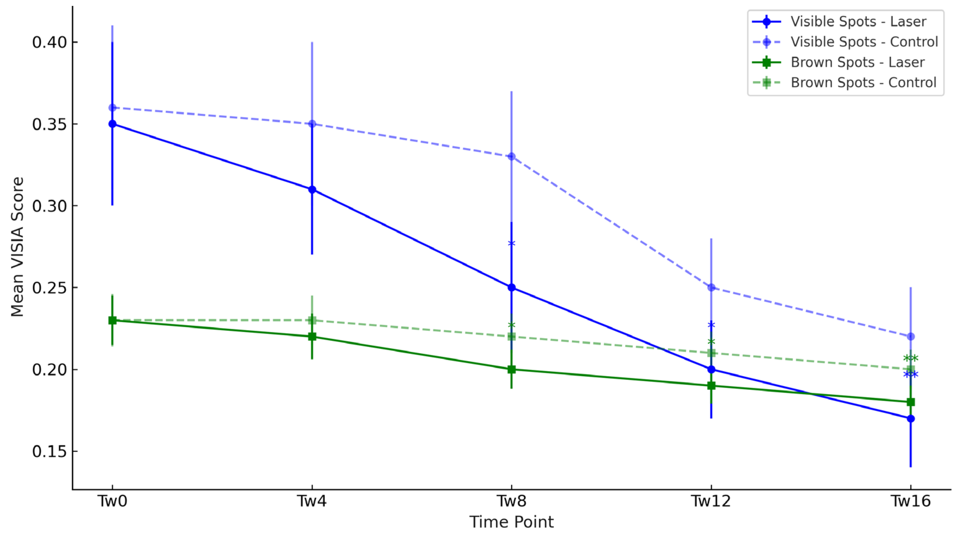
The mean scores for all three assessed VISIA CR® parameters (Visible Spots, UV Spots, and Brown Spots) are detailed in Table 3 and graphically illustrated in Figure 4, which depicts the temporal evolution of pigmentation parameters over the 16-week period.
Table 3. Mean (± Standard Deviation) VISIA CR® Scores (Visible, UV, and Brown Spots) by treatment group at each study timepoint. # Indicates significant difference compared to baseline (Tw0) within the same group (intra-group comparison, Wilcoxon signed-rank test). * p < 0.05, ** p < 0.01 vs. the control group (inter-group comparison, Mann–Whitney U test).
Figure 4. Mean Visible, UV, and Brown spot scores over the 16-week study period in both treatment groups (Laser vs. Control). Asterisks indicate statistically significant differences between groups at the same timepoint, placed above the laser group curve. * Between-group comparisons were performed using the Mann–Whitney U test. * p < 0.05, ** p < 0.01, *** p < 0.001.
The mean scores for all three assessed VISIA CR® parameters are detailed in Table 3.
A statistically significant reduction in Visible Spot and Brown Spot scores was observed in both groups over the 16-week period. In the Laser group, this improvement from baseline became significant starting at week 8 (Tw8) (p < 0.05), while in the Control group, significance was reached at week 12 (Tw12) (p < 0.05).
In contrast, for the UV Spots parameter, which represents deeper, subclinical photodamage, no statistically significant improvement from baseline was observed in either the Laser group or the Control group at any timepoint during the study (p > 0.05).
Inter-group analysis confirmed the superior efficacy of the Laser group for Visible Spots and Brown Spots, with significantly lower (better) scores compared to the Control group from week 8 onwards (p < 0.05). No significant difference between the groups was found for UV Spot scores.

3.2. Clinical Severity Assessment (mMASI Scores) and Patient-Reported Outcomes (DLQI Scores)

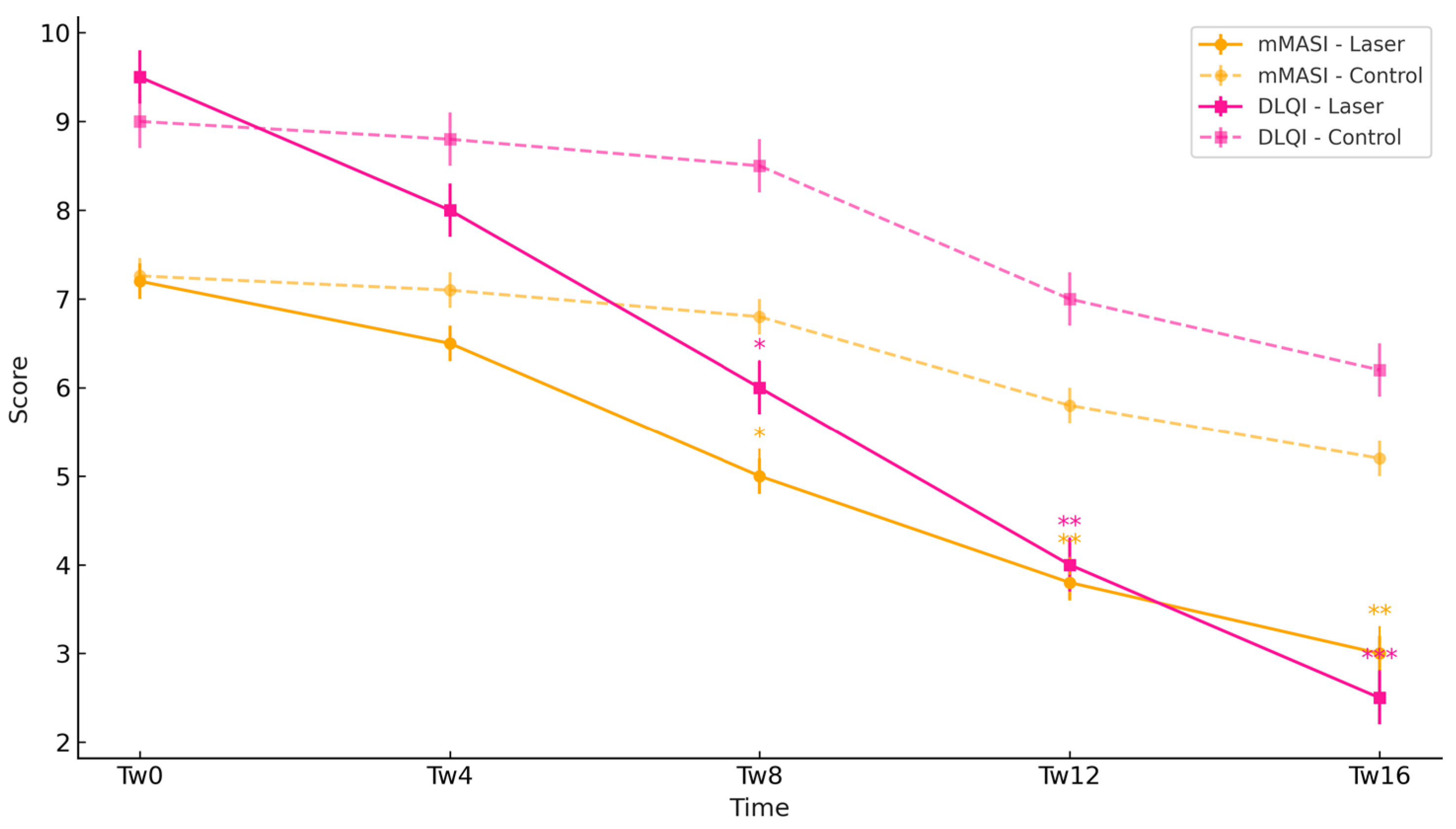
A similar pattern of early efficacy for the Laser group and later efficacy for the Control group was observed for clinical and patient-reported outcomes, as detailed in Table 4. And illustrated in Figure 5. These data are summarized in Table 4 and graphically depicted in Figure 5, illustrating the trajectory of mMASI and DLQI scores over time in both treatment groups.
Table 4. Mean (± Standard Deviation) mMASI and DLQI scores by treatment group at each study timepoint. # Indicates significant difference compared to baseline (Tw0) within the same group (intra-group comparison, Wilcoxon signed-rank test). * p < 0.05, ** p < 0.01, *** p < 0.001 vs. the control group (inter-group comparison, Mann–Whitney U test).
Figure 5. Mean mMASI and DLQI scores over the 16-week study period in both treatment groups (Laser vs. Control). Asterisks indicate statistically significant differences between groups at the same timepoint, placed above the laser group curve. * Between-group comparisons were performed using the Mann–Whitney U test. * p < 0.05, ** p < 0.01, *** p < 0.001.
The Laser group demonstrated a statistically significant reduction in mMASI scores from week 8 (p < 0.05), while the Control group showed significant improvement starting from week 12 (p < 0.05). For DLQI, a similar pattern was observed, with the Laser group showing significant improvement from week 8 and the Control group from week 12. Between-group comparisons assessed using the Mann–Whitney U test, confirmed that the Laser group had significantly lower (better) mMASI and DLQI scores than the Control group from week 8 onwards, with increasing significance at week 12 (p < 0.01) and week 16 (p < 0.001).
These differences are visually indicated in the figures by asterisks placed above the Laser group curves at each timepoint where statistical significance was observed.

4. Discussion

Hyperpigmentation disorders such as melasma, brown spots, and post-inflammatory hyperpigmentation (PIH) are among the most challenging dermatologic conditions to treat due to their multifactorial etiology, chronicity, and high tendency for recurrence (Table 5). Conventional therapies often fall short, particularly when pigmentation extends into the dermis. In this context, laser-assisted drug delivery (LADD) has emerged as an advanced strategy to enhance the penetration and efficacy of topical agents [].
Table 5. Summary of key characteristics and comparative profile of topical 2-MNG-serum, fractional CO2 laser-assisted 2-MNG-serum delivery, and Q-switched laser alone for hyperpigmentation treatment. The table includes mechanisms of action, target depth, efficacy, effect on deep pigmentation, risk of post-inflammatory hyperpigmentation (PIH), healing time, patient comfort, cost considerations, optimal indications, and overall safety.
Our study demonstrated that while 2-MNG-containing serum monotherapy is effective, its combination with fractional CO2 laser yields a significantly faster onset of efficacy. The mean reduction in the mMASI score from 7.20 to 3.00 in the laser group over 16 weeks provides a strong benchmark for comparison with other established treatments. The fractional CO2 laser offers a dual benefit: it targets and vaporizes pigment-laden melanocytes while simultaneously stimulating new, evenly pigmented skin growth, making it effective for solar lentigo, melasma, and PIH [].
These results are particularly relevant when contextualized with real-world and clinical studies of other depigmenting agents. For instance, Thiamidol, a powerful inhibitor of human tyrosinase [], demonstrated significant improvements in Melasma Area and Severity Index (MASI) scores versus vehicle over 24 weeks in a randomized controlled trial, with excellent tolerability [].
Topical Arbutin has also emerged as an effective and safer alternative to hydroquinone []. Studies have shown that creams containing alpha-arbutin, often in combination with agents like kojic acid, can achieve efficacy comparable to traditional therapies but with fewer adverse events and lower recurrence rates [].
Regarding device-based therapies, our protocol is competitive. While the Q-switched Nd:YAG laser is widely studied for melasma, it carries a non-negligible risk of rebound pigmentation, especially in patients with darker skin phototypes []. In contrast, although concerns about PIH have historically limited its use in melasma, recent improvements in HP-mode protocols and cooling technologies have rendered fractional CO2 a viable and well-tolerated option, even in patients with darker skin tones [,,].
In summary, while Thiamidol and arbutin formulations offer efficacious and generally well-tolerated alternatives to traditional depigmenting regimens, our study suggests that the combination of fractional CO2 laser and 2-MNG-serum may accelerate clinical improvement. This is particularly relevant for recalcitrant pigmentation, as it leverages both enhanced transdermal delivery and a novel mechanistic pathway, recently shown to be effective against hydroquinone 4% []. Table 4 provides a comparative overview of these different treatment modalities.
Several limitations of this study warrant consideration. The relatively small sample size, the 16-week follow-up period, the lack of a ‘laser only’ arm, and the focus on female participants with Fitzpatrick skin types I-III may limit the broader generalizability of our findings.

5. Conclusions

In conclusion, this study investigated the effectiveness of fractional CO2 laser combined with 2-MNG-containing serum in patients with mild-to-moderate melasma and/or facial hyperpigmentation. The group treated with laser and 2-MNG-serum showed a significantly greater reduction in visible and brown spots analyzed by VISIA-CR parameters. This outcome can be attributed to the laser’s role in facilitating transdermal delivery of the topical product, enhancing its efficacy. The group receiving only 2-MNG-serum topical treatment also demonstrated statistically significant improvement, though less pronounced. These findings suggest that combining CO2 laser with topical therapy offers a more effective strategy for managing facial hyperpigmentation and melasma, while monotherapy with 2-MNG remains a valid option for patients with milder cases.

Author Contributions

Conceptualization, A.D., E.A. and S.A.; methodology, A.D., E.A. and S.A.; software, A.D., E.A. and S.A.; validation, S.P.N. and G.P.; formal analysis, A.D., E.A. and S.A.; investigation, A.D., E.A. and S.A.; resources, A.D., E.A. and S.A.; data curation, A.D., E.A. and S.A.; writing—original draft preparation, A.D., E.A. and S.A.; writing—review and editing, A.D., E.A. and S.A.; visualization, A.D., E.A. and S.A.; supervision, A.D., E.A. and S.A.; project administration, G.P.; funding acquisition, S.P.N. All authors have read and agreed to the published version of the manuscript.

Funding

This research received no external funding.

Institutional Review Board Statement

The study was conducted in accordance with the Declaration of Helsinki. Ethical review and approval were waived for this study as it was an observational, real-life experience conducted in routine clinical practice, without any intervention beyond standard care.

Data Availability Statement

The data presented in this study are available upon request from the corresponding author. The data are not publicly available due to privacy and ethical restrictions.

Conflicts of Interest

Giovanni Pellacani (GP) and Annunziata Dattola (AD) have served as speakers or members of advisory boards for the following companies: AbbVie, Almirall, Leo Pharma, UCB Pharma, Celgene, Eli Lilly, Novartis, Janssen, Boehringer Ingelheim, Incyte, and L’Oréal. Stefano Nisticò (SM) has served as a speaker or consultant for: L’Oréal, Chiesi Farmaceutici, Difa Cooper, and DEKA. The other authors declare no conflicts of interest.

References

  1. Syder, N.C.; Quarshie, C.; Elbuluk, N. Disorders of Facial Hyperpigmentation. Dermatol. Clin. 2023, 41, 393–405. [Google Scholar] [CrossRef] [PubMed]
  2. Wang, R.F.; Ko, D.; Friedman, B.J.; Lim, H.W.; Mohammad, T.F. Disorders of hyperpigmentation. Part I. Pathogenesis and clinical features of common pigmentary disorders. J. Am. Acad. Dermatol. 2023, 88, 271–288. [Google Scholar] [CrossRef] [PubMed]
  3. Elbuluk, N.; Grimes, P.; Chien, A.; Hamzavi, I.; Alexis, A.; Taylor, S.; Gonzalez, N.; Weiss, J.; Desai, S.R.; Kang, S. The Pathogenesis and Management of Acne-Induced Post-inflammatory Hyperpigmentation. Am. J. Clin. Dermatol. 2021, 22, 829–836. [Google Scholar] [CrossRef] [PubMed]
  4. Espósito, A.C.C.; Cassiano, D.P.; da Silva, C.N.; Lima, P.B.; Dias, J.A.F.; Hassun, K.; Bagatin, E.; Miot, L.D.B.; Miot, H.A. Update on Melasma—Part I: Pathogenesis. Dermatol. Ther. 2022, 12, 1967–1988. [Google Scholar] [CrossRef]
  5. Zhao, L.; Fu, X.; Cheng, H. Prevention of Melasma During Pregnancy: Risk Factors and Photoprotection-Focused Strategies. Clin. Cosmet. Investig. Dermatol. 2024, 17, 2301–2310. [Google Scholar] [CrossRef]
  6. Bostan, E.; Cakir, A. The dermoscopic characteristics of melasma in relation to different skin phototypes, distribution patterns and wood lamp findings: A cross-sectional study of 236 melasma lesions. Arch. Dermatol. Res. 2023, 315, 1927–1938. [Google Scholar] [CrossRef]
  7. Ozbagcivan, O.; Akarsu, S.; Ikiz, N.; Semiz, F.; Fetil, E. Dermoscopic Differentiation of Facial Lentigo Maligna from Pigmented Actinic Keratosis and Solar Lentigines. Acta Dermatovenerol. Croat. 2019, 27, 146–152. [Google Scholar]
  8. Woolery-Lloyd, H.; Kammer, J.N. Treatment of hyperpigmentation. Semin. Cutan. Med. Surg. 2011, 30, 171–175. [Google Scholar] [CrossRef]
  9. Charoo, N.A. Hyperpigmentation: Looking beyond hydroquinone. J. Cosmet. Dermatol. 2022, 21, 4133–4145. [Google Scholar] [CrossRef]
  10. Nautiyal, A.; Wairkar, S. Management of hyperpigmentation: Current treatments and emerging therapies. Pigment Cell Melanoma Res. 2021, 34, 1000–1014. [Google Scholar] [CrossRef]
  11. Borelli, C.; Fischer, S. Chemical Peelings zur Behandlung von Melasma, Pigmentstörungen und Hyperpigmentierungen: Indikationen, Effektivität und Risiken. Hautarzt 2020, 71, 950–959. [Google Scholar] [CrossRef] [PubMed]
  12. Lai, D.; Zhou, S.; Cheng, S.; Liu, H.; Cui, Y. Laser therapy in the treatment of melasma: A systematic review and meta-analysis. Lasers Med. Sci. 2022, 37, 2099–2110. [Google Scholar] [CrossRef] [PubMed]
  13. Ortiz, A.E.; Goldman, M.P.; Fitzpatrick, R.E. Ablative CO2 lasers for skin tightening: Traditional versus fractional. Dermatol. Surg. 2014, 40 (Suppl. S12), S147–S151. [Google Scholar] [CrossRef] [PubMed]
  14. Zhao, S.; Wang, M.; Lai, X.; Yan, Y. Efficacy and Safety of Ablative Fractional Laser in Melasma: A Meta-analysis and Systematic Review. Lasers Med. Sci. 2024, 39, 71. [Google Scholar] [CrossRef]
  15. Sextius, P.; Warrick, E.; Prévot-Guéguiniat, A.; Lereaux, G.; Boirre, F.; Baux, L.; Hassine, S.B.; Qiu, J.; Huang, X.; Xu, J.; et al. 2-Mercaptonicotinoyl glycine, a new potent melanogenesis inhibitor, exhibits a unique mode of action while preserving melanocyte integrity. Pigment Cell Melanoma Res. 2024, 37, 462–479. [Google Scholar] [CrossRef]
  16. de Dormael, R.; Sextius, P.; Bourokba, N.; Mainguene, E.; Tachon, R.; Gaurav, K.; Jouni, H.; Bastien, P.; Diridollou, S. 2-Mercaptonicotinoyl glycine prevents UV-induced skin darkening and delayed tanning in healthy subjects: A randomized controlled clinical study. J. Cosmet. Dermatol. 2024, 23, 1745–1752. [Google Scholar] [CrossRef]
  17. Muller, B.; Flament, F.; Jouni, H.; Sextius, P.; Tachon, R.; Wang, Y.; Wang, H.; Qiu, H.; Qiu, J.; Amar, D.; et al. A Bayesian network meta-analysis of 14 molecules inhibiting UV daylight-induced pigmentation. J. Eur. Acad. Dermatol. Venereol. 2024, 38, 1566–1574. [Google Scholar] [CrossRef]
  18. Demessant-Flavigny, A.L.; Petkar, G.; Jodun, D.; Le Dantec, G.; Le Floc’h, C.; Kerob, D. Efficacy of a 2-MNG-Containing Depigmenting Serum in the Treatment of Post-Inflammatory Hyperpigmentation. J. Cosmet. Dermatol. 2025, 24, e16735. [Google Scholar] [CrossRef]
  19. Passeron, T.; Kerob, D.; Le Dantec, G.; Demessant-Flavigny, A.L.; do Nascimer, A.R.; Moura, R.; Salah, S.; Feiges, M.; Fernandez, E.; Alexis, A. Efficacy and Tolerability of a New Facial 2-Mercaptonicotinoyl Glycine-Containing Depigmenting Serum versus Hydroquinone 4% over 3-Month Treatment of Facial Melasma. Dermatol. Ther. 2025, in press. [Google Scholar] [CrossRef]
  20. Wu, W.; Su, Q.; Zhang, Y.; Du, Y.; Hu, Y.; Wang, F. Novel 532-nm Q-switched Nd:YAG laser for the treatment of melasma and rejuvenation: A prospective, randomized controlled comparison with 1064-nm Q-switched Nd:YAG laser. Int. J. Dermatol. 2024, 63, 1242–1251. [Google Scholar] [CrossRef]
  21. Duteil, L.; Queille-Roussel, C.; Lacour, M.; Zastrow, L. Clinical proof of concept for Thiamidol, a new powerful inhibitor of human tyrosinase. J. Investig. Dermatol. 2014, 134, S62. [Google Scholar]
  22. Khemis, A.; Kaiafa, A.; Queille-Roussel, C.; Duteil, L. A randomized, double-blind, vehicle-controlled study to evaluate the efficacy of a topical formulation containing Thiamidol in the treatment of melasma. J. Cosmet. Dermatol. 2020, 19, 1106–1112. [Google Scholar]
  23. Draelos, Z.D. Skin lightening preparations and the hydroquinone controversy. Dermatol. Ther. 2010, 20, 308–313. [Google Scholar] [CrossRef] [PubMed]
  24. El-Sinbawy, Z.G.; Abdelnabi, N.M.; Sarhan, N.E.; Elgarhy, L.H. Clinical & ultrastructural evaluation of the effect of fractional CO2 laser on facial melasma. Ultrastruct. Pathol. 2019, 43, 135–144. [Google Scholar] [CrossRef] [PubMed]
  25. Kim, C.; Gao, J.C.; Moy, J.; Lee, H.S. Fractional CO2 laser and adjunctive therapies in skin of color melasma patients. JAAD Int. 2022, 8, 118–123. [Google Scholar] [CrossRef] [PubMed]
  26. Hermanns, J.F.; Piérard, G.E. Skin Colour and Ethnic Variations. In Textbook of Aging Skin; Springer: Berlin/Heidelberg, Germany, 2006; pp. 303–314. [Google Scholar]
Disclaimer/Publisher’s Note: The statements, opinions and data contained in all publications are solely those of the individual author(s) and contributor(s) and not of MDPI and/or the editor(s). MDPI and/or the editor(s) disclaim responsibility for any injury to people or property resulting from any ideas, methods, instructions or products referred to in the content.

Article Metrics

Citations

Article Access Statistics

Multiple requests from the same IP address are counted as one view.