Article

Multifunctional Effects of N-Carbamylglutamate on Skin-Related Cells: Antioxidant, Anti-Aging, Anti-Melanogenic and Anti-Inflammatory Activities

1
Department of Otolaryngology—Head and Neck Surgery, Chonnam National University Medical School and Chonnam National University Hospital, 42 Jaebong-ro, Dong-gu, Gwangju 61469, Republic of Korea
2
Department of Biocosmetics, Dongshin University, 185, Gunjae-ro, Naju 58245, Jeonnam, Republic of Korea
3
BIO-FD&C Co., Ltd., 106, Sandan-gil, Hwasun 58141, Jeonnam, Republic of Korea
*
Author to whom correspondence should be addressed.
These authors contributed equally to this work.
Cosmetics 2025, 12(6), 250; https://doi.org/10.3390/cosmetics12060250
Submission received: 25 September 2025 / Revised: 16 October 2025 / Accepted: 1 November 2025 / Published: 7 November 2025
(This article belongs to the Special Issue Skin Anti-Aging Strategies)

Abstract

Skin aging is accelerated by both environmental factors—including ultraviolet (UV) radiation and pollution—and intrinsic processes such as chronic inflammaging. N-carbamylglutamate (NCG), an arginine precursor known for its benefits for gut and reproductive health, has not been extensively studied in dermatological applications. To explore its suitability as a multifunctional cosmetic ingredient, this study examines the protective role of NCG in counteracting UV-stimulated oxidative and inflammatory responses in skin cells. NCG significantly reduced UV-induced reactive oxygen species (ROS), indicating strong antioxidant properties. It also inhibited matrix metalloproteinase (MMP) activity, preserving collagen integrity and reducing wrinkle formation. In addition, NCG suppressed nitric oxide (NO) production and downregulated key inflammatory mediators—including cyclooxygenase-2 (COX-2), inducible nitric oxide synthase (iNOS), tumor necrosis factor-alpha (TNF-α), and interleukin-6 (IL-6)—highlighting its anti-inflammatory potential. Furthermore, NCG reduced melanin production and the expression of melanogenesis-related factors such as the microphthalmia-associated transcription factor (MITF), tyrosinase-related protein 1 (TRP-1), and TRP-2. These findings support the role of NCG as a promising multifunctional cosmetic ingredient with antioxidant, anti-inflammatory, anti-wrinkle, and skin-brightening properties.

Graphical Abstract

1. Introduction

Skin aging is a multifactorial biological process influenced by both intrinsic physiological factors and extrinsic environmental stressors, ultimately leading to structural and functional deterioration of the skin [1,2]. Clinically, skin aging presents as wrinkle formation, reduced elasticity, and hyperpigmentation—conditions that compromise both skin health and appearance, negatively impacting quality of life. Emerging evidence has identified inflammaging, a state of chronic, low-grade inflammation, as a pivotal contributor to skin aging. This chronic inflammatory state is exacerbated by environmental insults such as ultraviolet (UV) radiation, pollution, and lipopolysaccharide (LPS), a bacterial endotoxin [3,4,5]. Among these, UV radiation is considered the most potent contributor, inducing DNA damage, oxidative stress, and inflammatory cytokine production, which degrade extracellular matrix (ECM) components like collagen and elastin [6,7,8,9,10,11]. Although UVA penetrates more deeply into the dermis, UVB exerts stronger biological effects by directly inducing oxidative stress, inflammation, and photoaging-related responses such as MMP activation and collagen degradation. Accordingly, UVB irradiation has been widely employed as a representative in vitro model for studying photoaging in human dermal fibroblasts (HDFs) [12,13]. Similarly, LPS exposure activates innate immunity, stimulating the release of inflammatory mediators that not only accelerate skin aging but also contribute to hyperpigmentation [14,15,16]. Additionally, α-melanocyte-stimulating hormone (α-MSH) upregulation promotes melanogenesis, thereby exacerbating pigmentary disorders like melasma and lentigines [17,18].
Given this mechanistic understanding, there is a growing demand for functional cosmetic ingredients capable of preventing or ameliorating inflammaging-related skin aging. Although several anti-aging agents such as retinoids, peptides, and hydroxy acids (AHA, BHA) are currently in use, these compounds often present limitations including skin irritation, suboptimal safety profiles, and diminished long-term efficacy [19,20,21]. Therefore, the development of novel functional materials with enhanced safety and efficacy remains an urgent priority.
N-carbamylglutamate (NCG), an analog of N-acetylglutamate, serves as an effective precursor for endogenous arginine synthesis and has demonstrated anti-inflammatory and antioxidant effects in systemic studies. Mechanistically, NCG is known to suppress the ERK1/2–mTOR–S6K1 signaling pathway, downregulating pro-inflammatory cytokines such as interleukin-1β (IL-1β), interleukin-6 (IL-6), and interleukin-8 (IL-8) [22,23,24]. Despite these benefits, NCG’s potential in skin health has not been systematically evaluated.
The present study investigates the biological effects and underlying mechanisms of NCG in skin cells. We focused on its antioxidant, anti-inflammatory, anti-wrinkle, and anti-melanogenic activities using in vitro models exposed to UVB and LPS. Our aim was to validate NCG as a multifunctional cosmetic agent capable of alleviating inflammaging and enhancing skin health.

2. Materials and Methods

2.1. Chemicals and Reagents

The following reagents were used in this study, sourced from the respective manufacturers: NCG (C4375) from Sigma-Aldrich Corporation (St. Louis, MO, USA); Dulbecco’s Modified Eagle Medium (DMEM) and phosphate-buffered saline (PBS) from Lonza Group Ltd. (Walkersville, MD, USA). For primary fibroblast culture, fibroblast medium (FM), fibroblast growth supplement (FGS), and penicillin-streptomycin (P/S) were purchased from ScienCell (Carlsbad, CA, USA). 3-(4,5-dimethylthiazol-2-yl)-2,5-diphenyltetrazolium bromide (MTT), dimethyl sulfoxide (DMSO), α-MSH, arbutin, and lipopolysaccharide (LPS) were also obtained from Sigma-Aldrich. ELISA kits for human Pro-MMP-1, Pro-MMP-3, and Pro-Collagen I alpha 1 were purchased from R&D Systems (Minneapolis, MN, USA). Antibodies used in Western blotting were sourced as follows: GAPDH from Enogen Biotechnology (New York, NY, USA); TRP-1, TRP-2, MMP-1, JNK, p-JNK, TNF-α, NF-κB, and p38 from Santa Cruz Biotechnology (Dallas, TX, USA); MITF, p-p38 MAPK, and ERK from Cell Signaling Technology (Beverly, MA, USA); COX-2, IL-6, and iNOS from Invitrogen (Carlsbad, CA, USA).

2.2. Cell Culture

B16F10 melanoma cells, human epidermal keratinocytes (HaCaTs), and RAW 264.7 macrophages were obtained from the American Type Culture Collection (ATCC, Manassas, VA, USA). Cells were maintained in DMEM supplemented with 10% fetal bovine serum (FBS) and 1% penicillin-streptomycin in a humidified incubator at 37 °C with 5% CO2. HDFs were obtained from ScienCell (Carlsbad, CA, USA).

2.3. Cell Viability Assay

The MTT assay was conducted to determine cell viability. Following seeding into 96-well plates, cells were treated with varying doses of NCG. After incubation (24 h for HaCaT and RAW 264.7; 72 h for B16F10 and HDF), MTT solution (0.5 mg/mL) was added and incubated for 3 h. DMSO was then added to dissolve the resulting formazan crystals, and absorbance was measured at 570 nm using a Multiskan Sky spectrophotometer (Thermo Fisher Scientific, Waltham, MA, USA).

2.4. Analysis of Intracellular Reactive Oxygen Species (ROS) Reduction Activity

HaCaT cells were cultured for 24 h in 24-well plates prior to further treatment. Based on our preliminary experiments, UVB irradiation at an intensity of 15 mJ/cm2 was selected because this level effectively induced oxidative stress without causing cytotoxicity in HaCaTs, consistent with previous studies employing similar UVB doses in keratinocyte-based oxidative stress models [25,26]. After UVB exposure, cells were treated with various NCG concentrations and incubated for an additional 24 h. ROS levels were assessed using CellROX™ Orange Reagent (Thermo Fisher Scientific). Cells were stained with 5 µM CellROX™ for 30 min in the dark, and fluorescence was visualized using the EVOS™ M5000 imaging system with appropriate filters (excitation/emission: 545/565 nm). Fluorescence intensity was quantified from three randomly selected fields per condition.

2.5. Measurement of Melanin Content

B16F10 melanoma cells were seeded in 6-well plates and incubated overnight. Cells were subsequently exposed to varying concentrations of NCG for 72 h. After treatment, the cells were washed with PBS, harvested by trypsinization, and centrifuged at 13,000 rpm for 5 min. Following centrifugation, the collected cell pellets were treated with 1 N NaOH containing 10% DMSO and maintained at 80 °C for 1 h to ensure complete solubilization. Readings at 475 nm were used to determine the level of absorbance.

2.6. Determination of Cellular Tyrosinase Activity

The tyrosinase activity within B16F10 melanoma cells was assessed according to a previously described method with slight modifications [27]. Cells were plated in 6-well plates and treated with different concentrations of NCG for a duration of 72 h. Supernatants obtained after centrifugation (13,000 rpm, 5 min, 4 °C) were diluted with 0.1 M phosphate buffer (pH 6.8) and aliquoted into 96-well plates. L-DOPA (1 mg/mL) was added, and samples were incubated for 1 h prior to reading absorbance at 475 nm.

2.7. Type 1 Procollagen Synthesis

Type I procollagen was quantified using an enzyme immunoassay specific to the Procollagen Type I C-peptide (PIP), following the manufacturer’s protocol. HDFs were plated and incubated for 24 h, exposed to UVB (10 mJ/cm2), and treated with different NCG concentrations. Collected culture supernatants were centrifuged (12,000 rpm, 10 min, 4 °C). Antibody-POD and supernatant were added, incubated, washed, and reacted with substrate. Reaction was stopped using 1 N H2SO4, and absorbance was read at 450 nm.

2.8. Inhibitory Activity of MMP-1 and MMP-3

The inhibitory effects of NCG on MMP-1 and MMP-3 were evaluated using ELISA kits specific to human pro-MMP-1 and pro-MMP-3, respectively. HDFs were seeded into 6-well plates and cultured. Following UVB irradiation, cells were treated with NCG for 72 h. The culture media were collected and centrifuged to obtain the supernatant. Thereafter, 100 μL of the collected supernatant along with the RD1-52 reagent was dispensed into each well and allowed to react for 2 h. The wells were then emptied, followed by the addition of either anti-human pro-MMP-1 or pro-MMP-3 conjugate antibodies, which were incubated for another 2 h. Following this, the wells were washed, and 100 μL of substrate solution was added and incubated for 20 min. The reaction was terminated using stop solution. Absorbance was measured at 450 nm with a reference at 540 nm, and the final value was calculated as the difference between the two readings.

2.9. Measurement Nitric Oxide (NO) Inhibitory Activity

The inhibitory effect of NCG on NO production was assessed by measuring nitrite accumulation via the Griess reaction. RAW 264.7 macrophage cells were seeded into 96-well plates, stimulated with lipopolysaccharide (LPS, 1 μg/mL), and subsequently treated with various concentrations of NCG. Following incubation, equal volumes of the culture supernatant and Griess reagent were mixed in a new 96-well plate and allowed to react at room temperature for 20 min. The absorbance was measured at 540 nm to quantify nitrite levels, which reflect NO production.

2.10. Western Blot Analysis

Western blotting was conducted with minor modifications to established protocols [28]. Cells were seeded and maintained under standard culture conditions, then treated with NCG. Following incubation, cells were lysed, and the resulting lysates were centrifuged to isolate the supernatant. Following quantification using a protein assay method, quantities of each protein sample were loaded onto SDS–PAGE gels and subsequently transferred to polyvinylidene difluoride (PVDF) membranes for detection. Following 1 h of blocking in 5% skim milk diluted in TBST, the membranes were incubated with specific primary antibodies at 4 °C for approximately 24 h. (as detailed in Table 1). Membranes were washed four times with TBST and subsequently incubated at room temperature for 1 h with secondary antibodies. Protein bands were visualized using an enhanced chemiluminescence (ECL) detection system.

2.11. Statistical Analysis

All experiments were independently repeated three times, and the data are expressed as mean ± standard deviation (SD). To evaluate statistical differences, analyses were conducted in SPSS (v27.0; IBM, Chicago, IL, USA). Group differences were examined using one-way ANOVA or Student’s t-test where appropriate. Statistical significance was set at * p < 0.05, ** p < 0.01, and *** p < 0.001.

3. Results

3.1. Effect of NCG on Cell Viability

The cytotoxicity of NCG was evaluated in HaCaT, HDF, B16F10, and RAW 264.7 cells using the MTT assay. NCG exhibited no cytotoxic effects at concentrations ranging from 100–1000 µM in HaCaT and RAW 264.7 cells, 100–500 µM in B16F10 cells, and 25–200 µM in HDFs. These results confirm that NCG is biocompatible within the tested ranges for each cell type (Figure 1a–d).

3.2. Effects of NCG on Intracellular and Mitochondrial ROS Levels

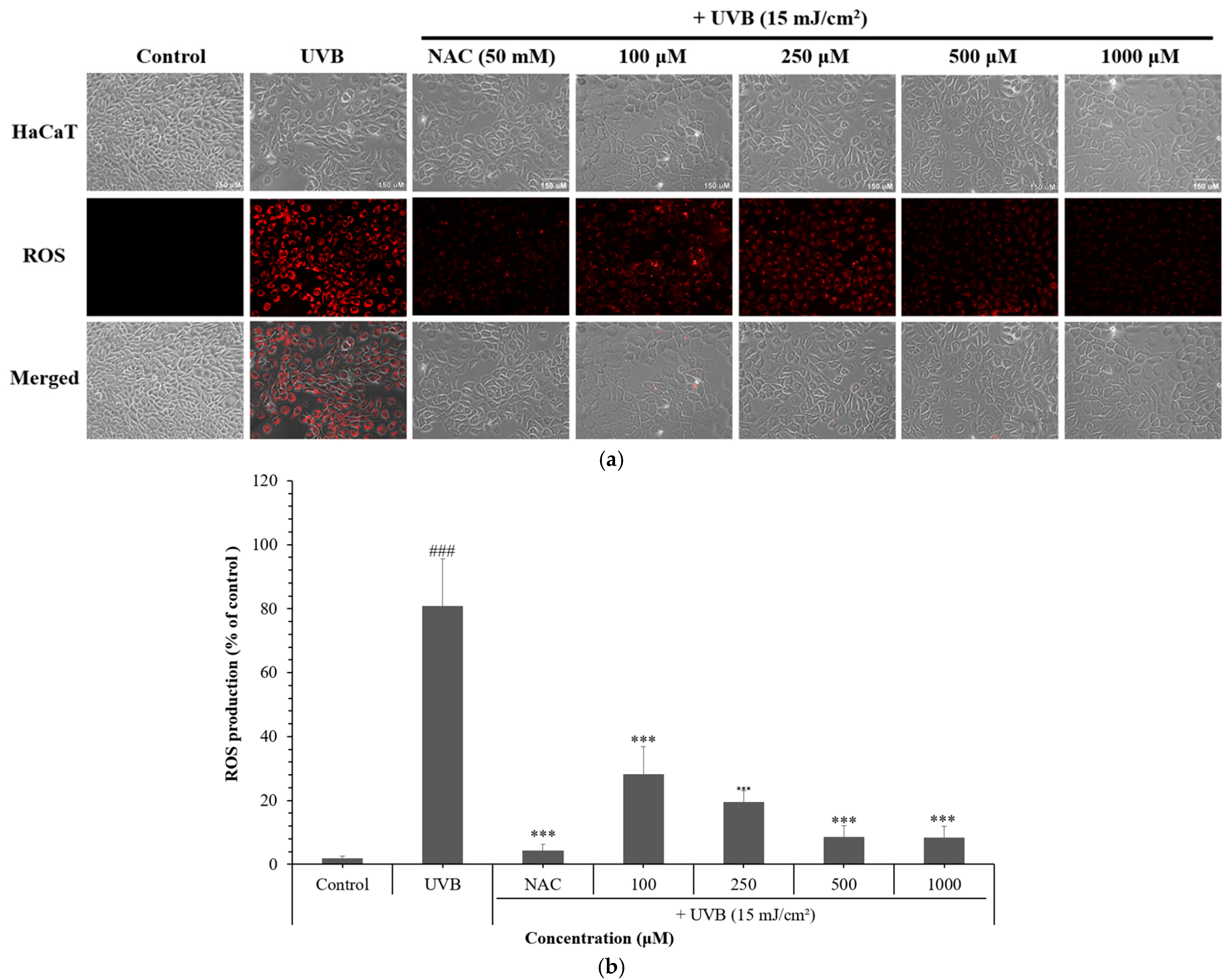
To assess the antioxidant properties of NCG, ROS production was induced in HaCaT cells using UVB irradiation (15 mJ/cm2). Post-irradiation treatment with NCG significantly suppressed intracellular ROS levels in a dose-dependent manner. Notably, concentrations between 500–1000 µM reduced ROS generation by over 90%. These findings demonstrate the potent ROS-scavenging capacity of NCG. Importantly, 500 µM NCG exhibited a ROS scavenging effect comparable to that of 50 mM NAC, a well-established antioxidant used as a positive control (Figure 2). These findings demonstrate the potent ROS-scavenging capacity of NCG, even at much lower concentrations than conventional antioxidants.

3.3. Effects of NCG on UVB-Induced Skin Aging in HDFs

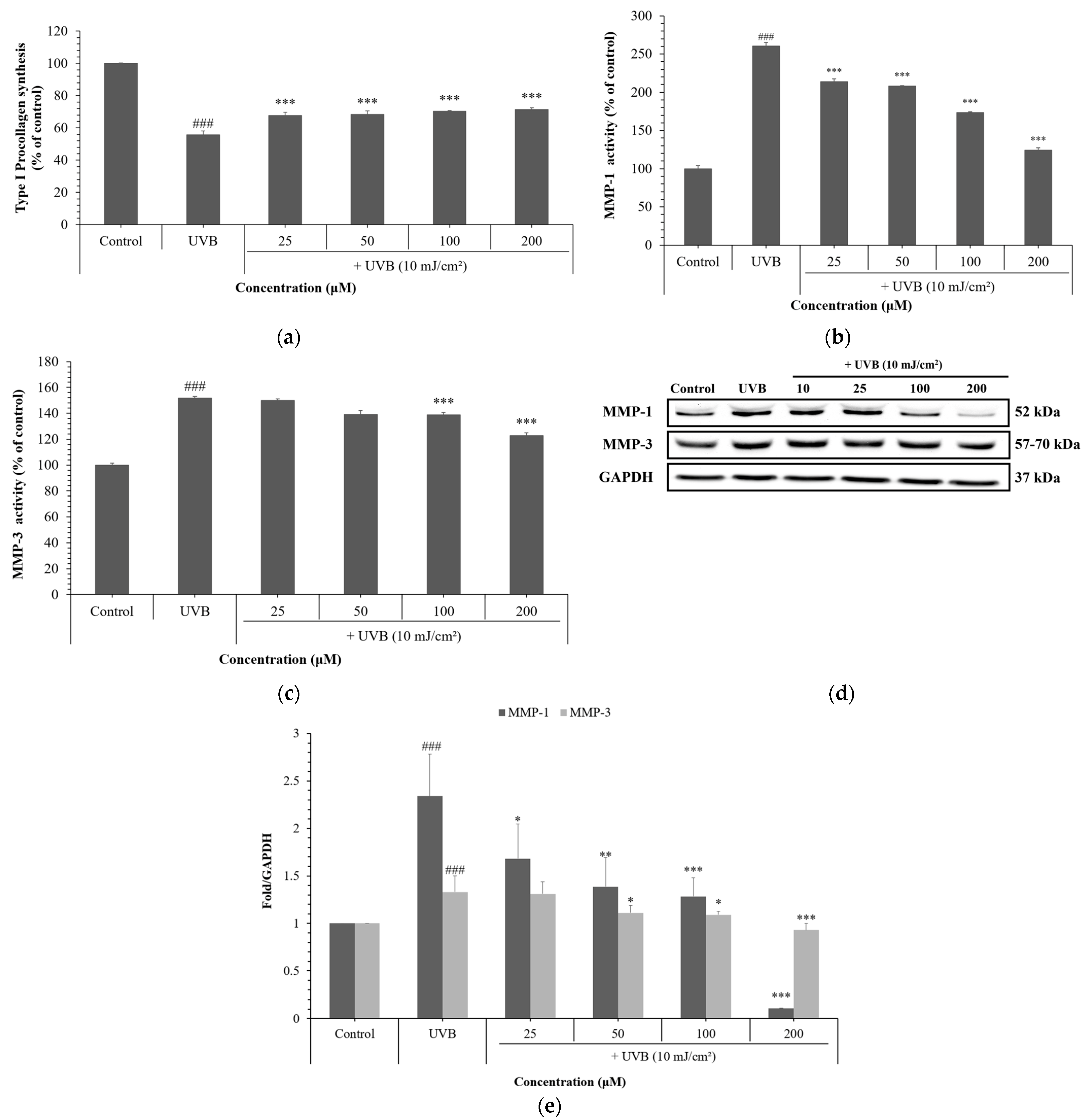
To evaluate NCG’s protective effects against UVB-induced dermal aging, HDFs were exposed to UVB and subsequently treated with NCG. NCG significantly restored type I procollagen synthesis, achieving up to a 30% increase compared to UVB-treated controls (Figure 3a). Additionally, NCG inhibited enzymatic activities of MMP-1 and MMP-3 by more than 53% (Figure 3b,c). Western blot analysis confirmed reduced protein expression of both MMPs following NCG treatment, suggesting a robust protective effect on ECM integrity (Figure 3d,e).

3.4. Effects of NCG on Melanogenesis in B16F10 Cells

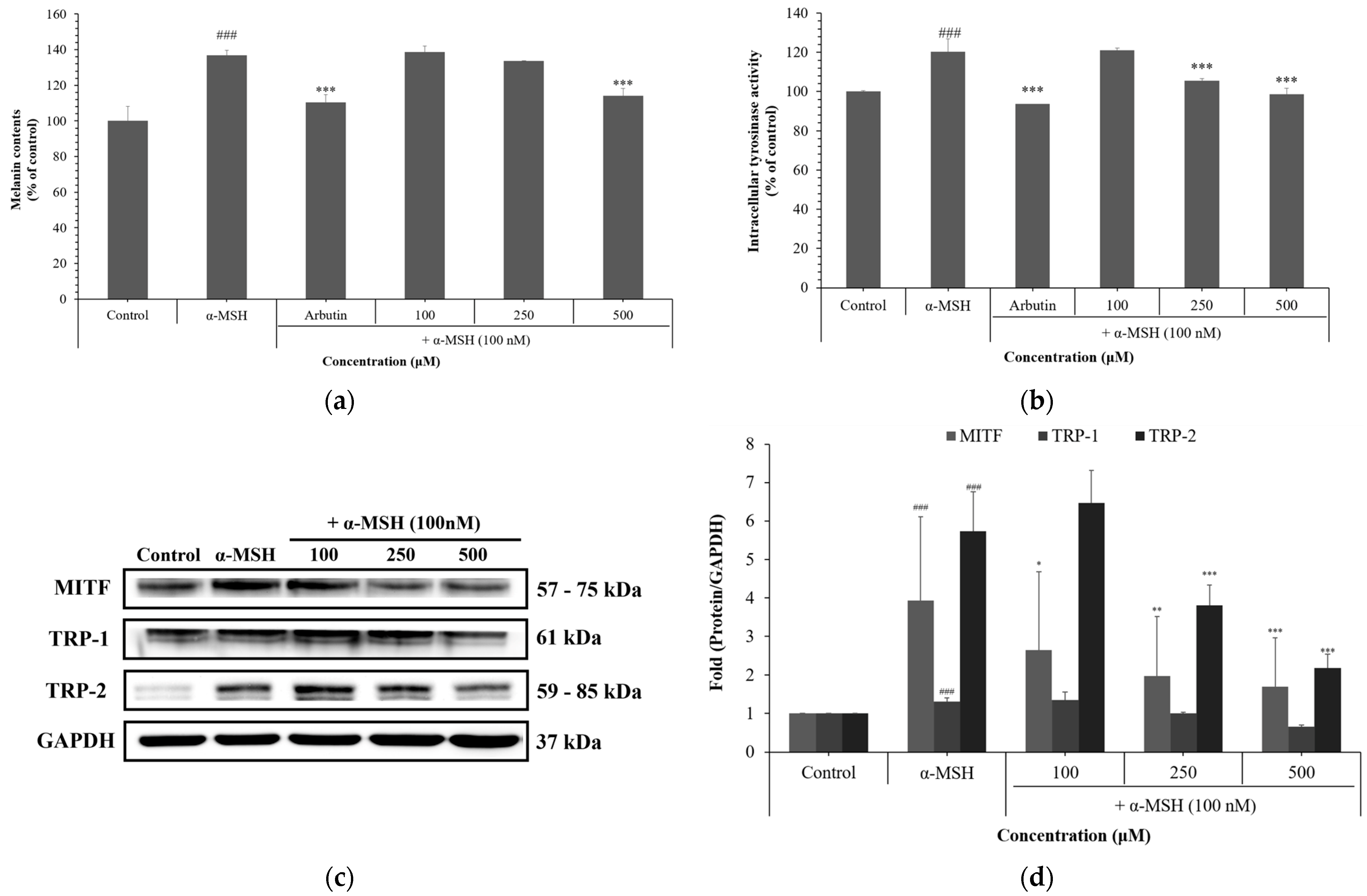
To determine the impact of NCG on melanogenesis, B16F10 cells were stimulated with α-MSH (100 nM), and arbutin (400 µM) was used as a positive control. NCG treatment significantly inhibited melanin synthesis and reduced intracellular tyrosinase activity by 18.3%. Western blot analysis revealed that NCG downregulated melanogenic proteins including microphthalmia-associated transcription factor (MITF), tyrosinase-related protein (TRP-1), and 2 (TRP-2) by over 50%. Notably, 500 µM NCG exhibited anti-melanogenic effects comparable to those of the positive control arbutin, both in melanin content reduction and tyrosinase inhibition assays, indicating that NCG has a similar level of efficacy at a comparable concentration (Figure 4). These results highlight the ability of NCG to attenuate melanin production by modulation of key melanogenic pathways.

3.5. Anti-Inflammatory Effects of NCG

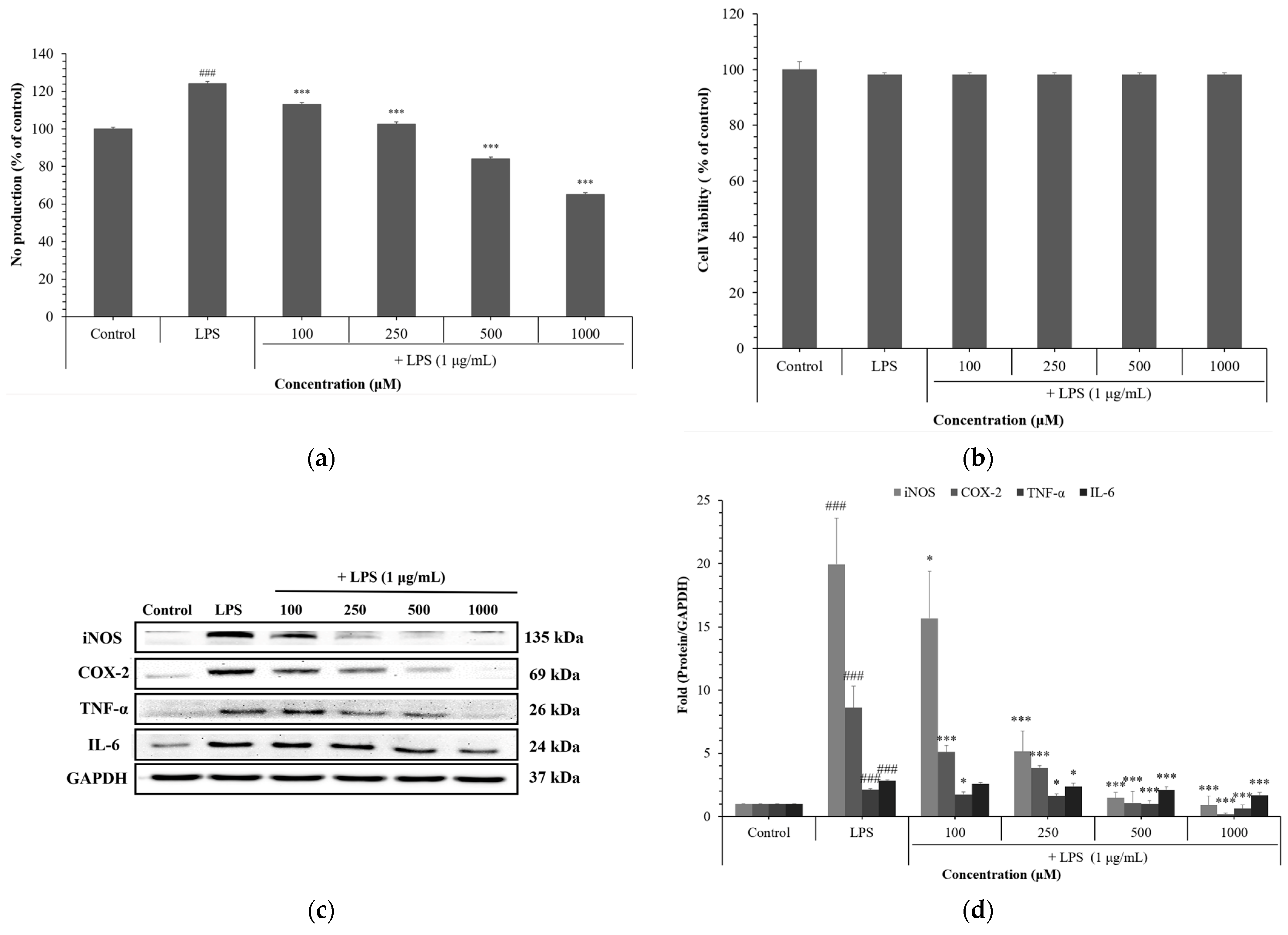
The anti-inflammatory potential of NCG was assessed in RAW 264.7 macrophages stimulated with LPS. NCG significantly decreased NO production in a dose-dependent manner, with a maximum reduction of 47.5% at 1000 µM. Importantly, this reduction in NO levels did not affect cell viability. NCG effectively inhibited the protein expression of key inflammatory mediators—cyclooxygenase-2 (COX-2), inducible Nitric Oxide Synthase (iNOS), tumor necrosis factor-alpha (TNF-α), and IL-6—as confirmed by Western blotting, indicating a strong anti-inflammatory response at the molecular level (Figure 5).

4. Discussion

Skin aging is a complex biological process influenced by intrinsic and extrinsic factors, including genetic background, oxidative stress, and chronic inflammation [29]. Among these, pro-inflammatory cytokines have emerged as central mediators of structural degradation and functional decline in the skin. Notably, the concept of “inflammaging”—chronic, low-grade inflammation associated with aging has garnered increasing attention as a critical contributor of skin deterioration [30,31].
Prolonged inflammatory stimuli also influence melanogenesis [32,33]. Inflammatory cytokines such as IL-6 and TNF-α stimulate melanocyte activity, leading to increased melanin synthesis [34,35,36,37]. While melanin serves as a protective pigment against UV radiation, its excessive accumulation may exacerbate oxidative stress by interacting with reactive species, further damaging skin cells and contributing to pigmentation disorders such as melasma and freckles [38,39].
Oxidative stress induced by UV radiation and environmental pollutants promotes ROS generation, leading to inflammation, hyperpigmentation, and degradation of extracellular matrix components [40,41,42,43]. Specifically, ROS trigger the expression of MMPs, such as MMP-1 and MMP-3, which degrade collagen and elastin fibers, reducing dermal elasticity and accelerating wrinkle formation [44,45].
MITF is a key transcriptional regulator of melanogenesis [46]. It governs the expression of tyrosinase and related proteins (TRP-1 and TRP-2), which are essential enzymes in melanin biosynthesis [47]. Overactivation of MITF contributes to melanin overproduction and the development of hyperpigmentation disorders [48].
Inflammaging is further perpetuated by transcriptional regulators including iNOS, COX-2, TNF-α, and IL-6. iNOS generates nitric oxide (NO), COX-2 synthesizes pro-inflammatory prostaglandins, and cytokines such as TNF-α and IL-6 amplify inflammatory cascades [49,50]. Chronic overexpression of these mediators disrupts skin homeostasis, impairs the skin barrier, and accelerates aging.
Our study demonstrates that NCG modulates these interconnected biological pathways: First, NCG attenuated UVB-induced ROS generation, reducing oxidative stress and preventing dermal damage. Although the precise mechanism remains to be fully elucidated, this antioxidant effect may result from both direct ROS scavenging activity and the modulation of endogenous antioxidant systems, such as glutathione metabolism or redox-sensitive signaling pathways. Based on these findings, we believe that NCG exerts various biological effects—such as anti-inflammatory, anti-melanogenic, and anti-aging activities—by suppressing UVB-induced ROS accumulation. This hypothesis is supported by previous studies reporting that NCG enhances antioxidant capacity through glutathione metabolism and redox-sensitive pathways [51,52,53]. Second, it suppressed MMP-1 and MMP-3 activity while enhancing type I procollagen synthesis, thereby preserving ECM structure and skin elasticity. Third, NCG modulated melanogenesis by downregulating MITF, tyrosinase, and TRP-1 expression, leading to reduced melanin production. Finally, NCG significantly downregulated iNOS, COX-2, TNF-α, and IL-6, thereby alleviating inflammation and maintaining skin homeostasis. In addition, although the intracellular uptake and skin permeability of NCG were not directly assessed in this study, the observed cellular effects suggest that NCG may be bioavailable and capable of penetrating the skin barrier to exert its effects in dermal cells. Future studies employing uptake assays, skin diffusion models, or reconstructed human skin systems will be essential to validate its delivery and efficacy in realistic dermatological applications. To gain a more comprehensive understanding of these biological effects, we plan to explore the involvement of key regulatory signaling pathways—such as NF-κB, MAPKs (p38, JNK, ERK), and CREB—in future studies. Identifying these upstream modulators will help elucidate the precise mechanism of action of NCG and further validate its potential as a targeted bioactive compound.
Taken together, these results position NCG as a promising multifunctional compound capable of targeting multiple mechanisms of skin aging. Its simultaneous antioxidant, anti-inflammatory, anti-wrinkle, and anti-melanogenic effects offer considerable potential for use in dermocosmetic applications. Further formulation studies and clinical evaluations will be instrumental in commercializing NCG-based skin health products.

5. Conclusions

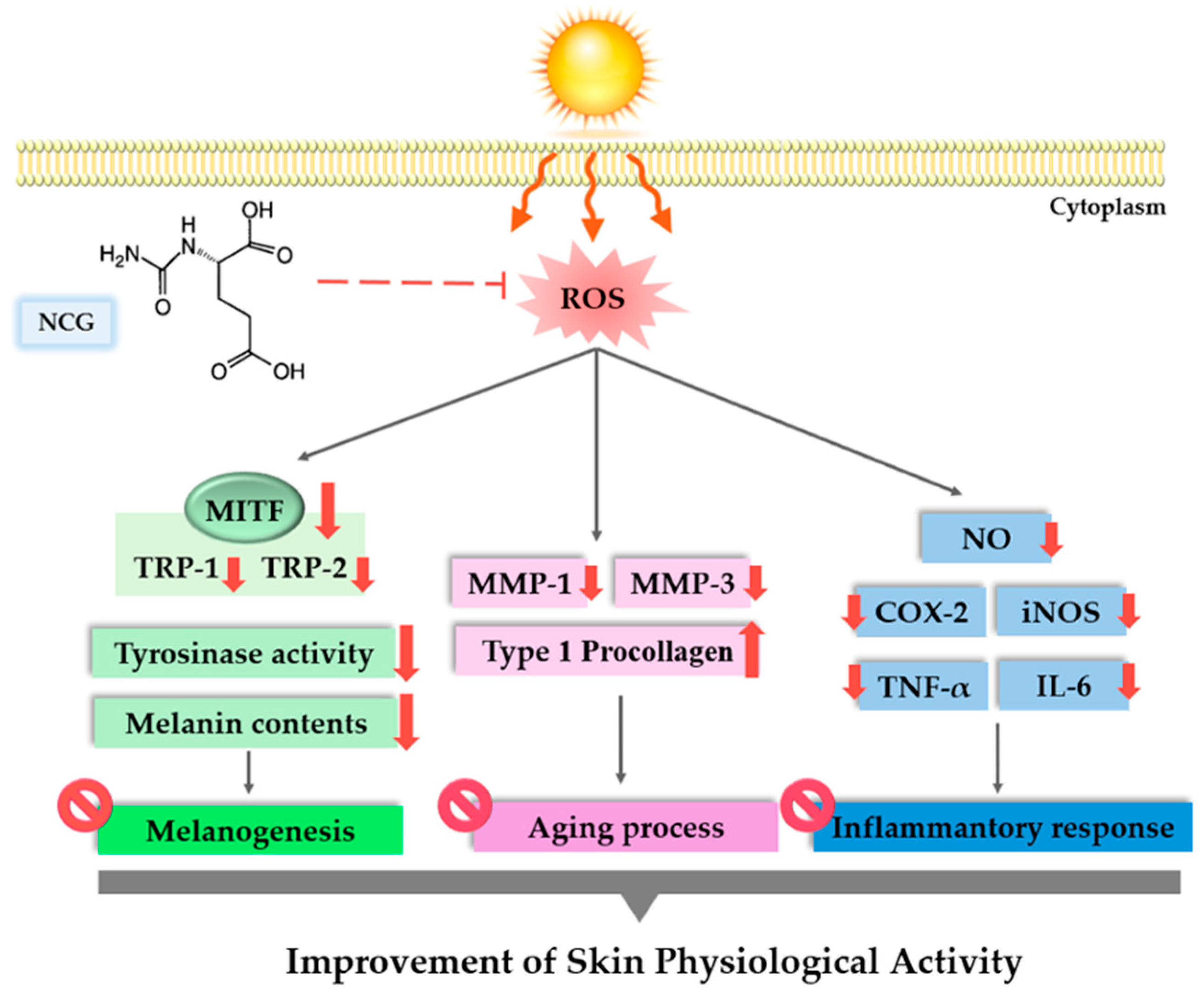
Collectively, our findings demonstrate that NCG possesses antioxidant, anti-inflammatory, and anti-melanogenic properties that contribute to the attenuation of inflammaging and skin aging (Figure 6). The current in vitro data highlight NCG’s potential as a safe and multifunctional active compound for use in cosmetic and dermatological products. Still, in vivo validation and clinical assessments remain necessary before it can be considered for practical application in commercial formulations.

Author Contributions

Conceptualization, methodology, and writing—original draft preparation: S.R.C.; investigation and writing—review and editing: N.R.S.; investigation and methodology: S.Y.S., K.M.K., J.H.B. and S.J.K. (Seon Ju Kim); methodology: D.H.J. and S.J.K. (Su Jung Kim); project administration and writing—review and editing: K.M.P. All authors have read and agreed to the published version of the manuscript.

Funding

This research was supported by a Korea Innovation Foundation (INNOPOLIS) grant funded by the Korean government (Ministry of Science and ICT) through the “Science and Technology Project that Opens the Future of the Region” (grant number: 2021-DD-UP-0380). This research was supported by the Regional Innovation System & Education (RISE) program through the Jeollanamdo RISE center, funded by the Ministry of Education (MOE) and the Jeollanamdo, Republic of Korea (2025-RISE-14-001).

Institutional Review Board Statement

Not applicable.

Informed Consent Statement

Not applicable.

Data Availability Statement

Data available on request due to restrictions.

Acknowledgments

During the preparation of this manuscript, the authors used ChatGPT 4o for the purposes of preparing the manuscript for general editing of this paper. The authors have reviewed and edited the output and take full responsibility for the content of this publication.

Conflicts of Interest

S.J.K. and D.H.J. are employees of BIO-FD&C Co., Ltd. BIO-FD&C provided no funding for this study. The company had no role in the study design; data collection, analysis, or interpretation; manuscript preparation; or the decision to submit for publication.

References

  1. Shin, S.H.; Lee, Y.H.; Rho, N.K.; Park, K.Y. Skin aging from mechanisms to interventions: Focusing on dermal aging. Front. Physiol. 2023, 14, 1195272. [Google Scholar] [CrossRef]
  2. Park, S. Biochemical, structural and physical changes in aging human skin, and their relationship. Biogerontology 2022, 23, 275–288. [Google Scholar] [CrossRef]
  3. Ding, Y.; Jiratchayamaethasakul, C.; Lee, S.H. Protocatechuic aldehyde attenuates UVA-induced photoaging in human dermal fibroblast cells by suppressing MAPKs/AP-1 and NF-κB signaling pathways. Int. J. Mol. Sci. 2020, 21, 4619. [Google Scholar] [CrossRef]
  4. Pilkington, S.M.; Bulfone-Paus, S.; Griffiths, C.E.; Watson, R.E. Inflammaging and the Skin. J. Investig. Dermatol. 2021, 141, 1087–1095. [Google Scholar] [CrossRef]
  5. Yadav, E. Inflammation and Aging: The Skin Inflammasome in the Context of Longevity Science. J. Cell. Immunol. 2025, 7, 37–42. [Google Scholar] [CrossRef]
  6. Salminen, A.; Kaarniranta, K.; Kauppinen, A. Photoaging: UV radiation-induced inflammation and immunosuppression accelerate the aging process in the skin. Inflamm. Res. 2022, 71, 817–831. [Google Scholar] [CrossRef] [PubMed]
  7. Singh, D.; Rai, V.; Agrawal, D.K. Regulation of collagen I and collagen III in tissue injury and regeneration. Cardiol. Cardiovasc. Med. 2023, 7, 5. [Google Scholar] [CrossRef] [PubMed]
  8. Ha, A.T.; Cho, J.Y.; Kim, D. MLK3 regulates inflammatory response via activation of AP-1 pathway in HEK293 and RAW264. 7 cells. Int. J. Mol. Sci. 2022, 23, 10874. [Google Scholar] [CrossRef] [PubMed]
  9. Gorman, A.; Golovanov, A.P. Lipopolysaccharide structure and the phenomenon of low endotoxin recovery. Eur. J. Pharm. Biopharm. 2022, 180, 289–307. [Google Scholar] [CrossRef]
  10. Tang, X.; Yang, T.; Yu, D.; Xiong, H.; Zhang, S. Current insights and future perspectives of ultraviolet radiation (UV) exposure: Friends and foes to the skin and beyond the skin. Environ. Int. 2024, 185, 108535. [Google Scholar] [CrossRef]
  11. Sharma, M.R.; Mitrani, R.; Werth, V.P. Effect of TNFα blockade on UVB-induced inflammatory cell migration and collagen loss in mice. J. Photochem. Photobiol. B Biol. 2020, 213, 112072. [Google Scholar] [CrossRef] [PubMed]
  12. Cavinato, M.; Jansen-Dürr, P. Molecular mechanisms of UVB-induced senescence of dermal fibroblasts and its relevance for photoaging of the human skin. Exp. Gerontol. 2017, 94, 78–82. [Google Scholar] [CrossRef]
  13. Wang, P.W.; Hung, Y.C.; Lin, T.Y.; Fang, J.Y.; Yang, P.M.; Chen, M.H.; Pan, T.L. Comparison of the biological impact of UVA and UVB upon the skin with functional proteomics and immunohistochemistry. Antioxidants 2019, 8, 569. [Google Scholar] [CrossRef]
  14. Rybnikář, M.; Malaník, M.; Šmejkal, K.; Švajdlenka, E.; Shpet, P.; Babica, P.; Dall’Acqua, S.; Ondřej, J.; Treml, J. Dibenzocyclooctadiene Lignans from Schisandra chinensis with Anti-Inflammatory Effects. Int. J. Mol. Sci. 2024, 25, 3465. [Google Scholar] [CrossRef]
  15. Tamagawa-Mineoka, R.; Ueta, M.; Arakawa, Y.; Nakanishi, M.; Nishigaki, H.; Katoh, N. Roles of interferon regulatory factor 3 in skin inflammation: Possible involvement of regulatory mechanisms. Exp. Dermatol. 2023, 32, 715–717. [Google Scholar] [CrossRef] [PubMed]
  16. Tong, W.; Chen, X.; Song, X.; Chen, Y.; Jia, R.; Zou, Y.; Li, L.; Yin, Z.; He, C.; Liang, X.; et al. Resveratrol inhibits LPS-induced inflammation through suppressing the signaling cascades of TLR4-NF-κB/MAPKs/IRF3. Exp. Ther. Med. 2020, 19, 1824–1834. [Google Scholar] [CrossRef] [PubMed]
  17. Bayazid, A.B.; Jang, Y.A.; Jeong, S.A.; Lim, B.O. Cypress tree (Chamaecyparis obtusa) Bark extract inhibits melanogenesis through repressing CREB and MITF signalling pathways in α-MSH-stimulated B16F10 cells. Food Agric. Immunol. 2022, 33, 498–510. [Google Scholar] [CrossRef]
  18. Shin, S.Y.; Sun, S.O.; Ko, J.Y.; Oh, Y.S.; Cho, S.S.; Park, D.H.; Park, K.M. New synthesized Galloyl-RGD inhibits Melanogenesis by regulating the CREB and ERK signaling pathway in B16F10 melanoma cells. Photochem. Photobiol. 2020, 96, 1321–1331. [Google Scholar] [CrossRef]
  19. Cha, S.Y.; Woo, I.K.; Cha, Y.J.; Lee, N.K.; Jang, H.J.; Paik, H.D. Anti-melanogenic and Antioxidant Activities of Lactiplantibacillus plantarum Strains in Skin Cells via the CREB/MITF and Nrf2/HO-1 Pathways. Probiotics Antimicrob. Proteins 2025, 17. [Google Scholar] [CrossRef]
  20. Prakoeswa, F.R.S.; Maharani, F.; Satria, Y.A.A.; Awanis, G.S.; Febrianty, A.F. Topical anti-aging agents: State-of-the-art review. J. Med. Sci. 2023, 55, 372–383. [Google Scholar] [CrossRef]
  21. Milani, M.; Colombo, F. Skin anti-aging effect of oral vitamin a supplementation in combination with topical retinoic acid treatment in comparison with topical treatment alone: A randomized, prospective, Assessor-blinded, parallel trial. Cosmetics 2023, 10, 144. [Google Scholar] [CrossRef]
  22. Mew, N.A.; Payan, I.; Daikhin, Y.; Nissim, I.; Nissim, I.; Tuchman, M.; Yudkoff, M. Effects of a single dose of N-carbamylglutamate on the rate of ureagenesis. Mol. Genet. Metab. 2009, 98, 325–330. [Google Scholar] [CrossRef]
  23. Hu, Y.; Shao, D.; Wang, Q.; Xiao, Y.; Zhao, X.; Shen, Y.; Shan, Z.; Haibing, T.; Shi, S. Effects of dietary N-carbamylglutamate supplementation on growth performance, tissue development and blood parameters of yellow-feather broilers. Poult. Sci. 2019, 98, 2241–2249. [Google Scholar] [CrossRef]
  24. Hu, N.; Mao, P.; Xiong, X.; Ma, Z.; Xie, Z.; Gao, M.; Wu, Q.; Ma, W. Effect of N-Carbamylglutamate Supplementation on Growth Performance, Jejunal Morphology, Amino Acid Transporters, and Antioxidant Ability of Weaned Pigs. Animals 2023, 13, 3183. [Google Scholar] [CrossRef]
  25. Zhu, X.; Li, N.; Wang, Y.; Ding, L.; Chen, H.; Yu, Y.; Shi, X. Protective effects of quercetin on UVB irradiation-induced cytotoxicity through ROS clearance in keratinocyte cells. Oncol. Rep. 2017, 37, 209–218. [Google Scholar] [CrossRef]
  26. Kong, Y.H.; Xu, S.P. Juglanin administration protects skin against UVB-induced injury by reducing Nrf2-dependent ROS generation. Int. J. Mol. Med. 2020, 46, 67–82. [Google Scholar] [CrossRef]
  27. Kavinda, M.H.D.; Park, J.; Kim, N.; Choi, Y.H.; Kim, G.Y. Anti-melanogenic properties of FBCC–EP850 derived from Carex pumila Thunb. Asian Pac. J. Trop. Biomed. 2024, 14, 477–485. [Google Scholar] [CrossRef]
  28. Pillai-Kastoori, L.; Schutz-Geschwender, A.R.; Harford, J.A. A systematic approach to quantitative Western blot analysis. Anal. Biochem. 2020, 593, 113608. [Google Scholar] [CrossRef] [PubMed]
  29. Krutmann, J.; Schikowski, T.; Morita, A.; Berneburg, M. Environmentally-induced (extrinsic) skin aging: Exposomal factors and underlying mechanisms. J. Investig. Dermatol. 2021, 141, 1096–1103. [Google Scholar] [CrossRef]
  30. Naharro-Rodriguez, J.; Bacci, S.; Hernandez-Bule, M.L.; Perez-Gonzalez, A.; Fernandez-Guarino, M. Decoding Skin Aging: A Review of Mechanisms, Markers, and Modern Therapies. Cosmetics 2025, 12, 144. [Google Scholar] [CrossRef]
  31. He, X.; Gao, X.; Xie, W. Research progress in skin aging and immunity. Int. J. Mol. Sci. 2024, 25, 4101. [Google Scholar] [CrossRef]
  32. Fu, C.; Chen, J.; Lu, J.; Yi, L.; Tong, X.; Kang, L.; Pei, S.; Ouyang, Y.; Jiang, L.; Ding, Y.; et al. Roles of inflammation factors in melanogenesis. Mol. Med. Rep. Mol. 2020, 21, 1421–1430. [Google Scholar] [CrossRef]
  33. Thawabteh, A.M.; Jibreen, A.; Karaman, D.; Thawabteh, A.; Karaman, R. Skin pigmentation types, causes and treatment—A review. Molecules 2023, 28, 4839. [Google Scholar] [CrossRef] [PubMed]
  34. Emanuelli, M.; Sartini, D.; Molinelli, E.; Campagna, R.; Pozzi, V.; Salvolini, E.; Simonetti, O.; Campanati, A.; Offidani, A. The double-edged sword of oxidative stress in skin damage and melanoma: From physiopathology to therapeutical approaches. Antioxidants 2022, 11, 612. [Google Scholar] [CrossRef] [PubMed]
  35. Hossain, M.R.; Ansary, T.M.; Komine, M.; Ohtsuki, M. Diversified stimuli-induced inflammatory pathways cause skin pigmentation. Int. J. Mol. Sci. 2021, 22, 3970. [Google Scholar] [CrossRef]
  36. Di Costanzo, L.; Scala, E.; Caiazzo, G.; Lembo, S.; Marino, R.; Megna, M.; Patri, A.; Di Caprio, R.; Balato, A. Possible role of BMP-4 in the hyper-pigmentation of psoriatic plaques after anti-TNF-α treatment. Exp. Ther. Med. 2019, 18, 4120–4124. [Google Scholar] [CrossRef]
  37. Markiewicz, E.; Karaman-Jurukovska, N.; Mammone, T.; Idowu, O.C. Post-inflammatory hyperpigmentation in dark skin: Molecular mechanism and skincare implications. Clin. Cosmet. Investig. Dermatol. 2022, 15, 2555–2565. [Google Scholar] [CrossRef] [PubMed]
  38. Bocheva, G.; Slominski, R.M.; Janjetovic, Z.; Kim, T.K.; Böhm, M.; Steinbrink, K.; Reiter, R.J.; Kleszczyński, K.; Slominski, A.T. Protective role of melatonin and its metabolites in skin aging. Int. J. Mol. Sci. 2022, 23, 1238. [Google Scholar] [CrossRef]
  39. Fahad, D.; Mohammed, M.T. Oxidative stress: Implications on skin diseases. Plant Arch. 2020, 20, 4150–4157. [Google Scholar]
  40. Papaccio, F.; Caputo, S.; Bellei, B. Focus on the contribution of oxidative stress in skin aging. Antioxidants 2022, 11, 1121. [Google Scholar] [CrossRef]
  41. Sparavigna, A. Role of the extracellular matrix in skin aging and dedicated treatment-state of the art. Plast. Aesthetic Res. 2020, 7, 14. [Google Scholar] [CrossRef]
  42. Xing, X.; Dan, Y.; Xu, Z.; Xiang, L. Implications of oxidative stress in the pathogenesis and treatment of hyperpigmentation disorders. Oxid. Med. Cell. Longev. 2022, 2022, 7881717. [Google Scholar] [CrossRef]
  43. Ansary, T.M.; Hossain, M.R.; Kamiya, K.; Komine, M.; Ohtsuki, M. Inflammatory molecules associated with ultraviolet radiation-mediated skin aging. Int. J. Mol. Sci. 2021, 22, 3974. [Google Scholar] [CrossRef]
  44. Loo, Y.C.; Hu, H.C.; Yu, S.Y.; Tsai, Y.H.; Korinek, M.; Wu, Y.C.; Chang, F.R.; Chen, Y.J. Development on potential skin anti-aging agents of Cosmos caudatus Kunth via inhibition of collagenase, MMP-1 and MMP-3 activities. Phytomedicine 2023, 110, 154643. [Google Scholar] [CrossRef] [PubMed]
  45. Wan, J.; Zhang, G.; Li, X.; Qiu, X.; Ouyang, J.; Dai, J.; Min, S. Matrix metalloproteinase 3: A promoting and destabilizing factor in the pathogenesis of disease and cell differentiation. Front. Physiol. 2021, 12, 663978. [Google Scholar] [CrossRef] [PubMed]
  46. Chung, Y.C.; Kim, M.J.; Kang, E.Y.; Kim, Y.B.; Kim, B.S.; Park, S.M.; Hyun, C.G. Anti-melanogenic effects of hydroxyectoine via mitf inhibition by jnk, p38, and akt pathways in b16f10 melanoma cells. Nat. Prod. Commun. 2019, 14, 1934578X19858523. [Google Scholar] [CrossRef]
  47. Lim, J.W.; Ha, J.H.; Jeong, Y.J.; Park, S.N. Anti-melanogenesis effect of dehydroglyasperin C through the downregulation of MITF via the reduction of intracellular cAMP and acceleration of ERK activation in B16F1 melanoma cells. Pharmacol. Rep. 2018, 70, 930–935. [Google Scholar] [CrossRef]
  48. Nguyen, N.T.; Fisher, D.E. MITF and UV responses in skin: From pigmentation to addiction. Pigment. Cell Melanoma Res. 2019, 32, 224–236. [Google Scholar] [CrossRef]
  49. Zhang, Z.; Li, L.; Huang, G.; Zhou, T.; Zhang, X.; Leng, X.; Chen, Z.; Lin, J. Embelia Laeta aqueous extract suppresses acute inflammation via decreasing COX-2/iNOS expression and inhibiting NF-κB pathway. J. Ethnopharmacol. 2021, 281, 114575. [Google Scholar] [CrossRef]
  50. Heo, Y.J.; Lee, N.; Choi, S.E.; Jeon, J.Y.; Han, S.J.; Kim, D.J.; Kang, Y.; Lee, K.W.; Kim, H.J. Amphiregulin Induces iNOS and COX-2 Expression through NF-κB and MAPK Signaling in Hepatic Inflammation. Mediat. Inflamm. 2023, 2023, 2364121. [Google Scholar] [CrossRef]
  51. Xiao, L.; Cao, W.; Liu, G.; Fang, T.; Wu, X.; Jia, G.; Chen, X.; Zhao, H.; Wang, J.; Wu, C.; et al. Arginine, N-carbamylglutamate, and glutamine exert protective effects against oxidative stress in rat intestine. Anim. Nutr. 2016, 2, 242–248. [Google Scholar] [CrossRef] [PubMed]
  52. Liu, G.; Xiao, L.; Cao, W.; Fang, T.; Jia, G.; Chen, X.; Zhao, H.; Wu, C.; Wang, J. Changes in the metabolome of rats after exposure to arginine and N-carbamylglutamate in combination with diquat, a compound that causes oxidative stress, assessed by 1 H NMR spectroscopy. Food Funct. 2016, 7, 964–974. [Google Scholar] [CrossRef] [PubMed]
  53. Gu, F.F.; Jiang, L.Y.; Wang, D.M.; Zhao, F.Q.; Liu, J.X. Supplementation with N-carbamoylglutamate during the transition period improves the function of neutrophils and reduces inflammation and oxidative stress in dairy cows. J. Dairy Sci. 2022, 105, 5786–5795. [Google Scholar] [CrossRef] [PubMed]
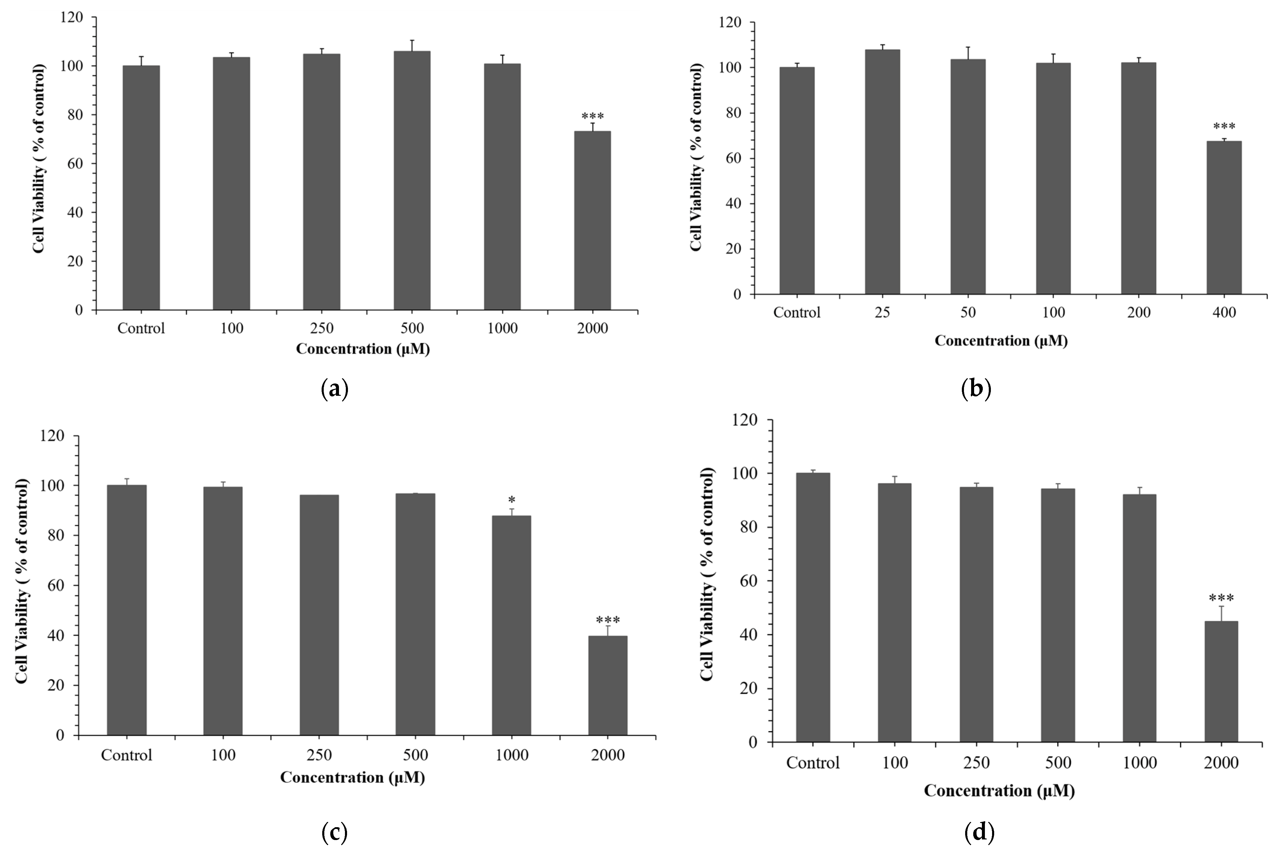
Figure 1. Effects of NCG on cell viability. (a) HaCaTs, (b) HDFs, (c) B16F10 cells, and (d) RAW 264.7 macrophages. Cells were treated with NCG and incubated for 24 h (HaCaTs, RAW 264.7) or 72 h (B16F10, HDF). Data are presented as mean ± SD based on triplicate experiments. * p < 0.05, *** p < 0.001 vs. control.
Cosmetics 12 00250 g001
Figure 2. Effects of NCG on intracellular and mitochondrial ROS levels. (a) Representative fluorescence images of ROS production in HaCaTs exposed to UVB (15 mJ/cm2) and treated with NCG (100–1000 µM) for 24 h. NAC (N-acetylcysteine, 50 mM) was used as a positive control. Images were captured under identical acquisition settings at 20×; scale bar = 150 µm (applies to all images in each row). (b) Quantification of intracellular ROS production expressed as a percentage of control. Data are presented as mean ± SD from at least three independent experiments. ### p < 0.001 vs. control; *** p < 0.001 vs. UVB.
Cosmetics 12 00250 g002
Figure 3. Effects of NCG on UVB-induced skin aging in HDFs. (a) Type I procollagen synthesis measured by ELISA. (b) MMP-1 enzymatic activity measured by ELISA. (c) MMP-3 enzymatic activity measured by ELISA. (d) Protein expression levels of MMP-1 and MMP-3 analyzed by Western blot. (e) Densitometric quantification of Western blot bands from panel (d), showing relative expression of MMP-1 and MMP-3. The results are expressed as the mean ± standard deviation from three independent experiments. ### p < 0.001 vs. control; * p < 0.05, ** p < 0.01, *** p < 0.001 vs. UVB-treated group.
Cosmetics 12 00250 g003
Figure 4. Effects of NCG on melanogenesis in B16F10 melanoma cells. (a) Melanin content after treatment with NCG (100–500 μM) for 72 h. (b) Intracellular tyrosinase activity following NCG treatment. (c) Protein expression of MITF, TRP-1, and TRP-2 determined by Western blot; normalized to GAPDH. (d) Densitometric analysis of protein levels. As a standard for comparison, arbutin was included as the positive control. Experimental data are displayed as mean ± standard deviation, based on three independently repeated experiments. ### p < 0.001 vs. control; * p < 0.05, ** p < 0.01, *** p < 0.001 vs. α-MSH group.
Cosmetics 12 00250 g004
Figure 5. Anti-inflammatory effects of NCG in RAW 264.7 macrophages. (a) Cell viability after NCG treatment (100–1000 μM) for 24 h. (b) NO production measured by the Griess assay. (c) Protein expression levels of iNOS, COX-2, IL-6, and TNF-α analyzed by Western blot; normalized to GAPDH. (d) Quantification of Western blot bands. The results are presented as mean ± SD obtained from three independent experiments. Statistical significance was indicated as follows: ### p < 0.001 vs. control; * p < 0.05, *** p < 0.001 vs. LPS group.
Cosmetics 12 00250 g005
Figure 6. Summary of the molecular mechanism by which NCG enhances skin physiological activity through anti-melanogenic, anti-aging, and anti-inflammatory pathways.
Cosmetics 12 00250 g006
Table 1. Primary antibodies used in Western blot analysis.
Target ProteinCatalog No.Host SpeciesDilution Ratio
GAPDHE12-057Mouse1:5000
TRP-1ab178676Rabbit1:10,000
TRP-2ab2211144Rabbit1:1000
MITF#12590Rabbit1:1000
MMP-1sc-58377Mouse1:400
MMP-3AF513Goat1:5000
TNF-α#12744Mouse1:200
COX-2#35-8200Mouse1:500
IL-6#P620Rabbit1:1000
iNOS#PA1-036Rabbit1:1000
Disclaimer/Publisher’s Note: The statements, opinions and data contained in all publications are solely those of the individual author(s) and contributor(s) and not of MDPI and/or the editor(s). MDPI and/or the editor(s) disclaim responsibility for any injury to people or property resulting from any ideas, methods, instructions or products referred to in the content.