Abstract
Two-dimensional (2D) van der Waals materials have been actively investigated for broadband, high-sensitivity, low-power-consumption photodetection owing to their highly customizable band structures and fast interfacial charge transfers. Studying photocurrent generation mechanisms provides insights into charge carrier dynamics in WSe2-based detectors, linking spatial factors (e.g., photocurrent generation/collection) with interfacial band alignment. Here, we employ scanning photocurrent microscopy to spatially resolve the processes of photocurrent generation and collection in WSe2-based composite structures. Photocurrent polarity and magnitude at interface reflects interfacial band alignment and potential gradients at metal–WSe2 and WSe2–In2Se3 junctions. Strong electric fields at metal–WSe2 interfaces drive more efficient electron–hole separation and yield higher photocurrents, compared with WSe2–In2Se3 interfaces. The photodetector exhibits broadband detection capabilities from visible to infrared light, achieving a high responsivity of 17.7 A/W and an excellent detectivity of 3.7 × 10¹² Jones, as well as fast response times of <113 µs. Furthermore, object imaging with a resolution better than 0.5 mm was successfully demonstrated, highlighting the potential of this photoresponse for practical imaging applications. This work reveals that photocurrent is distributed with a clear dependence on device configuration, offering a new avenue for optimizing 2D material-based photoelectric devices.
1. Introduction
Transition metal dichalcogenides (TMDCs), exemplified by monolayer tungsten diselenide (WSe2), have emerged as promising optoelectronic materials due to their layer-dependent bandgaps, high carrier mobilities, and strong light–matter interactions [1]. Beyond their potential in ultrathin electronics, WSe2-based heterostructures exhibit exceptional photoresponse characteristics, making them ideal candidates for next-generation photodetection systems [2]. Unlike zero-bandgap graphene, WSe2 possesses a direct bandgap (~1.6 eV) in monolayer form, enabling efficient photon absorption across visible to near-infrared spectra [3,4]. The direct bandgap also facilitates fast exciton generation and dissociation, which is essential for high-performance photodetectors. Moreover, WSe2 is an intrinsic p-type semiconductor, which is highly valuable for enabling hole transport, realizing complementary device architectures, and enhancing charge separation efficiency in electronic and optoelectronic systems [5]. Furthermore, vertical stacking with other 2D materials (e.g., graphene) creates tailored interfacial band alignments, which are crucial for efficient photocurrent generation and extraction [6,7]. Such heterostructures allow for precise control over charge transfer dynamics, enhancing both responsivity and detectivity in optoelectronic devices [8].
Recent advances in WSe2-based photodetectors [9] have established three dominant architectures: photoconductors, phototransistors, and photodiodes, each leveraging distinct operational mechanisms to optimize performance. Electrically, WSe2 exhibits intrinsic p-type conductivity yet allows dynamic carrier-type modulation through doping, electrostatic gating, or contact engineering [10], enabling flexible integration with both Schottky and Ohmic contacts. Photoconductive configurations employ these interfacial designs to regulate carrier transport through Schottky barrier modulation [11], achieving high responsivities, microsecond-scale response times, and low dark currents. Breakthroughs in van der Waals heterojunctions (WSe2/MoS2, WSe2/In2Se3, WSe2/graphene) exploit type-II band alignment or vertical p-n junctions to generate built-in potentials [12,13,14]. These heterostructures leverage interfacial charge transfer efficiencies and built-in electric fields to enhance carrier separation, with WSe2/In2Se3 systems demonstrating broadband detection and long interlayer exciton lifetimes [11,15]. Graphene–WSe2 hybrids further enable fast photocarrier extraction through ballistic transport channels. Such engineered interfaces position WSe2 as a versatile platform for next-generation optoelectronics, including field-effect transistors [16], photovoltaic devices achieving external quantum efficiency. Nevertheless, critical gaps persist in our understanding of photocurrent dynamics within WSe2-based detectors. The tripartite interplay between interfacial band engineering, multidimensional carrier routing, and spatially heterogeneous potential landscapes remains largely uncharted, demanding systematic investigation through advanced spatial-resolution techniques.
The spatially resolved scanning photocurrent (SPCM) technique, previously applied to 1D nanostructures (carbon nanotubes/nanowires) [17,18,19] with constrained current paths, contrasts with WSe2’s 2D architecture, enabling multidirectional photocurrent pathways. In this study, we constructed WSe2-based detectors via dry-transfer methods, and found that laser-localized photocurrent generation and electrode-specific collection revealed potential-dependent transport dynamics of photon-generated carriers. Compared with pure WSe2 and the WSe2–In2Se3 heterojunction, the multiple-pair metal-WSe2 heterojunction exhibited an enhancement in photoresponse of nearly two orders of magnitude, highlighting the effectiveness of the asymmetric multiple-metal–WSe2 configuration for high-performance visible-to-infrared photodetection, particularly under low-light conditions and in high-resolution imaging applications.
2. Device Fabrication and Characterization
To preserve the integrity of WSe2 and optimize the interface quality, all devices were fabricated on Si/SiO2 substrates (300 nm oxide) with laser-direct-written Au electrodes (electron-beam evaporated, 50 nm thick). Three types of device architectures were prepared: (i) A photodetector structure with a single pair of electrodes, where dry-transferred WSe2 flakes were aligned onto Au electrodes to form metal–semiconductor Schottky junctions. (ii) A photodetector structure with multiple interdigitated electrodes, where WSe2 flakes were dry-transferred onto pre-patterned arrays to form multiple metal–semiconductor junctions. (iii) A vertical van der Waals heterostructure, formed by sequentially stacking mechanically exfoliated WSe2 and In2Se3 flakes between Au electrodes using a PDMS-assisted dry-transfer method. This technique ensures clean interfaces and well-controlled charge transport across the heterojunction. This methodology avoids photolithographic damage and guarantees clean van der Waals contacts, which are essential for preserving the intrinsic properties of the layered materials and achieving high-performance and reproducible photoresponse.
Optical and spectroscopic measurements were performed using an Olympus optical microscope and a 532 nm micro-Raman system. All electrical and optoelectronic measurements were conducted in ambient atmosphere at room temperature. A dual-channel digital source meter (Keithley 6482; METATEST CORPORATION, Nanjing, China) was used to simultaneously apply bias and measure current. Photocurrent signals were collected using a lock-in amplifier (MStarter 200; METATEST CORPORATION, Nanjing, China), and spatial photocurrent mapping was carried out by scanning the sample in both lateral directions using a precision-controlled probe stage. Devices were securely mounted on a custom sample holder to ensure measurement stability.
3. Results and Discussion
To investigate the photoresponse performance of WSe2-based devices, we fabricated a WSe2 detector with a pair of electrodes (Device 1). Figure 1a,b present the schematic diagram and corresponding optical microscope image of the WSe2 device with a metal–semiconductor–metal (MSM) structure. The morphology of the WSe2 flakes is clearly observed, the electrodes are uniformly covered by the material, and the overall device structure appears intact.
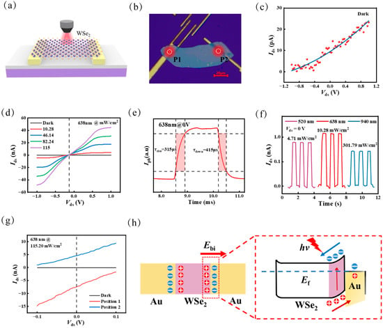
Figure 1.
Photoresponse of WSe2 detector with a pair of electrodes. (a) Schematic diagram of the detector and (b) the corresponding optical microscope image. (c) Current–voltage curves measured in the dark, illustrating Schottky contacts. (d) Photocurrent characteristics of the device under different incident light power densities at 638 nm. (e) Rise and fall times of the device at zero bias voltage. (f) Photoresponses to 520 nm, 638 nm, and 940 nm lasers at zero bias voltage. (g) Dark and locally illuminated I-V curves at P1 and P2 show position-dependent photocurrent polarity induced by Schottky contacts. (h) Energy band alignment of metal-WSe2 contacts.
Figure 1c displays the output current–voltage characteristic curves under dark conditions. The rectifying behavior of the Au/WSe2/Au device shows voltage-dependent nonlinearity in the dark state, indicating the presence of an inherent electric field. Figure 1d shows the I–V characteristics of the device under different light power densities at 638 nm, as well as in the dark. The photocurrent amplitude also increases with rising light intensity (see Figure S1 in the Supporting Information for more power-dependent and bias-dependent photoresponse data at 638 nm). To further characterize the advantages of our device in terms of response speed, the rise and fall times, typically defined as the duration required for the photocurrent to transition from 10% to 90% of its maximum value, are shown in Figure 1e. The rise time is ~370 μs, and the fall time is ~405 μs. This rapid response speed is attributed to fast separation and transportation of photogenerated carriers facilitated by the strong built-in electric field at the metal–WSe2 Schottky junction. Figure 1f presents the time-resolved photoresponse of the device under laser illumination ranging from visible to near-infrared light of 520–940 nm at zero external bias, further confirming that the device maintains a strong photoresponse capability in the near-infrared range (see Figures S2 and S3 in the Supporting Information for power-dependent photoresponse at 520 nm and 940 nm, respectively).
Figure 1g presents the photocurrent generated at positions P1 and P2 under focused optical illumination, highlighting the spatial dependence of the photoresponse. The photocurrent polarity observed at zero bias voltage, 4.6 nA at position P1 and −7.5 nA at position P2, indicates the presence of a built-in electric field at the interface. Analysis of the zero-bias photocurrent direction indicates that the internal electric field points from WSe2 flakes toward the Au electrode. The schematic diagrams of the energy band structure are illustrated in Figure 1h. A Schottky barrier is formed due to the difference in work function between Au and WSe2 [20], leading to band bending and the formation of a built-in electric field. The inherent symmetry of the dual Schottky junction architecture leads to photocurrent cancellation under global illumination; however, localized excitation breaks this symmetry, enabling a net photoresponse that provides a robust basis for probing the influence of composite structures on photodetector performance.
To further confirm the influence of metal–WSe2 Schottky junctions on the photoresponse, a WSe2-based photodetector (Device 2) with multiple electrode pairs was fabricated. Figure 2a illustrates the schematic structure of the device, while Figure 2b shows an optical microscope image of a representative device fabricated based on this design. The interdigitated electrode gaps are uniformly distributed, and the WSe2 flake extends across multiple electrode fingers, forming a large effective photosensitive area to facilitate a more comprehensive analysis of the photoresponse mechanisms. In the lower panel of Figure 2a, adjacent WSe2 electrodes may be seen to generate opposing built-in fields, enabling directional separation of photogenerated carriers for enhanced photocurrent. The scanning photocurrent microscopy characterization under zero bias shown in Figure 2c reveals spatially localized photocurrent at WSe2/Au interfaces under 638 nm illumination. The mapped photoresponse exhibits five polarity inversion regions between neighboring electrode pairs, governed by counter-oriented built-in potentials across multiple WSe2/Au Schottky junctions. This spatial photoresponse pattern directly correlates with interfacial band bending dynamics, demonstrating the critical role of electrode architecture in steering carrier transport pathways.
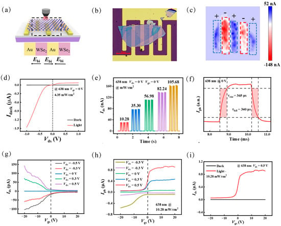
Figure 2.
Photoresponse of WSe2 photodetector with multiple pairs of electrodes. (a) Schematic diagram of Device 2 with multiple built-in electric field of opposite polarity. (b) Corresponding optical microscope image. (c) Photocurrent mapping under 638 nm laser illumination at zero bias voltage. (d) Current–voltage curves under 638 nm laser illumination and dark condition. (e) Time-resolved photoresponse under different incident-light power densities at zero bias voltage. (f) Rise and fall times under 638 nm light illumination at zero bias voltage. (g) Transfer curves at different bias voltages under dark condition. (h) Gated voltage-dependent photocurrent under 638 nm light illumination and dark condition at a bias voltage of 0.5 V. (i) Gated voltage-dependent photocurrent at various bias voltages under 638 nm light illumination.
The I–V curves of the device under dark conditions and 638 nm laser illumination are shown in Figure 2d. Across the entire voltage of −1 V to 1 V, the photocurrent under 638 nm illumination is significantly higher than the dark current, indicating a strong photoresponse. Time-resolved zero-bias photoresponse measurements were performed under various incident-light power densities at 638 nm, as shown in Figure 2e (see Figure S4 in the Supporting Information for the time-resolved photoresponse at 0.5 V under varying optical power). As the light intensity increases, the photocurrent response amplitude gradually increases, demonstrating good power-dependent behavior. Figure 2f depicts the temporal response at zero bias voltage, with rise and fall times of approximately 365 and 340 μs, respectively, indicating rapid carrier dynamics under drift conditions (see Figure S5 in the Supporting Information for temporal photoresponse under 520 nm and 940 nm illumination).
Gate voltage modulation is an effective strategy for optimizing the performance of photodetectors [1]. Figure 2g shows the transfer characteristics of Device 2 at different bias voltages in dark condition. A significant suppression of the dark current occurs as the gate voltage sweeps from −20 V to 20 V, at both positive and negative bias voltage. Specifically, the dark current of the device decreases by 97.2% (from 142 pA to 4 pA) at 0.5 V and by 96% (from −102 pA to −5 pA) at –0.5 V. The transfer characteristics of the device under illumination are presented in Figure 2h. The photocurrent displays different modulation behaviors at positive and negative bias voltages. At a positive bias voltage of 0.5 V, the photocurrent increases by five-fold, from 0.17 μA to 0.9 μA, while at a negative bias voltage of −0.5 V, the photocurrent decreases by approximately 95%, from −0.56 μA to −0.08 μA. Moreover, the photocurrent saturates rapidly under positive gate voltage. In Figure 2i, as the gate voltage increases, the photocurrent increases while the dark current decreases, leading to a 509-fold improvement in the on–off ratio, which rises from 369 at −20 V to 188,000 at 20 V. Thus, by leveraging interdigitated electrode architectures and gate modulation, we systematically probed the interfacial role of Au/WSe2 junctions, and established photocurrent performance optimization strategies through composite structural engineering.
Raman spectroscopy was performed to verify the formation of the heterostructure. Figure 3c presents the Raman spectra of WSe2 (purple line), In2Se3 (yellow line), and the WSe2/In2Se3 heterostructure region (green line). The results show characteristic E2g and A1g peaks of WSe2 near 250 cm−1 [21], while In2Se3 exhibits distinct peaks in the range of 80–200 cm−1. The heterojunction spectrum retains both material signatures with preserved crystallinity, demonstrating non-destructive van der Waals integration. Figure 3d reveals the formation of two distinct built-in electric fields arising from the differences in bandgap and electron affinity among WSe2, Au, and In2Se3. The WSe2/In2Se3 interface exhibits type-II band alignment, with the conduction and valence bands of In2Se3 positioned below those of WSe2. This built-in field directed from WSe2 to In2Se3 spatially separates photogenerated electrons into In2Se3 and holes into WSe2. Concurrently, the Au/WSe2 contact with a Schottky junction from Au to WSe2 enhances hole extraction to the electrode via work-function mismatch. These two fields synergistically suppress recombination and improve charge collection [22]. Their coexistence creates an asymmetric potential gradient across the device, enabling bias-free photocarrier separation/transport and supporting self-powered operation.
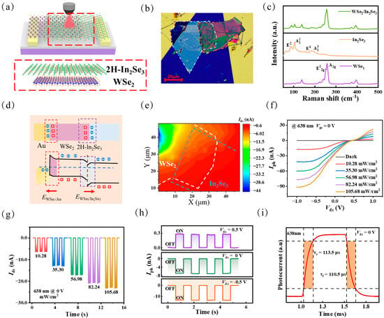
Figure 3.
Photoresponse of the WSe2/In2Se3 heterojunction. (a) Schematic diagram of the WSe2/In2Se3 heterostructure. (b) The corresponding optical microscope image. (c) Raman spectra of WSe2 (purple line), In2Se3 (yellow line), and the WSe2/In2Se3 heterostructure (green line). (d) Schematic energy band diagrams of the device under 638 nm illumination at zero bias voltage. (e) Photocurrent mapping under 638 nm illumination at zero bias voltage. (f) I–V curves of the device under different light power densities of 638 nm and dark condition. (g) Time-resolved photoresponse under 638 nm laser illumination at different incident powers at bias voltage of −1 V. (h) Time-resolved photoresponse under 638 nm illumination at applied bias voltages of −0.5 V, 0 V, 0.5 V. (i) Temporal photoresponse showing rise and fall times of approximately 113 μs and 110 μs, respectively, at zero bias voltage.
Figure 3e displays the spatial photocurrent distribution under 638 nm illumination at zero bias voltage. The observed asymmetry in photocurrent distribution reflects the asymmetric built-in electric fields of the device. A strong photoresponse region (−30–44 nA) appears at the metal–WSe2 contact interface, while the WSe2/In2Se3 region diminishes. Figure 3f shows the current–voltage curves of the device under 638 nm laser illumination at various optical power densities, as well as in the dark (see Figure S6 in the Supporting Information for the current–voltage characteristics under 940 nm illumination at different power levels). Compared with the WSe2-based devices, the heterostructure device exhibits a significantly lower dark current across the entire bias range, primarily due to the strong internal built-in electric field, which effectively suppresses leakage and reduces power consumption.
Moreover, time-resolved zero bias photoresponse in Figure 3g demonstrates strong optical power dependence, with photocurrent increasing steadily as optical power rises to 638 nm. External bias modulation (−0.5 V to +0.5 V) reveals distinct carrier transport regimes in Figure 3h: negative bias enhances photocurrent, while positive bias suppresses carrier extraction through potential barrier elevation (see Figure S7 in the Supporting Information for power-dependent photoresponse at different bias voltages). This suggests that the direction of the external electric field significantly modulates the separation and transport of photogenerated carriers. Additionally, Figure 3i presents the response time of the device under 638 nm illumination with a bias voltage of 0 V (see Figure S8 in the Supporting Information for response time at −0.5 V). The fast photoresponse, with a time of about 110 μs, indicates excellent dynamic performance. These findings further highlight the device’s excellent zero-bias photoresponse performance, attributable to the synergistic contributions from intrinsic material properties, electrode-induced Schottky barriers, heterointerface effects, and additional built-in electric fields.
To comprehensively evaluate the photoresponse performance of the three devices, the value of Iph, the Iph/Idark ratio, and the response time of each device under 638 nm illumination at zero bias voltage was calculated. As shown in Figure 4a, the photocurrents of Device 1, Device 2, and Device 3 under 638 nm illumination at zero bias voltage were measured to be 6 nA, 5 nA, and 7 nA, respectively. Figure 4b presents the light-to-dark current ratios of the devices, which reached 640, 11388, and 16,661 in the cases of Device 1, Device 2, and Device 3, respectively. Furthermore, the response times of the three devices, as illustrated in Figure 4c, were 375 μs, 365 μs, and 113 μs. The rapid response of Device 3 is attributed to its optimized carrier transport pathways.
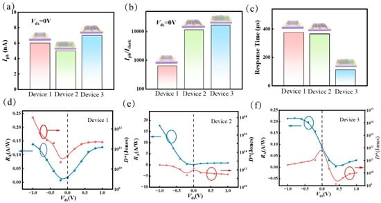
Figure 4.
Comparison of photoresponse, responsivity and specific detectivity of the three devices. (a) Photocurrents, (b) photocurrent/dark current ratios, and (c) response times of Device 1, Device 2, and Device 3 under 638 nm at zero bias voltage. Responsivity and specific detectivity of (d) Device 1, (e) Device 2, and (f) Device 3 under 638 nm illumination at zero bias voltage.
To further assess the performance of the three devices, we quantitatively calculated the responsivity and specific detectivity of each device. Responsivity (R) is an important indicator for evaluating the performance of photodetectors. It is defined as: R = Iph/(Pin·S) [23], where Iph, Pin, and S represent the photocurrent, the illumination power, and the effective illumination area, respectively. Specific detectivity (D*) is another critical parameter for assessing the performance of photodetectors. This is defined as: D* = (S1/2·R)/(2q·Idark) 1/2 [24], where q is the elementary charge and Idark is the dark current. The variations in responsivity (R) and specific detectivity (D*) for the three devices under varying bias voltages are presented in Figure 4.
As shown in Figure 4d, Device 1 reaches its highest responsivity of 0.14 A/W and detectivity of 3.7 × 10¹² Jones at a bias of −1 V. In the case of Device 2 (Figure 4e), D* decreases from 3.66 × 10¹² Jones at −1 V to 3.03 × 10¹¹ Jones at 1 V, while R increases from 0.058 A/W to 17.7 A/W over the same voltage range. In contrast, Device 3 in Figure 4f displays a decrease in responsivity from −1 V to 0.4 V, followed by an increase to 0.03 A/W at 1 V. Meanwhile, D* exhibits triphasic behavior: it initially rises from −1 V to 0 V (peaking at 2.16 × 10¹² Jones), then declines until 0.4 V, and subsequently increases again towards 1 V. Overall, the detectivity across the devices spans over two orders of magnitude, from 1.32 × 10⁹ Jones to 2.16 × 10¹² Jones. Specifically, the detectivity’s peak-valley behavior versus bias voltage originates from heterojunction internal and external electric field modulation. At zero bias, detectivity peaks as the device relies solely on built-in fields from heterojunction and Schottky contacts. This minimizes dark current (suppressing noise) while maintaining sufficient field strength for efficient photocarrier separation without external bias, maximizing signal-to-noise ratio. Conversely, a detectivity valley emerges at bias voltage of ~0.4 V, where the applied field partially cancels the built-in field, degrading carrier separation efficiency and lowering photocurrent. These results highlight the crucial role of self-driven operation, enhanced carrier separation induced by Schottky barriers, and efficient charge transport across the heterointerface. The synergistic effects of these factors collectively contribute to the observed high photocurrent, rapid photoresponse, and low-power photoelectric detection capabilities. A comparison of the performance of the device fabricated in this study with recently reported photodetectors based on WSe2 and other two-dimensional TMD materials is provided in Table S1 of the Supporting Information [25,26,27,28,29,30,31,32,33,34,35].
To validate the stability and imaging performance of our device, we carried out long-term stability testing over 1000 s. The results shown in Figure 5a reveal photocurrent decay under 638 nm illumination at zero bias, with consistent response amplitudes throughout multiple switching cycles. To further demonstrate the imaging capability of our photodetector, we constructed an imaging system for a demonstration experiment, as illustrated in Figure 5b. The imaging system consists of a laser source, lens, focusing optics, target object, stepper motor, lock-in amplifier, preamplifier, and a computer. The laser beam is emitted from the source, focused by the optics, and then directed onto the object. Under the precise control of the stepper motor, the object moves point-by-point in both horizontal (x) and vertical (y) directions to ensure complete scanning coverage of the entire area. The transmitted light passing through the object is collected by our photodetector, which converts the optical signal into an electrical signal. The generated photocurrent is processed through a preamplifier and a lock-in amplifier, then collected and recorded by the computer along with positional information. Finally, based on the spatial distribution of the photocurrent, the contour and features of the target object are reconstructed.
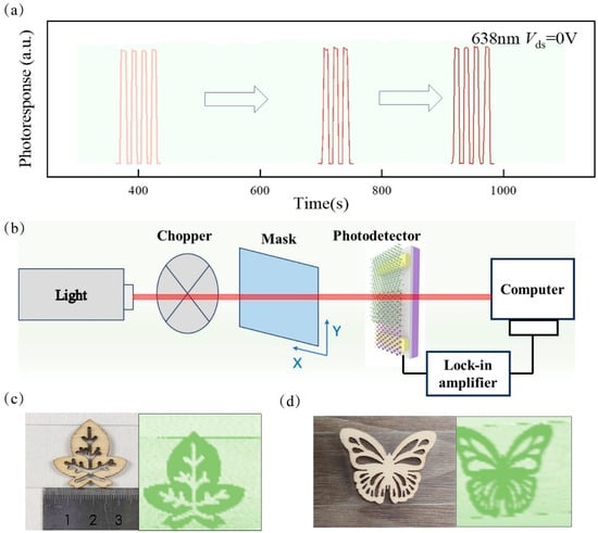
Figure 5.
Photoresponse stability and imaging applications. (a) On/off switching behavior of the device under 638 nm illumination at zero bias voltage, demonstrating temporal stability. (b) Schematic diagram of the imaging system using the WSe2/In2Se3 heterojunction photodetector as the imaging unit. High-resolution transmission imagings of (c) leaf-shaped and (d) butterfly-shaped structures under 638 nm illumination.
The leaf and butterfly both feature complex boundaries and intricate details, such as fine edge contours and internal cut patterns, which presented a greater challenge for our detector during point-by-point scanning and imaging. The real picture and the imaging of the leaf are shown in Figure 5c, where the size of the leaf pattern is 30 mm × 25 mm. The real picture and the imaging of the butterfly are shown in Figure 5d, with a pattern size of 35 mm × 30 mm. Even under zero-bias conditions, the photodetector successfully achieved high-quality imaging of these complex-shaped patterns, producing images with high contrast and clearly defined contours of <0.5 mm resolution. These results confirm the stable and excellent detection performance of our device and demonstrate the application potential of the photodetector, laying a solid foundation for the development of low-power, high-resolution image sensors.
4. Conclusions
This study presents a systematic design and investigation of a WSe2-based photodetectors featuring three structures: a paired-electrode WSe2 device, an interdigitated-electrode WSe2 device, and a WSe2/In2Se3 heterojunction. Scanning photocurrent imaging of WSe2 enables deeper understanding of its photoresponse characteristics across two-terminal and multiterminal device architectures. We anticipate that the incorporation of spectrally resolved techniques will provide a powerful multi-resolution platform for understanding the properties of other two-dimensional materials in a much more detailed fashion. Additionally, comparing the mapping images of WSe2-based devices helps elucidate the respective roles of metal contacts and heterojunctions. These findings lay a solid foundation for optimizing the low-power, high-sensitivity performance of WSe2-based electronic and nanomechanical devices.
Supplementary Materials
The following supporting information can be downloaded at: https://www.mdpi.com/article/10.3390/coatings15060672/s1, Figure S1. (a) Zero-bias photocurrent as a function of the light powers under 638 nm radiation. (b) Photoresponse of device 1 with different bias voltages under 638 nm illumination; Figure S2. (a) Current−voltage curves of device 1 in the dark and under 520 nm illumination with different powers. (b) The temporal photocurrent at different incident powers without any bias voltage under 520 nm radiation; Figure S3. (a) Current−voltage curves of device 1 in the dark and under 940 nm illumination with different powers. (b) The temporal photocurrent at different incident powers without any bias voltage under 940 nm radiation; Figure S4. The temporal photocurrent of device 2 under different incident powers of 638 nm radiation at bias voltage of 0.5 V; Figure S5. Device 2 exhibits time-dependent zero-bias photoresponse switching characteristics under illumination at 520 nm and 940 nm; Figure S6. Current−voltage curves of device 3 under 940 nm illumination with different powers; Figure S7. Photocurrent in device 3 as a function of incident 638 nm light power at different bias voltages; Figure S8. Rising and falling time of a single period photoresponse curve of device 3 under 638 nm at a bias voltage of −0.5 V; Table S1. The key performance of TMDs photodetectors.
Author Contributions
Methodology, Y.Z. and W.T.; Software, F.Z.; Validation, S.N.; Formal analysis, X.F. and Y.M.; Investigation, Z.H. and C.L.; Resources, Y.Z., S.M. and G.L.; Data curation, Y.Z. and C.P.; Writing – original draft, Y.Z. and C.L.; Supervision, X.C. All authors have read and agreed to the published version of the manuscript.
Funding
This research was funded by the National Natural Science Foundation of China (62204249 and 62005249), Open Fund of State Key Laboratory of Infrared Physics (SITP-NLIST-YB-2023-13), Natural Science Foundation of Zhejiang Province (LZ24F050006, LQ20F050005), and the Research Funds of Hangzhou Institute for Advanced Study, UCAS (B02006C019025, B02006C021010).
Institutional Review Board Statement
Not applicable.
Informed Consent Statement
Not applicable.
Data Availability Statement
Relevant data supporting the key findings of this study are available in the article and Supplementary Information file. All raw data generated in this study are available from the corresponding authors upon reasonable request.
Conflicts of Interest
The authors declare no conflicts of interest.
References
- Baugher, B.W.H.; Churchill, H.O.H.; Yang, Y.; Jarillo-Herrero, P. Optoelectronic Devices Based on Electrically Tunable p–n Diodes in a Monolayer Dichalcogenide. Nat. Nanotechnol. 2014, 9, 262–267. [Google Scholar] [CrossRef] [PubMed]
- Tang, Y.; Wang, Z.; Wang, P.; Wu, F.; Wang, Y.; Chen, Y.; Wang, H.; Peng, M.; Shan, C.; Zhu, Z.; et al. WSe2 Photovoltaic Device Based on Intramolecular p–n Junction. Small 2019, 15, 1805545. [Google Scholar] [CrossRef]
- Wu, R.; Tao, Q.; Dang, W.; Liu, Y.; Li, B.; Li, J.; Zhao, B.; Zhang, Z.; Ma, H.; Sun, G.; et al. Van Der Waals Epitaxial Growth of Atomically Thin 2D Metals on Dangling-Bond-Free WSe2 and WS2. Adv. Funct. Mater. 2019, 29, 1806611. [Google Scholar] [CrossRef]
- Li, X.; Chen, J.; Yu, F.; Chen, X.; Lu, W.; Li, G. Achieving a Noise Limit with a Few-Layer WSe2 Avalanche Photodetector at Room Temperature. Nano Lett. 2024, 24, 13255–13262. [Google Scholar] [CrossRef]
- Pendurthi, R.; Sakib, N.U.; Sadaf, M.U.K.; Zhang, Z.; Sun, Y.; Chen, C.; Jayachandran, D.; Oberoi, A.; Ghosh, S.; Kumari, S.; et al. Monolithic three-dimensional integration of complementary two-dimensional field-effect transistors. Nat. Nanotechnol. 2024, 19, 970–977. [Google Scholar] [CrossRef] [PubMed]
- Kim, K.; Larentis, S.; Fallahazad, B.; Lee, K.; Xue, J.; Dillen, D.C.; Corbet, C.M.; Tutuc, E. Band Alignment in WSe2–Graphene Heterostructures. ACS Nano 2015, 9, 4527–4532. [Google Scholar] [CrossRef] [PubMed]
- Liu, M.; Wei, J.; Qi, L.; An, J.; Liu, X.; Li, Y.; Shi, Z.; Li, D.; Novoselov, K.S.; Qiu, C.-W.; et al. Photogating-assisted tunneling boosts the responsivity and speed of heterogeneous WSe2/Ta2NiSe5 photodetectors. Nat. Commun. 2024, 15, 141. [Google Scholar] [CrossRef]
- Hu, C.; Dong, D.; Yang, X.; Qiao, K.; Yang, D.; Deng, H.; Yuan, S.; Khan, J.; Lan, Y.; Song, H.; et al. Synergistic Effect of Hybrid PbS Quantum Dots/2D-WSe2 Toward High Performance and Broadband Phototransistors. Adv. Funct. Mater. 2017, 27, 1603605. [Google Scholar] [CrossRef]
- Cheng, Q.; Pang, J.; Sun, D.; Wang, J.; Zhang, S.; Liu, F.; Chen, Y.; Yang, R.; Liang, N.; Lu, X.; et al. WSe2 2D P-type Semiconductor-based Electronic Devices for Information Technology: Design, Preparation, and Applications. InfoMat 2020, 2, 656–697. [Google Scholar] [CrossRef]
- Furchi, M.M.; Pospischil, A.; Libisch, F.; Burgdörfer, J.; Mueller, T. Photovoltaic Effect in an Electrically Tunable van Der Waals Heterojunction. Nano Lett. 2014, 14, 4785–4791. [Google Scholar] [CrossRef]
- Zhou, C.; Zhang, S.; Lv, Z.; Ma, Z.; Yu, C.; Feng, Z.; Chan, M. Self-Driven WSe2 Photodetectors Enabled with Asymmetrical van Der Waals Contact Interfaces. Npj 2D Mater. Appl. 2020, 4, 46. [Google Scholar] [CrossRef]
- Liang, J.; Xu, K.; Toncini, B.; Bersch, B.; Jariwala, B.; Lin, Y.; Robinson, J.; Fullerton-Shirey, S.K. Impact of Post-Lithography Polymer Residue on the Electrical Characteristics of MoS2 and WSe2 Field Effect Transistors. Adv. Mater. Interfaces 2019, 6, 1801321. [Google Scholar] [CrossRef]
- Lee, I.; Rathi, S.; Lim, D.; Li, L.; Park, J.; Lee, Y.; Yi, K.S.; Dhakal, K.P.; Kim, J.; Lee, C.; et al. Gate-Tunable Hole and Electron Carrier Transport in Atomically Thin Dual-Channel WSe2/MoS2 Heterostructure for Ambipolar Field-Effect Transistors. Adv. Mater. 2016, 28, 9519–9525. [Google Scholar] [CrossRef]
- Patel, A.B.; Machhi, H.K.; Chauhan, P.; Narayan, S.; Dixit, V.; Soni, S.S.; Jha, P.K.; Solanki, G.K.; Patel, K.D.; Pathak, V.M. Electrophoretically Deposited MoSe2/WSe2 Heterojunction from Ultrasonically Exfoliated Nanocrystals for Enhanced Electrochemical Photoresponse. ACS Appl. Mater. Interfaces 2019, 11, 4093–4102. [Google Scholar] [CrossRef]
- Cheng, R.; Li, D.; Zhou, H.; Wang, C.; Yin, A.; Jiang, S.; Liu, Y.; Chen, Y.; Huang, Y.; Duan, X. Electroluminescence and Photocurrent Generation from Atomically Sharp WSe2/MoS2 Heterojunction p–n Diodes. Nano Lett. 2014, 14, 5590–5597. [Google Scholar] [CrossRef]
- Wang, Z.; Jian, J.; Weng, Z.; Wu, Q.; Li, J.; Zhou, X.; Kong, W.; Xu, X.; Lin, L.; Gu, X.; et al. 2D Programmable Photodetectors Based on WSe2/h-BN/Graphene Heterojunctions. Adv. Sci. 2025, 2417300. [Google Scholar] [CrossRef] [PubMed]
- Freitag, M.; Martin, Y.; Misewich, J.A.; Martel, R.; Avouris, P. Photoconductivity of Single Carbon Nanotubes. Nano Lett. 2003, 3, 1067–1071. [Google Scholar] [CrossRef]
- Lee, E.J.H.; Balasubramanian, K.; Weitz, R.T.; Burghard, M.; Kern, K. Contact and Edge Effects in Graphene Devices. Nat. Nanotechnol. 2008, 3, 486–490. [Google Scholar] [CrossRef]
- Ni, S.; Pan, C.; Li, X.; Zhu, F.; Mi, S.; Fan, X.; Zhang, R.; Zhang, X.; Guan, H.; Zhu, H.; et al. Tunable Drift–Diffusion Synergy in Suspended Te Nanowires for Multistate Photodetection. Nano Lett. 2025, 25, 5899–5907. [Google Scholar] [CrossRef]
- Li, D.; Chen, M.; Sun, Z.; Yu, P.; Liu, Z.; Ajayan, P.M.; Zhang, Z. Two-Dimensional Non-Volatile Programmable p–n Junctions. Nat. Nanotechnol. 2017, 12, 901–906. [Google Scholar] [CrossRef]
- Del Corro, E.; Terrones, H.; Elias, A.; Fantini, C.; Feng, S.; Nguyen, M.A.; Mallouk, T.E.; Terrones, M.; Pimenta, M.A. Excited Excitonic States in 1L, 2L, 3L, and Bulk WSe2 Observed by Resonant Raman Spectroscopy. ACS Nano 2014, 8, 9629–9635. [Google Scholar] [CrossRef] [PubMed]
- Shang, H.; Gao, F.; Dai, M.; Hu, Y.; Wang, S.; Xu, B.; Wang, P.; Gao, B.; Zhang, J.; Hu, P. Light-Induced Electric Field Enhanced Self-Powered Photodetector Based on Van der Waals Heterojunctions. Small Methods 2023, 7, 2200966. [Google Scholar] [CrossRef]
- Sun, J.; Han, M.; Peng, M.; Zhang, L.; Liu, D.; Miao, C.; Ye, J.; Pang, Z.; He, L.; Wang, H.; et al. Stoichiometric Effect on Electrical and Near-Infrared Photodetection Properties of Full-Composition-Range GaAs1−xSbx Nanowires. Nano Res. 2021, 14, 3961–3968. [Google Scholar] [CrossRef]
- Zeng, L.; Chen, Q.; Zhang, Z.; Wu, D.; Yuan, H.; Li, Y.; Qarony, W.; Lau, S.P.; Luo, L.; Tsang, Y.H. Multilayered PdSe2/Perovskite Schottky Junction for Fast, Self-Powered, Polarization-Sensitive, Broadband Photodetectors, and Image Sensor Application. Adv. Sci. 2019, 6, 1901134. [Google Scholar] [CrossRef] [PubMed]
- Dixit, V.; Nair, S.; Joy, J.; Vyas, C.U.; Patel, A.B.; Chauhan, P.; Sumesh, C.K.; Narayan, S.; Jha, P.K.; Solanki, G.K.; et al. Growth and application of WSe2 single crystal synthesized by DVT in thin film hetero-junction photodetector. Eur. Phys. J. B 2019, 92, 137. [Google Scholar] [CrossRef]
- Sun, M.; Fang, Q.; Xie, D.; Sun, Y.; Qian, L.; Xu, J.; Xiao, P.; Teng, C.; Li, W.; Ren, T.; et al. Heterostructured graphene quantum dot/ WSe2/Si photodetector with suppressed dark current and improved detectivity. Nano Res. 2018, 11, 3233–3243. [Google Scholar] [CrossRef]
- Ouyang, B.; Chang, C.; Zhao, L.D.; Wang, Z.L.; Yang, Y. Thermo-photoelectric coupled effect induced electricity in N-type SnSe:Br single crystals for enhanced self-powered photodetectors. Nano Energy 2019, 66, 104111. [Google Scholar] [CrossRef]
- Zhang, S.; Hao, Y.; Gao, F.; Wu, X.; Hao, S.; Qiu, M.; Zheng, X.; Wei, Y.; Hao, G. Controllable growth of wafer-scale two-dimensional WS2 with outstanding optoelectronic properties. Npj 2D Mater. Appl. 2023, 11, 015007. [Google Scholar] [CrossRef]
- Wang, Z.; Zeng, P.; Hu, S.; Wu, X.; He, J.; Wu, Z.; Wang, W.; Zheng, P.; Zheng, H.; Zheng, L.; et al. Broadband photodetector based on ReS2/graphene/WSe2 heterostructure. Nanotechnology 2021, 32, 465201. [Google Scholar] [CrossRef]
- Xue, H.; Dai, Y.; Kim, W.; Wang, Y.; Bai, X.; Qi, M.; Halonen, K.; Lipsanen, H.; Sun, Z. High photoresponsivity and broadband photodetection with a band-engineered WSe2/SnSe2 heterostructure. Nanoscale 2019, 11, 3240–3247. [Google Scholar] [CrossRef]
- Lin, P.; Yang, J. Tunable WSe2/WS2 van der Waals heterojunction for self-powered photodetector and photovoltaics. J. Alloys Compd. 2020, 842, 155890. [Google Scholar] [CrossRef]
- Liu, B.; Zhao, C.; Chen, X.; Zhang, L.; Li, Y.; Yan, H.; Zhang, Y. Self-powered and fast photodetector based on graphene/MoSe2/Au heterojunction. Superlattices Microstruct. 2019, 130, 87–92. [Google Scholar] [CrossRef]
- Ji, X.; Bai, Z.; Luo, F.; Zhu, M.; Guo, C.; Zhu, Z.; Qin, S. High-performance photodetectors based on MoTe2–MoS2 van der Waals heterostructures. ACS Omega 2022, 7, 10049–10055. [Google Scholar] [CrossRef] [PubMed]
- Zou, Z.; Li, D.; Liang, J.; Zhang, X.; Liu, H.; Zhu, C.; Yang, X.; Li, L.; Zheng, B.; Sun, X.; et al. Epitaxial synthesis of ultrathin β-In2Se3/MoS2 heterostructures with high visible/near-infrared photoresponse. Nanoscale 2020, 12, 6480–6488. [Google Scholar] [CrossRef]
- Yu, M.W.; Lin, Y.T.; Wu, C.H.; Wang, T.J.; Cyue, J.H.; Kikkawa, J.; Ishii, S.; Lu, T.C.; Chen, K.P. Role of defects in the photoluminescence and photoresponse of WS2–graphene heterodevices. Appl. Surf. Sci. 2024, 642, 158541. [Google Scholar] [CrossRef]
Disclaimer/Publisher’s Note: The statements, opinions and data contained in all publications are solely those of the individual author(s) and contributor(s) and not of MDPI and/or the editor(s). MDPI and/or the editor(s) disclaim responsibility for any injury to people or property resulting from any ideas, methods, instructions or products referred to in the content. |
© 2025 by the authors. Licensee MDPI, Basel, Switzerland. This article is an open access article distributed under the terms and conditions of the Creative Commons Attribution (CC BY) license (https://creativecommons.org/licenses/by/4.0/).