Article

Development of Chitosan/Squid Skin Gelatin Hydrolysate Films: Structural, Physical, Antioxidant, and Antifungal Properties

by
Dulce Alondra Cuevas-Acuña
1,2,
Maribel Plascencia-Jatomea
1,
Hisila del Carmen Santacruz-Ortega
3,
Wilfrido Torres-Arreola
1 and
Josafat Marina Ezquerra-Brauer
1,*
1
Department of Research and Postgraduate Studies in Food, University of Sonora, Hermosillo 83000, Mexico
2
Department of Health Sciences, University of Sonora, Cd. Obregon 85199, Mexico
3
Polymer and Materials Research Department, University of Sonora, Hermosillo 83000, Mexico
*
Author to whom correspondence should be addressed.
Coatings 2021, 11(9), 1088; https://doi.org/10.3390/coatings11091088
Submission received: 15 August 2021 / Revised: 1 September 2021 / Accepted: 6 September 2021 / Published: 8 September 2021
(This article belongs to the Section Bioactive Coatings and Biointerfaces)

Abstract

:
Chitosan (85% deacetylated, viscosity > 400 MPa, and molecular weight of 570.3 kDa)/squid gelatin hydrolysates (SGH) were prepared by incorporating SGHs (10%, 20%, and 40%) into chitosan films. SGH were obtained from squid skin gelatin by hydrolysis with Alcalase. The effects of adding SGH on the physical, chemical structure, mechanical, degradability, antioxidant, and antifungal properties of the chitosan films were evaluated. Films containing SGH were opaquer and more colored than the reference. Scanning electron microscope imaging showed that the surface sections of the CH/SGH films were smooth and homogeneous, though a small amount of insoluble microparticles was observed. Atomic force microscopy indicated that the surface roughness of the chitosan films increased with the addition of SGH. Fourier-transform infrared spectroscopy and nuclear magnetic resonance spectroscopy suggested an excellent compatibility of the components due to hydrogen bonding. The flexibility and in vitro degradability of the films increased as the SGH content increased. The 2,2′-azino-bis(3-ethylbenzothiazoline-6-sulfonate acid and 1,1-diphenyl-2-picrylhydrazyl scavenging rate of films increased with the addition of SGH. Moreover, films containing 20% SGH improved the fungistatic activity against Aspergillus parasiticus of chitosan films. The chitosan/SGH composite films have the potential for use in food packaging.

1. Introduction

There has been growing interest in developing biodegradable packaging material with adequate mechanical and functional properties from biopolymers [1]. Among other biopolymer films, chitosan-based films have been widely explored. Chitosan is an amino polysaccharide prepared by the deacetylation of chitin [2], which is a naturally abundant mucopolysaccharide found in the shells or walls of invertebrates, fungi, and yeasts [3]. Chitosan has many desirable features such as renewability, non-toxicity, biocompatibility, biodegradability, and antimicrobial preservative proprieties [1]. However, the application of chitosan films is limited due to their physical and low antioxidant properties [4]. Therefore, it is possible to improve the antioxidant and antifungal properties of chitosan films by incorporating natural antioxidant and antibacterial agents such as protein hydrolysates to chitosan, which can modify their physical and bioactive properties and expand their application in food products.
Gelatin is one of the most promising proteins that can be used as environmentally friendly materials [5]. Gelatin is a soluble protein derived from collagen, the major structural component of connective tissue, and it is widely used in several fields such as surgical, pharmaceutical, food, and tissue engineering practice [6]. Gelatin functionality is observed in both the gelatin and hydrolysate forms. Hydrolysates are obtained via the chemical or enzymatic decomposition of proteins; during this process, peptides of different sizes and free amino acids are produced. In addition to their protein nutritive value, these compounds also possess several functional and bioactive properties [7].
There is a high global interest in seafood by-product recovery because they are valuable sources of several bioactive chemical compounds. Among them, squid by-products are an important source of bioactive compounds. Several biological activities have been reported for protein hydrolysates prepared from squid by-products [8]. However, the incorporation of seafood by-product hydrolysates to edible films has been limited. Giménez et al. reported that the antioxidant activity of gelatin films could be improved by the addition of squid hydrolysates, though the mechanical properties were also affected [9]. Nuanmano et al. revealed that fish gelatin hydrolysates could be used as plasticizers in fish myofibrillar films, and the plasticizing properties were found to depend on the degree of hydrolysis [10]. de Morais Lima et al. found that merging fish hydrolysates to a film based on chitosan and xanthan gum improved the film’s antioxidant activity, although a high concentration of the hydrolysates led to decreases in its tensile strength [11]. da Roche et al. found that incorporating fish hydrolysates into agar films improved biochemical and microbiological parameters in the last stage of flounder fillets during chilled storage [12].
On the other hand, several foods products, such as cereals, are usually infected by fungi and can lead to serious health complications in humans and animals. Fungal colonization is frequently caused by the genera Aspergillus. Among the Aspergillus genera, A. parasiticus was reported as producing aflatoxins and commonly grow on foods during storage [13]. Preventing fungal growth can be achieved using chitosan films [14]. Likewise, the incorporation of protein hydrolysates improves the antimicrobial properties of chitosan films [15].
Although several studies have investigated chitosan films containing protein hydrolysates’ chemical, physical, structural, and biological properties, reports regarding chitosan composite films containing squid gelatin hydrolysates (SGH) have not been reported. Therefore, the focus of this work was to evaluate the potential application of squid gelatin hydrolysates (SGH) as additives in the preparation of chitosan films. As the aim of the study, the effects of different levels of SGH on the physical, color, mechanical, microstructural, antioxidant, and antifungal against the toxigenic fungi Aspergillus parasiticus (strain ATCC 16992) properties of chitosan films were examined. Moreover, compatibility among components was evaluated by Fourier-transform infrared spectroscopy and nuclear magnetic resonance spectrum analysis.
The new technologies are based on the necessity for natural additives and the integral use of food products, including their by-products. Then, the data presented will provide essential information for the effective use of squid by-product hydrolysates as a natural antimicrobial additive in chitosan films, which makes possible the development of active packaging for future application in food and agriculture areas, mainly for control of socioeconomic important toxigenic fungi.

2. Materials and Methods

2.1. Raw Material and Reagents

Squid (Dosidicus gigas) specimens previously hand-captured by jigging were purchased from local fisherman (Guaymas, Sonora, Mexico). The specimens were transported on ice to the laboratory within 12 h of capture. Skin was manually removed, packed in polyethylene bags, and stored at freezing temperature (−25 °C) for a time of no longer than 30 days. Chitosan that was ≥75% deacetylated from shrimp shells with a high viscosity (>400 MPa s) and a high molecular weight (570.3 kDa) was obtained from Fluka (BioChemika, Tokyo, Japan). Alcalase was acquired from Calbiochem (Darmstadt, Germany). ABTS (2,2′-azino-bis-(3-ethylbenzothiazoline-6-sulfonic acid)), AAPH (2,2′-azobis-(2-amidinopropane) dihydrochloride)) and Trolox (6-hydroxy-2,5,7,8-tetramethylchroman-2-carboxylic acid) were obtained from Sigma Chemical, Co. (Mexico City, Mexico). All other chemicals used in this study were of analytical grade and commercially available.

2.2. Gelatin Extraction and Preparation of Hydrolysates (SGH)

All operations were performed at 4 °C. Gelatin was extracted according to a previously described method [16] that involved preliminary extraction with 0.1 M NaOH (ratio 1:3, w/v). Samples were stirred for 12 h and centrifuged at 10,000× g for 45 min to remove non-collagenous proteins. The pellets were sequentially treated with tap water until a neutral pH was achieved, heated at 60 °C for 8 h, and centrifuged at 5200× g for 20 min. The collected gelatin samples were then frozen at −80 °C and lyophilized.
The freeze-dried gelatin was enzymatically hydrolyzed in a 1 L thermos equipped with a stirrer and a pH meter (T50, Mettler Toledo, Mexico City, Mexico). Freeze-dried gelatin (2.5% w/w) in a 100 mM phosphate buffer (pH 8) was hydrolyzed by Alcalase (enzyme/substrate ratio; 0.012 AU/g). The vessels were maintained at 55 °C and hydrolyzed under constant mixing for 60 min [16]. The pH was maintained at 8.0, and the volume of consumed 1 N NaOH was recorded. Afterward, the reaction was stopped by heating the mixture at 90 °C for 15 min and then centrifuging it at 2000× g for 15 min at 4 °C. The supernatant was retained and dialyzed against a 100 mM phosphate buffer pH 8.0 for 24 h, and then it was centrifuged at 2000× g for 15 min at 4 °C. The hydrolysates were frozen at −80 °C and lyophilized.
The degree of hydrolysis (DH) was calculated as the consumption (in mL) of standard alkali (1 N NaOH) required to maintain the reaction mixture at a pH of 8.0, and α and total peptide bond values of 1.13 and 8.6 meq/g, respectively, were used for protein (htot) [17]. The amino acid composition and molecular weight of the hydrolysates were determined using HPLC [18] and SDS-PAGE (4% stacking gel and 16% separating gel) [19], respectively.
The antioxidant activity of SGH was measured by the 2,2′-azino-bis(3-ethylbenzothiazoline-6-sulfonate acid) (ABTS) radical and oxygen radical antioxidant capacity (ORAC) assays according to a previous study [20,21]. After the ABTS˙+ was generated (5 mL of 7 mM ABTS with 88 µL of 0.139 mM K2S2O8), it was diluted (50 mM phosphate buffer at pH 7.4) to obtain a solution with absorbance of 0.700 at 734 nm. Then, 100 µL of SGH (0.5 mg/mL) were mixed with the diluted ABTS˙+ solution (2.9 mL). The absorbance was monitored at 734 nm for 5 min. The ORAC assay was performed by preparing an antioxidant mixture: 100 μL of SGH (0.5 mg/mL), 1.7 mL of a 75 mM phosphate buffer (pH 7.3), 100 μL of 0.2 M 2,20-Azobis (2-amidinopropane) dihydrochloride (AAPH), and 100 μL of 4.21 μM fluorescein. Fluorescence was measured and recorded at the emission wavelength of 515 nm and excitation wavelength of 540 nm with a Cary Eclipse fluorescence spectrophotometer (Agilent Technologies, Mexico City, Mexico) every 5 min for a total period of 60 min. For each assay, the samples were analyzed in triplicate, and results are expressed as µM TE/mg protein.

2.3. Preparation of Chitosan/SGH Films

The films were prepared by the casting method [22]. The SGH (2% w/v) and chitosan (CH) (2% w/v) solutions were separately prepared by dissolving lyophilized SGH or CH in 0.1 M acetic acid at room temperature with mechanical stirring overnight to mix well. The film-forming solutions were blended over 24 h. The resultant solutions were degassed, and 30–40 mL of them were poured into Petri dishes and dried at 25 °C under vacuum conditions for 1–3 days to obtain films with uniform thickness. The final compositions of chitosan/SGH (w/w) were 100/0, 90/10, 80/20, and 60/40. The obtained films were stored in desiccators at room temperature.

2.4. Characterization of the Properties of the Films

2.4.1. Optical Properties, SEM, and AFM

The color of the films was determined using a Hunter lab ColorQuest II Spectrophotometer (Hunter Associates Laboratory, Inc., Reston, VA, USA). The color parameters are expressed as lightness (L*), redness/greenness (a*), and yellowness/blueness (b*). The total color difference (ΔE*) was calculated with Equation (1) [23]. The presented values are the average of ten measurements.
Δ E * = ( L L * ) 2 + ( a a * ) 2 + ( b b * ) 2
where L*, a*, and b* are the differences between the color parameters of the films and L, a, b are values related to the standard color parameter of a white surface (L = 100.32; a = −5.42; b = 5.5).
The surface morphology of films was observed using SEM (JSM 5400LV scanning electron microscope, Peabody, MA, USA) at an acceleration voltage of 15 kV, at 1.5 kV under low-vacuum pressure (65 KPa). HCC films were coated with a thin layer (13 mm) of carbon paper tape and 20 nm gold coating before being imaged.
The three-dimensional images of the film surfaces were obtained with AFM using an Autoprobe CP (Park Scientific Instruments, Santa Clara, CA, USA) in contact mode and commercial Si3N4 tips at ambient pressure and room temperature. Images were analyzed with XEI software (version 1.8, 2011, Park Systems Corp., Santa Clara, CA, USA).

2.4.2. FT-IR and 1H NMR

FT-IR spectra were obtained at 24 ± 1 °C using a Nicolet FT-IR spectrophotometer (Nicolet Instrument Corp., Madison, WI, USA), with an average of 16 scans in the range of 4000–400 cm−1, and resolution of 0.4/cm. FT-IR spectra were obtained from 1 mg lyophilized samples mixed with 100 mg of dry potassium bromide.
1H NMR spectra were obtained using a Bruker Avance 400 nuclear magnetic spectrometer (Billerica, MA, USA) operating at 400 MHz. One milligram of a lyophilized sample was dissolved in 0.5 mL of a 1% (v/v) deuterated hydrochloride acid solution (DCl 40% in D2O) with deuterated water (D2O) in NMR tubes. Dimethylsilapentane-5-sulfonic acid (DSS) was used as a reference. The spectral window was 20 ppm at 24 ± 1 °C.

2.4.3. Thickness, Mechanical Properties and Enzymatic Degradation

Thickness measurements were carried out with a micrometer (Fowler 0-1”, Newton, MA, USA) at nine random positions on each film. Only films with smooth surface and thickness showing a standard deviation of less than 10% were selected, and some symmetrical sections were subjected to characterization and averaged. The values of these sections were used for mechanical analysis.
The tensile strength (MPa), elongation at break (%), and elastic modulus (MPa) of the films were determined with the ASTM-D882 standard test [24]. Briefly, samples were preconditioned at 25 °C and 50% RH in a desiccator for 24 h before analysis. Each film specimen (5 mm × 80 mm) was fixed between the separated grips (20 mm) of the texture analyzer (United Model SSTM-5KN, Fullerton, CA, USA) with the crosshead speed set to 10 mm/s. Tensile strength was calculated as the maximum load divided by the initial cross-sectional area of the sample and expressed in MPa. Elongation at break was calculated as the ratio of the elongation at the point of the sample rupture and the initial length of samples multiplied by 100. Finally, the elastic modulus was determined as the slope of the stress–strain curve in the elastic deformation region. The results are expressed as the means of at least five measurements.
The films’ biodegradation was determined by monitoring the weight change of the sample in a lysozyme solution over time [25]. Three specimens of each film were cut into triangles (2 cm × 2 cm × 2 cm). The initial weight of the films was determined. Samples were sterilized in UV light and then placed in a sterile phosphate-buffered saline solution with 10 mg of egg white lysozyme and incubated at 37 °C. The lysozyme solution was changed every week; samples were taken every seven days for 30 days. After each sampling, the films were washed with distilled water and allowed to dry, and then the remaining weight of these films was settled. The results are expressed as the remaining weight (%).

2.5. Antioxidant Activity

The antioxidant capacity of the films was evaluated with the ABTS radical scavenging and 1,1-diphenyl-2-picrylhydrazyl (DPPH) assays according to previous works [26]. We added 0.1 g films cut into small pieces to the ABTS assay, and the antioxidant compounds were extracted with a hydroalcoholic mixture under overnight stirring [26]. Each sample was assessed in triplicate, and the results are expressed as µM TE per 100 mg of film. For DPPH, 0.1 g films were dissolved in distilled water followed by the addition of ethanol, and then the solutions were centrifuged at 4000× g for 10 min at room temperature [26]. Next, 500 mL of the samples were mixed with 2 mL of 0.06 mM DPPH in ethanol. The mixture was then vigorously mixed and allowed to stand at room temperature in the dark for 30 min. The absorbance of the mixture was measured at 517 nm. Each sample was assessed in triplicate, and the results are expressed as the percentage of radical scavenging activity (%RSA).

2.6. Antifungal Activity

The antifungal activity of the films was determined by measuring the radial growth and diameter of spores and hyphae of Aspergillus parasiticus. Samples of films were cut into 5 mm × 5 mm squares, and the samples were sterilized on both sides by ultraviolet light radiation for 5 min [27].
Aspergillus parasiticus was elected because of its high toxins production. The fungi were activated in agar dextrose medium (Difco, Lawrence, KS, USA), and incubated at 25 ± 2 °C for 5 days. Spores were harvested by pouring a sterile solution of 0.1% (v/v) Tween 80 into the flask and stirring with a magnetic bar over 5 min. The spore concentration was determined using a Neubauer chamber (Brand, Whetheim, Germany) and adjusted at a final concentration of 106 spores/mL. Czapek culture medium was used for spores’ germination [14].
To evaluate the effect of the films on the fungi radial growth, the inoculum was directly placed on the agar and the film squares were placed over the inoculum. An inoculum with a concentration of 1 × 10³ spores/mL was deposited and incubated at 25 °C. The diameter of the A. parasiticus was manually measured at 4, 8 and 12 h of incubation. The Fungistatic index was obtained by Equation (2) [27].
F u n g i s t a t i c   i n d e x   ( % ) = 1 [ R i R c ]   100  
where Rc is the mean colony radius of control media, and Ri is the colony radius of chitosan film.
The diameters of spores and hyphae was determined by image analysis using an Olympus CX31 microscope equipped with an integrated Infinity camera (Media Cybernetics, Rockville, MD, USA) at 40× objective. Image Pro Plus (version 6.3, 2010, Media Cybernetics, Inc., Rockville, MD, USA) software was used to calculate the diameters of spores and hyphae.

2.7. Data Analysis

A completely randomized design was used for the experiments. Statistics were determined with the SPSS® program (Version 20, 2011, SPSS Statistical Software, Inc., Chicago, IL, USA). Tukey multiple comparison tests were used to determine significant differences (p < 0.05) between treatments.

3. Results

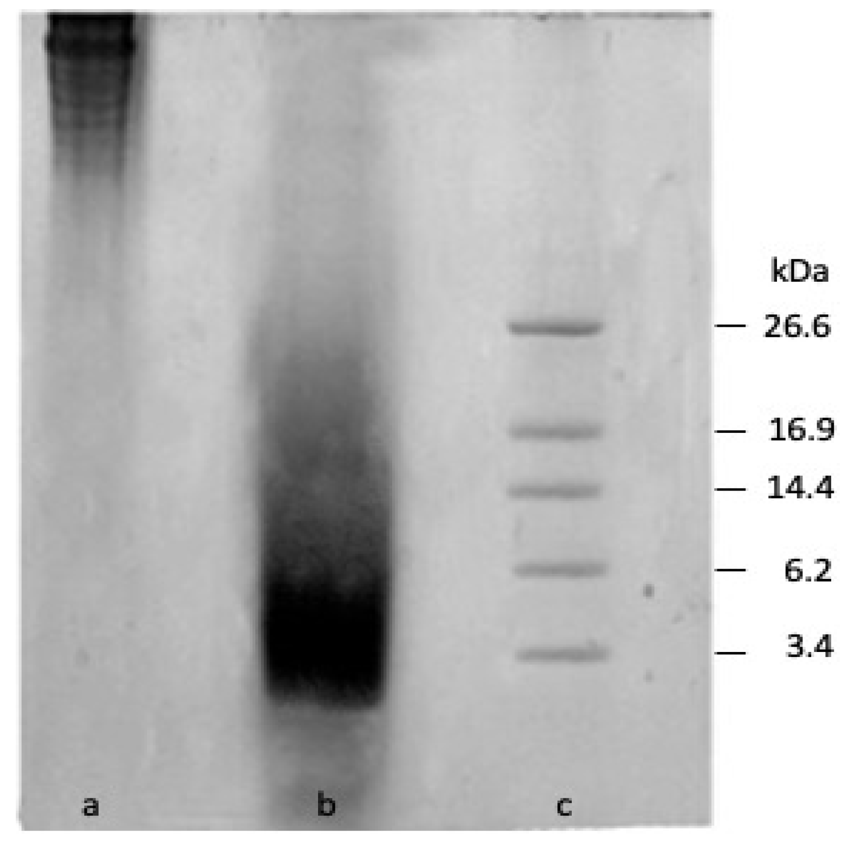
3.1. Characterisation of Gelatine Hydrolysate

The gelatin yield was 7.2 ± 1.5% (expressed as dry gelatin per 100 g of skin), and the percentage of true gelatin was 65.8 ± 2.2% with a DH of 25 ± 1.3%. These results agree with others obtained from the same squid species [28]. The obtained DH value was similar to previously reported values [13,26,27]. The proteolytic degradation of the squid skin gelatin after 60 min of hydrolysis was predominated by peptides smaller than 14 KDa (Figure 1). This behavior has previously been reported [16,29,30].
The obtained hydrolysates were rich in arginine, alanine, proline, and hydroxyproline. A similar amino acid composition was previously detected in squid hydrolysates [9,28,30]. These residues are expected to benefit the free radical scavenging and interaction of polypeptide chains with chitosan due to an increase in exposure to carboxylic acids as potential negative groups available for the polyelectrolyte complex. Moreover, the detection of hydrophobic amino acids of glycine (270.3/1000), alanine (129.6/1000), valine (22.6/1000), leucine (26.1/1000), isoleucine (15.3/1000), proline (109.9/1000), phenylalanine (19.9/1000), methionine (12.2/1000), and tryptophan (29.2/1000), as well as the presence of charged amino acids such as arginine (58.7/1000), histidine (9/1000), and lysine (14.7/1000), could stimulate the antifungal activity of the obtained hydrolysates [31].
The reduction of ABTS radicals in SGH was 118.3 ± 4.1 μM TE/mg protein, and the ORAC value was 253.1 ± 15.9 μM TE/mg. These values were higher than those previously reported for ascorbic acid (90.5 μM TE by ABTS and 40.4 μM TE by ORAC) and gallic acid (161 μM TE by ABTS and 111 μM TE by ORAC) but lower than that reported for β-carotene (240 μM TE by TEAC and 582 μM TE by ORAC) [32]. Therefore, the results suggest that squid skin hydrolysates have the capacity to scavenge free radicals, thus avoiding lipid oxidation through chain-breaking reactions [33], and the ability to donate a hydrogen atom to peroxyl radicals [34].

3.2. Properties of Films

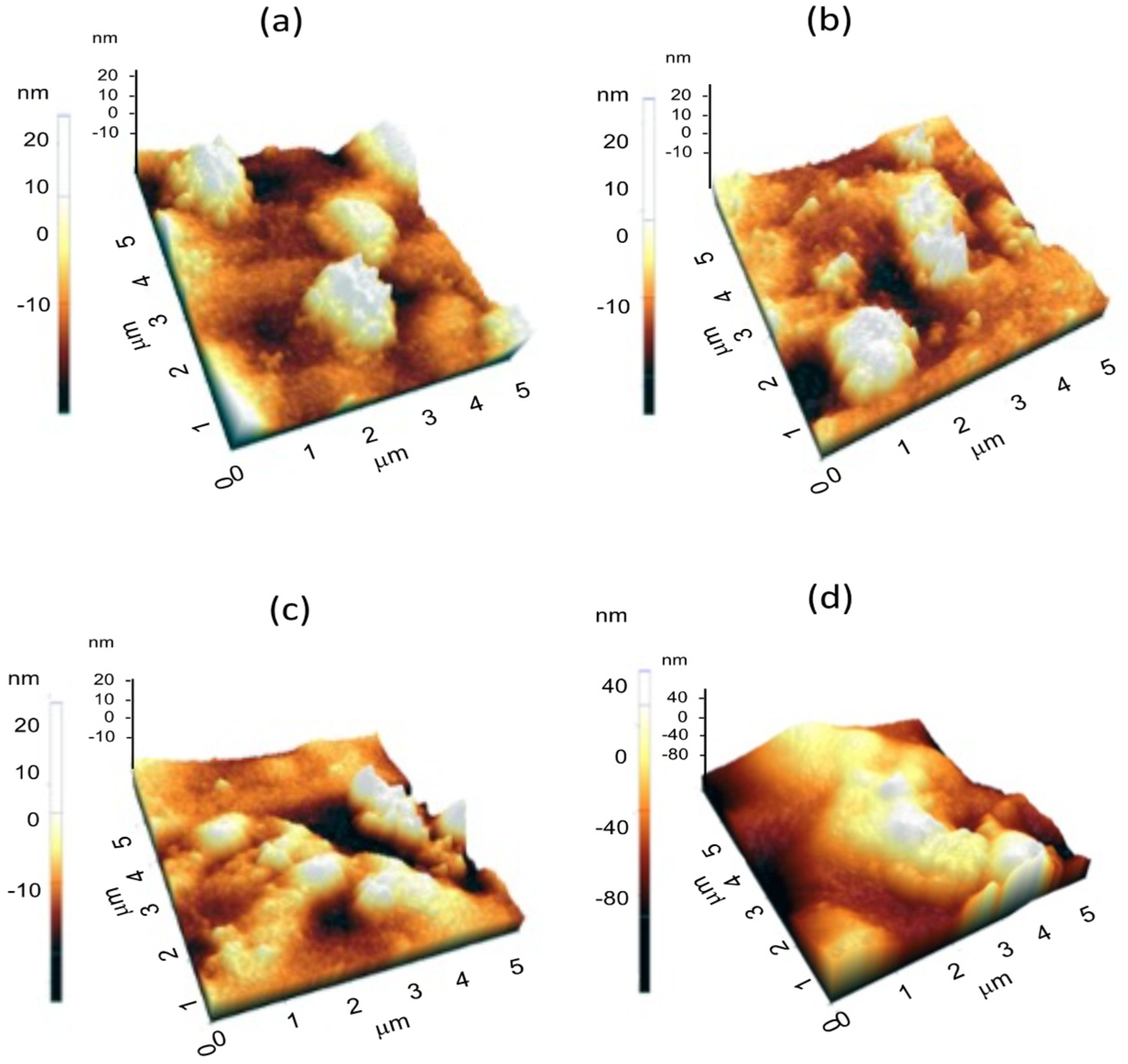
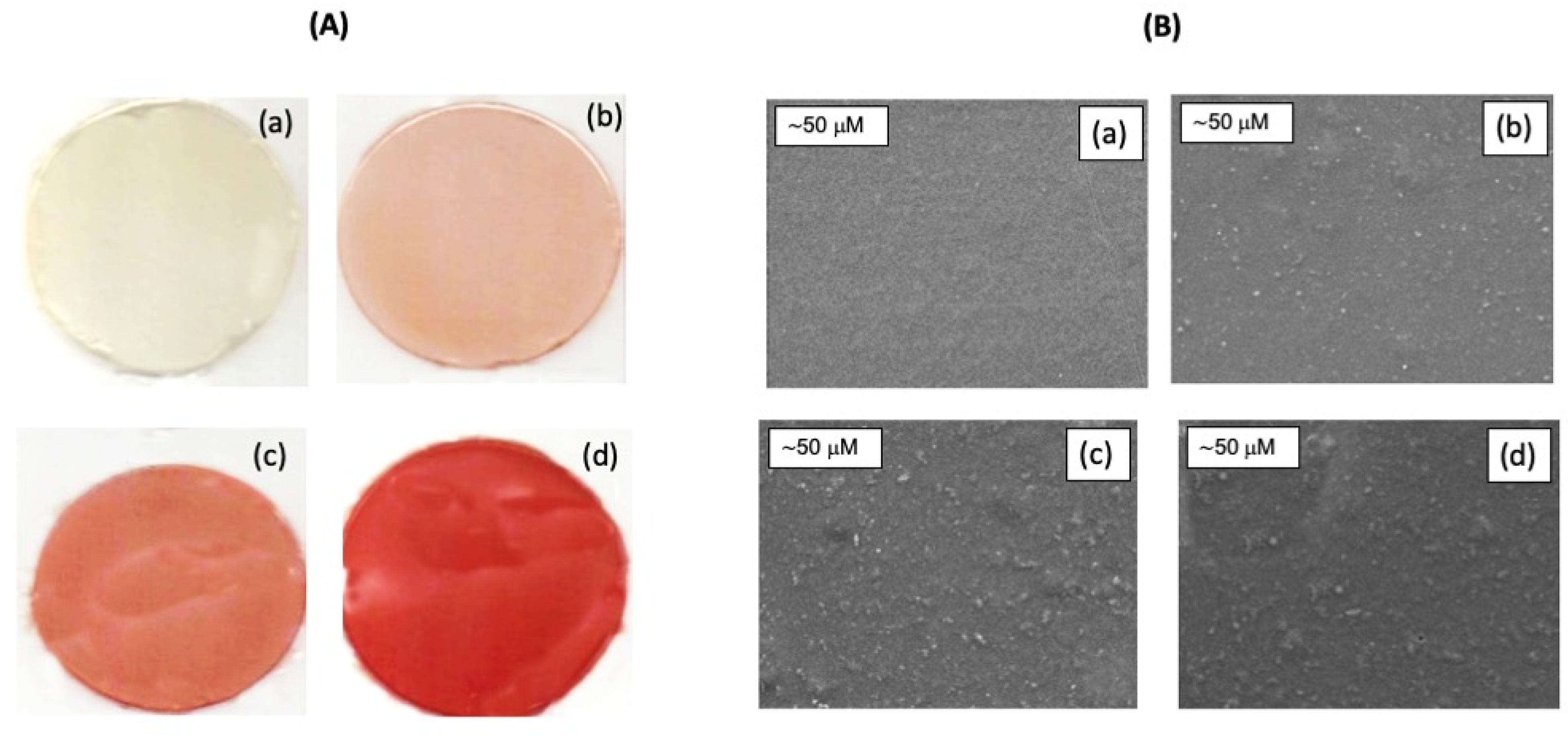
3.2.1. Color, SEM, and AFM

The film appearance, which influences the application of obtained films, can be observed in Figure 2A. The spectrophotometrically measured optical properties indicated that chitosan films with SGH incorporation were opaquer and more colored than the reference (Table 1). The increase in the opacity of the films could have been due to pigment substances present in squid skin [35] that remained in the gelatin after the hydrolysis process. The total color difference (ΔE*) increased significantly with an increase in SGH concentration, which indicated that the films with higher concentrations of SGH showed more red and less yellow color. The reddish color may be attributed to the presence of pigments in SGH, as well as the presence of amino groups in the peptides produced during the hydrolysis process, which may have interacted with the carbonyl groups of chitosan via the Maillard reaction (particularly during the drying of films), thus affecting the chitosan film’s color [36]. The pigmented molecules identified in squid skin belong to the ommochrome family [35]. Pigments from squid skin have been reported to contain active biological ommochromes with strong antioxidant and antimicrobial activity [8,35]. Therefore, the presence of pigments in SGH could enhance their bioactive properties.
Microstructure is another indicator of the characteristics of the obtained films. The cross-section morphology of film showed that the surface sections of the CH/SGH films were smooth and homogeneous, though a small amount of insoluble microparticles was observed (Figure 2B). Higher porosity and rougher surface values were detected in chitosan films with higher SGH contents (Figure 2A,B,b–d). This could be associated with the increase in the cross-linking via covalent and non-covalent bonding between hydrolysates and chitosan as the hydrolysates concentration increase, resulting in greater intermolecular aggregation and consequently producing some irregularities on the film’s surface [37]. Rough surfaces were detected in chitosan films with the addition of corn or casein peptides, and the films seemed to be fragmented when soy peptides were added [15]. These results suggest that squid gelatin hydrolysates interact well with chitosan. The roughness values estimated by the three-dimensional images of the film surfaces were obtained with AFM are shown in Table 2.
AFM (Figure 3) showed that the surface roughness of the chitosan films increased with the addition of SGH, which indicated closely spaced irregularities that imply that chitosan films with high concentrations of hydrolysates have weaker organizational structures.

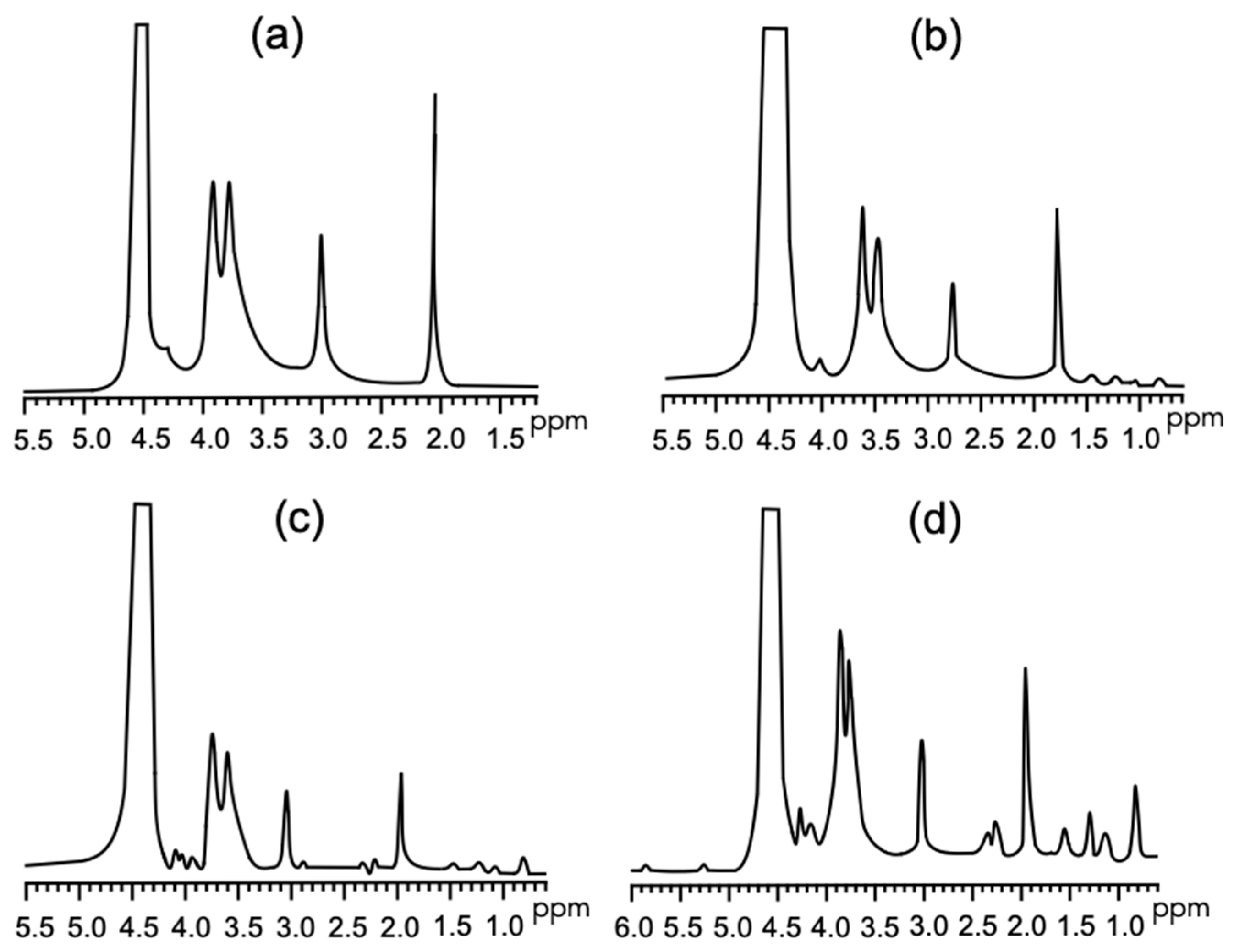
3.2.2. FT-IR and 1H NMR

The FT-IR spectra of the films containing 0%, 10%, 20%, and 40% SGH are shown in Figure 4. The spectrum of chitosan (Figure 4a) showed characteristic chitosan bands at around 3405, 1653, 1378, 1135, and 900 cm−1, which corresponded to the stretching vibrations of bonds of the amino group (NH), the stretching of the O–H links, the protonation of the amino groups of chitosan under acidic conditions, the stretching vibrations of the C–O primary alcohol group (–CH2OH), and sucrose glycoside linkage (C–O–C), respectively [11,15,27]. New signal bands were not observed in the chitosan/SGH films’ spectra (Figure 4b–d), indicating that no covalent bonds were formed. However, the displacement of peaks around 3400 and 1370 cm−1, as well as the modification of the band amplitude of the peak at around 1653 cm−1 that occurred as the amount of added SGH increased, suggests the occurrence of hydrogen bonding interactions among groups belonging to SGH (NH2, C=O, and OH) and chitosan (NH2 and OH).
The chemical interactions between chitosan and SGH were also analyzed by 1H NMR (Figure 5). The spectrum of chitosan (Figure 5a) showed the three protons of N-acetylglucosamine (GlcN) appeared at 1.89–2.05 ppm, whereas the peak at 3.01 ppm represents the H-2 proton of glucosamine; this indicates that chitosan was partially deacetylated (GlcNac). In the spectra region between 3.5 and 4 ppm, several bands and partially overlapping non-anomeric proton signals were observed. The band at 4.7 ppm indicates the proton of D2O [38]. The spectra in Figure 5b–d show the peaks of the chitosan film as a function of SGH content. The shifts of peaks from 1.89–2.05 to 2.88 ppm (GlcN) and from 3.01 to 3.46 ppm (GlcNac) indicate that the GlcN and GlcNac proton environments were modified due to chemical interactions, such as hydrogen bonding with SGH.

3.2.3. Thickness, Mechanical Properties and Enzymatic Degradation

Film thickness was in the range of 29.6 and 34.6 μm for all films (Table 3). The incorporation of SGH significantly decreases the thickness (p < 0.05) of chitosan films. The mechanical properties of the films are summarized in Table 3. The tensile strength (TS), elongation at break (EB), and elastic modulus (EM) decreased as the SGH content increased. The correlation between higher reductions of TS as larger additions of SGH may be attributed to a disruption in the interaction between chitosan molecules caused by the high MW compounds [14,39], and EB reduction led to a decrease in the free volume of the chitosan chains [40]. The film TS values were in the range of 77–47 MPa, which is comparable to low-density polyethylene food packaging. The EM values were like those of a chitosan film with a plasticizer (glycerol) [41], thus indicating that the addition of SGH to the chitosan films produced more flexible materials. Moreover, the incorporation of SGH induced an increase in film degradability, as measured by enzymatic degradation for 30 days (Table 3).

3.3. Antioxidant and Antifungal Activity

The three chitosan films containing SGH showed better scavenging activity against ABTS and DPPH radicals than that of chitosan films without SGH (Table 4). In both assays, the greatest activity (p < 0.05) was detected in the film with the highest SGH concentration. The ABTS assay indicated that the addition of SGH increased antioxidant activity by up to 900%, while the DPPH assay indicated an increase of up to 300% times. These results implied that SGHs were released from the composite to the medium and became available to exert antioxidant activity. Previous studies have been demonstrated that chitosan has the capacity to interact with free radicals through ionic interaction with its amino groups [3]. Moreover, different authors reported strong antioxidant activity of squid hydrolysates [8] studied by ABTS and DPPH tests. Li et al. observed that that the incorporation of casein peptides significantly increased the antioxidant activity of the composite films [15], possibly due to the presence of more amino groups that could donate more electrons.
The inhibitory effect of chitosan films on the growth of A. parasiticus was determined by measuring the radial growth (Table 4). The fungistatic activity of the chitosan films was not significantly improved with the addition of 10% SGH. Whereas the addition of 20% SGH enhance the antifungal activity of films. The fungistatic index increase by 34% (p < 0.05). However, films containing 40% of SGH colonies developed a higher radius than control (Table 4), which can be attributed to the weaker organizational structures detected in these films. Meanwhile, the higher fungistatic values can be due to the synergistic effect of chitosan and peptides [15] present in SGH, which limits the growth of fungus. It was observed that the fungus germinated in the absence of a chitosan film, and the development of hyphae was observed after 8 h of inoculation. The chitosan films inhibited the spores’ germinations by 12 h (Table 3), and hyphae development was not detected (Figure 6). The antifungal property of chitosan can be attributed to the presence of amino side groups that chelate calcium ions, which are essential minerals for fungus development [14,42]. The addition of 10% or 20% SGH to the chitosan films did not affect the fungistatic properties of A. parasiticus. Moreover, it was observed that chitosan films with 20% SGH induced a decrease in spore germination size. However, chitosan films with 40% SGH induced an increase in both fungus spores and hyphae size. This could have been because the films with higher SGH contents showed higher porosity and surface roughness values, as well as presenting weaker organization. It has been previously established that the loss of the physical integrity of a material can favor the transit of nutrients to spores and therefore facilitate the development of fungus [27].

4. Conclusions

The hydrolysates obtained from squid gelatin skin by enzymatic hydrolysis were incorporated into chitosan films to form chitosan/squid gelatin hydrolysates. The results indicated that the concentration of SGH affects the color, mechanical, microstructural, antioxidant, and antifungal against the toxigenic fungi Aspergillus parasiticus properties of chitosan films. The films with 40% SGH showed a redder color. SEM and AFM images indicated homogenous dispersion of CH/SGH films, though insoluble microparticles were observed. The formation of chemical bonds between chitosan and squid gelatin hydrolysates enhanced the intermolecular inter among the components, increasing stiffness and in vitro degradability. The antioxidant analysis proved that incorporation of SGH determined significant improvement of the film properties. Among the four films, 80 CH/20 SGH possessed better antifungal activities. Therefore, 80 CH/20 SGH can be suggested as a promising material for food packaging. Nevertheless, further research is needed, such as applying chitosan/squid gelatin hydrolysates films to cereals products and evaluating their control toxigenic fungi effectiveness.

Author Contributions

Formal analysis D.A.C.-A.; writing—original draft preparation D.A.C.-A.; conceptualization of the study J.M.E.-B.; helped with the proper establishment of the chemical structural analysis H.d.C.S.-O.; writing—review and editing the appropriate explanation of the chemical structural analysis H.d.C.S.-O.; supported with the establishment conditions of gelatin and hydrolysates productions W.T.-A.; writing—review and editing the appropriate explanation of the properties of the obtained gelatin and hydrolysates W.T.-A.; assisted with the chitosan films M.P.-J.; writing—review and editing the appropriate explanation of the microbiological results M.P.-J.; supervision and funding acquisition J.M.E.-B. All authors have read and agreed to the published version of the manuscript.

Funding

This research was funded by Consejo Nacional de Ciencia y Tecnología (CONACyT-Mexico), grant number 154046, and D.A.C.A. acknowledges a doctoral fellowship from CONACyT.

Institutional Review Board Statement

Not applicable.

Informed Consent Statement

Not applicable.

Data Availability Statement

Data supporting the reported results can be found at the following link: https://drive.google.com/drive/folders/1N8H6gPHSBYXX9uN9awC-rmX_Q63c5twq?usp=sharing (accessed on 8 September 2021).

Acknowledgments

The authors thank Alexel Burgara of the University of Sonora for his technical assistance.

Conflicts of Interest

The authors declare no conflict of interest.

Abbreviations

SGHsquid gelatin hydrolysates
CHfilms from chitosan
CH/SGHfilms from chitosan and squid gelatin hydrolysates
ABTS2,2′-azino-bis(3-ethylbenzothiazoline-6-sulfonate acid
DPPH1,1-diphenyl-2-picrylhydrazy
AAPH (2,2′-azobis-(2-amidinopropane) dihydrochloride))
Trolox(6-hydroxy-2,5,7,8-tetramethylchroman-2-carboxylic acid)
ORACoxygen radical antioxidant capacity

References

  1. Bhargava, N.; Sharanagat, V.S.; Mor, R.S.; Kumar, K. Active and intelligent biodegradable packaging films using food and food waste-derived bioactive compound: A review. Trends Food Sci. Technol. 2020, 105, 385–401. [Google Scholar] [CrossRef]
  2. Srinivasa, P.C.; Tharanathan, R.N. Chitin/chitosan-safe, ecofriendly packaging materials with multiple potential uses. Food Rev. Int. 2007, 23, 53–72. [Google Scholar] [CrossRef]
  3. Younes, I.; Rinaudos, M. Chitin and chitosan preparation from marine sources. Structure, properties and applications. Mar. Drugs 2015, 13, 1133–1174. [Google Scholar] [CrossRef] [PubMed] [Green Version]
  4. Yen, M.T.; Yang, J.H.; Mau, J.L. Antioxidant properties of chitosan from crab shells. Carbohydr. Polym. 2008, 4, 840–844. [Google Scholar] [CrossRef]
  5. Nur Hanani, Z.A.; Roos, Y.H.; Kerry, J.P. Use and application of gelatin as potential biodegradable packaging materials for food products. Int. J. Biol. Macromol. 2014, 71, 94–102. [Google Scholar] [CrossRef]
  6. Gómez-Guillen, M.C.; Giménez, B.; López-Caballero, M.E.; Montero, M.P. Functional and bioactive properties of collagen and gelatin from alternative sources: A review. Food Hydrocoll. 2011, 25, 1813–1827. [Google Scholar] [CrossRef] [Green Version]
  7. Tkaczewska, J. Peptides and protein hydrolysates as food preservatives and bioactive components of edible films and coatings-A review. Trends Food Sci. Technol. 2020, 106, 298–311. [Google Scholar] [CrossRef]
  8. Ezquerra-Brauer, J.M.; Aubourg, S.P. Recent trends for the employment of jumbo squid (Dosidicus gigas) by-products as a source of bioactive compounds with nutritional, functional and preservative applications: A review. Int. J. Food Sci. Technol. 2019, 54, 987–998. [Google Scholar] [CrossRef] [Green Version]
  9. Giménez, B.; Estaca, J.G.; Alemán, A.; Guillén, M.C.G.; Montero, M. Improvement of the antioxidant properties of squid skin gelatin films by the addition of hydrolysates from squid gelatin. Food Hydrocoll. 2009, 23, 1322–1327. [Google Scholar] [CrossRef] [Green Version]
  10. Nuanmanto, S.; Prodpran, T.; Benjakul, S. Potential use of gelatin hydrolysate as plasticizer in fish myofibrillar protein film. Food Hydrocoll. 2015, 47, 61–68. [Google Scholar] [CrossRef]
  11. de Morais Lima, M.; Bianchini, D.; Guerrra Dias, A.; da Rosa Zavareze, E.; Prentice, C.; da Silveira Moreira, A. Biodegrdable films based on chitosan, xanthan gum, and fish protein hydrolysate. J. Appl. Polym. Sci. 2017, 134, 44899. [Google Scholar] [CrossRef]
  12. da Roche, M.; Alemán, A.; Baccan, G.C.; López-Caballero, M.E.; Gómez-Guillen, C.; Montero, P. Anti-inflammatory, antioxidant, and antimicrobial effects of underutilized fish protein hydrolysate. J. Aquat. Food Prod. Technol. 2018, 27, 592–608. [Google Scholar] [CrossRef] [Green Version]
  13. Marroquín-Cardona, A.G.; Johnson, N.M.; Phillips, T.D.; Hayes, A.W. Mycotoxins in a changing global environment-A review. Food Chem. Toxicol. 2014, 69, 220–230. [Google Scholar] [CrossRef] [PubMed]
  14. Cota-Arriola, O.; Cortez-Rocha, M.O.; Burgos-Hernández, A.; Ezquerra-Brauer, J.M.; Plascencia-Jatomea, M. Controlled release matrices and micro/nanoparticles of chitosan with antimicrobial potential: Development of new strategies for microbial control in agriculture. J. Sci. Food Agric. 2013, 93, 1525–1536. [Google Scholar] [CrossRef] [PubMed]
  15. Li, C.; Pei, J.; Zhu, S.; Song, Y.; Xiong, X.; Xue, F. Development of chitosan/peptide films: Physical, antibacterial and antioxidant properties. Coatings 2020, 10, 1193. [Google Scholar] [CrossRef]
  16. Cuevas-Acuña, D.A.; Robles-Sánchez, R.M.; Torres-Arreola, W.; Márquez-Ríos, E.; Ezquerra-Brauer, J.M. Collagen from jumbo squid fin: Extracting conditions and influence of the protease system on collagen hydrolysate antioxidant activity. CyTA-J. Food 2016, 14, 193–199. [Google Scholar] [CrossRef] [Green Version]
  17. Adler-Nissen, J. Enzymatic Hydrolysis of Food Proteins, 1st ed.; Adler-Nisse, J., Ed.; Elsevier: New York, NY, USA, 1986; pp. 9–131. [Google Scholar]
  18. Vazquez-Ortiz, F.A.; Higuera-Ciapara, I.; Caire, G.; Hernández-Watanabe, G. High performance liquid chromatographic determination of free amino acids in shrimp. J. Liq. Chromatogr. 1995, 10, 2050–2068. [Google Scholar] [CrossRef]
  19. Laemmli, U.K. Cleavage of structural proteins during the assembly of the head of bacteriophage T4. Nature 1970, 227, 680–685. [Google Scholar] [CrossRef]
  20. Re, R.; Pellegrini, N.; Proteggente, A.; Pannala, A.; Yang, M.; Rice- Evans, C. Antioxidant activity applying an improved ABTS radical cation decolorisation assay. Free Radic. Biol. Med. 1999, 26, 1231–1237. [Google Scholar] [CrossRef]
  21. Prior, R.L.; Hoang, H.; Gu, L.; Wu, X.; Bacchiocca, M.; Howard, L.; Hampsch-Woodill, M.; Huang, D.; Ou, B.; Jacob, R. Assays for hydrophilic and lipophilic antioxidant capacity (oxygen radical absorbance capacity (ORACFL) of plasma and other biological and food samples. J. Agric. Food Chem. 2003, 5, 3273–3279. [Google Scholar] [CrossRef]
  22. Arias-Moscoso, J.L.; Soto-Valdez, H.; Plascencia-Jatomea, M.; Vidal-Quintanar, R.L.; Rouzaud-Sández, O.; Ezquerra-Brauer, J.M. Composites of chitosan with acid-soluble collagen from jumbo squid (Dosidicus gigas) by-products. Polym. Int. 2011, 60, 924–931. [Google Scholar] [CrossRef]
  23. Moreno-Osorio, L.; Garcia, M.; Villalobos-Carbajal, R. Effect of polygodial on mechanical, optical and barrier properties of chitosan films. J. Food Process. Preserv. 2010, 34, 219–234. [Google Scholar] [CrossRef]
  24. ASTM D882-10. Standard test methods for tensile properties of thin plastic sheeting. In Annual Book of American Society for Testing and Materials Standards; ASTM International: West Conshohocken, PA, USA, 2010. [Google Scholar] [CrossRef]
  25. Han, T.; Nwe, N.; Furuike, T.; Tokura, S.; Tamura, H. Methods of N-acetylated chitosan scaffolds and its in vitro biodegradation by lysozyme. J. Biomed. Sci. Eng. 2012, 5, 15–23. [Google Scholar] [CrossRef] [Green Version]
  26. Byun, Y.; Kim, Y.T.; Whiteside, S. Characterization of an antioxidant polylactic acid (PLA) film prepared with a-tocopherol, BHT and polyethylene glycol using film cast extruder. J. Food Eng. 2010, 100, 239–244. [Google Scholar] [CrossRef]
  27. Martínez-Camacho, A.P.; Cortez-Rocha, M.O.; Ezquerra-Brauer, J.M.; Rodríguez-Feliz, F.; Castillo-Ortega, M.M.; Yepiz-Gómez, M.; Plascencia-Jatomea, M. Chitosan chomposite films: Thermal, structural, mechanical and antifungal properties. Cabohydr. Polym. 2010, 82, 305–315. [Google Scholar] [CrossRef]
  28. Uriarte-Montoya, M.H.; Santacruz-Ortega, H.C.; Cinco-Moroyoqui, F.J.; Rouzaud-Sández, O.; Plascencia-Jatomea, M.; Ezquerra-Brauer, J.M. Giant squid gelatin: Chemical composition and biophysical characterization. Food Res. Int. 2011, 44, 3243–3249. [Google Scholar] [CrossRef]
  29. Suárez-Jiménez, G.M.; Robles-Sanchez, R.M.; Yepiz-Plascencia, G.; Burgos-Hernandez, A.; Ezquerra-Brauer, J.M. In vitro antioxidant, antimutagenic and antiproliferative activities of collagen hydrolysates of jumbo squid (Dosidicus gigas) byproducts. Food Sci. Technol. 2015, 35, 421–427. [Google Scholar] [CrossRef] [Green Version]
  30. Suárez-Jiménez, G.M.; Burgos-Hernández, A.; Torres-Arreola, W.; López-Saiz, C.M.; Velázquez-Contreras, C.A.; Ezquerra-Brauer, J.M. Bioactive peptides from collagen hydrolysates from squid (Dosidicus gigas) by-products fractionated by ultrafiltration. Int. J. Food Sci. Technol. 2019, 54, 1054–1061. [Google Scholar] [CrossRef]
  31. Gómez-Guillen, M.C.; López-Caballero, M.E.; Alemán, A.; López de Lacey, A.; Giménez, B.; Montero, P. Antioxidant and antimicrobial peptide fractions from squid and tuna skin gelatin. In Sea By-Products as Real Material: New Ways of Application; Le Bihan, A., Ed.; Transworld Research Network: Kerala, India, 2010; pp. 89–115. [Google Scholar]
  32. Zulueta, A.; Esteve, M.J.; Frígola, A. ORAC and TEAC assay comparison to measure the antioxidant capacity of food products. Food Chem. 2009, 114, 310–316. [Google Scholar] [CrossRef]
  33. You, L.; Zhao, M.; Regentein, J.M.; Ren, J. Changes in the antioxidant activity of loach (Misgurnusan guillicaudatus) protein hydrolysates during a simulated gastrointestinal digestion. Food Chem. 2010, 120, 810–816. [Google Scholar] [CrossRef]
  34. Prior, R.L.; Wu, X.; Schaich, K. Standardize method for the determination of antioxidant capacity and phenolics in foods and dietary supplements. J. Agric. Food Chem. 2005, 53, 4290–4302. [Google Scholar] [CrossRef] [PubMed]
  35. Chan-Higuera, J.E.; Carbonell-Barrachina, A.A.; Cárdenas-López, J.L.; Kačániová, M.; Burgos-Hernández, A.; Ezquerra-Brauer, J.M. Jumbo squid (Dosidicus gigas) skin pigments: Chemical analysis and evaluation of antimicrobial and antimutagenic potential. J. Microbiol. Biotechnol. Food Sci. 2019, 9, 349–353. [Google Scholar] [CrossRef]
  36. Xu, Z.Z.; Huang, G.Q.; Xu, T.C.; Liu, L.N.; Xiao, J.X. Comparative study of the Maillard reaction of chitosan oligosaccharide and glucose with soybean protein isolate. Int. J. Biol. Macromol. 2019, 131, 601–607. [Google Scholar] [CrossRef] [PubMed]
  37. Ocak, B. Film-forming ability of collagen hydrolysate extracted from leather solid wastes with chitosan. Environ. Sci Pollut. Res. Int. 2018, 25, 4643–4655. [Google Scholar] [CrossRef] [PubMed]
  38. Kassai, M.R. Determination of the degree of N-acetylation for chitin and chitosan by various NMR spectroscopy techniques: A review. Carbohyd. Polym. 2010, 79, 801–810. [Google Scholar] [CrossRef]
  39. Sothornvit, R.; Krochta, J.M. Oxygen permeability and mechanical properties of films from hydrolyzed whey protein. J. Agric. Food Chem. 2000, 48, 3913–3916. [Google Scholar] [CrossRef]
  40. Nogueira Campos, M.G.; Innocentini Mei, L.H.; Rodrigues Santos, A. Sorbitol-plasticized and neutralized chitosan membranes as skin substitutes. Mater. Res. 2015, 18, 781–790. [Google Scholar] [CrossRef] [Green Version]
  41. Lavorgna, M.; Piscitelli, F.; Mangiacapra, P.; Buonocore, G.C. Study of the combined effect of both clay and glycerol plasticizer on the properties of chitosan films. Carbohydr. Polym. 2010, 82, 291–298. [Google Scholar] [CrossRef]
  42. Sébastien, F.; Stéphane, G.; Copinet, A.; Coma, V. Novel biodegradable films made from chitosan and poly(lactic acid) with antifungal properties against mycotoxinogen strains. Carbohydr. Polym. 2006, 65, 185–193. [Google Scholar] [CrossRef]
Figure 1. Electrophoresis analysis of squid gelatin hydrolysates (Tricine-SDS-PAGE): a—gelatin, b—hydrolysates, and c—peptide molecular weight marker.
Coatings 11 01088 g001
Figure 2. Photograph (A) and SEM images of the morphologies of the chitosan (CH) and squid gelatin hydrolysates (SGH) films at 1000× magnification (B): (a) 100 CH/0 SGH, (b) 90 CH/10 SGH, (c) 80 CH/20 SGH, and (d) 60 CH/40 SGH.
Coatings 11 01088 g002
Figure 3. Images of the morphologies of chitosan (CH) and squid gelatin hydrolysates (SGH) films at 1000× magnification: (a) 100 CH/0 SGH, (b) 90 CH/10 SGH, (c) 80 CH/20 SGH, and (d) 60 CH/40 SGH.
Coatings 11 01088 g003
Figure 4. FT-IR spectra of chitosan (CH) and squid gelatin hydrolysates (SGH) films: (a)—100 CH/0 SGH, (b)—90 CH/10 SGH, (c)—80 CH/20 SGH, and (d)—60 CH/40 SGH.
Coatings 11 01088 g004
Figure 5. 1H NMR spectra of chitosan (CH) and squid gelatin hydrolysates (SGH) films: (a) 100 CH/0 SGH, (b) 90 CH/10 SGH, (c) 80 CH/20 SGH, and (d) 60 CH/40 SGH.
Coatings 11 01088 g005
Figure 6. Mycelium of Aspergillus parasiticus grown on chitosan (CH) and squid gelatin hydrolysates (SGH) films. At 12 h after incubation at 25 °C, at 40×: (a) 100 CH/0 SGH, (b) 90 CH/10 SGH, (c) 80 CH/20 SGH, and (d) 60 CH/40 SGH.
Coatings 11 01088 g006
Table 1. Color properties 1 of chitosan (CH) with squid gelatin hydrolysate (SGH) films.
Parameter100 CH/0 SGH90 CH/10 SGH80 CH/20 SGH60 CH/40 SGH
L79.7 ± 0.4 a31.6 ± 1.3 b16.4 ± 0.6 c13.5 ± 0.4 d
A−6.8 ± 0.2 c24.7 ± 0.7 a25.1 ± 0.3 a19.9 ± 0.3 b
B29.1 ± 1.6 a27.9 ± 0.9 a18.3 ± 0.5 b11.1 ± 0.2 c
ΔE*31.4 ± 1.0 c78.3 ± 1.2 b90.2 ± 1.3 a90.7 ± 0.8 a
1 Mean value ± standard deviation from ten separate samples. Mean values followed by different letters (a, b, c, d) indicate significant differences (p < 0.05).
Table 2. Roughness values estimated by the three-dimensional images of chitosan (CH) with squid gelatin hydrolysate (SGH) films surface obtained with AFM 1.
FilmAverage Roughness
100 CH/0 SGH3.11 ± 0.93 a
90 CH/10 SGH4.35 ± 1.11 ab
80 CH/20 SGH5.50 ± 0.05 c
60 CH/40 SGH8.38 ± 0.65 d
1 Mean value ± standard deviation from five separate samples. Mean values followed by different letters (a, b, c, d) indicate significant differences (p < 0.05).
Table 3. Thickness, tensile strength (TS), elongation at break (EB), elastic modulus (EM), and enzymatic degradation (ED) of chitosan (CH) with squid gelatin hydrolysate (SGH) films 1.
Parameter100 CH/0 SGH90 CH/10 SGH80 CH/20 SGH60 CH/40 SGH
Thickness (μM) 34.6 ± 1.1 a31.2 ± 1.3 b30.4 ± 1.5 b29.6 ± 1.6 b
TS (MPa)77.5 ± 5.7 a58.1 ± 5.3 b55.1 ± 2.3 b47.3 ± 5.7 c
EB (%)23.1 ± 3.0 a12.6 ± 1.9 b5.8 ± 1.0 c4.5 ± 1.2 c
EM (MPa)2625 ± 242 a2129 ± 141 b2392 ± 238 b1869 ± 145 c
ED (%) 287.2 ± 2.7 a72.8 ± 2.5 b56.4 ± 3.1 c35.8 ± 2.8 d
1 Mean value ± standard deviation from five separate samples. Mean values followed by different letters (a, b, c, d) indicate significant differences (p < 0.05). 2. Enzymatic degradation after 30 days.
Table 4. Antioxidant activity 1 and fungistatic activity 2 of chitosan (CH) with squid gelatin hydrolysate (SGH) films.
Assay100 CH/0 SGH90 CH/10 SGH80 CH/20 SGH60 CH/40 SGH
ABTS (μM TE/100 mg)3.3 ± 1.2 a30.1 ± 3.9 b57.7 ± 2.2 c88.9 ± 2.5 d
DPPH (%)2.8 ± 0.4 a6.1 ± 0.8 b6.6 ± 1.0 b,c8.7 ± 1.7 c
Spore size (μm)51 ± 2.5 b50 ± 2.2 b42 ± 2.6 c91 ± 9.0 a
Hyphae size (μm)NDNDND47.8 ± 5.9 a
Fungistatic index (%) 325 ± 3.9 b28 ± 1.6 b34 ± 3.1 a0 ± 0.4 c
1 Mean value ± standard deviation from five separate samples. Mean values followed by different letters (a, b, c, d) indicate significant differences (p < 0.05). 2. Fungistatic activity of films after 12 h. 3. Results are represented as percentage of inhibition of radial growth of control media (3.27 mm/h).
Publisher’s Note: MDPI stays neutral with regard to jurisdictional claims in published maps and institutional affiliations.

