Abstract
Background: Oral infections such as dental caries and candidiasis are mediated by resilient biofilms, which are increasingly tolerant to conventional antimicrobials. This study investigated the antimicrobial and antibiofilm effects of Salvia officinalis and Nigella sativa CO2 extracts against Streptococcus mutans and Candida albicans, with emphasis on synergistic interactions. Methods: Extracts were analyzed using gas chromatography–mass spectrometry analysis (GC–MS) and evaluated through planktonic MIC/MBC assays, time–kill kinetics, and biofilm models (MBIC/MBEC, biomass, metabolic activity). A novel three-dimensional checkerboard (3D-CB) and fractional inhibitory concentration index (FICI) approach was applied to optimize extract ratios, concentrations, and exposure times. Results: S. officinalis extract showed greater activity against S. mutans (MIC 256 mg/L; MBC 512 mg/L), while N. sativa was more effective against C. albicans (MIC 256 mg/L; MFC 512 mg/L). Both extracts reduced biofilm biomass and metabolic activity by over 70% at higher doses. Synergy was confirmed at ratios of 70:30 (S. officinalis: N. sativa) for S. mutans (FICI 0.38) and 40:60 for C. albicans (FICI 0.42). The achieved synergistic effect further decreased MBEC values fourfold and prolonged post-antibiotic effects. Conclusions: Synergistic S. officinalis–N. sativa formulations enhanced antimicrobial activity against oral pathogens in both planktonic and biofilm states, supporting their potential as next-generation dental antimicrobials.
1. Introduction
Oral diseases such as caries, gingivitis, and periodontitis remain persistent global challenges, driven by the activity of complex microbial biofilms that colonize hard and soft tissues in the oral cavity [,]. These conditions are not only highly prevalent but also strongly associated with systemic health risks, ranging from cardiovascular disease to diabetes. Current chemotherapeutic strategies, including chlorhexidine rinses, fluoride agents, and conventional antibiotics, are increasingly constrained by microbial tolerance, biofilm resilience, and undesirable side effects such as mucosal irritation and dysbiosis of the oral microbiota []. From a technological microbiology perspective, the development of new antimicrobial systems must therefore address two critical needs: selective inhibition of pathogenic microorganisms and modulation of microbial community dynamics without disrupting ecological balance [,].
Plant-derived antimicrobials have emerged as valuable tools in microbial control strategies, offering structural diversity and multifactorial mechanisms of action. Salvia officinalis (sage) is particularly noteworthy for its essential oils and polyphenolic compounds, which interact with microbial membranes and enzymatic systems, thereby suppressing growth of Streptococcus mutans, Lactobacillus spp., and other cariogenic organisms [,]. Beyond growth inhibition, S. officinalis derivatives exhibit capacity to interfere with quorum sensing and oxidative stress responses—two hallmarks of microbial survival within biofilms. This positions S. officinalis as a candidate for functional integration into dental care formulations designed to manage oral microbial ecosystems. In parallel, Nigella sativa (black cumin) has gained attention as a source of thymoquinone and related bioactives with strong antimicrobial and antibiofilm activities. Studies have shown that N. sativa disrupts extracellular polysaccharide synthesis and inhibits enzymes such as glucosyltransferase, thereby impairing the structural integrity of cariogenic biofilms [,]. From a microbiological engineering viewpoint, these properties are particularly significant because they target not only planktonic pathogens but also sessile microbial populations that drive chronic oral infections. Moreover, N. sativa derivatives demonstrate immunomodulatory and anti-inflammatory effects, supporting tissue healing in periodontitis and enhancing the overall therapeutic potential of botanical interventions [,].
While each extract demonstrates independent antimicrobial activity, the concept of phytochemical synergy provides a compelling technological solution. Synergy allows for reduced dosages of individual agents, mitigates cytotoxicity risks, and increases the probability of overcoming adaptive resistance mechanisms. In microbial systems, synergistic effects often arise when membrane-disrupting compounds facilitate intracellular penetration of secondary bioactives, or when multiple targets within biofilm regulation pathways are simultaneously inhibited. Preliminary evidence suggests that combinations of S. officinalis with other herbal extracts produce additive or synergistic inhibition of biofilm-forming pathogens []. By extension, pairing S. officinalis with N. sativa could provide a dual-mode antimicrobial platform: S. officinalis-derived terpenoids destabilizing microbial membranes and N. sativa-derived thymoquinone impairing EPS synthesis and enzymatic virulence. The application of pharmacodynamic modeling is essential in translating these observations into technological practice. Techniques such as checkerboard microdilution, time–kill kinetics, and isobologram analysis provide quantitative measures of interaction, enabling determination of fractional inhibitory concentration indices and dose–response relationships. Integrating these tools into dental microbiology research ensures that potential formulations are not only empirically active but also scientifically optimized for microbial control in the oral environment [].
In the search for safer and more sustainable alternatives to synthetic antimicrobials, plant-derived compounds, especially polyphenols and terpenoids—have gained renewed attention for their potential in oral health applications. A recent review by Arzani et al. [] highlights that phenolic compounds, terpenes, and terpenoids are the most abundant and pharmacologically active phytochemicals in medicinal plants with roles in preventing dental caries, gingivitis, and periodontitis, emphasizing their relevance to oral microbial ecology. In parallel, Touati et al. [] critically assessed the utility of essential oils for biofilm control, noting that synergy among volatile compounds, disruption of quorum sensing, and EPS degradation are key mechanisms by which complex phytochemical mixtures can outperform single compounds. Despite these advances, many studies remain limited to simple checkerboard assays and lack temporal dynamics or process reproducibility in extraction. Moreover, pharmacodynamic modeling of time–kill kinetics for natural antimicrobials is underexplored. Onita et al. [] emphasize the need to adapt PK/PD frameworks originally developed for antibiotics to novel compounds. Therefore, combining optimized extraction methods (e.g., supercritical CO2 co-extraction), multidimensional synergy screening, and pharmacodynamic modeling constitutes a timely approach in technological microbiology to move from “proof-of-concept” to scalable, reproducible formulations.
The objective of this study is therefore to investigate the pharmacodynamic interactions between S. officinalis and N. sativa extracts against key oral pathogens, with emphasis on growth inhibition, biofilm disruption, and potential synergism. By grounding this work in technological microbiology, we aim to provide a scientific framework for developing next-generation botanical antimicrobials that align with the dual demands of clinical efficacy and microbial ecosystem stewardship.
2. Results
2.1. Chemical Characterization of Plant Samples
The five most abundant compounds in S. officinalis and N. sativa CO2 extracts are presented in Table 1. In the S. officinalis CO2 extract, the dominant compounds were cis-thujone (19.9%), camphor (15.8%), trans-thujone (13.3%), and 1,8-cineole (11.3%), followed by camphene (6.3%) and 23 other compounds (Supplementary Table S1). In the N. sativa CO2 extract, the most abundant compound was p-cymene (47.2%), followed by cis-4-methoxythujane (7.9%), 2-butyl-2-octenal (6.0%), longifolene (5.4%), γ-terpinene (4.7%), and 50 additional compounds (Supplementary Table S2).
Table 1.
Top five identified compounds in plant extracts based on GC-MS analysis.
2.2. Antimicrobial Activity
2.2.1. Planktonic MIC and MBC Values
As can be seen in Table 2, both extracts exhibited inhibitory activity against S. mutans and C. albicans, although S. officinalis demonstrated lower MIC values against the bacterial strain, while N. sativa showed higher potency against the yeast. As expected, chlorhexidine and amoxicillin exhibited potent antibacterial activity against S. mutans with MIC and MBC values of 2 and 4 mg/L, and 2 and 3 mg/L, respectively, which were significantly lower than those of plant extracts. Similarly, nystatin (MIC 2 mg/L; MFC 4 mg/L) and fluconazole (MIC 1 mg/L; MFC 2 mg/L) showed strong fungicidal activity against C. albicans. While the CO2 extracts required higher concentrations to achieve similar effects, their selective and synergistic actions remain noteworthy in the context of natural product development.
Table 2.
Minimum inhibitory concentration (MIC), minimum bactericidal concentration (MBC), and minimum fungicidal concentration (MFC) values of Salvia officinalis and Nigella sativa CO2 extracts and positive controls against oral pathogens (Streptococcus mutans and Candida albicans). Values are expressed in mg/L.
2.2.2. Time–Kill Kinetics
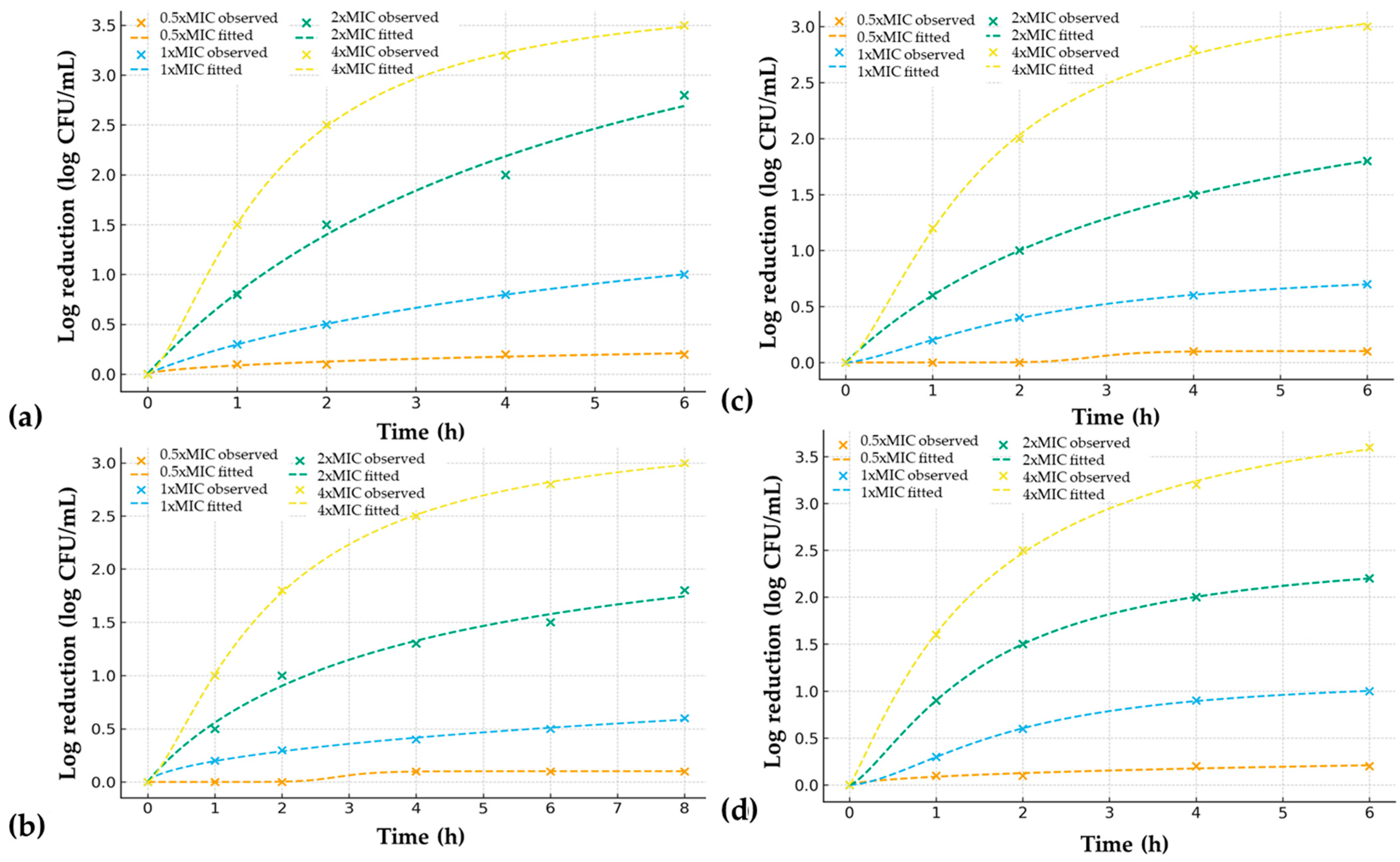
The time–kill assays confirmed the concentration- and time-dependent antimicrobial action of both extracts (Table 3, Figure 1). At 1× MIC, both extracts achieved bacteriostatic or fungistatic activity, while 2× MIC resulted in significant CFU reductions. The reduction of 99.9% (>3 log10 reduction) was achieved after 6 h with S. officinalis against S. mutans and after 8 h with N. sativa against C. albicans. At 4× MIC, both extracts demonstrated rapid bactericidal/fungicidal effects within 2–4 h. The post-antibiotic effect (PAE) revealed a regrowth delay of 2.5 h for S. mutans and 1.5 h for C. albicans following 1 h exposure to S. officinalis at 2× MIC. For N. sativa, the PAE was 2.0 h for C. albicans and 1.0 h for S. mutans under the same conditions. At the 24 h sampling point, no detectable CFU were observed for S. mutans and C. albicans treated with ≥2× MIC of either extract, confirming complete microbial elimination by the end of the assay.
Table 3.
Time–kill kinetics and post-antibiotic effect (PAE) of S. officinalis and N. sativa CO2 extracts against S. mutans and C. albicans. Values represent log10 CFU reductions at each time point (0–8 h) for the indicated extract concentration (0.5×–4× MIC). PAE (h) indicates the post-antibiotic effect, defined as the delay in logarithmic regrowth between treated and untreated cultures following 1 h exposure at 2× MIC (Section 4.4).
Figure 1.
Time–kill kinetics of extracts against oral pathogens. Observed data points (dots) and fitted sigmoidal (lines) curves are shown for (a) S. officinalis versus S. mutans; (b) S. officinalis versus C. albicans; (c) N. sativa versus S. mutans; and (d) N. sativa versus C. albicans.
The dynamic effects of S. officinalis and N. sativa CO2 extracts on planktonic growth were further examined using time–kill assays modeled with a four-parameter logistic function (Figure 1). Both extracts exhibited clear concentration- and time-dependent killing profiles against S. mutans and C. albicans. For S. mutans, exposure to S. officinalis resulted in rapid and pronounced activity, with reduction of 99,9% within 6 h at 4× MIC and a t50 of approximately 1.35 h. At 2× MIC, bactericidal levels (>3 log10 reduction) were reached by 5.17 h, while sub-MIC exposure produced minimal inhibition. N. sativa also demonstrated bactericidal activity, although the onset was slightly slower (t50 ≈ 4 h at 2× MIC), consistent with the higher MIC/MBC values observed in Table 2. Against C. albicans, the activity profiles were reversed. N. sativa exhibited superior fungicidal kinetics, reducing viable counts below detection within a few hours at 4× MIC (t50 ≈ 1.52 h at 2× MIC), whereas S. officinalis required at least double time to achieve comparable effects at 2× MIC. Both extracts showed concentration-dependent post-antifungal effects, with delayed regrowth observed after removal of the extract at 2× MIC. Overall, the sigmoidal fits confirmed that the antimicrobial action of both extracts followed steep, dose-dependent trajectories, reflected by Hill slopes > 2 at bactericidal/fungicidal concentrations. These findings corroborate the MIC/MBC results and highlight distinct pharmacodynamic profiles: S. officinalis exerted faster bactericidal action against S. mutans, while N. sativa displayed stronger fungicidal activity against C. albicans.
To further quantify the pharmacodynamic characteristics of the observed killing curves, time–kill data were fitted to a four-parameter logistic model. The derived kinetic parameters, including t50 (time to half-maximal effect), Hill slope (steepness of response), and residual statistics (R2, RMSE, MAE, SSres), are summarized in Table 4. These values provide a quantitative basis for comparing the bactericidal and fungicidal profiles of S. officinalis and N. sativa, complementing the visual assessment of time–kill curves in Figure 1. Time–kill kinetics of S. mutans and C. albicans exposed to extracts were fitted using a four-parameter sigmoidal model. The experimental data aligned well with the logistic function, with coefficients of determination (R2) ranging from 0.946 to 0.99 across all treatments, confirming the suitability of the model despite the limited number of time points (Figure 1). For S. mutans, S. officinalis displayed concentration-dependent killing, with t50 decreasing from 8.32 h at 1× MIC to 5.17 h at 2× MIC and 1.35 h at 4× MIC. Complete kill (≥3 log10 CFU reduction) was observed within 6 h at 4× MIC. In contrast, N. sativa required shorter exposure, with t50 values of 2.22 h at 1× MIC and 4 h at 2× MIC, but still achieved complete bactericidal activity at 4× MIC within a few hours.
Table 4.
Kinetic modeling parameters and verification statistics for time–kill kinetics of Salvia officinalis and Nigella sativa CO2 extracts against S. mutans and C. albicans.
Against C. albicans, N. sativa showed superior antifungal kinetics compared with S. officinalis. The N. sativa extract achieved t50 = 1.52 h at 2× MIC and 1.57 h at 4× MIC. In comparison, S. officinalis required longer exposure. Hill slope values reflect the extracts’ sharp transition between sublethal and lethal concentrations, a characteristic often associated with membrane-active natural products. Overall, the kinetic modeling corroborated the MIC/MBC results: S. officinalis was more potent against S. mutans, while N. sativa was more effective against C. albicans. Importantly, the sigmoidal model provided additional quantitative descriptors enabling comparison of pharmacodynamic profiles across treatments.
2.3. Biofilm Assays
For S. mutans, S. officinalis exhibited an MBIC of 512 mg/L and an MBEC of 1024 mg/L, whereas chlorhexidine and amoxicillin inhibited biofilm formation at 4 mg/L and 3 mg/L and eradicated biofilms at 8 mg/L and 7 mg/L, respectively (Table 5). Biomass inhibition by S. officinalis reached 72 ± 4% at 2× MBIC, while controls achieved >90% inhibition at 2× MBIC. EPS quantification confirmed that S. officinalis reduced total carbohydrate content by 60 ± 5%, with insoluble glucans reduced by 55 ± 4%, suggesting interference with GtfB/C-mediated glucan synthesis.
Table 5.
Biofilm inhibitory and eradication activity of Salvia officinalis and Nigella sativa extracts compared with chlorhexidine, nystatin, and fluconazole (mean ± SD).
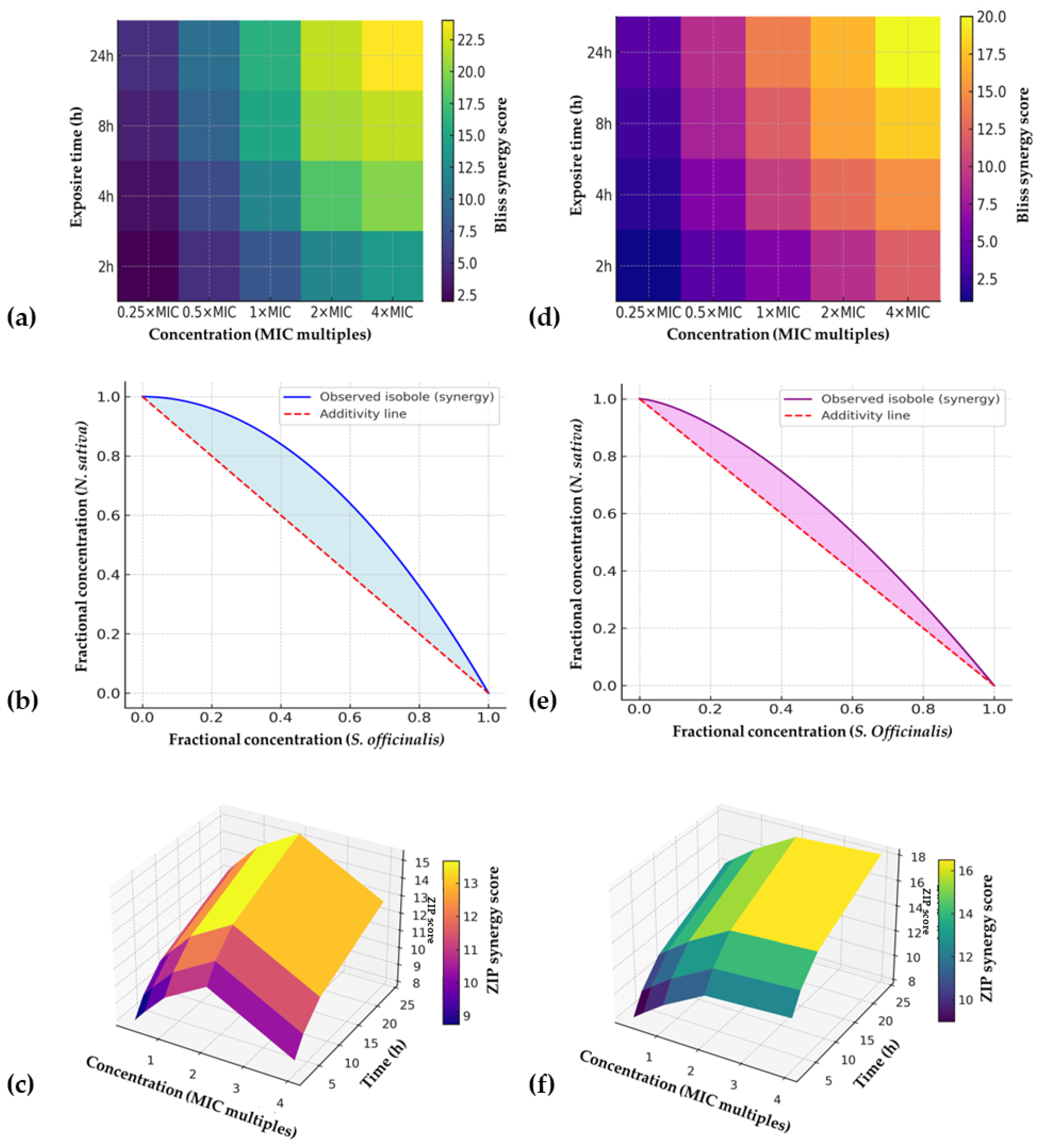
For C. albicans, N. sativa displayed an MBIC of 512 mg/L and MBEC of 1024 mg/L, compared with nystatin (MBIC 2 mg/L; MBEC 4 mg/L) and fluconazole (MBIC 1 mg/L; MBEC 2 mg/L). N. sativa extract reduced biofilm biomass by 68 ± 5% and metabolic activity by 71 ± 4% at 2× MBIC, while nystatin achieved >90% reductions. As the assessment of biofilm biomass inhibition rate, crystal violet staining assays confirmed dose-dependent biomass reduction. At 2× MBIC, S. officinalis inhibited S. mutans biofilm formation by 72 ± 4%, while N. sativa reduced biomass by 55 ± 6%. Conversely, for C. albicans, N. sativa achieved stronger inhibition (68 ± 5%) compared with S. officinalis (50 ± 7%). Treatment of 48 h pre-formed biofilms revealed partial to substantial eradication depending on concentration and exposure time (Figure 2). At 2× MBEC, S. officinalis eradicated 74 ± 3% of S. mutans biomass, with a 2.8 log10 CFU/mL reduction, while N. sativa achieved 63 ± 4% biomass removal and a 2.2 log10 CFU/mL reduction. For C. albicans, N. sativa produced a 70 ± 5% eradication and 3.0 log10 CFU/mL reduction, whereas S. officinalis yielded 58 ± 6% biomass removal and 2.0 log10 CFU/mL reduction. Similarly, resazurin assays showed that metabolic activity was significantly impaired in both microbial biofilms. At 2× MBIC, S. mutans biofilms treated with S. officinalis exhibited a 65 ± 5% reduction in metabolic activity, compared with 48 ± 6% for N. sativa. In contrast, C. albicans biofilms treated with N. sativa showed a 71 ± 4% reduction, compared with 52 ± 5% for S. officinalis.
Figure 2.
Synergy mapping of S. officinalis and N. sativa extracts against oral pathogens using the three-dimensional checkerboard approach. (a) Bliss synergy heatmap for S. mutans (70:30 ratio) showing concentration–time–response surfaces; (b) Loewe isobologram for S. mutans, demonstrating concave deviation below the additivity line consistent with synergy; (c) ZIP synergy surface for S. mutans, integrating concentration and exposure time effects; (d) Bliss synergy heatmap for C. albicans (40:60 ratio); (e) Loewe isobologram for C. albicans; (f) ZIP synergy surface for C. albicans. Note: Synergy was defined as FICI ≤ 0.5 and ZIP synergy score > 10. Heatmaps and surfaces represent mean synergy scores from three independent experiments (n = 3) with 95% confidence intervals.
2.4. Three-Dimensional Checkerboard Synergy Optimization
The three-dimensional checkerboard approach revealed pronounced synergy between S. officinalis and N. sativa extracts, particularly against S. mutans. At a 70:30 ratio, synergy was evident across exposure times, with FICI values of 0.31 ± 0.05 and ZIP synergy scores of 15.2 ± 2.1. Bliss scores confirmed that observed inhibitory effects exceeded additive predictions by 18–22%. Loewe isobolograms demonstrated concave curves, further supporting synergy. Against C. albicans, synergy was strongest at a 40:60 ratio (FICI 0.38 ± 0.06; ZIP 12.8 ± 1.9), with Bliss excess up to 16%. In contrast, ratios skewed heavily toward one extract often yielded additive or indifferent effects. Importantly, no antagonism was detected at any ratio, concentration, or time point.
2.5. Pharmacodynamic Synergy
The three-dimensional checkerboard experiments revealed that the combined use of S. officinalis and N. sativa extracts produced markedly stronger antimicrobial effects against both S. mutans and C. albicans than when applied individually (Table 6, Figure 2). For S. mutans, the most pronounced activity was observed at a 70:30 ratio of S. officinalis to N. sativa extract where the minimum inhibitory concentration decreased from 256 mg/L and 512 mg/L for the single extracts, respectively, to only 128 mg/L in combination. A similar trend was recorded for C. albicans, where a 40:60 ratio provided the optimal outcome, lowering the MIC from 512 mg/L for S. officinalis and 256 mg/L for N. sativa to 128 mg/L for the mixture. In both cases, the combinations also shortened the time required to achieve complete killing; S. mutans biofilms were eradicated within 4 h at 2× MIC of the optimized blend, compared with 6 h for the S. officinalis extract alone, while C. albicans biofilms were eliminated within 6 h compared with 8 h for N. sativa alone.
Table 6.
Synergistic effects of S. officinalis-N. sativa extracts in 3D checkerboard optimization.
Importantly, post-antibiotic effects were extended following exposure to the optimized mixtures. For S. mutans, regrowth was delayed by 3.5 h, while C. albicans showed a delay of 3.0 h, with both values exceeding those of the individual extracts. In parallel, the optimization of co-extraction parameters using supercritical CO2 demonstrated that a process carried out at 35 MPa and 45 °C with 5% ethanol as a co-solvent and a 60:40 feed ratio (S. officinalis–N. sativa) produced an extract with superior activity. This optimized co-extract displayed MIC values of 128 mg/L against S. mutans and 96 mg/L against C. albicans, while MBEC values were reduced fourfold compared with single extracts. Biofilm biomass was eradicated by more than 80% in both microorganisms, and metabolic activity was suppressed by more than 75%, indicating not only structural but also functional impairment of biofilms. Pharmacodynamic modeling provided consistent evidence of synergy. The fractional inhibitory concentration indices were 0.38 for S. mutans and 0.42 for C. albicans, both well below the threshold of 0.5 that indicates synergism. Bliss independence analysis confirmed these findings, with synergy scores of +22% for bacteria and +25% for yeast, while the ZIP model returned positive synergy scores of +15 and +18, respectively. Loewe additivity surfaces visualized these interactions as clear synergy ridges, validating the presence of cooperative effects between the two plant extracts. Overall, the 3D checkerboard approach demonstrated that both blending and optimized co-extraction strategies significantly enhanced antimicrobial efficacy, positioning these plant-derived mixtures as strong candidates for next-generation oral antimicrobials.
3. Discussion
This study confirmed that S. officinalis and N. sativa CO2 extracts display distinct but complementary antimicrobial effects against S. mutans and C. albicans. The planktonic results demonstrated that S. officinalis was more effective against S. mutans, while N. sativa exerted stronger antifungal activity. This pattern is supported by prior research on S. officinalis terpenoids, including thujones, camphor, and cineole, which have been associated with membrane disruption and enzyme inhibition in Gram-positive bacteria []. Conversely, the antifungal activity of N. sativa has been consistently linked to thymoquinone, anethole, and carvacrol, which interfere with ergosterol biosynthesis and oxidative stress responses in Candida spp. []. However, discrepancies in reported MIC values across studies must be noted. Beheshti-Rouy et al. [] reported strong S. mutans inhibition using a S. officinalis mouthwash, while Ntondini et al. [] found S. officinalis essential oil active on titanium surfaces at considerably lower MICs than those reported for crude CO2 extracts, highlighting that extraction method and phytochemical profile strongly influence potency. The MIC/MBC values of chlorhexidine/amoxicillin and nystatin/fluconazole were, as expected, an order of magnitude lower than those of S. officinalis and N. sativa. This disparity is consistent with their established role as gold-standard synthetic antimicrobials in oral care [,]. However, the plant extracts offer distinct advantages such as lower cytotoxicity risks at optimized ratios, potential for reduced resistance pressure, and the capacity for synergy. Thus, although the potency of crude extracts is lower than that of synthetic controls, the pharmacodynamic benefits of synergy and co-extraction strategies strengthen their candidacy for next-generation dental formulations [].
The time–kill and PAE assays reinforced the concentration- and time-dependent action of both extracts. Although modest, PAEs of 1–3.5 h are significant in oral care contexts where dosing intervals and contact times are limited. Comparable findings were reported for thymoquinone and N. sativa oil in synergy with antibiotics, where enhanced killing curves were observed in time–kill models []. However, few studies have explicitly measured PAE for botanical antimicrobials, making our results novel but requiring careful interpretation. In vivo, salivary washout and dynamic microbial competition may reduce apparent PAE durations. The biofilm assays demonstrated that both extracts inhibited biomass formation and partially eradicated mature biofilms. S. officinalis was more effective against S. mutans, while N. sativa displayed stronger antibiofilm activity against C. albicans. Previous reports align with this trend: S. officinalis essential oils caused biofilm structural disruption on dental materials [], while N. sativa seed oil impaired EPS production and reduced oral biofilm stability []. Nonetheless, the obtained MBEC values remained higher than planktonic MICs, a well-recognized phenomenon reflecting the tolerance of sessile cells []. Importantly, the discrepancy between biomass and CFU reduction observed here suggests that extracts may preferentially target EPS structure or metabolically active cells, consistent with dual modes of action proposed for phytochemicals. The biofilm assays highlighted the superior efficacy of synthetic comparators, as expected, but also confirmed meaningful activity of CO2 extracts at higher concentrations. Chlorhexidine/amoxicillin completely suppressed S. mutans biofilm biomass and glucan synthesis at single-digit mg/L concentrations, whereas S. officinalis achieved >70% biomass inhibition and ~60% reduction in EPS and insoluble glucans at ≥512 mg/L. This suggests that S. officinalis interferes with GtfB/C-mediated extracellular glucan synthesis, a critical virulence mechanism of S. mutans, in line with previous findings that phytochemicals can downregulate gtf genes or destabilize EPS matrices [,]. Similarly, while nystatin and fluconazole eradicated C. albicans biofilms at low mg/L, N. sativa reduced biomass and metabolic activity by ~70%, consistent with reports that thymoquinone and phenolic compounds impair fungal adhesion and hyphal development [,]. These data reinforce the concept that plant extracts are less potent but mechanistically complementary, and their synergy could be exploited to reduce reliance on high-dose synthetic agents.
The most innovative aspect of this study was the application of a three-dimensional checkerboard optimization. Unlike conventional two-dimensional designs, this approach incorporated ratio, concentration, and exposure time, enabling the identification of synergy “hot spots.” Optimal ratios (70:30 for S. mutans and 40:60 for C. albicans) consistently yielded FICI values below 0.5, supported by positive Bliss and ZIP scores, and synergy ridges on Loewe surfaces. Comparable synergistic effects have been reported for essential oil mixtures [,,], but rarely with the added temporal dimension. Mechanistically, the synergy likely arises from membrane perturbation by S. officinalis terpenoids facilitating the penetration of N. sativa phenolics, or complementary inhibition of bacterial and fungal survival pathways. Previous work has documented similar cooperative effects between thymoquinone and antibiotics []. The optimized co-extraction (35 MPa, 45 °C, 5% ethanol, 60:40 feed) further validated the technological feasibility of producing a single, standardized product with enhanced antimicrobial activity. Reduced MBEC values and improved eradication rates (>80%) emphasize the practical relevance of process optimization. However, variability in raw plant material, potential antagonistic effects at untested ratios, and the gap between in vitro and in vivo conditions must be addressed before clinical application. Additionally, polymicrobial oral biofilms and host factors may substantially alter efficacy, as highlighted in broader oral microbiome studies [,]. These synergy findings extend prior reports of additive or synergistic effects of plant extracts [,]. Unlike conventional two-dimensional checkerboards, the inclusion of time as a third axis allowed dynamic evaluation of pharmacodynamics, revealing that synergy was most consistent at intermediate exposure times. The agreement across models strengthens the robustness of the synergy claim, particularly since multiple frameworks were used and supported by bootstrap confidence intervals. Importantly, our predefined thresholds minimized the risk of overinterpreting statistical noise as synergy. Mechanistically, synergy likely results from S. officinalis terpenoids perturbing cell membranes, facilitating penetration of thymoquinone and other N. sativa compounds that target intracellular pathways [].
The synergistic behavior we observed between S. officinalis and N. sativa extracts aligns with the emerging concept that complex phytochemical mixtures can yield supra-additive effects through complementary modes of action. Touati et al. [] summarized multiple instances where essential oils act synergistically by disrupting cell membranes, suppressing quorum sensing, degrading EPS, and facilitating penetration of secondary compounds. In a broader context, Rasheed et al. [] reviewed recent advances in plant-based antimicrobials, noting that extraction methods and synergistic fractionation are increasingly recognized as pivotal for maximizing bioactivity. From a pharmacodynamic modeling perspective, our use of a sigmoidal four-parameter model provides quantitative descriptors (t50, Hill slope) that can be compared across conditions. While antibiotic PK/PD modeling is well established, there is increasing recognition of its application to natural compounds [,,], to inform dose/exposure relationships. Our results thus contribute to bridging this methodological gap. Importantly, from a technological microbiology viewpoint, the reproducibility and standardization of botanical extracts is often undervalued. Variability in plant origin, harvest season, extraction conditions, and co-extraction ratios can dramatically affect phytochemical profiles. The presented co-extraction optimization (35 MPa, 45 °C, 5% ethanol) aims to produce a more consistent synergy-enhanced extract batch to batch. As Sun et al. [] observed, phytochemical strategies must address not only potency but process reproducibility to facilitate translation. Translating such extraction protocols into industrial scales, while maintaining synergy, is a key engineering challenge in the development of next-generation dental antimicrobials.
These findings support the working hypothesis that phytochemical synergy between S. officinalis and N. sativa extracts can be exploited for dental antimicrobial applications. They also extend the broader understanding of natural antimicrobials by demonstrating that rational, multidimensional optimization strategies can yield superior efficacy compared with single extracts or simple blends. Future research should focus on validating these results in multispecies biofilm models, performing mechanistic omics-based studies, and translating optimized co-extracts into delivery formats such as oral rinses or implant coatings. Although this study demonstrates the promising synergistic potential of S. officinalis and N. sativa CO2 extracts against S. mutans and C. albicans, several limitations must be acknowledged. First, only mono-species planktonic and static biofilm models were tested. Oral infections are typically polymicrobial, with complex cross-feeding, competition, and quorum-sensing interactions; thus, the activity observed here may differ in multispecies biofilms. Second, we focused on standard laboratory strains rather than recent clinical isolates, which may vary in susceptibility. Third, the study did not address host-related variables such as salivary proteins, pH cycling, or shear stress, all of which can influence antimicrobial efficacy in vivo. Fourth, cytotoxicity and selectivity data remain preliminary, and further testing on oral keratinocytes, fibroblasts, and commensal microbiota is needed to establish safety. Finally, batch-to-batch variability of botanical extracts poses challenges for reproducibility, and chemical stability during storage and formulation was not investigated. Future research should therefore prioritize: (i) validation in multispecies plaque biofilm models that more closely mimic clinical conditions; (ii) ex vivo studies using saliva-coated hydroxyapatite or enamel specimens; (iii) mechanistic investigations using transcriptomics, proteomics, and EPS structural analysis to clarify modes of action; (iv) cytotoxicity, hemolysis, and selectivity assays to assess host safety; (v) stability and standardization studies to ensure reproducibility; and (vi) preclinical in vivo studies or pilot clinical trials to evaluate translational potential. Integration of these steps will help bridge the gap between in vitro efficacy and clinical application, moving toward evidence-based development of plant-derived, synergy-optimized dental antimicrobials.
4. Materials and Methods
4.1. Materials
The commercial CO2 extracts of S. officinalis and N. sativa (Alekpharm Ltd., Belgrade, Serbia) were used in this study. Their volatile profiles were evaluated using gas chromatography–mass spectrometry (GC–MS) with a coupled system consisting of an Agilent 7890 GC, an Agilent 5975C mass selective detector (MSD), a flame ionization detector (FID), and an HP-5MS column (Agilent 19091S-433) (Agilent Technologies, Santa Clara, CA, USA). Compound identification was based on linear retention indices (RI) relative to C8–C32 n-alkanes and comparison with reference spectra from the Wiley 7, NIST 17, and Adams databases. The relative percentages of volatile compounds were determined by FID peak area normalization.
4.2. Microorganisms and Culture Conditions
Reference oral pathogens were selected to represent bacterial and fungal challenges: Streptococcus mutans ATCC 25175 (caries-associated, from carious dentine) and Candida albicans ATCC 64124 (oral candidiasis isolate). S. mutans was cultured in brain–heart infusion (BHI) broth supplemented with 5% horse serum at 37 °C under aerobic conditions, while C. albicans was maintained in Sabouraud dextrose broth at 30 °C with shaking. Cultures were standardized to mid-logarithmic phase (OD600 ≈ 0.1, ~106 CFU/mL) prior to antimicrobial testing.
4.3. Antimicrobial Susceptibility Testing
Antimicrobial efficacy of the extracts was evaluated by agar well diffusion and broth microdilution. Using agar diffusion technique, standardized microbial suspensions (106 CFU/mL) were spread on selective agar plates. Wells (6 mm) were loaded with 20 µL of extract at defined concentrations. Inhibition zones were measured after 24–48 h of incubation. Using broth microdilution, minimum inhibitory concentration (MIC) and minimum bactericidal/fungicidal concentration (MBC/MFC) were determined according to CLSI guidelines. The antimicrobial parameters were determined as follows: MIC—the lowest concentration showing no visible growth after incubation; MBC or MFC—the lowest concentration causing ≥99.9% (≥3 log10) reduction in viable cell counts compared with the initial inoculum. Serial two-fold dilutions ranged from 16 to 2048 mg/L. MIC was the lowest concentration with no visible growth, while MBC/MFC was the lowest concentration producing ≥99.9% reduction in CFU compared with the starting inoculum. For benchmarking, 0.12% chlorhexidine digluconate and amoxicillin (Sigma-Aldrich, Saint Louis, MO, USA) was used as a positive control for bacterial assays, while nystatin (100 IU/mL) and fluconazole (32 mg/L) served as positive controls for fungal assays. Concentrations were prepared according to CLSI guidelines. Vehicle controls (DMSO ≤ 1% v/v) were included in all experiments to confirm the absence of solvent effects.
4.4. Time–Kill Kinetics and Post-Antibiotic Effect
Time–kill assays were performed by exposing logarithmic-phase cultures to extract concentrations of 0.5×, 1×, 2×, and 4× the minimum inhibitory concentration (MIC). At predetermined time points (0, 1, 2, 4, 6, and 8 h), aliquots were removed, serially diluted, and plated for CFU enumeration. The data are expressed as log CFU/mL compared with the initial inoculum, representing the extent of viable-cell reduction at each concentration and sampling interval. Each column in Table 3 corresponds to one of these multiples of MIC (0.5×–4×), illustrating the progressive decline in viable counts with increasing extract concentrations. To determine the post-antibiotic effect (PAE), cultures were exposed to 2× MIC of each extract for 1 h, washed twice with phosphate-buffered saline to remove residual compound, and transferred to antimicrobial-free medium. Regrowth was monitored in parallel with untreated controls. The PAE was defined as the time delay (in hours) between treated and control cultures in reaching a 1 log CFU/mL increase after drug removal. In Table 3, the final column (“PAE [h] at 2× MIC”) reports these regrowth delays, while the preceding columns (“0.5×, 1×, 2×, 4× MIC”) list the observed reductions in log10 CFU/mL at each time point (0–8 h). This layout allows visualization of both the immediate killing kinetics and the residual post-exposure suppression produced by each extract. To provide a quantitative description of the pharmacodynamic behavior, the experimental data were further fitted to a four-parameter sigmoidal model, describing the time-dependent decrease in viable counts. The parameters derived from these nonlinear regressions: t50 (time to half-maximal effect), Hill slope (steepness of response), and verification statistics (R2, RMSE, MAE, and SSres) are summarized in Table 4.
4.5. Biofilm Inhibition and Eradication Assays
Biofilm assays were carried out on hydroxyapatite discs (simulating dental enamel) and titanium coupons (simulating implant surfaces). The minimum biofilm inhibitory concentration (MBIC) was defined as the lowest concentration of extract that completely prevented visible biofilm formation. In contrast, the minimum biofilm eradication concentration (MBEC) represented the lowest concentration capable of eradicating a pre-formed biofilm. For biofilm prevention, sterile discs/coupons were pre-treated with extract concentrations ranging from sub-MIC to 2× MIC, inoculated with microbial suspensions (107 CFU/mL), and incubated for 24 h. In the case of biofilm eradication testing, mature 48 h biofilms were exposed to extracts for 15, 30, or 60 min. Biofilm biomass was quantified with crystal violet staining (OD590), and viability was assessed using CFU recovery and resazurin metabolic activity assays. Biofilm extracellular polymeric substances (EPSs) were quantified using the phenol–sulfuric acid assay for total carbohydrates. Insoluble glucans were measured after alkali extraction with 1 M NaOH, followed by the same colorimetric method. The activity of S. mutans glucosyltransferases (GtfB/C) was inferred from glucan yield relative to untreated control biofilms. Chlorhexidine (0.12%), amoxicillin (0.12%), nystatin (100 IU/mL), and fluconazole (32 mg/L) were included as positive controls for bacterial and fungal biofilms.
4.6. Three-Dimensional Checkerboard Synergy Optimization
The synergistic potential between S. officinalis and N. sativa CO2 extracts was evaluated using an innovative three-dimensional checkerboard (3D-CB) methodology. This approach was designed to simultaneously assess the effects of compositional ratio, concentration, and exposure time on antimicrobial activity. Extract ratios were systematically varied from 90:10 to 10:90 (v/v), while concentrations ranged from one-quarter to four times the minimum inhibitory concentration (up to 4× MIC). The exposure time was adjusted between 2 and 24 h for planktonic cultures and between 15 and 60 min for biofilm assays. Fractional inhibitory concentration indices (FICI) were calculated to interpret the nature of interactions, with FICI values ≤ 0.5 indicating synergy, values between >0.5 and 1.0 suggesting additivity, >1.0–4.0 indicating indifference, and values >4.0 representing antagonism.
To achieve a more comprehensive evaluation, several mathematical models were applied, including the Loewe additivity, Bliss independence, Highest Single Agent (HSA), and Zero Interaction Potency (ZIP) models. Loewe isobolograms were generated for selected ratio–concentration combinations to visualize synergistic or antagonistic trends. All computational analyses and synergy modeling were performed using the SynergyFinder 3.0 platform (ICR/Harvard, R package v4.3), implementing a 1000-fold bootstrap procedure to estimate 95% confidence intervals. Predefined acceptance criteria for robust synergy were set as FICI ≤ 0.5 and ZIP synergy score >10. The experimental workflow consisted of two complementary streams. In Stream A, focused on pharmacodynamic synergy, extract ratios (0–100%), concentrations (0.125×–4× MIC), and exposure times (0.5–24 h for planktonic and 15–60 min for biofilm cells) were systematically modulated to generate three-dimensional dose–response surfaces. In Stream B, aimed at optimizing co-extraction parameters, supercritical CO2 extraction conditions were varied along three axes: pressure (25–40 MPa), temperature (35–55 °C), and ethanol co-solvent fraction (0–10% v/v).
4.7. Statistical Analysis
All experiments were performed in triplicate on three independent time points. Results are expressed as mean ± standard deviation (SD). Data were analyzed using one-way ANOVA with Tukey’s post hoc test. A p-value < 0.05 was considered statistically significant. Time–kill kinetics data were analyzed using a four-parameter logistic (sigmoidal) model to describe microbial reduction over time:
Nmin is the lower asymptote (maximal kill), Nmax the upper asymptote (initial baseline), t50 the time required to achieve 50% of the maximal effect, and h the Hill coefficient describing the steepness of the curve. Nonlinear regression was performed using the least squares method (GraphPad Prism v10.0 and R v4.3). Each experimental dataset (extract × pathogen × concentration) was fitted individually. Model verification included calculation of the coefficient of determination (R2), root mean square error (RMSE), mean absolute error (MAE), and residual sum of squares (SSres). Residuals were inspected to exclude systematic deviations. Fits were considered acceptable if R2 ≥ 0.90 and residuals were randomly distributed. Comparisons of kinetic parameters (t50, Hill slope, and Nmin) between concentrations and extracts were conducted using extra sum-of-squares F-tests, with significance set at p < 0.05. Biological significance was interpreted according to CLSI guidelines, where a ≥3 log10 CFU reduction within 24 h was considered bactericidal/fungicidal.
5. Conclusions
This study demonstrated that S. officinalis and N. sativa CO2 extracts exhibit significant antimicrobial potential against both S. mutans and C. albicans in planktonic and biofilm states. S. officinalis was more effective against bacterial growth and biofilms, while N. sativa displayed stronger antifungal properties. Importantly, when combined, the two extracts acted synergistically, lowering MIC and MBEC values, enhancing time–kill kinetics, and extending post-antibiotic effects. The application of a three-dimensional checkerboard approach allowed the identification of optimal ratios and co-extraction parameters that maximized antimicrobial efficacy, particularly against biofilm-associated cells. These findings highlight the potential of phytochemical synergy as a basis for the development of next-generation dental antimicrobials. By integrating pharmacodynamic modeling with innovative extraction technologies, this work provides a framework for the rational design of plant-based formulations that could complement or replace conventional oral care agents. Future studies should explore broader pathogen panels, in vivo safety and efficacy, and formulation into clinically applicable products such as rinses, gels, or implant coatings.
Supplementary Materials
The following supporting information can be downloaded at https://www.mdpi.com/article/10.3390/antibiotics14111100/s1, Supplementary Table S1. Chemical profiling of Salvia officinalis CO2 extract; Supplementary Table S2. Chemical profiling of Nigella sativa CO2 extract.
Author Contributions
Conceptualization, A.T. and L.T.; methodology, L.T.; validation, L.T., M.A. and M.A.; formal analysis, O.Š. and M.A.; investigation, L.T.; resources, M.A. and L.T.; data curation, O.Š.; writing—original draft preparation, L.T., O.Š. and M.A.; writing—review and editing, A.T. and A.M. All authors have read and agreed to the published version of the manuscript.
Funding
This research was funded by the Ministry of Science, Technological Development and Innovation of the Republic of Serbia under the following grant numbers: 451-03-136/2025-03/200134, 451-03-137/2025-03/200134 and 451-03-136/2025-03/200032.
Institutional Review Board Statement
Not applicable.
Informed Consent Statement
Not applicable.
Data Availability Statement
The original contributions presented in the study are included in the article and Supplementary Materials; further inquiries can be directed to the corresponding authors.
Conflicts of Interest
The authors declare no conflicts of interest.
References
- Sabella, F.M.; de Feiria, S.N.; Ribeiro, A.D.; Theodoro, L.H.; Höfling, J.F.; Parisotto, T.M.; Duque, C. Exploring the interplay between oral diseases, microbiome, and chronic diseases driven by metabolic dysfunction in childhood. Front. Dent. Med. 2021, 17, 718441. [Google Scholar] [CrossRef]
- Sedghi, L.; DiMassa, V.; Harrington, A.; Lynch, S.V.; Kapila, Y.L. The oral microbiome: Role of key organisms and complex networks in oral health and disease. Periodontology 2021, 87, 107–131. [Google Scholar] [CrossRef]
- Alrashdan, M.S.; Leao, J.C.; McCullough, M.; Porter, S. The effects of antimicrobial mouthwashes on systemic disease: What is the evidence? Int. Dent. J. 2023, 1, S82–S88. [Google Scholar] [CrossRef]
- Boyd, N.K.; Teng, C.; Frei, C.R. Brief overview of approaches and challenges in new antibiotic development: A focus on drug repurposing. Front. Cell. Infect. Microbiol. 2021, 11, 684515. [Google Scholar] [CrossRef]
- Coates, A.; Hu, Y.; Bax, R.; Page, C. The future challenges facing the development of new antimicrobial drugs. Nat. Rev. Drug Discov. 2002, 1, 895–910. [Google Scholar] [CrossRef] [PubMed]
- Zouine, N.; El Ghachtouli, N.; El Abed, S.; Koraichi, S.I. A comprehensive review on medicinal plant extracts as antibacterial agents: Factors, mechanism insights and future prospects. Sci. Afr. 2024, 1, e02395. [Google Scholar] [CrossRef]
- Ghorbani, A.; Esmaeilizadeh, M. Pharmacological properties of Salvia officinalis and its components. J. Trad. Complem. Med. 2017, 7, 433–440. [Google Scholar] [CrossRef] [PubMed]
- Mulat, M.; Banicod, R.J.; Tabassum, N.; Javaid, A.; Karthikeyan, A.; Jeong, G.J.; Kim, Y.M.; Jung, W.K.; Khan, F. Multiple Strategies for the Application of Medicinal Plant-Derived Bioactive Compounds in Controlling Microbial Biofilm and Virulence Properties. Antibiotics 2025, 14, 555. [Google Scholar] [CrossRef]
- Zeineldin, M.; Esmael, A.; Al-Hindi, R.R.; Alharbi, M.G.; Ashenafi Bekele, D.; Teklemariam, A.D. Beyond the risk of biofilms: An up-and-coming battleground of bacterial life and potential antibiofilm agents. Life 2023, 13, 503. [Google Scholar] [CrossRef]
- Majdalawieh, A.F.; Fayyad, M.W. Immunomodulatory and anti-inflammatory action of Nigella sativa and thymoquinone: A comprehensive review. Int. Immunopharmacol. 2015, 28, 295–304. [Google Scholar] [CrossRef]
- Sharma, S.; Mohler, J.; Mahajan, S.D.; Schwartz, S.A.; Bruggemann, L.; Aalinkeel, R. Microbial biofilm: A review on formation, infection, antibiotic resistance, control measures, and innovative treatment. Microorganisms 2023, 11, 1614. [Google Scholar] [CrossRef]
- Scoffone, V.C.; Barbieri, G.; Irudal, S.; Trespidi, G.; Buroni, S. New antimicrobial strategies to treat multi-drug resistant infections caused by gram-negatives in cystic fibrosis. Antibiotics 2024, 13, 71. [Google Scholar] [CrossRef]
- Huang, R.Y.; Pei, L.; Liu, Q.; Chen, S.; Dou, H.; Shu, G.; Yuan, Z.X.; Lin, J.; Peng, G.; Zhang, W.; et al. Isobologram analysis: A comprehensive review of methodology and current research. Fron. Pharm. 2019, 10, 1222. [Google Scholar] [CrossRef]
- Arzani, V.; Soleimani, M.; Fritsch, T.; Jacob, U.M.; Calabrese, V.; Arzani, A. Plant polyphenols, terpenes, and terpenoids in oral health. Open Med. 2025, 20, 20251183. [Google Scholar] [CrossRef] [PubMed]
- Touati, A.; Mairi, A.; Ibrahim, N.A.; Idres, T. Essential Oils for Biofilm Control: Mechanisms, Synergies, and Translational Challenges in the Era of Antimicrobial Resistance. Antibiotics 2025, 14, 503. [Google Scholar] [CrossRef]
- Onita, T.; Ishihara, N.; Yano, T. PK/PD-Guided Strategies for Appropriate Antibiotic Use in the Era of Antimicrobial Resistance. Antibiotics 2025, 14, 92. [Google Scholar] [CrossRef] [PubMed]
- Delamare, A.P.; Moschen-Pistorello, I.T.; Artico, L.; Atti-Serafini, L.; Echeverrigaray, S. Antibacterial activity of the essential oils of Salvia officinalis L. and Salvia triloba L. cultivated in South Brazil. Food Chem. 2007, 100, 603–608. [Google Scholar] [CrossRef]
- Khan, A.; Ahmad, A.; Khan, L.A.; Padoa, C.J.; van Vuuren, S.; Manzoor, N. Effect of two monoterpene phenols on antioxidant defense system in Candida albicans. Microb. Pathog. 2015, 80, 50–56. [Google Scholar] [CrossRef] [PubMed]
- Beheshti-Rouy, M.; Azarsina, M.; Rezaie-Soufi, L.; Alikhani, M.Y.; Roshanaie, G.; Komaki, S. The antibacterial effect of sage extract (Salvia officinalis) mouthwash against Streptococcus mutans in dental plaque: A randomized clinical trial. Iran. J. Microbiol. 2015, 7, 173. [Google Scholar]
- Ntondini, S.S.; Lenetha, G.; Dzogbewu, T.C. Antimicrobial Activity of Salvia officinalis against Streptococcus mutans Causing Dental Implant Failure: An in vitro Study. J. Int. Oral Health 2021, 13, 499–507. [Google Scholar] [CrossRef]
- Gloria-Garza, M.A.; Reyna-Martínez, G.R.; Jiménez-Salas, Z.; Campos-Góngora, E.; Kačániová, M.; Aguirre-Cavazos, D.E.; Bautista-Villarreal, M.; Leos-Rivas, C.; Elizondo-Luevano, J.H. Medicinal Plants Against Dental Caries: Research and Application of Their Antibacterial Properties. Plants 2025, 14, 1390. [Google Scholar] [CrossRef]
- Vaou, N.; Stavropoulou, E.; Voidarou, C.; Tsigalou, C.; Bezirtzoglou, E. Towards advances in medicinal plant antimicrobial activity: A review study on challenges and future perspectives. Microorganisms 2021, 9, 2041. [Google Scholar] [CrossRef]
- Badger-Emeka, L.I.; Emeka, P.M.; Ibrahim, H.I. A molecular insight into the synergistic mechanism of Nigella sativa (black cumin) with β-lactam antibiotics against clinical isolates of methicillin-resistant Staphylococcus aureus. Appl. Sci. 2021, 11, 3206. [Google Scholar] [CrossRef]
- Jo, A.; Kim, H.E. Antibacterial Effects of Black Cumin Seed Oil on Oral Microcosm Biofilms. Microorganisms 2024, 12, 2098. [Google Scholar] [CrossRef] [PubMed]
- Herrera-Candelario, L.; De la Rosa-García, S.; Zarza, E.; Guillen-Navarro, K.; Alvarez-Lemus, M.A.; Gómez-Cornelio, S. Sessile culture as a strategy to enhance the production of emulsifying exopolysaccharides by Glutamicibacter sp. XHA18. Int. Biodeterior. Biodegradation. 2026, 206, 106213. [Google Scholar] [CrossRef]
- Stipp, R.N.; Goncalves, R.B.; Höfling, J.F.; Smith, D.J.; Mattos-Graner, R.O. Transcriptional analysis of gtfB, gtfC, and gbpB and their putative response regulators in several isolates of Streptococcus mutans. Oral Microbiol. Immunol. 2008, 23, 466–473. [Google Scholar] [CrossRef]
- Kashi, M.; Varseh, M.; Hariri, Y.; Chegini, Z.; Shariati, A. Natural compounds: New therapeutic approach for inhibition of Streptococcus mutans and dental caries. Front. Pharmacol. 2025, 16, 1548117. [Google Scholar] [CrossRef] [PubMed]
- Nouri, N.; Mohammadi, S.R.; Beardsley, J.; Aslani, P.; Ghaffarifar, F.; Roudbary, M.; Rodrigues, C.F. Thymoquinone antifungal activity against Candida glabrata Oral isolates from patients in intensive care units-an in vitro study. Metabolites 2023, 13, 580. [Google Scholar] [CrossRef]
- Stein, C.; Makarewicz, O.; Bohnert, J.A.; Pfeifer, Y.; Kesselmeier, M.; Hagel, S.; Pletz, M.W. Three dimensional checkerboard synergy analysis of colistin, meropenem, tigecycline against multidrug-resistant clinical Klebsiella pneumonia isolates. PLoS ONE 2015, 10, e0126479. [Google Scholar] [CrossRef]
- Fratini, F.; Mancini, S.; Turchi, B.; Friscia, E.; Pistelli, L.; Giusti, G.; Cerri, D. A novel interpretation of the Fractional Inhibitory Concentration Index: The case Origanum vulgare L. and Leptospermum scoparium JR et G. Forst essential oils against Staphylococcus aureus strains. Microbiol. Res. 2017, 195, 11–17. [Google Scholar] [CrossRef]
- Repash, E.M.; Pensabene, K.M.; Palenchar, P.M.; Eggler, A.L. Solving the problem of assessing synergy and antagonism for non-traditional dosing curve compounds using the DE/ZI method: Application to Nrf2 activators. Front. Pharm. 2021, 12, 686201. [Google Scholar] [CrossRef]
- Dera, A.A.; Ahmad, I.; Rajagopalan, P.; Al Shahrani, M.; Saif, A.; Alshahrani, M.Y.; Alraey, Y.; Alamri, A.M.; Alasmari, S.; Makkawi, M.; et al. Synergistic efficacies of thymoquinone and standard antibiotics against multi-drug resistant isolates. Saudi Med. J. 2021, 42, 196. [Google Scholar] [CrossRef]
- Vaou, N.; Stavropoulou, E.; Voidarou, C.; Tsakris, Z.; Rozos, G.; Tsigalou, C.; Bezirtzoglou, E. Interactions between medical plant-derived bioactive compounds: Focus on antimicrobial combination effects. Antibiotics 2022, 11, 1014. [Google Scholar] [CrossRef]
- Hannan, M.A.; Rahman, M.A.; Sohag, A.A.; Uddin, M.J.; Dash, R.; Sikder, M.H.; Rahman, M.S.; Timalsina, B.; Munni, Y.A.; Sarker, P.P.; et al. Black cumin (Nigella sativa L.): A comprehensive review on phytochemistry, health benefits, molecular pharmacology, and safety. Nutrients 2021, 13, 1784. [Google Scholar] [CrossRef] [PubMed]
- Rasheed, H.A.; Rehman, A.; Karim, A.; Al-Asmari, F.; Cui, H.; Lin, L. A comprehensive insight into plant-derived extracts/bioactives: Exploring their antimicrobial mechanisms and potential for high-performance food applications. Food Biosci. 2024, 59, 104035. [Google Scholar] [CrossRef]
- Pereira, L.C.; Fátima, M.A.; Santos, V.V.; Brandão, C.M.; Alves, I.A.; Azeredo, F.J. Pharmacokinetic/pharmacodynamic modeling and application in antibacterial and antifungal pharmacotherapy: A narrative review. Antibiotics 2022, 11, 986. [Google Scholar] [CrossRef] [PubMed]
- Šovljanski, O.; Saveljić, A.; Aćimović, M.; Šeregelj, V.; Pezo, L.; Tomić, A.; Ćetković, G.; Tešević, V. Biological Profiling of Essential Oils and Hydrolates of Ocimum basilicum var. Genovese and var. Minimum Originated from Serbia. Processes 2022, 10, 1893. [Google Scholar]
- Zhang, L.; Xie, H.; Wang, Y.; Wang, H.; Hu, J.; Zhang, G. Pharmacodynamic parameters of pharmacokinetic/pharmacodynamic (PK/PD) integration models. Front. Vet. Sci. 2022, 9, 860472. [Google Scholar] [CrossRef]
- Sun, S.; Yu, Y.; Jo, Y.; Han, J.H.; Xue, Y.; Cho, M.; Bae, S.J.; Ryu, D.; Park, W.; Ha, K.T.; et al. Impact of extraction techniques on phytochemical composition and bioactivity of natural product mixtures. Front. Pharm. 2025, 16, 1615338. [Google Scholar] [CrossRef]
Disclaimer/Publisher’s Note: The statements, opinions and data contained in all publications are solely those of the individual author(s) and contributor(s) and not of MDPI and/or the editor(s). MDPI and/or the editor(s) disclaim responsibility for any injury to people or property resulting from any ideas, methods, instructions or products referred to in the content. |
© 2025 by the authors. Licensee MDPI, Basel, Switzerland. This article is an open access article distributed under the terms and conditions of the Creative Commons Attribution (CC BY) license (https://creativecommons.org/licenses/by/4.0/).