Microfluidic Sensors for Micropollutant Detection in Environmental Matrices: Recent Advances and Prospects


Abstract
1. Introduction
2. Recent Advances in Microfluidic Sensor Fabrication and Design
2.1. Materials for Microfluidic Device Fabrication
2.2. Microfabrication Techniques
2.3. Design Strategies for Enhanced Sensitivity and Specificity
3. Application of Microfluidic-Based Sensors for Micropollutant Detection
3.1. Micropollutants
3.2. Environmental Applications of Microfluidic Sensors
3.2.1. Agricultural Pesticides

| Micropollutant Type | Chip Materials | Detection Method | Target Micropollutant | LOD | Portability | References |
|---|---|---|---|---|---|---|
| Agricultural Pesticides | LCD-fabricated microfluidic mixer chip | Colorimetric detection with a photodetector | Organophosphorus pesticide residues | 0.045 mg·L−1 | +/(battery-powered) | [101] |
| 3D microfluidic paper analytical device (3D μPAD) | Enzyme-free ratiometric fluorescence | Carbaryl, Glyphosate | 1.11 μM (Carbaryl), 0.63 μM (Glyphosate) | +/(Smartphone-assisted portable system with custom app) | [102] | |
| Microfluidic chip, aptamer-functionalized DNA hydrogel and AuNPs | Colorimetric and electrochemical | Malathion | 56 nM (colorimetric), 5 nM (electrochemical) | +/(Smartphone-based colorimetric sensor) | [103] | |
| Microfluidic glass chip | LC-ECD-SERS integrated detection | Phenylurea Herbicides | 9.9 μM–138.8 μM | –/(Lab-based, complex instrumentation) | [104] | |
| 3D paper-based chip with metal–organic frameworks | Dual-mode: fluorescence and colorimetric detection | CPF | 0.028 ng/mL (fluorescence), 0.043 ng/mL (colorimetric) | +/(Field-deployable, portable readout) | [105] | |
| μ-PAD, polystyrene-AuNP microparticles | Smartphone-integrated colorimetric aptasensor | Imidacloprid, Carbendazim | 3.12/1.56 ppm | +/(Smartphone-based portable system) | [106] | |
| Polyimide (PI) film with LIG-Au electrodes and PDMS channels | Electrochemical detection via smartphone | Methyl Parathion | 0.000646 μM | +/(Capillary-driven, on-site detection) | [107] | |
| Personal Care Products | Paper-based μPAD with AgNPs/MWCNT ink; ZnO-modified zones | Dual: Electrochemical & LDI-MS detection via capillary action | BPA | 0.35 μM (electrochemical), 0.01 pM (LDI-MS) | −/(Requires LDI-MS; not field-portable) | [108] |
| Agarose membrane with natural deep eutectic solvents | Green microfluidic-based liquid-phase microextraction (LPME) | Parabens | 0.011 μg/mL | −/(Lab-bound setup, no integrated readout) | [109] | |
| 2D Ni-BDC-NH2 MOF grown via LbL-LPE on Si/SiO2 with microelectrode arrays in a microfluidic chip | Electrochemical impedance spectroscopy (EIS) | DiBP | 1–20 μg/mL (range) | −/(Bench-scale fabrication, not field-portable) | [110] | |
| Screen-printed paper device with buckypaper working electrode | Electrochemical detection via potentiostat | Phthalates | 12.64 ppm | +/(Screen-printed, low-voltage, field-tested) | [111] | |
| Heavy Metals | Portable microfluidic chip with DNAzyme and magnetic/polystyrene microparticles | Visual thermometer-like readout via DNAzyme-based CHA | Cd2+ | 11.3 nM | +/(Compact chip with visual readout for on-site detection) | [112] |
| PMMA and microfluidic chip and electronics | Chemiluminescence | Cd2+ | 0.207 µg/L | +/(Portable workstation with remote control and on-site applicability) | [113] | |
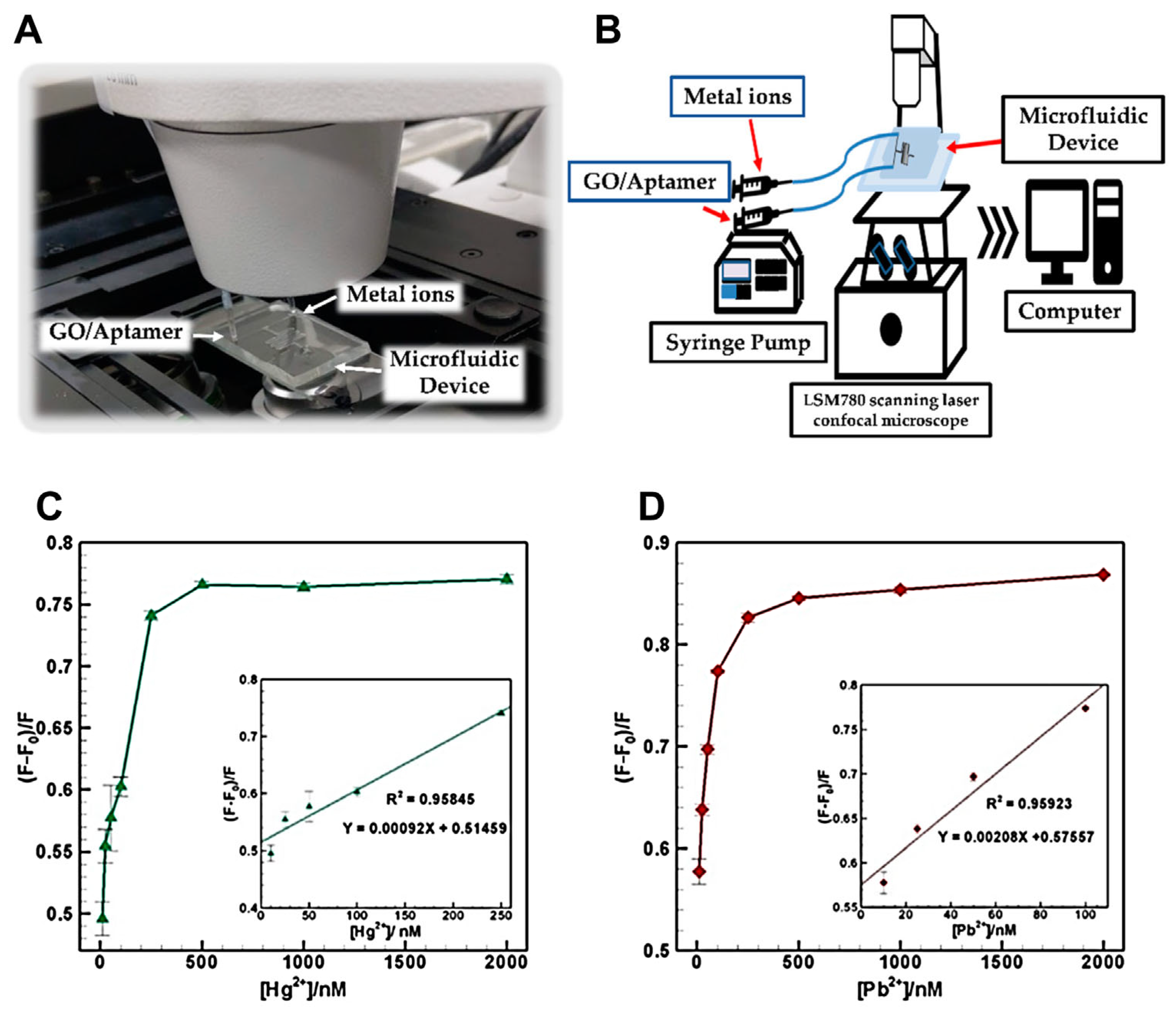
| Microfluidic chip, graphene oxide (GO), aptamers labeled with FAM/HEX dyes | FRET-based fluorescence quenching by GO-aptamer interactions | Hg2+ and Pb2+ | 0.70 ppb (Hg2+), 0.53 ppb (Pb2+) | +/(Compact PDMS chip enabling on-site fluorescence sensing) | [114] | |
| 3D-printed microfluidic device, silver nanoparticles (AgNPs@BS), leucomalachite green dye | Naked-eye colorimetric detection (color change) | Hg2+ and As3+ | 0.089 mg/L (Hg2+), 0.042 mg/L (As3+) | +/(3D-printed portable chip with smartphone quantification) | [115] | |
| Hydrophobin chimera on polystyrene multiwell plates | Fluorescence decrease coupled with machine learning analysis | Hg2+ | 0.3 nM (seawater), 0.4 nM (tap water) | +/(Smartphone-assisted portable biosensor with ML support) | [116] | |
| PDMS microchannel and organic probes | Fluorescence | Hg2+, Pb2+, Cr3+, Cu2+ | 0.89 nM (Hg2+), 9.60 nM (Pb2+), 5.45 nM (Cr3+), 1.77 nM (Cu2+) | +/(Smartphone-based detection system) | [117] | |
| Microplastics & Nanoplastics | PDMS microchannel with copper microwire electrodes | Electrical resistance measurement under DC electrophoretic force | Polystyrene microplastics (1–10 μm) | Not specified | −/(Lab-based setup) | [118] |
| Microfluidic channel integrated with microwave electric inductive-capacitive resonator | Microwave sensor detecting S11 resonance frequency shift | Microplastics | Not specified | +/(Compact, suitable for real-time use) | [119] | |
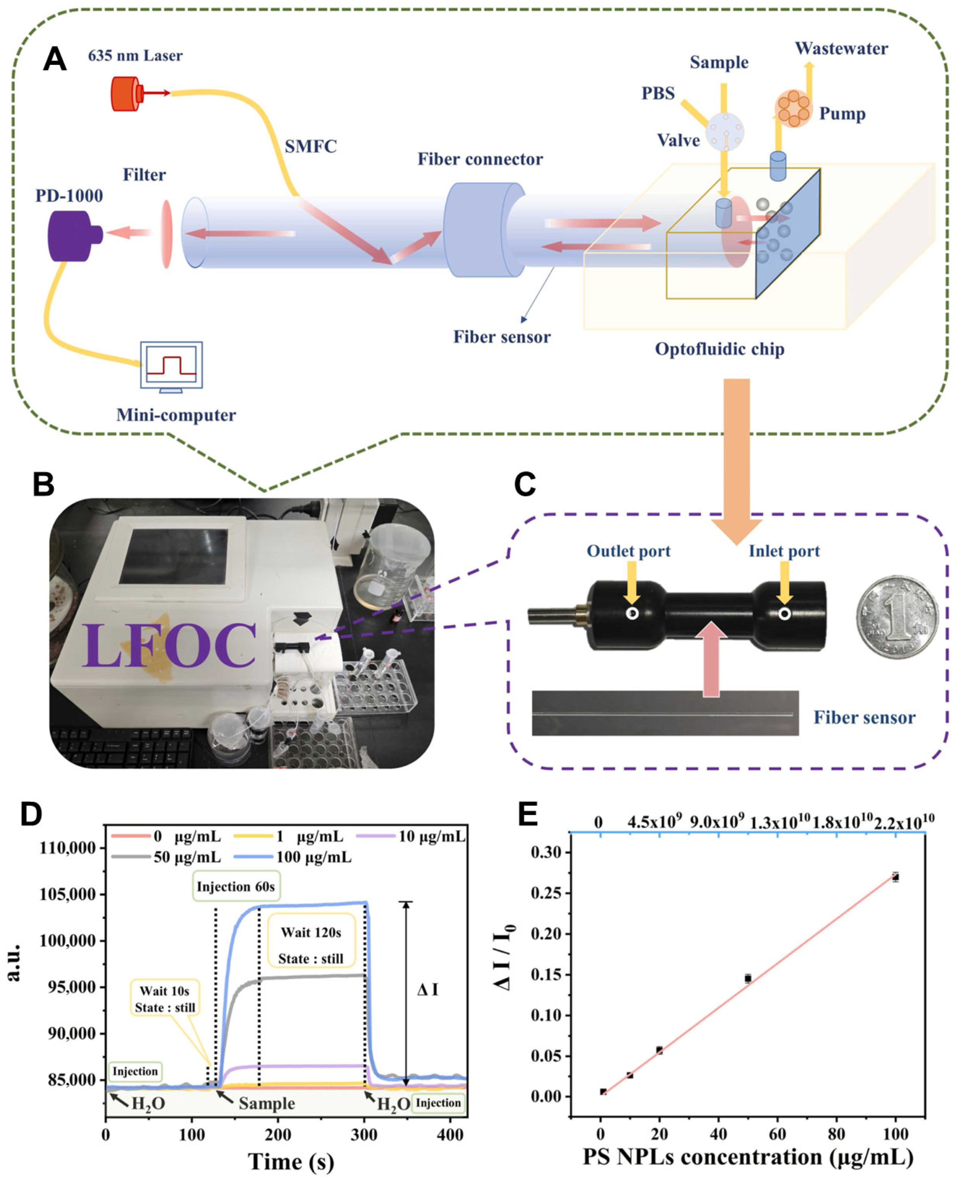
| Fiber-embedded optofluidic chip | 180° laser-backscattered signal quantification | PS, PE, PET, PP, PMMA | 0.23 μg/mL (PS) | +/(Compact, no reagents, field applicable) | [120] | |
| Droplet-based microfluidic system (PDMS) | ML-assisted real-time droplet image analysis | Polystyrene MPs (3–50 μm) | Not specified | +/(AI-assisted, portable, field validated) | [121] | |
| Agarose-based microfluidic chip with micropost arrays | ML-assisted Raman spectroscopy | Polystyrene NPs (100 nm) | 6.25 µg/mL | +/(Scalable, AI-integrated, seawater-compatible) | [122] | |
| PFAS | Janus droplet-based microfluidic chip | Real-time optical sensing, PCA, and Random Forest analysis | Four PFAS compounds | Not specified | +/(ML) | [123] |
| MOF (Cr-MIL-101) + Interdigitated microelectrodes (IDμE) | Electrochemical impedance spectroscopy | PFOS | 0.5 ng/L | –/(Lab-based setup; lacks field integration) | [124] |
3.2.2. Personal Care Products (PCPs)
3.2.3. Heavy Metals and Inorganic Contaminants
3.2.4. Microplastics and Nanoplastics
3.2.5. Per- and Polyfluoroalkyl Substances (PFAS)
4. Integration of Microfluidic Sensors with Emerging IT Technologies
4.1. Smartphone-Based Platforms
4.2. Artificial Intelligence and Machine Learning
5. Conclusions and Outlook
Author Contributions
Funding
Conflicts of Interest
References
- Wołowicz, A.; Munir, H.M.S. Emerging organic micropollutants as serious environmental problem: A comprehensive review. Sci. Total Environ. 2025, 958, 177948. [Google Scholar] [CrossRef] [PubMed]
- Ganguly, A.; Nanda, S.; Das, K.; Ghanty, S.; Biswas, G.; Mandi, M.; Mukherjee, S.; Paramanik, M.; Rajak, P. Micropollutants in environment: Sources, ecotoxicity, and strategies for remediation. In Ecosystem Management: Climate Change and Sustainability; Wiley: Hoboken, NJ, USA, 2024; pp. 453–492. [Google Scholar]
- Ahmed, M.; Johir, M.; Ngo, H.H.; Guo, W.; Zhou, J.; Belhaj, D.; Moni, M. Methods for the analysis of micro-pollutants. In Current Developments in Biotechnology and Bioengineering; Elsevier: Amsterdam, The Netherlands, 2020; pp. 63–86. [Google Scholar]
- Chen, P.; Wang, J.; Xue, Y.; Wang, C.; Sun, W.; Yu, J.; Guo, H. From challenge to opportunity: Revolutionizing the monitoring of emerging contaminants in water with advanced sensors. Water Res. 2024, 265, 122297. [Google Scholar] [CrossRef] [PubMed]
- Abdelhamid, M.A.; Ki, M.-R.; Pack, S.P. Biominerals and bioinspired materials in biosensing: Recent advancements and applications. Int. J. Mol. Sci. 2024, 25, 4678. [Google Scholar] [CrossRef] [PubMed]
- Berlanda, S.F.; Breitfeld, M.; Dietsche, C.L.; Dittrich, P.S. Recent advances in microfluidic technology for bioanalysis and diagnostics. Anal. Chem. 2020, 93, 311–331. [Google Scholar] [CrossRef] [PubMed]
- Kim, J.S.; Son, H.; Han, J.; Cha, H.; Lee, J.H.; Pack, S.P.; Lee, C.-S. Evaluation of mitochondrial activity via cellular interactions between adrenal and neuronal cells in a microfluidic coculture device. Biotechnol. Bioprocess Eng. 2025, 30, 1–18. [Google Scholar] [CrossRef]
- Song, H.; Son, H.; Hua, H.Q.H.; Kim, J.S.; Lee, J.H.; Pack, S.P.; Lee, C.-S. Microfluidic assessment of corticosterone-induced mitochondrial dysfunction in neuronal cells. Biochem. Eng. J. 2025, 215, 109607. [Google Scholar] [CrossRef]
- Hajam, M.I.; Khan, M.M. Microfluidics: A concise review of the history, principles, design, applications, and future outlook. Biomater. Sci. 2024, 12, 218–251. [Google Scholar] [CrossRef] [PubMed]
- Abdelhamid, M.A.; Son, R.G.; Park, K.S.; Pack, S.P. Oriented multivalent silaffin-affinity immobilization of recombinant lipase on diatom surface: Reliable loading and high performance of biocatalyst. Colloids Surf. B Biointerfaces 2022, 219, 112830. [Google Scholar] [CrossRef] [PubMed]
- Park, K.S.; Park, T.-I.; Lee, J.E.; Hwang, S.-Y.; Choi, A.; Pack, S.P. Aptamers and nanobodies as new bioprobes for SARS-CoV-2 diagnostic and therapeutic system applications. Biosensors 2024, 14, 146. [Google Scholar] [CrossRef] [PubMed]
- Abdelhamid, M.A.; Ki, M.-R.; El-Hafeez, A.A.A.; Son, R.G.; Pack, S.P. Tailored functionalized protein nanocarriers for cancer therapy: Recent developments and prospects. Pharmaceutics 2023, 15, 168. [Google Scholar] [CrossRef] [PubMed]
- Park, T.-I.; Yang, A.H.; Kanth, B.K.; Pack, S.P. Aptamers as diagnostic and therapeutic agents for aging and age-related diseases. Biosensors 2025, 15, 232. [Google Scholar] [CrossRef] [PubMed]
- Jo, H.; Ju, S.; Kim, M.; Beon, J.; Jang, S.-Y.; Pack, S.P.; Son, C.Y.; Kim, J.-S.; Oh, S.S. Aptamer-guided, hydrolysis-resistant deoxyoxanosine enables epitope-and moiety-selective conjugation to nonengineered proteins even in complex environments. J. Am. Chem. Soc. 2025, 147, 9328–9340. [Google Scholar] [CrossRef] [PubMed]
- Park, K.S.; Cha, H.; Niu, J.; Soh, H.T.; Lee, J.H.; Pack, S.P. DNA-controlled protein fluorescence: Design of aptamer-split peptide hetero-modulator for GFP to respond to intracellular ATP levels. Nucleic Acids Res. 2024, 52, 8063–8071. [Google Scholar] [CrossRef] [PubMed]
- Razavi, Z.; Soltani, M.; Pazoki-Toroudi, H.; Chen, P. CRISPR-microfluidics nexus: Advancing biomedical applications for understanding and detection. Sens. Actuators A Phys. 2024, 376, 115625. [Google Scholar] [CrossRef]
- O’Kennedy, R.; Fitzgerald, J.; Cassedy, A.; Crawley, A.; Zhang, X.; Carrera, S. Applications of antibodies in microfluidics-based analytical systems: Challenges and strategies for success. J. Micromech. Microeng. 2018, 28, 063001. [Google Scholar] [CrossRef]
- Whitesides, G.M. The origins and the future of microfluidics. Nature 2006, 442, 368–373. [Google Scholar] [CrossRef] [PubMed]
- Yew, M.; Ren, Y.; Koh, K.S.; Sun, C.; Snape, C. A review of state-of-the-art microfluidic technologies for environmental applications: Detection and remediation. Glob. Chall. 2019, 3, 1800060. [Google Scholar] [CrossRef] [PubMed]
- Zhu, X.; Wang, K.; Yan, H.; Liu, C.; Zhu, X.; Chen, B. Microfluidics as an emerging platform for exploring soil environmental processes: A critical review. Environ. Sci. Technol. 2022, 56, 711–731. [Google Scholar] [CrossRef] [PubMed]
- Aryal, P.; Hefner, C.; Martinez, B.; Henry, C.S. Microfluidics in environmental analysis: Advancements, challenges, and future prospects for rapid and efficient monitoring. Lab Chip 2024, 24, 1175–1206. [Google Scholar] [CrossRef] [PubMed]
- Adampourezare, M.; Asadpour-Zeynali, K.; de la Guardia, M.; Dolatabadi, J.E.N. The design of paper-based electroanalytical microfluidic device coupled with post-synthesized molecularly imprinted polymers (rGO/Au@ Ag2S/PANI/polyacrylamide) for the detection of streptomycin. Sens. Actuators Rep. 2025, 9, 100297. [Google Scholar] [CrossRef]
- Escobedo, C.; Brolo, A. Synergizing microfluidics and plasmonics: Advances, applications, and future directions. Lab Chip 2025, 25, 1256–1281. [Google Scholar] [CrossRef] [PubMed]
- Abdullaev, S.S.; Althomali, R.H.; Khan, A.R.; Jabbar, H.S.; Aggarwal, S.; Mustafa, Y.F.; Khlewee, I.H. Integrating of analytical techniques with enzyme-mimicking nanomaterials for the fabrication of microfluidic systems for biomedical analysis. Talanta 2024, 273, 125896. [Google Scholar] [CrossRef] [PubMed]
- Liu, Y.; Yang, G.; Hui, Y.; Ranaweera, S.; Zhao, C.X. Microfluidic nanoparticles for drug delivery. Small 2022, 18, 2106580. [Google Scholar] [CrossRef] [PubMed]
- Abdelhamid, M.A.; Pack, S.P. Biomimetic and bioinspired silicifications: Recent advances for biomaterial design and applications. Acta Biomater. 2021, 120, 38–56. [Google Scholar] [CrossRef] [PubMed]
- Temiz, Y.; Delamarche, E. Sub-nanoliter, real-time flow monitoring in microfluidic chips using a portable device and smartphone. Sci. Rep. 2018, 8, 10603. [Google Scholar] [CrossRef] [PubMed]
- Pujol-Vila, F.; Giménez-Gómez, P.; Santamaria, N.; Antúnez, B.; Vigués, N.; Díaz-González, M.; Jiménez-Jorquera, C.; Mas, J.; Sacristán, J.; Muñoz-Berbel, X. Portable and miniaturized optofluidic analysis system with ambient light correction for fast in situ determination of environmental pollution. Sens. Actuators B Chem. 2016, 222, 55–62. [Google Scholar] [CrossRef]
- Mesquita, P.; Gong, L.; Lin, Y. Low-cost microfluidics: Towards affordable environmental monitoring and assessment. Front. Lab Chip Technol. 2022, 1, 1074009. [Google Scholar] [CrossRef]
- Liu, Y.; Zhang, X. Microfluidics-based plasmonic biosensing system based on patterned plasmonic nanostructure arrays. Micromachines 2021, 12, 826. [Google Scholar] [CrossRef] [PubMed]
- Chałupniak, A.; Merkoçi, A. Toward integrated detection and graphene-based removal of contaminants in a lab-on-a-chip platform. Nano Res. 2017, 10, 2296–2310. [Google Scholar] [CrossRef]
- Debnath, S.; Ghosh, R.; Mukhopadhyay, S.; Baskaran, K.V.; Chatterjee, P.B. Fabrication of a paper-based facile and low-cost microfluidic device and digital imaging technique for point-of-need monitoring of hypochlorite. Analyst 2023, 148, 4072–4083. [Google Scholar] [CrossRef] [PubMed]
- Khalaf, E.M.; Jabbar, H.S.; Romero-Parra, R.M.; Al-Awsi, G.R.L.; Budi, H.S.; Altamimi, A.S.; Gatea, M.A.; Falih, K.T.; Singh, K.; Alkhuzai, K.A. Smartphone-assisted microfluidic sensor as an intelligent device for on-site determination of food contaminants: Developments and applications. Microchem. J. 2023, 190, 108692. [Google Scholar] [CrossRef]
- Liu, J.; Du, H.; Huang, L.; Xie, W.; Liu, K.; Zhang, X.; Chen, S.; Zhang, Y.; Li, D.; Pan, H. AI-powered microfluidics: Shaping the future of phenotypic drug discovery. ACS Appl. Mater. Interfaces 2024, 16, 38832–38851. [Google Scholar] [CrossRef] [PubMed]
- Ren, K.; Zhou, J.; Wu, H. Materials for microfluidic chip fabrication. Acc. Chem. Res. 2013, 46, 2396–2406. [Google Scholar] [CrossRef] [PubMed]
- Bartali, R.; Lorenzelli, L.; Scarpa, M.; Morganti, E.; Collini, C.; Micheli, V.; Gottardi, G.; Gambetti, A.; Gambetti, G.; Coser, G. Super-hydrophilic PDMS and PET surfaces for microfluidic devices. Adv. Sci. Technol. 2013, 81, 96–100. [Google Scholar]
- Scott, S.M.; Ali, Z. Fabrication methods for microfluidic devices: An overview. Micromachines 2021, 12, 319. [Google Scholar] [CrossRef] [PubMed]
- Anushka; Bandopadhyay, A.; Das, P.K. Paper based microfluidic devices: A review of fabrication techniques and applications. Eur. Phys. J. Spec. Top. 2023, 232, 781–815. [Google Scholar] [CrossRef] [PubMed]
- Iliescu, C.; Taylor, H.; Avram, M.; Miao, J.; Franssila, S. A practical guide for the fabrication of microfluidic devices using glass and silicon. Biomicrofluidics 2012, 6, 16505-1–16505-16. [Google Scholar] [CrossRef] [PubMed]
- Xu, Z.; Jaiswal, A.; Liu, X.; Yang, Z.; Yin, Q.; Kong, K.V.; Yong, K.T. Microfluidics evolution and surface functionalization: A pathway to enhanced heavy metal ion detection. Adv. Sens. Res. 2024, 3, 2400008. [Google Scholar] [CrossRef]
- Carvell, T.; Burgoyne, P.; Fraser, A.R.; Bridle, H. Categorising hybrid material microfluidic devices. Front. Lab Chip Technol. 2024, 3, 1412290. [Google Scholar] [CrossRef]
- Zhou, W.; Dou, M.; Timilsina, S.S.; Xu, F.; Li, X. Recent innovations in cost-effective polymer and paper hybrid microfluidic devices. Lab Chip 2021, 21, 2658–2683. [Google Scholar] [CrossRef] [PubMed]
- Xu, J.; Harasek, M.; Gföhler, M. From soft lithography to 3D printing: Current status and future of microfluidic device fabrication. Polymers 2025, 17, 455. [Google Scholar] [CrossRef] [PubMed]
- Gao, S.; Xu, T.; Wu, L.; Zhu, X.; Li, C.; Li, X. A one-step soft lithography technique for making microfluidic PDMS chips with macro-scale structures. In Proceedings of the 2024 IEEE 37th International Conference on Micro Electro Mechanical Systems (MEMS), Austin, TX, USA, 21–25 January 2024; pp. 1170–1173. [Google Scholar]
- Yang, L.; Li, W.; Ding, X.; Zhao, Y.; Qian, X.; Shang, L. Biomimetic mineralized organic–inorganic hybrid scaffolds from microfluidic 3D printing for bone repair. Adv. Funct. Mater. 2025, 35, 2410927. [Google Scholar] [CrossRef]
- Yajun, Z.; Jingji, L.; Xie, Y.; Liang, K.; Zhang, Z.; Yang, C.; Yiqiang, F. Superhydrophobic treatment of PDMS-based microfluidic devices using CO2 laser ablation. Microfluid. Nanofluidics 2024, 28, 8. [Google Scholar] [CrossRef]
- Zhao, X.; Cui, C.; Ma, L.; Ding, Z.; Hou, J.; Xiao, Y.; Liu, B.; Qi, B.; Zhang, J.; Wei, J. Laser-ablated acoustofluidics-driven paper devices for controllable chemical engineering in color display applications. Chem. Eng. J. 2024, 480, 148245. [Google Scholar] [CrossRef]
- Su, W.; Cook, B.S.; Fang, Y.; Tentzeris, M.M. Fully inkjet-printed microfluidics: A solution to low-cost rapid three-dimensional microfluidics fabrication with numerous electrical and sensing applications. Sci. Rep. 2016, 6, 35111. [Google Scholar] [CrossRef] [PubMed]
- Askarzadeh, N.; Mohammadi, J.; Rabbani, H.; Hosseini, M. Lab-on-a-chip: A Road Map of Silicon Chemistry to Multipurpose Microfluidic Chips; Detection Science Series; Royal Society of Chemistry: Cambridge, UK, 2024; Volume 25, pp. 33–72. [Google Scholar]
- Sima, F.; Sugioka, K.; Vázquez, R.M.; Osellame, R.; Kelemen, L.; Ormos, P. Three-dimensional femtosecond laser processing for lab-on-a-chip applications. Nanophotonics 2018, 7, 613–634. [Google Scholar] [CrossRef]
- Orazi, L.; Siciliani, V.; Pelaccia, R.; Oubellaouch, K.; Reggiani, B. Ultrafast laser micromanufacturing of microfluidic devices. Procedia CIRP 2022, 110, 122–127. [Google Scholar] [CrossRef]
- Kumar, A.; Panda, D.; Gangawane, K.M. Microfabrication: Techniques and technology. Microfabr. Nanofabrication Precis. Manuf. 2024, 11, 47. [Google Scholar]
- Kumar, A.S.; Venkatesalu, S.; Dilliyappan, S.; Pasupulla, A.P.; Prathap, L.; Palaniyandi, T.; Baskar, G.; Ravi, M.; Sugumaran, A. Microfluidics as diagnostic tools. Clin. Chim. Acta 2024, 556, 117841. [Google Scholar] [CrossRef] [PubMed]
- Wong, W.D.; Majnis, M.F.; Lai, C.W.; Sagadevan, S.; Julkapli, N.M. Enhancement of mixing and reaction efficiency of various fluids applications at different microfluidic configuration and design. Chem. Eng. Process. Process Intensif. 2024, 198, 109729. [Google Scholar] [CrossRef]
- Haghparas, Z.; Tabalvandani, M.B.; Arghavani, P.; Hosseini, S.B.; Badieirostami, M.; Habibi-Rezaei, M.; Moosavi-Movahedi, A.A. Modeling the navigating forces behind BSA aggregation in a microfluidic chip. Soft Matter 2025, 21, 989–1001. [Google Scholar] [CrossRef] [PubMed]
- Song, Y.; Zhou, Y.; Zhang, K.; Fan, Z.; Zhang, F.; Wei, M. Microfluidic programmable strategies for channels and flow. Lab Chip 2024, 24, 4483–4513. [Google Scholar] [CrossRef] [PubMed]
- Bezinge, L.; Shih, C.J.; Richards, D.A.; deMello, A.J. Electrochemical paper-based microfluidics: Harnessing capillary flow for advanced diagnostics. Small 2024, 20, 2401148. [Google Scholar] [CrossRef] [PubMed]
- Lin, C.-C.; Hsu, J.-L.; Lee, G.-B. Sample preconcentration in microfluidic devices. Microfluid. Nanofluidics 2011, 10, 481–511. [Google Scholar] [CrossRef]
- Wei, Q.; Dong, Q.; Pu, H. Multiplex surface-enhanced Raman scattering: An emerging tool for multicomponent detection of food contaminants. Biosensors 2023, 13, 296. [Google Scholar] [CrossRef] [PubMed]
- Punnoy, P.; Aryal, P.; Hefner, C.E.; Brack, E.; Rodthongkum, N.; Potiyaraj, P.; Henry, C.S. Smartphone-assisted dual-sided capillary microfluidic device for multiplex detection of heavy metals and nutrients in drinking water. Anal. Chim. Acta 2025, 1356, 344031. [Google Scholar] [CrossRef] [PubMed]
- Cao, Y.; Zhang, B.; Zhu, Z.; Xin, X.; Wu, H.; Chen, B. Microfluidic based whole-cell biosensors for simultaneously on-site monitoring of multiple environmental contaminants. Front. Bioeng. Biotechnol. 2021, 9, 622108. [Google Scholar] [CrossRef] [PubMed]
- Gupta, A.; Kumar, S.; Bajpai, Y.; Chaturvedi, K.; Johri, P.; Tiwari, R.K.; Vivekanand, V.; Trivedi, M. Pharmaceutically active micropollutants: Origin, hazards and removal. Front. Microbiol. 2024, 15, 1339469. [Google Scholar] [CrossRef] [PubMed]
- Klančič, V.; Gobec, M.; Jakopin, Ž. Environmental contamination status with common ingredients of household and personal care products exhibiting endocrine-disrupting potential. Environ. Sci. Pollut. Res. 2022, 29, 73648–73674. [Google Scholar] [CrossRef] [PubMed]
- Sossalla, N.A.; Nivala, J.; Reemtsma, T.; Schlichting, R.; König, M.; Forquet, N.; van Afferden, M.; Müller, R.A.; Escher, B.I. Removal of micropollutants and biological effects by conventional and intensified constructed wetlands treating municipal wastewater. Water Res. 2021, 201, 117349. [Google Scholar] [CrossRef] [PubMed]
- Ianes, J.; Piraldi, S.; Cantoni, B.; Antonelli, M. Micropollutants removal, residual risk, and costs for quaternary treatments in the framework of the urban wastewater treatment directive. Water Res. X 2025, 29, 100334. [Google Scholar] [CrossRef]
- Srinivasulu, M.; Chandra, M.S.; Gooty, J.M.; Madhavi, A. Personal care products—Fragrances, cosmetics, and sunscreens—In the environment. In Environmental Micropollutants; Elsevier: Amsterdam, The Netherlands, 2022; pp. 131–149. [Google Scholar]
- Gul, B.; Naseem, M.K.; Malik, W.-u.-N.; Gurmani, A.R.; Mehmood, A.; Rafique, M. Environmental micropollutants and their impact on human health with special focus on agriculture. In Hazardous Environmental Micro-pollutants, Health Impacts and Allied Treatment Technologies; Springer: Berlin/Heidelberg, Germany, 2022; pp. 1–19. [Google Scholar]
- Ghirardini, A.; Grillini, V.; Verlicchi, P. A review of the occurrence of selected micropollutants and microorganisms in different raw and treated manure–environmental risk due to antibiotics after application to soil. Sci. Total Environ. 2020, 707, 136118. [Google Scholar] [CrossRef] [PubMed]
- Tiwari, B.; Drogui, P.; Tyagi, R.D. Removal of emerging micro-pollutants from pharmaceutical industry wastewater. In Current Developments in Biotechnology and Bioengineering; Elsevier: Amsterdam, The Netherlands, 2020; pp. 457–480. [Google Scholar]
- Liu, M.; Lv, J.; Qin, C.; Zhang, H.; Wu, L.; Guo, W.; Guo, C.; Xu, J. Chemical fingerprinting of organic micropollutants in different industrial treated wastewater effluents and their effluent-receiving river. Sci. Total Environ. 2022, 838, 156399. [Google Scholar] [CrossRef] [PubMed]
- Zeng, E.Y. Persistent Organic Pollutants (POPs): Analytical Techniques, Environmental Fate and Biological Effects; Elsevier: Amsterdam, The Netherlands, 2015; Volume 67. [Google Scholar]
- Zhang, Z.; Lu, Y.; Gao, S.; Wu, S. Sustainable and efficient wastewater treatment using cellulose-based hydrogels: A review of heavy metal, dye, and micropollutant removal applications. Separations 2025, 12, 72. [Google Scholar] [CrossRef]
- Thakur, D.; Ganguly, R. Occurrence and fate of micropollutants in air. In Environmental Micropollutants; Elsevier: Amsterdam, The Netherlands, 2022; pp. 305–313. [Google Scholar]
- Estevez, E.; del Carmen Cabrera, M.; Fernández-Vera, J.R.; Molina-Díaz, A.; Robles-Molina, J.; del Pino Palacios-Díaz, M. Monitoring priority substances, other organic contaminants and heavy metals in a volcanic aquifer from different sources and hydrological processes. Sci. Total Environ. 2016, 551, 186–196. [Google Scholar] [CrossRef] [PubMed]
- Abdelhamid, M.A.; Khalifa, H.O.; Yoon, H.J.; Ki, M.-R.; Pack, S.P. Microbial immobilized enzyme biocatalysts for multipollutant mitigation: Harnessing nature’s toolkit for environmental sustainability. Int. J. Mol. Sci. 2024, 25, 8616. [Google Scholar] [CrossRef] [PubMed]
- Chakraborty, A.; Adhikary, S.; Bhattacharya, S.; Dutta, S.; Chatterjee, S.; Banerjee, D.; Ganguly, A.; Rajak, P. Pharmaceuticals and personal care products as emerging environmental contaminants: Prevalence, toxicity, and remedial approaches. ACS Chem. Health Saf. 2023, 30, 362–388. [Google Scholar] [CrossRef]
- Włodarczyk-Makuła, M. Selected organic micropollutants in the aquatic environment. Desalination Water Treat. 2024, 317, 100061. [Google Scholar] [CrossRef]
- Srnovršnik, T.; Virant-Klun, I.; Pinter, B. Polycystic ovary syndrome and endocrine disruptors (bisphenols, parabens, and triclosan)—A systematic review. Life 2023, 13, 138. [Google Scholar] [CrossRef] [PubMed]
- Punniyakotti, P.; Vinayagam, S.; Rajamohan, R.; Priya, S.; Moovendhan, M.; Sundaram, T. Environmental fate and ecotoxicological behaviour of pesticides and insecticides in non-target environments: Nanotechnology-based mitigation strategies. J. Environ. Chem. Eng. 2024, 5, 113349. [Google Scholar] [CrossRef]
- Kumar, A.; Deepika; Tyagi, D.; Tarkeshwar; Kapinder. Organic micropollutants in agricultural system: Ecotoxicity, risk assessment and detection methods. In Organic Micropollutants in Aquatic and Terrestrial Environments; Springer: Berlin/Heidelberg, Germany, 2024; pp. 265–293. [Google Scholar]
- Rebai, H.; Sholkamy, E.N.; Abdelhamid, M.A.; Prakasam Thanka, P.; Aly Hassan, A.; Pack, S.P.; Ki, M.-R.; Boudemagh, A. Soil actinobacteria exhibit metabolic capabilities for degrading the toxic and persistent herbicide metribuzin. Toxics 2024, 12, 709. [Google Scholar] [CrossRef] [PubMed]
- Chaitanya, M.; Arora, S.; Pal, R.S.; Ali, H.S.; El Haj, B.; Logesh, R. Assessment of environmental pollutants for their toxicological effects of human and animal health. In Organic Micropollutants in Aquatic and Terrestrial Environments; Springer: Berlin/Heidelberg, Germany, 2024; pp. 67–85. [Google Scholar]
- Akhtar, A.B.T.; Naseem, S.; Yasar, A.; Naseem, Z. Persistent organic pollutants (POPs): Sources, types, impacts, and their remediation. In Environmental Pollution and Remediation; Springer: Berlin/Heidelberg, Germany, 2021; pp. 213–246. [Google Scholar]
- Amobonye, A.; Bhagwat, P.; Raveendran, S.; Singh, S.; Pillai, S. Environmental impacts of microplastics and nanoplastics: A current overview. Front. Microbiol. 2021, 12, 768297. [Google Scholar] [CrossRef] [PubMed]
- Schwarzenbach, R.P.; Escher, B.I.; Fenner, K.; Hofstetter, T.B.; Johnson, C.A.; Von Gunten, U.; Wehrli, B. The challenge of micropollutants in aquatic systems. Science 2006, 313, 1072–1077. [Google Scholar] [CrossRef] [PubMed]
- Gould, S.L.; Winter, M.J.; Norton, W.H.; Tyler, C.R. The potential for adverse effects in fish exposed to antidepressants in the aquatic environment. Environ. Sci. Technol. 2021, 55, 16299–16312. [Google Scholar] [CrossRef] [PubMed]
- Hazra, M.; Joshi, H.; Williams, J.B.; Watts, J.E. Antibiotics and antibiotic resistant bacteria/genes in urban wastewater: A comparison of their fate in conventional treatment systems and constructed wetlands. Chemosphere 2022, 303, 135148. [Google Scholar] [CrossRef] [PubMed]
- Chen, M.; Jin, X.; Guo, C.; Liu, Y.; Zhang, H.; Wang, J.; Dong, G.; Liu, N.; Guo, W.; Giesy, J.P. Micropollutants but high risks: Human multiple stressors increase risks of freshwater ecosystems at the megacity-scale. J. Hazard. Mater. 2023, 460, 132497. [Google Scholar] [CrossRef] [PubMed]
- Narwal, N.; Katyal, D.; Kataria, N.; Rose, P.K.; Warkar, S.G.; Pugazhendhi, A.; Ghotekar, S.; Khoo, K.S. Emerging micropollutants in aquatic ecosystems and nanotechnology-based removal alternatives: A review. Chemosphere 2023, 341, 139945. [Google Scholar] [CrossRef] [PubMed]
- Rai, M.; Pant, G.; Pant, K.; Aloo, B.N.; Kumar, G.; Singh, H.B.; Tripathi, V. Microplastic pollution in terrestrial ecosystems and its interaction with other soil pollutants: A potential threat to soil ecosystem sustainability. Resources 2023, 12, 67. [Google Scholar] [CrossRef]
- Ghosh, A.; Manna, M.C.; Jha, S.; Singh, A.K.; Misra, S.; Srivastava, R.C.; Srivastava, P.P.; Laik, R.; Bhattacharyya, R.; Prasad, S. Impact of soil-water contaminants on tropical agriculture, animal and societal environment. Adv. Agron 2022, 176, 209–274. [Google Scholar]
- Richard, G.; Sawyer, W.E.; Sharipov, A. Environmental impacts of air pollution. In Sustainable Strategies for Air Pollution Mitigation: Development, Economics, and Technologies; Springer: Berlin/Heidelberg, Germany, 2024; pp. 47–76. [Google Scholar]
- Asuku, A.O.; Ayinla, M.T.; Ajibare, A.J.; Adeyemo, M.B.; Adeyemo, R.O. Heavy metals and emerging contaminants in foods and food products associated with neurotoxicity. In Emerging Contaminants in Food and Food Products; CRC Press: Boca Raton, FL, USA, 2024; pp. 236–250. [Google Scholar]
- Kalenik, S.; Zaczek, A.; Rodacka, A. Air pollution-induced neurotoxicity: The relationship between air pollution, epigenetic changes, and neurological disorders. Int. J. Mol. Sci. 2025, 26, 3402. [Google Scholar] [CrossRef] [PubMed]
- Thakur, M.; Rachamalla, M.; Niyogi, S.; Datusalia, A.K.; Flora, S.J.S. Molecular mechanism of arsenic-induced neurotoxicity including neuronal dysfunctions. Int. J. Mol. Sci. 2021, 22, 10077. [Google Scholar] [CrossRef] [PubMed]
- Larsson, D.J.; Flach, C.-F. Antibiotic resistance in the environment. Nat. Rev. Microbiol. 2022, 20, 257–269. [Google Scholar] [CrossRef] [PubMed]
- Barathe, P.; Kaur, K.; Reddy, S.; Shriram, V.; Kumar, V. Antibiotic pollution and associated antimicrobial resistance in the environment. J. Hazard. Mater. Lett. 2024, 5, 100105. [Google Scholar] [CrossRef]
- Khalifa, H.O.; Abdelhamid, M.A.; Oreiby, A.; Mohamed, M.-Y.I.; Ramadan, H.; Elfadadny, A.; Pack, S.P.; Yoo, H.S.; Habib, I. Fire under the ashes: A descriptive review on the prevalence of methicillin-resistant Staphylococcus aureus in the food supply chain. J. Agric. Food Res. 2024, 19, 101606. [Google Scholar] [CrossRef]
- Azmy, L.; Al-Olayan, E.; Abdelhamid, M.A.; Zayed, A.; Gheda, S.F.; Youssif, K.A.; Abou-Zied, H.A.; Abdelmohsen, U.R.; Ibraheem, I.B.; Pack, S.P. Antimicrobial activity of Arthrospira platensis-mediated gold nanoparticles against Streptococcus pneumoniae: A metabolomic and docking study. Int. J. Mol. Sci. 2024, 25, 10090. [Google Scholar] [CrossRef] [PubMed]
- Khalifa, H.O.; Oreiby, A.; Abdelhamid, M.A.; Ki, M.-R.; Pack, S.P. Biomimetic antifungal materials: Countering the challenge of multidrug-resistant fungi. Biomimetics 2024, 9, 425. [Google Scholar] [CrossRef] [PubMed]
- Xie, J.; Pang, H.; Sun, R.; Wang, T.; Meng, X.; Zhou, Z. Development of rapid and high-precision colorimetric device for organophosphorus pesticide detection based on microfluidic mixer chip. Micromachines 2021, 12, 290. [Google Scholar] [CrossRef] [PubMed]
- Yang, Y.; Tong, C.; Zhou, R.; Qin, Z.; Xu, J.; Liao, C.; Zhang, S.; Shi, S.; Guo, Y. Hinge-like paper-based dual-channel enzyme-free ratiometric fluorescent microfluidic platform for simultaneous visual detection of carbaryl and glyphosate. Food Chem. 2024, 431, 137127. [Google Scholar] [CrossRef] [PubMed]
- Liu, Z.; Chen, R.; Wang, H.; Wang, C.; Zhang, X.; Yang, Y.; Pang, W.; Ren, S.; Yang, J.; Yang, C.; et al. A colorimetric/electrochemical microfluidic biosensor using target-triggered DNA hydrogels for organophosphorus detection. Biosens. Bioelectron. 2024, 263, 116558. [Google Scholar] [CrossRef] [PubMed]
- Wang, X.; Guo, Z.; Zhang, D.; Yan, Y.; Yu, Y.; Du, B.; Zhang, Z.; Wang, X. Integrating liquid chromatography-electrochemical detection-surface enhanced raman spectroscopy on microfluidic chip for phenylurea herbicides analysis. Sens. Actuators B Chem. 2024, 407, 135436. [Google Scholar] [CrossRef]
- Liu, S.; Zhang, M.; Chen, Q.; Ouyang, Q. Multifunctional metal–organic frameworks driven three-dimensional folded paper-based microfluidic analysis device for chlorpyrifos detection. J. Agric. Food Chem. 2024, 72, 14375–14385. [Google Scholar] [CrossRef] [PubMed]
- Ulloa-Gomez, A.M.; Waimin, J.F.; Yu, Y.-C.; Lucas, A.; Stanciu, L.A. A smartphone-integrated aptasensor for pesticide detection using gold-decorated microparticles. Microchim. Acta 2024, 191, 194. [Google Scholar] [CrossRef] [PubMed]
- Zhang, Q.; Ma, S.; Meng, W.; Zheng, Y.; Yin, L.; Wang, H.; Shi, H.; Zhang, K.; Su, S. Smartphone-based plant-wearable microfluidic sensor with self driven electrolyte for in-situ detection of methyl parathion. Sens. Actuators B Chem. 2024, 418, 136254. [Google Scholar] [CrossRef]
- Yukird, J.; Soum, V.; Kwon, O.-S.; Shin, K.; Chailapakul, O.; Rodthongkum, N. 3D paper-based microfluidic device: A novel dual-detection platform of bisphenol A. Analyst 2020, 145, 1491–1498. [Google Scholar] [CrossRef] [PubMed]
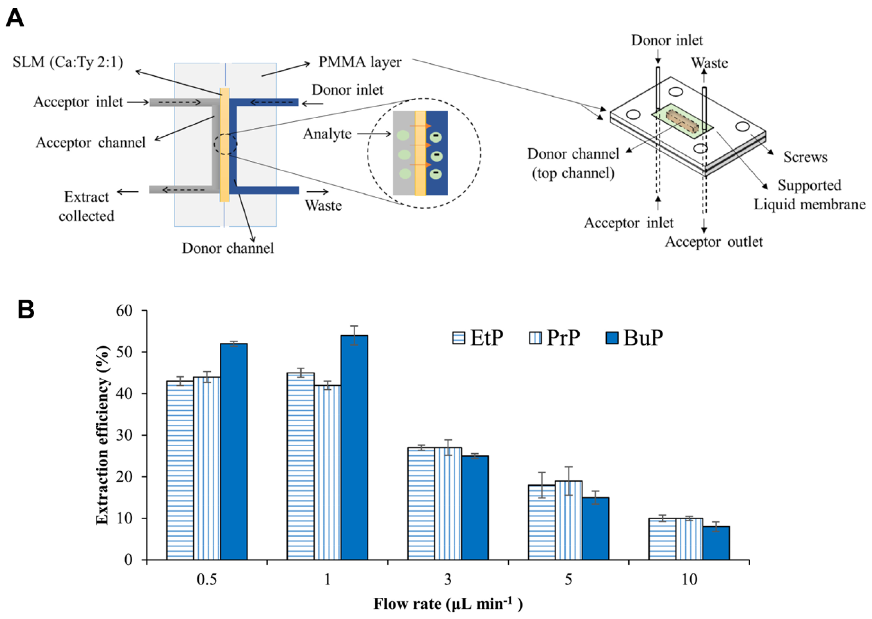
- Dowlatshah, S.; Saraji, M.; Ramos-Payán, M. A green microfluidic method based liquid phase microextraction for the determination of parabens in human urine samples. J. Chromatogr. A 2022, 1673, 463084. [Google Scholar] [CrossRef] [PubMed]
- Jiang, H.; Staeglich, B.; Knoch, J.; Kumar, S.; Dilbaghi, N.; Deep, A.; Ingebrandt, S.; Pachauri, V. Programming layer-by-layer liquid phase epitaxy in microfluidics for realizing two-dimensional metal–organic framework sensor arrays. Environ. Sci. Nano 2025, 12, 1849–1857. [Google Scholar] [CrossRef]
- Mohan, J.M.; Kumar, S.; Amreen, K.; Javed, A.; Dubey, S.K.; Goel, S. Disposable paper-based miniaturized device for sensing of phthalates. IEEE Sens. J. 2023, 23, 16189–16196. [Google Scholar] [CrossRef]
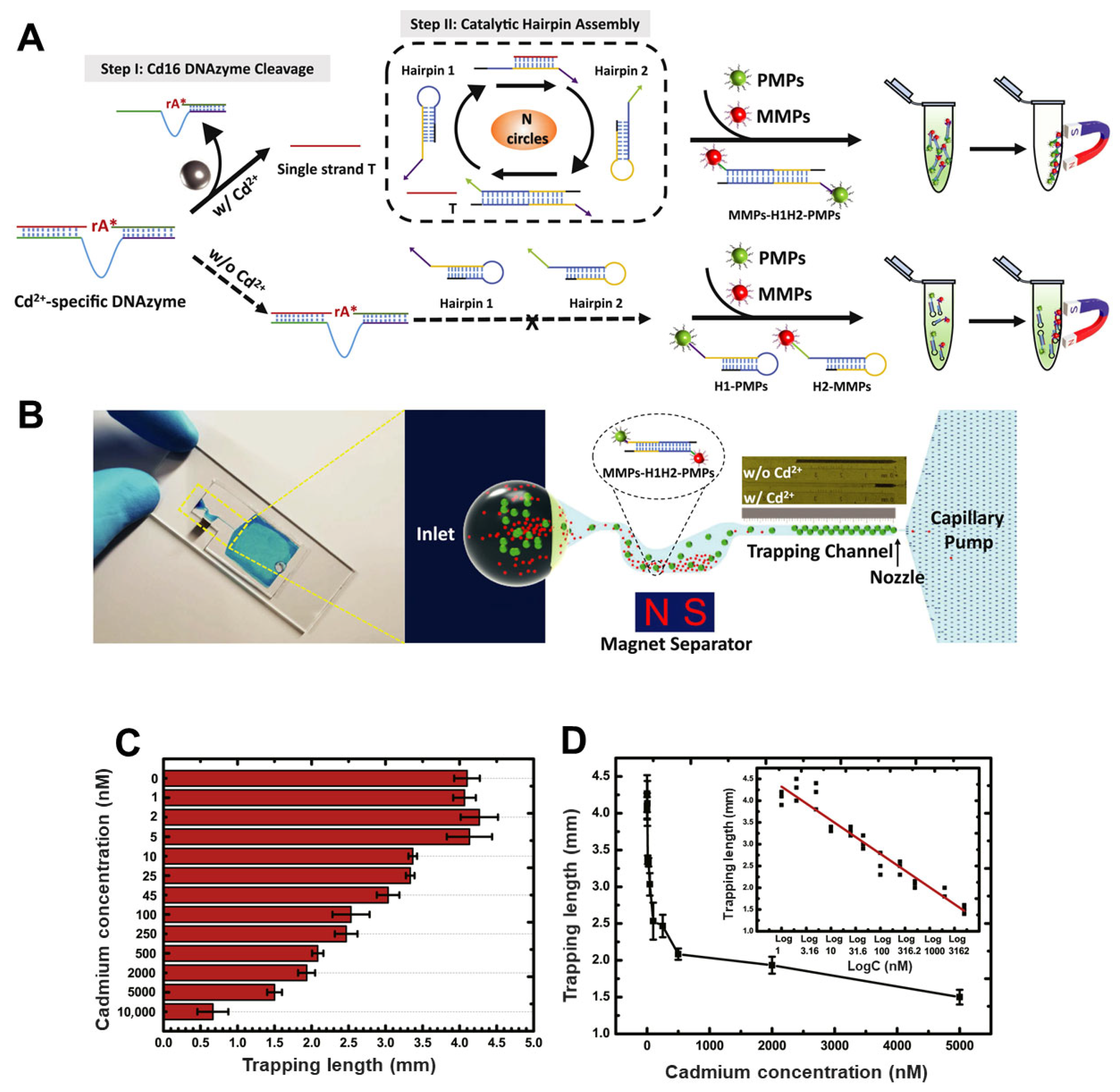
- Wang, G.; Wu, M.; Chu, L.T.; Chen, T.-H. Portable microfluidic device with thermometer-like display for real-time visual quantitation of cadmium (II) contamination in drinking water. Anal. Chim. Acta 2021, 1160, 338444. [Google Scholar] [CrossRef] [PubMed]
- Zeng, S.; Zhu, H.; Sohan, A.S.M.M.F.; Liu, J.; Wan, X.; Lin, X.; Yin, B. A remote-controlled portable workstation for highly sensitive and real-time chemiluminescent detection of cadmium. Food Chem. 2024, 452, 139549. [Google Scholar] [CrossRef] [PubMed]
- Huang, W.-H.; Mai, V.-P.; Wu, R.-Y.; Yeh, K.-L.; Yang, R.-J. A microfluidic aptamer-based sensor for detection of mercury (II) and lead (II) ions in water. Micromachines 2021, 12, 1283. [Google Scholar] [CrossRef] [PubMed]
- Al-aqbi, Z.T.; Abdulsahib, H.T.; Al-Doghachi, F.A. A portable microfluidic device-based colorimetric naked-eye sensors for determination of mercury and arsenic ions in river water samples. Plasmonics 2024, 19, 1–22. [Google Scholar] [CrossRef]
- Pennacchio, A.; Giampaolo, F.; Piccialli, F.; Cuomo, S.; Notomista, E.; Spinelli, M.; Amoresano, A.; Piscitelli, A.; Giardina, P. A machine learning-enhanced biosensor for mercury detection based on an hydrophobin chimera. Biosens. Bioelectron. 2022, 196, 113696. [Google Scholar] [CrossRef] [PubMed]
- Li, L.; Xu, S.; Li, X.; Gao, H.; Yang, L.; Jiang, C. Design of microfluidic fluorescent sensor arrays for real-time and synchronously visualized detection of multi-component heavy metal ions. Chem. Eng. J. 2024, 493, 152636. [Google Scholar] [CrossRef]
- Zabihihesari, A.; Khalili, A.; Farshchi-Heydari, M.-J.; Eilaghi, A.; Rezai, P. Simple microfluidic device for simultaneous extraction and detection of microplastics in water using DC electrical signal. New J. Chem. 2023, 47, 9050–9060. [Google Scholar] [CrossRef]
- Wen, H.; Zhao, Y.; Shi, T.; Li, M.; Li, T.; Xu, Y.; Jia, H.; Zhu, W.; Han, L.; Yan, S. Microfluidic microwave sensor for rapid detection of microplastics in water: Optimization, modeling, and performance evaluation. IEEE Sens. J. 2024, 24, 35599–35609. [Google Scholar] [CrossRef]
- Lu, Y.; Ji, T.; Xu, W.; Chen, D.; Gui, P.; Long, F. Rapid, sensitive, and non-destructive on-site quantitative detection of nanoplastics in aquatic environments using laser-backscattered fiber-embedded optofluidic chip. J. Hazard. Mater. 2024, 479, 135591. [Google Scholar] [CrossRef] [PubMed]
- Jeon, J.W.; Choi, J.W.; Shin, Y.; Kang, T.; Chung, B.G. Machine learning-integrated droplet microfluidic system for accurate quantification and classification of microplastics. Water Res. 2025, 274, 123161. [Google Scholar] [CrossRef] [PubMed]
- Gong, L.; Varela, B.; Eskandari, E.; Lombana, J.Z.; Biswas, P.; Ma, L.; Andreu, I.; Lin, Y. Machine Learning-driven Optical Microfiltration Device for Improved Nanoplastic Sampling and Detection in Water Systems. J. Hazard. Mater. 2025, 494, 138472. [Google Scholar] [CrossRef] [PubMed]
- Barua, B.; Dunham, L.K.; Gadh, A.; Savagatrup, S. Real-time detection and classification of PFAS using dynamic behaviors at liquid–liquid interfaces. RSC Appl. Interfaces 2024, 1, 1045–1056. [Google Scholar] [CrossRef]
- Cheng, Y.H.; Barpaga, D.; Soltis, J.A.; Shutthanandan, V.; Kargupta, R.; Han, K.S.; McGrail, B.P.; Motkuri, R.K.; Basuray, S.; Chatterjee, S. Metal–organic framework-based microfluidic impedance sensor platform for ultrasensitive detection of perfluorooctanesulfonate. ACS Appl. Mater. Interfaces 2020, 12, 10503–10514. [Google Scholar] [CrossRef] [PubMed]
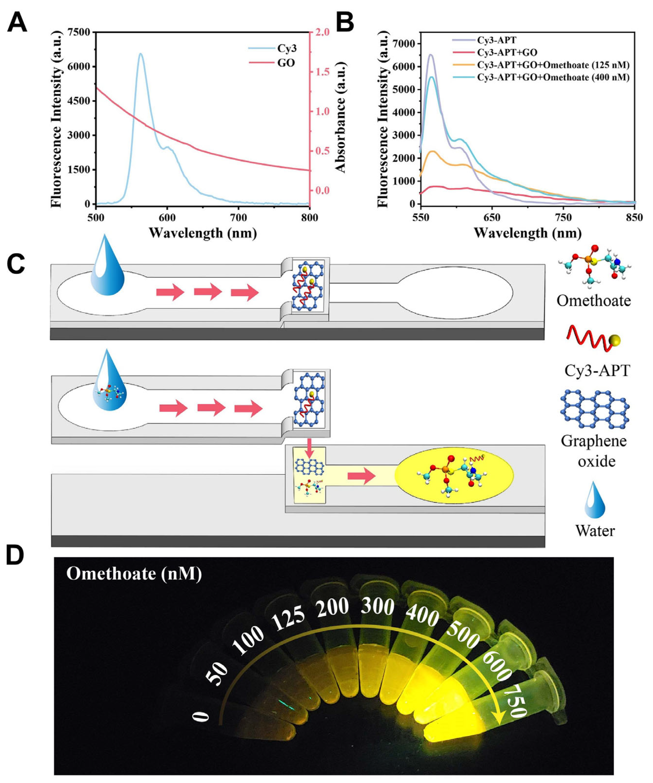
- Liu, S.; Zhao, J.; Wu, J.; Wang, L.; Yao, C.; Hu, J.; Zhang, H. A microfluidic paper-based fluorescent sensor integrated with a smartphone platform for rapid on-site detection of omethoate pesticide. Food Chem. 2025, 463, 141205. [Google Scholar] [CrossRef] [PubMed]
- Nagabooshanam, S.; Sharma, S.; Roy, S.; Mathur, A.; Krishnamurthy, S.; Bharadwaj, L.M. Development of field deployable sensor for detection of pesticide from food chain. IEEE Sens. J. 2020, 21, 4129–4134. [Google Scholar] [CrossRef]
- Teixeira, S.C.; Gomes, N.O.; Calegaro, M.L.; Machado, S.A.; de Oliveira, T.V.; Soares, N.d.F.F.; Raymundo-Pereira, P.A. Sustainable plant-wearable sensors for on-site, rapid decentralized detection of pesticides toward precision agriculture and food safety. Biomater. Adv. 2023, 155, 213676. [Google Scholar] [CrossRef] [PubMed]
- Cui, X.; Abd El-Aty, A.; Zhang, C.; Xu, L.; Liu, H.; Jia, H.; Wang, Y.; Cao, Z.; Salvador, J.-P.; She, Y. Enhanced bio-barcode immunoassay using droplet digital PCR for multiplex detection of organophosphate pesticides. J. Agric. Food Chem. 2021, 69, 11131–11141. [Google Scholar] [CrossRef] [PubMed]
- Zhang, M.; Si, Y.; Fu, Y.; An, J.; Zhang, Q.; Zhang, Y.; Zhang, H.; Yu, Y.; Zhang, D.; Fang, Y. Exploration of the Plant World: Application and Innovation of Plant-Wearable Sensors for Real-Time Detection. Crit. Rev. Anal. Chem. 2025, 1–17. [Google Scholar] [CrossRef]
- Kumar, M.; Sridharan, S.; Sawarkar, A.D.; Shakeel, A.; Anerao, P.; Mannina, G.; Sharma, P.; Pandey, A. Current research trends on emerging contaminants pharmaceutical and personal care products (PPCPs): A comprehensive review. Sci. Total Environ. 2023, 859, 160031. [Google Scholar] [CrossRef] [PubMed]
- Eze, E.M.; Aruoren, O.; Orogu, J.O.; Ukolobi, O.; Etaware, P.M.; Okpoghono, J. Effects of personal care products on surface water floating organisms. In Emergent Pollutants in Freshwater Plankton Communities; CRC Press: Boca Raton, FL, USA, 2024; pp. 106–120. [Google Scholar]
- Rehman, M.U.; Nisar, B.; Yatoo, A.M.; Sehar, N.; Tomar, R.; Tariq, L.; Ali, S.; Ali, A.; Rashid, S.M.; Ahmad, S.B. After effects of pharmaceuticals and personal care products (PPCPs) on the biosphere and their counteractive ways. Sep. Purif. Technol. 2024, 342, 126921. [Google Scholar] [CrossRef]
- Yang, Y.; Wu, R.S.S.; Tsang, Y.F. Revealing removal mechanisms of target personal care products in secondary wastewater treatment plants. J. Water Process Eng. 2024, 67, 106074. [Google Scholar] [CrossRef]
- Matesun, J.; Petrik, L.; Musvoto, E.; Ayinde, W.; Ikumi, D. Limitations of wastewater treatment plants in removing trace anthropogenic biomarkers and future directions: A review. Ecotoxicol. Environ. Saf. 2024, 281, 116610. [Google Scholar] [CrossRef] [PubMed]
- Silva, J.S.; de Lima Cruz, I.G.B.; Fragoso, W.D.; Lemos, S.G. Paper-based disposable electrochemical sensor from readily available materials for cost-effective triclosan detection. Microchem. J. 2024, 204, 111087. [Google Scholar] [CrossRef]
- Rahman, Z.; Singh, V.P. The relative impact of toxic heavy metals (THMs)(arsenic (As), cadmium (Cd), chromium (Cr)(VI), mercury (Hg), and lead (Pb)) on the total environment: An overview. Environ. Monit. Assess. 2019, 191, 419. [Google Scholar] [CrossRef] [PubMed]
- Sardar, K.; Ali, S.; Hameed, S.; Afzal, S.; Fatima, S.; Shakoor, M.B.; Bharwana, S.A.; Tauqeer, H.M. Heavy metals contamination and what are the impacts on living organisms. Greener J. Environ. Manag. Public Saf. 2013, 2, 172–179. [Google Scholar] [CrossRef]
- Hu, J.; Wang, L.; Song, Y.; Li, Y.; Shen, Y.; Gao, G.; Qin, L.; Wu, J.; Mulchandani, A. Ion imprinted polymers integrated into a multi-functional microfluidic paper-based analytical device for trace cadmium detection in water. Anal. Methods 2024, 16, 179–188. [Google Scholar] [CrossRef] [PubMed]
- Wang, W.; Ding, S.; Wang, Z.; Lv, Q.; Zhang, Q. Electrochemical paper-based microfluidic device for on-line isolation of proteins and direct detection of lead in urine. Biosens. Bioelectron. 2021, 187, 113310. [Google Scholar] [CrossRef] [PubMed]
- Pungjunun, K.; Praphairaksit, N.; Chailapakul, O. A facile and automated microfluidic electrochemical platform for the in-field speciation analysis of inorganic arsenic. Talanta 2023, 265, 124906. [Google Scholar] [CrossRef] [PubMed]
- Shin, S.; Jeon, B.; Kang, W.; Kim, C.; Choi, J.; Hong, S.C.; Lee, H.H. Characterization of microfluidic trap and mixer module for rapid fluorescent tagging of microplastics. Microfluid. Nanofluidics 2024, 28, 18. [Google Scholar] [CrossRef]
- Butement, J.T.; Wang, X.; Siracusa, F.; Miller, E.; Pabortsava, K.; Mowlem, M.; Spencer, D.; Morgan, H. Discrimination of microplastics and phytoplankton using impedance cytometry. ACS Sens. 2024, 9, 5206–5213. [Google Scholar] [CrossRef] [PubMed]
- Alatas, Y.C.; Tefek, U.; Salehin, S.; Alhmoud, H.; Hanay, M.S. Rapid differentiation between microplastic particles using integrated microwave cytometry with 3D electrodes. ACS Sens. 2025, 10, 1729–1735. [Google Scholar] [CrossRef] [PubMed]
- Shi, X.; Mao, T.; Huang, X.; Shi, H.; Jiang, K.; Lan, R.; Zhao, H.; Ma, J.; Zhao, J.; Xing, B. Capturing, enriching and detecting nanoplastics in water based on optical manipulation, surface-enhanced Raman scattering and microfluidics. Nat. Water 2025, 3, 449–460. [Google Scholar] [CrossRef]
- Martínez Vázquez, R.; Trotta, G.; Volpe, A.; Bernava, G.; Basile, V.; Paturzo, M.; Ferraro, P.; Ancona, A.; Fassi, I.; Osellame, R. Rapid prototyping of plastic lab-on-a-chip by femtosecond laser micromachining and removable insert microinjection molding. Micromachines 2017, 8, 328. [Google Scholar] [CrossRef] [PubMed]
- Wu, H.; Chen, J.; Yang, Y.; Yu, W.; Chen, Y.; Lin, P.; Liang, K. Smartphone-coupled three-layered paper-based microfluidic chips demonstrating stereoscopic capillary-driven fluid transport towards colorimetric detection of pesticides. Anal. Bioanal. Chem. 2022, 414, 1759–1772. [Google Scholar] [CrossRef] [PubMed]
- Ko, C.-H.; Liu, C.-C.; Huang, K.-H.; Fu, L.-M. Finger pump microfluidic detection system for methylparaben detection in foods. Food Chem. 2023, 407, 135118. [Google Scholar] [CrossRef] [PubMed]
- Yuan, M.; Li, C.; Zheng, Y.; Cao, H.; Ye, T.; Wu, X.; Hao, L.; Yin, F.; Yu, J.; Xu, F. A portable multi-channel fluorescent paper-based microfluidic chip based on smartphone imaging for simultaneous detection of four heavy metals. Talanta 2024, 266, 125112. [Google Scholar] [CrossRef] [PubMed]
- Sahin, F.; Celik, N.; Camdal, A.; Sakir, M.; Ceylan, A.; Ruzi, M.; Onses, M.S. Machine learning-assisted pesticide detection on a flexible surface-enhanced Raman scattering substrate prepared by silver nanoparticles. ACS Appl. Nano Mater. 2022, 5, 13112–13122. [Google Scholar] [CrossRef]
- Fdez-Sanromán, A.; Bernárdez-Rodas, N.; Rosales, E.; Pazos, M.; González-Romero, E.; Sanromán, M.Á. Biosensor technologies for water quality: Detection of emerging contaminants and pathogens. Biosensors 2025, 15, 189. [Google Scholar] [CrossRef] [PubMed]
- Saez, J.; Catalan-Carrio, R.; Owens, R.M.; Basabe-Desmonts, L.; Benito-Lopez, F. Microfluidics and materials for smart water monitoring: A review. Anal. Chim. Acta 2021, 1186, 338392. [Google Scholar] [CrossRef] [PubMed]
- Zhang, Y.; Li, J.; Jiao, S.; Li, Y.; Zhou, Y.; Zhang, X.; Maryam, B.; Liu, X. Microfluidic sensors for the detection of emerging contaminants in water: A review. Sci. Total Environ. 2024, 929, 172734. [Google Scholar] [CrossRef] [PubMed]


Disclaimer/Publisher’s Note: The statements, opinions and data contained in all publications are solely those of the individual author(s) and contributor(s) and not of MDPI and/or the editor(s). MDPI and/or the editor(s) disclaim responsibility for any injury to people or property resulting from any ideas, methods, instructions or products referred to in the content. |
© 2025 by the authors. Licensee MDPI, Basel, Switzerland. This article is an open access article distributed under the terms and conditions of the Creative Commons Attribution (CC BY) license (https://creativecommons.org/licenses/by/4.0/).
Share and Cite
Abdelhamid, M.A.A.; Ki, M.-R.; Yoon, H.J.; Pack, S.P. Microfluidic Sensors for Micropollutant Detection in Environmental Matrices: Recent Advances and Prospects. Biosensors 2025, 15, 474. https://doi.org/10.3390/bios15080474
Abdelhamid MAA, Ki M-R, Yoon HJ, Pack SP. Microfluidic Sensors for Micropollutant Detection in Environmental Matrices: Recent Advances and Prospects. Biosensors. 2025; 15(8):474. https://doi.org/10.3390/bios15080474
Chicago/Turabian StyleAbdelhamid, Mohamed A. A., Mi-Ran Ki, Hyo Jik Yoon, and Seung Pil Pack. 2025. "Microfluidic Sensors for Micropollutant Detection in Environmental Matrices: Recent Advances and Prospects" Biosensors 15, no. 8: 474. https://doi.org/10.3390/bios15080474
APA StyleAbdelhamid, M. A. A., Ki, M.-R., Yoon, H. J., & Pack, S. P. (2025). Microfluidic Sensors for Micropollutant Detection in Environmental Matrices: Recent Advances and Prospects. Biosensors, 15(8), 474. https://doi.org/10.3390/bios15080474