Electrochemical Biosensors for Detection of MicroRNA as a Cancer Biomarker: Pros and Cons

 , and
, and 
Abstract
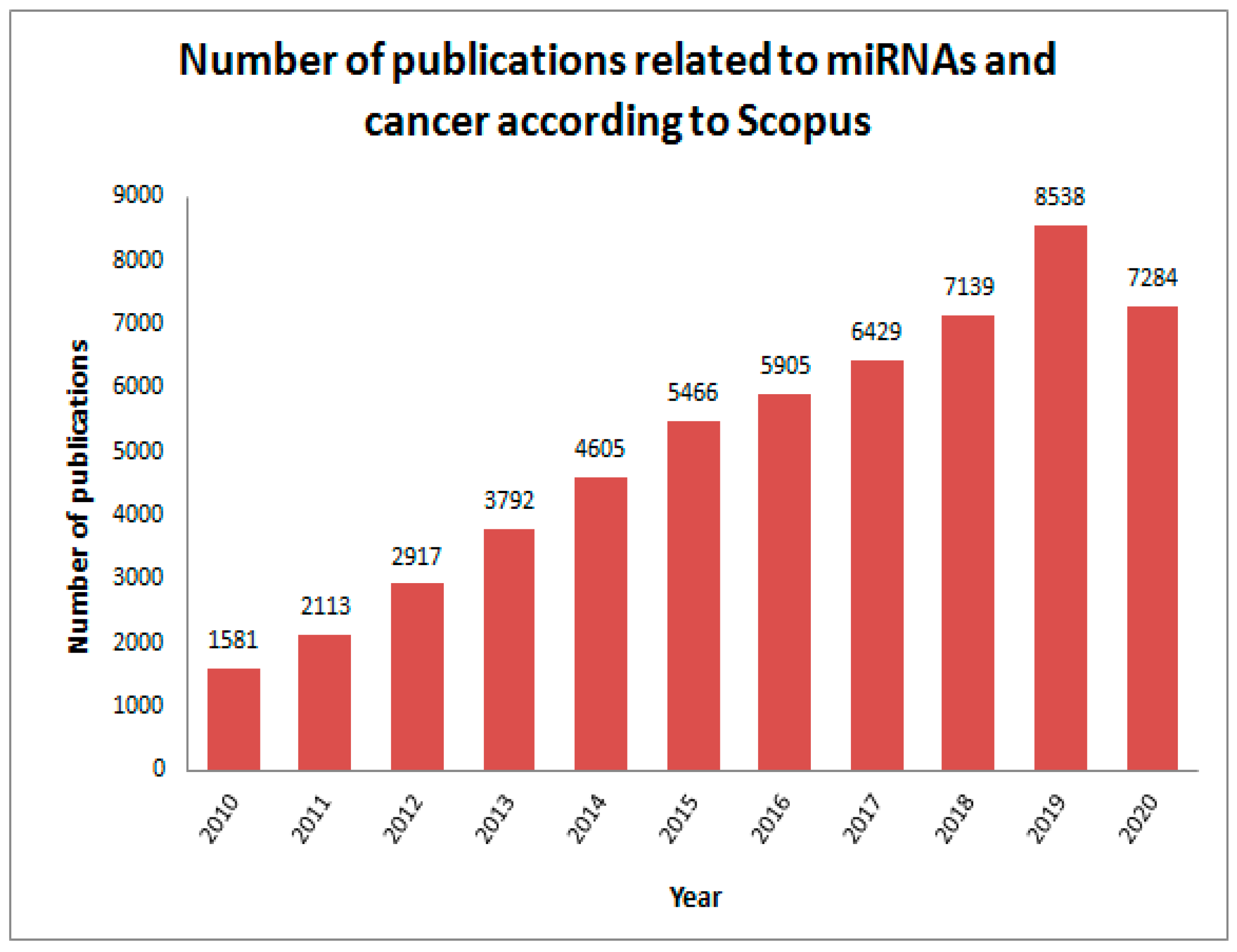
1. Introduction
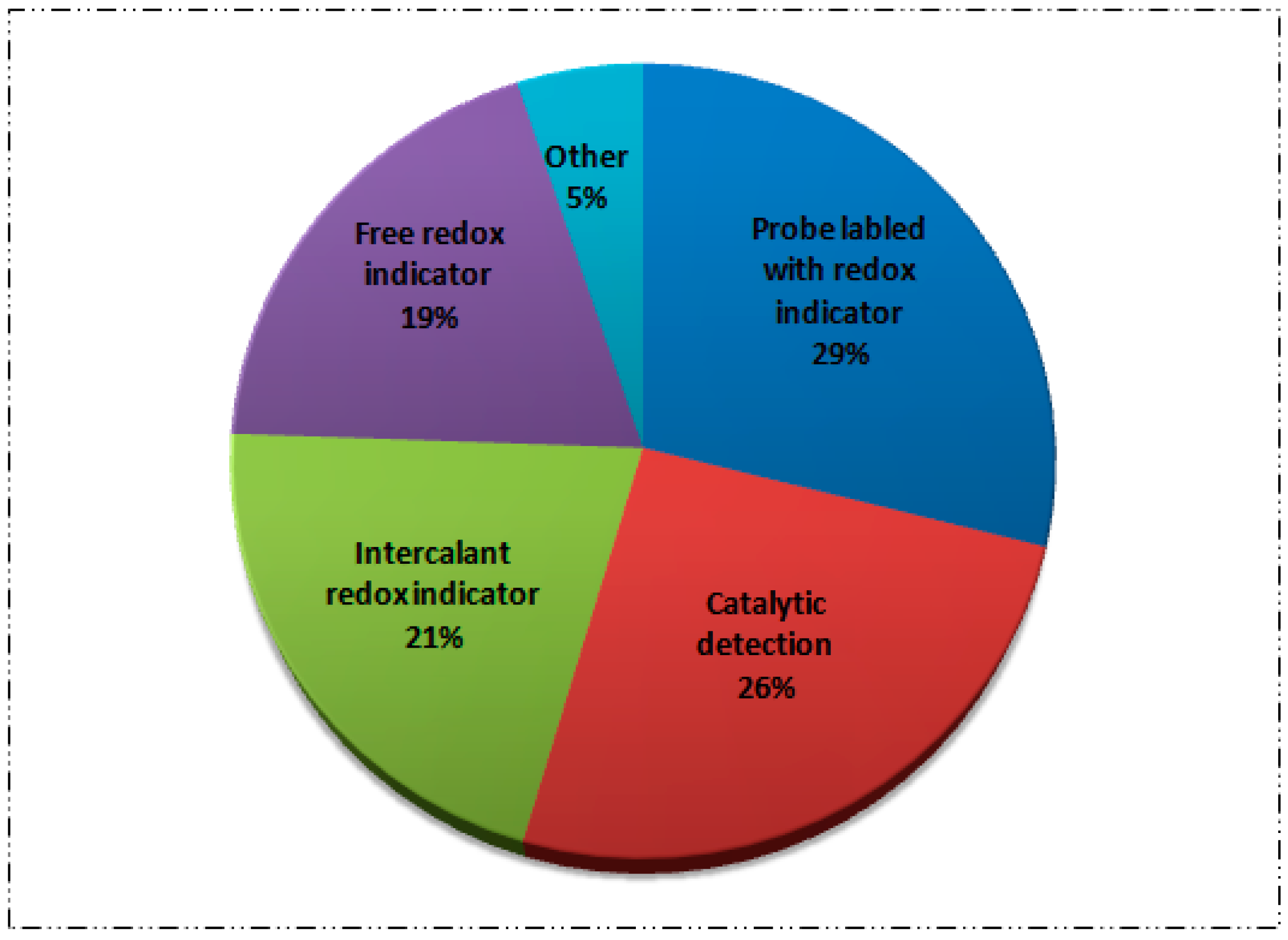
2. Electrochemical Biosensor Based on Electroactive Labeled Probe Sequence
2.1. Direct Labeling
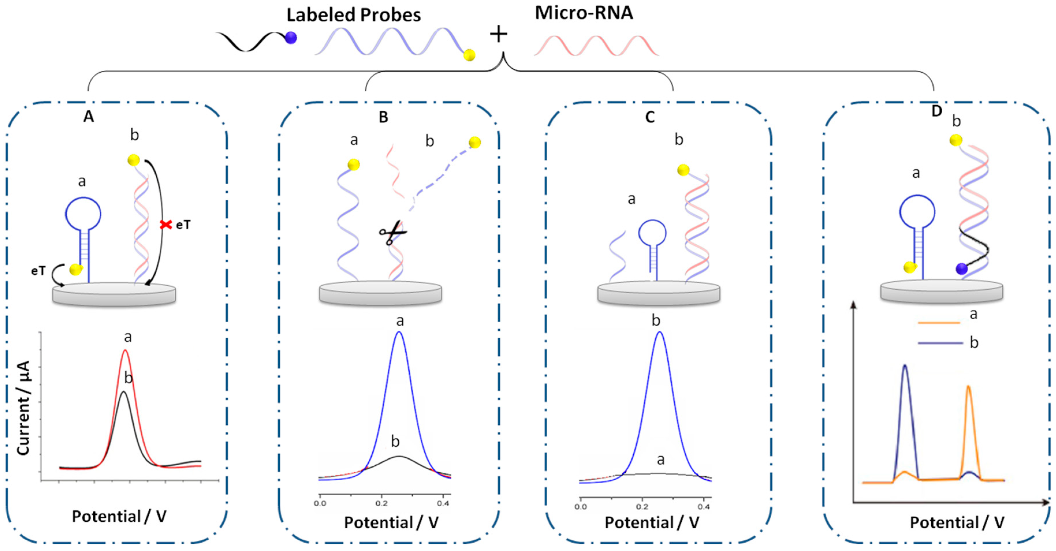
2.1.1. Basic Design
2.1.2. Response Based on the Elimination of the Labeled Probe
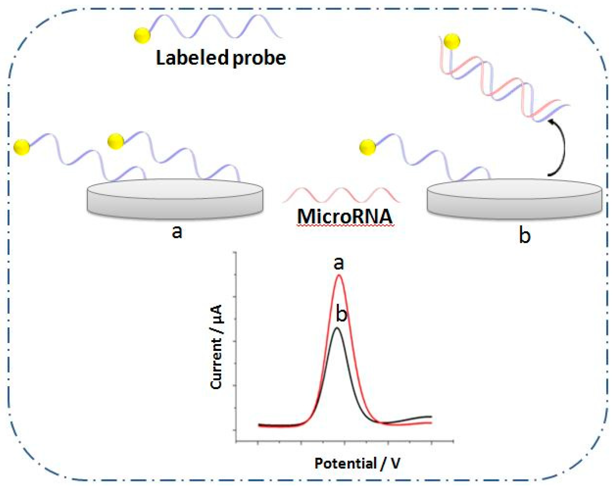
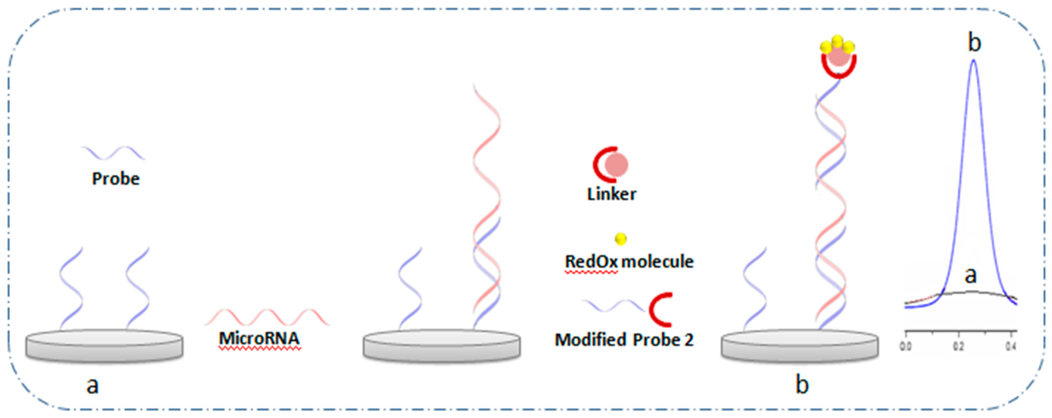
2.1.3. The Use of Secondary Probe Labeled with RedOx Molecule
2.1.4. Response Based on Two Labeled Probes
2.2. RedOx Molecules Linker to Nanocarriers
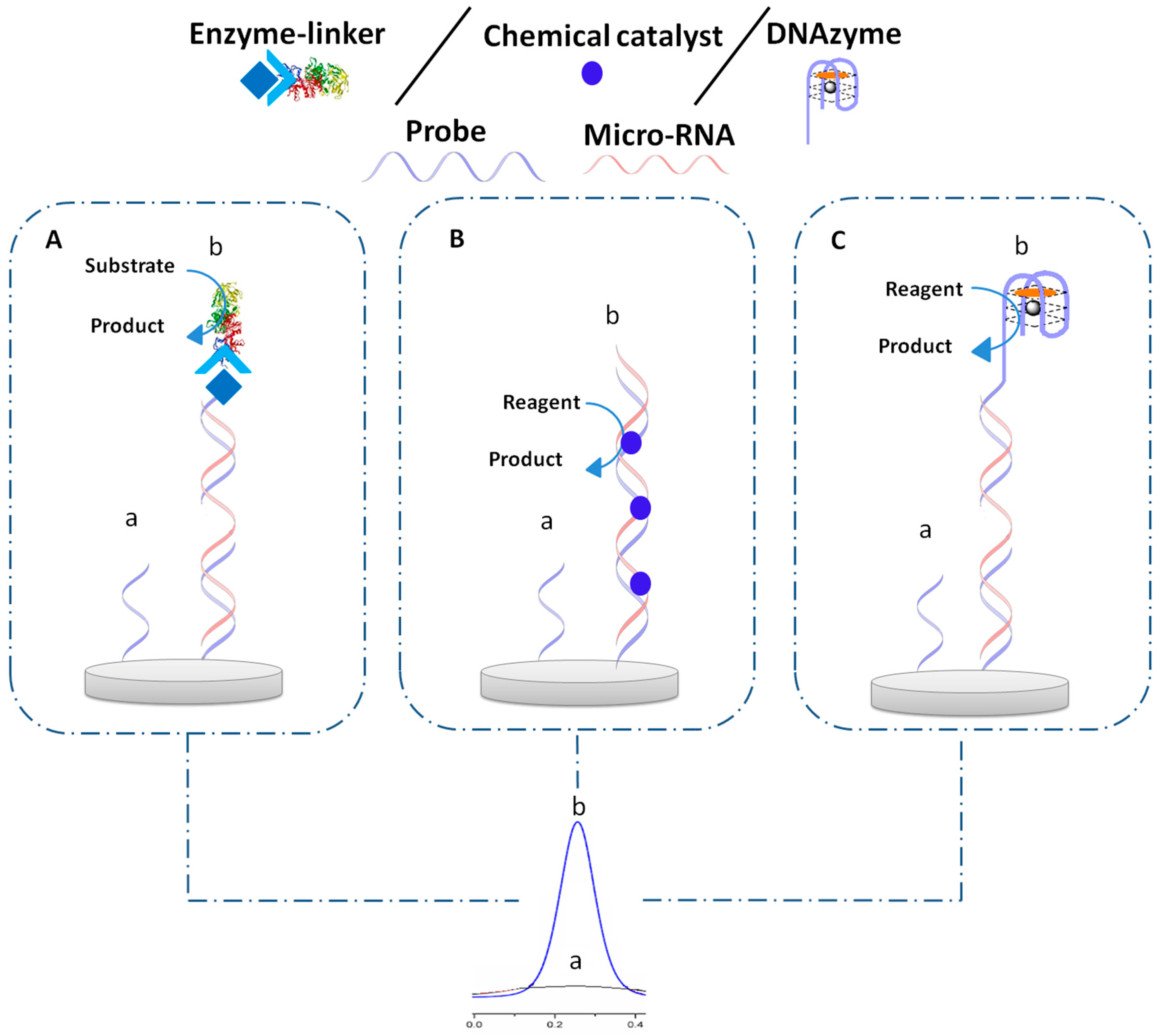
3. Electrochemical Biosensor Based on Catalysts
3.1. Enzyme
3.1.1. Enzyme–Steptavidin Binding
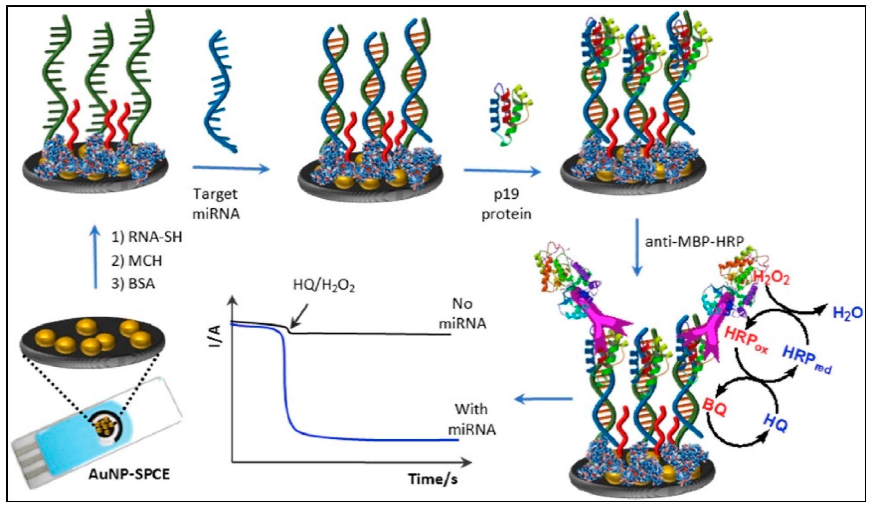
3.1.2. Enzyme–Protein Binding
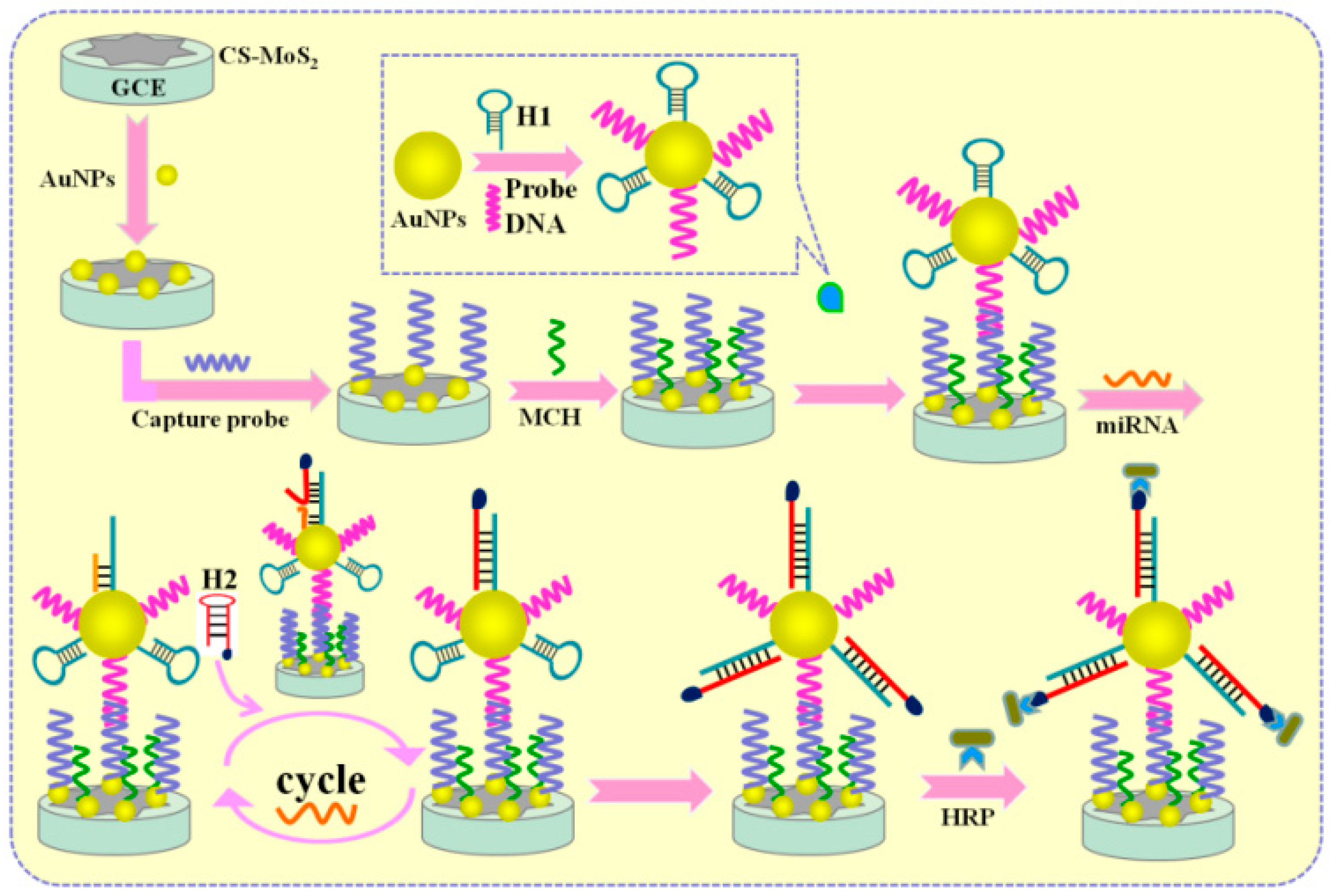
3.1.3. Other Types of Enzyme Binding
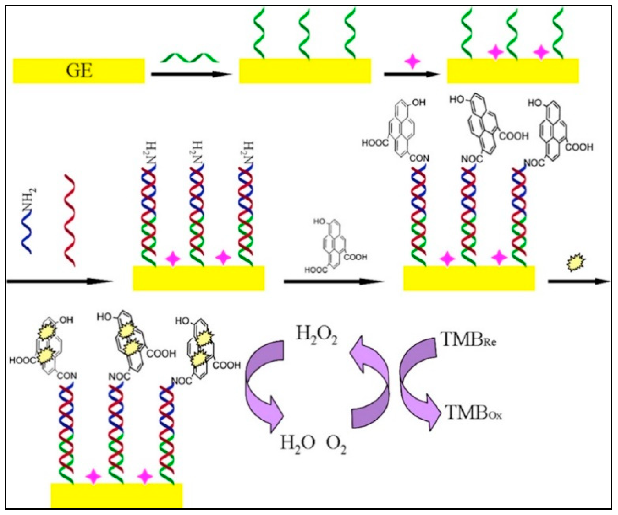
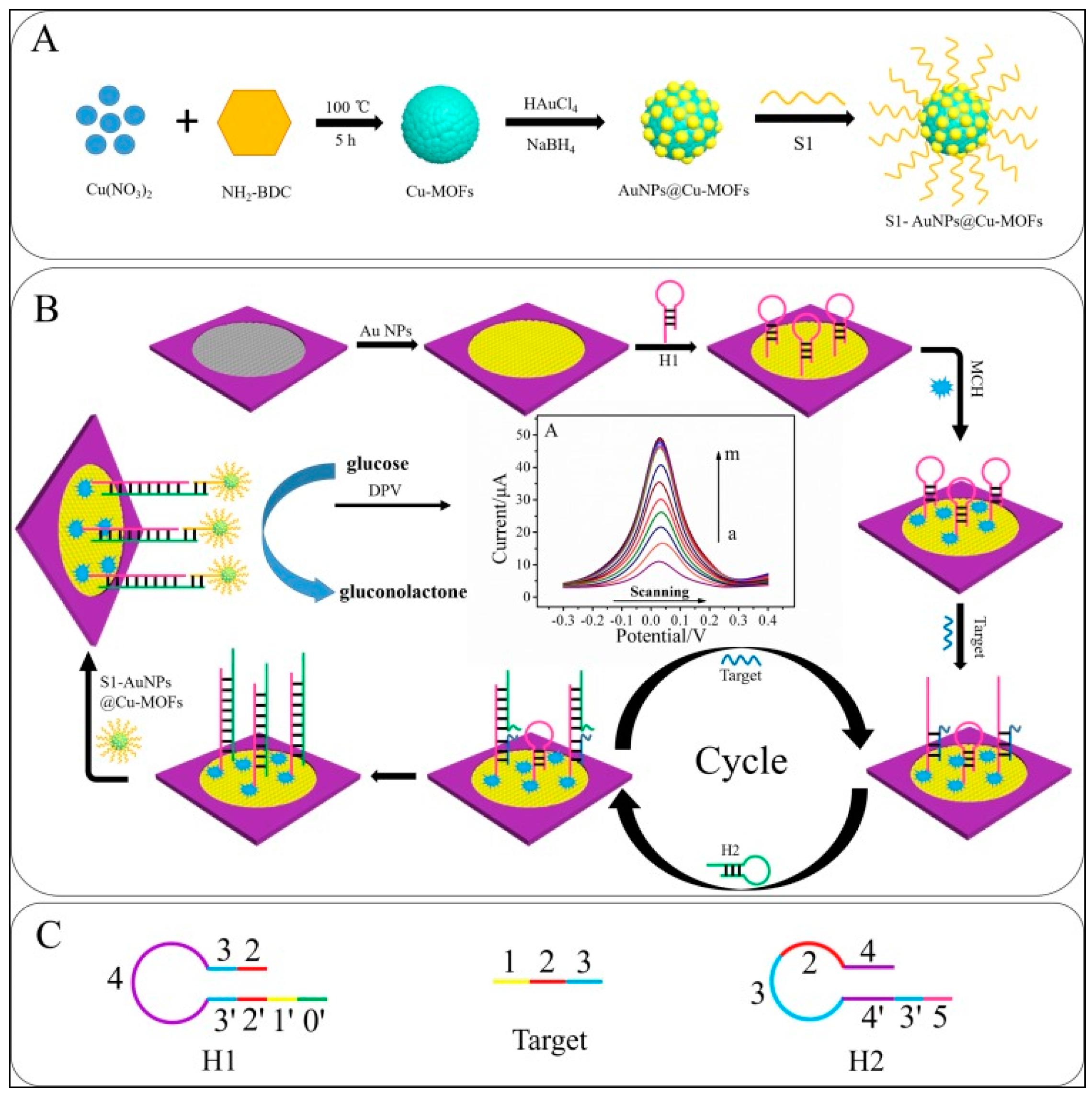
3.2. Chemical Catalysts
3.3. DNAzyme
4. Electrochemical Biosensor Based on RedOx Intercalating Agent
4.1. Direct Intercalation
4.1.1. Electroactive Molecule
4.1.2. Electroactive Metals Complex
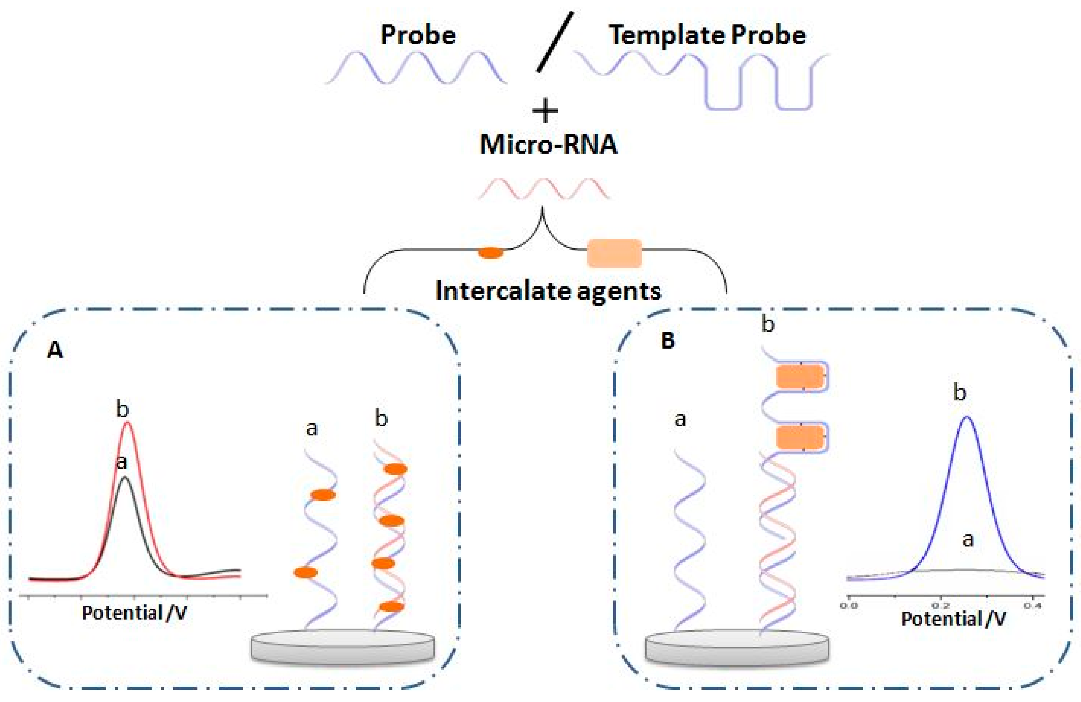
4.2. Intercalation via Template
4.3. Other Type of Intercalation
5. Electrochemical Label-Free Biosensing
5.1. Ferri/Ferrocyanide as Free RedOx Indicator
5.2. Hexaammineruthenium (II)/(III) Chloride as Free RedOx Indicator
6. Other Methods of MicroRNA Electrochemical Detection
6.1. Oxidation of Guanine
6.2. RedOx Current from Electrode Surface
6.3. Labeled MicroRNA
7. Conclusions and Future Perspectives
Author Contributions
Funding
Conflicts of Interest
References
- Roointan, A.; Mir, T.A.; Wani, S.I.; Rehman, M.; Hussain, K.K.; Ahmed, B.; Abrahim, S.; Savardashtaki, A.; Gandomani, G.; Gandomani, M.; et al. Early detection of lung cancer biomarkers through biosensor technology: A review. J. Pharm. Biomed. Anal. 2019, 164, 93–103. [Google Scholar] [CrossRef] [PubMed]
- Schootman, M.; Fuortes, L.; Aft, R. Prognosis of metachronous contralateral breast cancer according to stage at diagnosis: The importance of early detection. Breast Cancer Res. Treat. 2006, 99, 91–95. [Google Scholar] [CrossRef] [PubMed]
- Lytras, D.; Connor, S.; Bosonnet, L.; Jayan, R.; Evans, J.; Hughes, M.; Garvey, C.; Ghaneh, P.; Sutton, R.; Vinjamuri, S.; et al. Positron Emission Tomography Does Not Add to Computed Tomography for the Diagnosis and Staging of Pancreatic Cancer. Dig. Surg. 2005, 22, 55–62. [Google Scholar] [CrossRef] [PubMed]
- Portalez, D.; Mozer, P.; Cornud, F.; Renard-Penna, R.; Misrai, V.; Thoulouzan, M.; Malavaud, B. Validation of the European Society of Urogenital Radiology Scoring System for Prostate Cancer Diagnosis on Multiparametric Magnetic Resonance Imaging in a Cohort of Repeat Biopsy Patients. Eur. Urol. 2012, 62, 986–996. [Google Scholar] [CrossRef] [PubMed]
- Wagner, P.D.; Verma, M.; Srivastava, S. Challenges for Biomarkers in Cancer Detection. Ann. N. Y. Acad. Sci. 2004, 1022, 9–16. [Google Scholar] [CrossRef] [PubMed]
- Islam, N.; Masud, M.K.; Haque, H.; Hossain, S.A.; Yamauchi, Y.; Nguyen, N.-T.; Shiddiky, M.J. RNA Biomarkers: Diagnostic and Prognostic Potentials and Recent Developments of Electrochemical Biosensors. Small Methods 2017, 1, 1700131. [Google Scholar] [CrossRef]
- Mittal, S.; Kaur, H.; Gautam, N.; Mantha, A.K. Biosensors for breast cancer diagnosis: A review of bioreceptors, biotransducers and signal amplification strategies. Biosens. Bioelectron. 2017, 88, 217–231. [Google Scholar] [CrossRef]
- Mousa, S.A. Biosensors: The new wave in cancer diagnosis. Nanotechnol. Sci. Appl. 2010, 4, 1–10. [Google Scholar] [CrossRef]
- Hasanzadeh, M.; Shadjou, N.; De La Guardia, M. Early stage screening of breast cancer using electrochemical biomarker detection. TrAC Trends Anal. Chem. 2017, 91, 67–76. [Google Scholar] [CrossRef]
- Diaconu, I.; Cristea, C.; Harceaga, V.; Marrazza, G.; Berindan-Neagoe, I.; Săndulescu, R. Electrochemical immunosensors in breast and ovarian cancer. Clin. Chim. Acta 2013, 425, 128–138. [Google Scholar] [CrossRef]
- Yang, H.; Wang, J.; Yang, C.; Zhao, X.; Xie, S.; Ge, Z. Nano Pt@ZIF8 Modified Electrode and Its Application to Detect Sarcosine. J. Electrochem. Soc. 2018, 165, H247–H250. [Google Scholar] [CrossRef]
- Filella, X.; Fernández-Galán, E.; Bonifacio, R.F.; Foj, L. Emerging biomarkers in the diagnosis of prostate cancer. Pharm. Pers. Med. 2018, 11, 83–94. [Google Scholar] [CrossRef] [PubMed]
- Vaarala, M.H.; Porvari, K.; Lukkarinen, O.; Vihko, P. TheTMPRSS2 gene encoding transmembrane serine protease is overexpressed in a majority of prostate cancer patients: Detection of mutatedTMPRSS2 form in a case of aggressive disease. Int. J. Cancer 2001, 94, 705–710. [Google Scholar] [CrossRef]
- Elshafey, R.; Tlili, C.; Abulrob, A.; Tavares, A.C.; Zourob, M. Label-free impedimetric immunosensor for ultrasensitive detection of cancer marker Murine double minute 2 in brain tissue. Biosens. Bioelectron. 2013, 39, 220–225. [Google Scholar] [CrossRef] [PubMed]
- Laocharoensuk, R. Development of Electrochemical Immunosensors towards Point-of-care Cancer Diagnostics: Clinically Relevant Studies. Electroanalysis 2016, 28, 1716–1729. [Google Scholar] [CrossRef]
- Hasan, S.; Jacob, R.; Manne, U.; Paluri, R. Advances in pancreatic cancer biomarkers. Oncol. Rev. 2019, 13, 410. [Google Scholar] [CrossRef]
- Matsuoka, T.; Yashiro, M. Biomarkers of gastric cancer: Current topics and future perspective. World J. Gastroenterol. 2018, 24, 2818–2832. [Google Scholar] [CrossRef]
- Ashizawa, T.; Okada, R.; Suzuki, Y.; Takagi, M.; Yamazaki, T.; Sumi, T.; Aoki, T.; Ohnuma, S.; Aoki, T. Clinical significance of interleukin-6 (IL-6) in the spread of gastric cancer: Role of IL-6 as a prognostic factor. Gastric Cancer 2005, 8, 124–131. [Google Scholar] [CrossRef]
- Lou, J.; Zhang, L.; Lv, S.; Zhang, C.; Jiang, S. Biomarkers for Hepatocellular Carcinoma. Biomark. Cancer 2017, 9, 1–9. [Google Scholar] [CrossRef]
- Farzin, L.; Shamsipur, M. Recent advances in design of electrochemical affinity biosensors for low level detection of cancer protein biomarkers using nanomaterial-assisted signal enhancement strategies. J. Pharm. Biomed. Anal. 2018, 147, 185–210. [Google Scholar] [CrossRef]
- Li, J.; Sherman-Baust, A.C.; Tsai-Turton, M.; Bristow, R.E.; Roden, R.B.S.; Morin, P.J. Claudin-containing exosomes in the peripheral circulation of women with ovarian cancer. BMC Cancer 2009, 9, 244. [Google Scholar] [CrossRef] [PubMed]
- Altintas, Z.; Tothill, I. Biomarkers and biosensors for the early diagnosis of lung cancer. Sens. Actuators B Chem. 2013, 188, 988–998. [Google Scholar] [CrossRef]
- Arya, S.K.; Bhansali, S. Lung Cancer and Its Early Detection Using Biomarker-Based Biosensors. Chem. Rev. 2011, 111, 6783–6809. [Google Scholar] [CrossRef] [PubMed]
- Xie, Y.; Todd, N.W.; Liu, Z.; Zhan, M.; Fang, H.; Peng, H.; Alattar, M.; Deepak, J.; Stass, S.A.; Jiang, F. Altered miRNA expression in sputum for diagnosis of non-small cell lung cancer. Lung Cancer 2010, 67, 170–176. [Google Scholar] [CrossRef] [PubMed]
- Lim, E.H.; Zhang, S.-L.; Li, J.-L.; Yap, W.-S.; Howe, T.-C.; Tan, B.-P.; Lee, Y.-S.; Wong, D.; Khoo, K.-L.; Seto, K.-Y.; et al. Using whole genome amplification (WGA) of low-volume biopsies to assess the prognostic role of EGFR, KRAS, p53, and CMET mutations in advanced-stage non-small cell lung cancer (NSCLC). J. Thorac. Oncol. 2009, 4, 12–21. [Google Scholar] [CrossRef] [PubMed]
- Staden, R.-I.S.-V.; Comnea-Stancu, I.R.; Surdu-Bob, C.C. Molecular Screening of Blood Samples for the Simultaneous Detection of CEA, HER-1, NSE, CYFRA 21-1 Using Stochastic Sensors. J. Electrochem. Soc. 2017, 164, B267–B273. [Google Scholar] [CrossRef]
- Carr, O.; Raymundo-Pereira, P.A.; Shimizu, F.M.; Sorroche, B.P.; Melendez, M.E.; Pedro, R.D.O.; Miranda, P.B.; Carvalho, A.L.; Reis, R.M.; Arantes, L.M.; et al. Genosensor made with a self-assembled monolayer matrix to detect MGMT gene methylation in head and neck cancer cell lines. Talanta 2020, 210, 120609. [Google Scholar] [CrossRef]
- Lee, R.C.; Feinbaum, R.L.; Ambros, V. The C. elegans heterochronic gene lin-4 encodes small RNAs with antisense complementarity to lin-14. Cell 1993, 75, 843–854. [Google Scholar] [CrossRef]
- Negrini, M.; Ferracin, M.; Sabbioni, S.; Croce, C.M. MicroRNAs in human cancer: From research to therapy. J. Cell Sci. 2007, 120, 1833–1840. [Google Scholar] [CrossRef]
- Tang, X.; Tang, G.; Özcan, S. Role of microRNAs in diabetes. Biochim. Biophys. Acta BBA Bioenergy 2008, 1779, 697–701. [Google Scholar] [CrossRef]
- Chen, Y.-X.; Huang, K.-J.; Niu, K.-X. Recent advances in signal amplification strategy based on oligonucleotide and nanomaterials for microRNA detection—A review. Biosens. Bioelectron. 2018, 99, 612–624. [Google Scholar] [CrossRef] [PubMed]
- Keshavarz, M.; Behpour, M.; Rafiee-Pour, H.-A. Recent trends in electrochemical microRNA biosensors for early detection of cancer. RSC Adv. 2015, 5, 35651–35660. [Google Scholar] [CrossRef]
- Aziz, N.B.; Mahmudunnabi, R.G.; Umer, M.; Sharma, S.; Rashid, A.; Alhamhoom, Y.; Shim, Y.-B.; Salomon, C.; Shiddiky, M.J. MicroRNAs in ovarian cancer and recent advances in the development of microRNA-based biosensors. Analyst 2020, 145, 2038–2057. [Google Scholar] [CrossRef] [PubMed]
- Miodek, A.; Mejri-Omrani, N.; Khoder, R.; Korri-Youssoufi, H.; Mejri, N. Electrochemical functionalization of polypyrrole through amine oxidation of poly(amidoamine) dendrimers: Application to DNA biosensor. Talanta 2016, 154, 446–454. [Google Scholar] [CrossRef] [PubMed]
- Wang, Y.; Hsine, Z.; Sauriat-Dorizon, H.; Mlika, R.; Korri-Youssoufi, H. Structural and electrochemical studies of functionalization of reduced graphene oxide with alkoxyphenylporphyrin mono- and tetra- carboxylic acid: Application to DNA sensors. Electrochim. Acta 2020, 357, 136852. [Google Scholar] [CrossRef]
- Yazdanparast, S.; Benvidi, A.; Azimzadeh, M.; Tezerjani, M.D.; Ghaani, M.R. Experimental and theoretical study for miR-155 detection through resveratrol interaction with nucleic acids using magnetic core-shell nanoparticles. Microchim. Acta 2020, 187, 1–10. [Google Scholar] [CrossRef]
- Masud, M.K.; Umer, M.; Hossain, S.A.; Yamauchi, Y.; Nguyen, N.-T.; Shiddiky, M.J. Nanoarchitecture Frameworks for Electrochemical miRNA Detection. Trends Biochem. Sci. 2019, 44, 433–452. [Google Scholar] [CrossRef]
- Chen, C. Recent Advances in Nanomaterials-Based Electrochemical Biosensors for MicroRNAs Detection. Int. J. Electrochem. Sci. 2019, 14, 5174–5187. [Google Scholar] [CrossRef]
- Mujica, M.L.; Gallay, P.A.; Perrachione, F.; Montemerlo, A.E.; Tamborelli, L.A.; Vaschetti, V.M.; Reartes, D.F.; Bollo, S.; Rodríguez, M.C.; Dalmasso, P.R.; et al. New trends in the development of electrochemical biosensors for the quantification of microRNAs. J. Pharm. Biomed. Anal. 2020, 189, 113478. [Google Scholar] [CrossRef]
- Mohammadi, H.; Yammouri, G.; Amine, A. Current advances in electrochemical genosensors for detecting microRNA cancer markers. Curr. Opin. Electrochem. 2019, 16, 96–105. [Google Scholar] [CrossRef]
- Daneshpour, M.; Omidfar, K.; Ghanbarian, H. A novel electrochemical nanobiosensor for the ultrasensitive and specific detection of femtomolar-level gastric cancer biomarker miRNA-106a. Beilstein J. Nanotechnol. 2016, 7, 2023–2036. [Google Scholar] [CrossRef]
- Sun, E.; Wang, L.; Zhou, X.; Ma, C.; Sun, Y.; Lei, M.; Lu, B.; Han, R. Retracted Article: Graphene oxide/DNA-decorated electrode for the fabrication of microRNA biosensor. RSC Adv. 2015, 5, 69334–69338. [Google Scholar] [CrossRef]
- Yang, B.; Zhang, S.; Fang, X.; Kong, J. Double signal amplification strategy for ultrasensitive electrochemical biosensor based on nuclease and quantum dot-DNA nanocomposites in the detection of breast cancer 1 gene mutation. Biosens. Bioelectron. 2019, 142, 111544. [Google Scholar] [CrossRef] [PubMed]
- Rezaei, H.; Motovali-Bashi, M.; Radfar, S. An enzyme-free electrochemical biosensor for simultaneous detection of two hemophilia A biomarkers: Combining target recycling with quantum dots-encapsulated metal-organic frameworks for signal amplification. Anal. Chim. Acta 2019, 1092, 66–74. [Google Scholar] [CrossRef] [PubMed]
- Tian, R.; Ning, W.; Chen, M.; Zhang, C.; Li, Q.; Bai, J. High performance electrochemical biosensor based on 3D nitrogen-doped reduced graphene oxide electrode and tetrahedral DNA nanostructure. Talanta 2019, 194, 273–281. [Google Scholar] [CrossRef]
- Chen, Z.; Xie, Y.; Huang, W.; Qin, C.; Yu, A.; Lai, G. Exonuclease-assisted target recycling for ultrasensitive electrochemical detection of microRNA at vertically aligned carbon nanotubes. Nanoscale 2019, 11, 11262–11269. [Google Scholar] [CrossRef]
- Yammouri, G.; Mohammadi, H.; Amine, A. A Highly Sensitive Electrochemical Biosensor Based on Carbon Black and Gold Nanoparticles Modified Pencil Graphite Electrode for microRNA-21 Detection. Chem. Afr. 2019, 2, 291–300. [Google Scholar] [CrossRef]
- Jou, A.F.-J.; Chen, Y.-J.; Li, Y.; Chang, Y.-F.; Lee, J.-J.; Liao, A.T.; Ho, J.-A.A. Target-Triggered, Dual Amplification Strategy for Sensitive Electrochemical Detection of a Lymphoma-associated MicroRNA. Electrochim. Acta 2017, 236, 190–197. [Google Scholar] [CrossRef]
- Miao, P.; Jiang, Y.; Zhang, T.; Huang, Y.; Tang, Y. Electrochemical sensing of attomolar miRNA combining cascade strand displacement polymerization and reductant-mediated amplification. Chem. Commun. 2018, 54, 7366–7369. [Google Scholar] [CrossRef]
- Wang, T.; Viennois, E.; Merlin, D.; Wang, G. Microelectrode miRNA Sensors Enabled by Enzymeless Electrochemical Signal Amplification. Anal. Chem. 2015, 87, 8173–8180. [Google Scholar] [CrossRef]
- Miao, P.; Wang, B.; Yu, Z.; Zhao, J.; Tang, Y. Ultrasensitive electrochemical detection of microRNA with star trigon structure and endonuclease mediated signal amplification. Biosens. Bioelectron. 2015, 63, 365–370. [Google Scholar] [CrossRef] [PubMed]
- Ma, X.; Xu, H.; Qian, K.; Kandawa-Schulz, M.; Miao, W.; Wang, Y. Electrochemical detection of microRNAs based on AuNPs/CNNS nanocomposite with Duplex-specific nuclease assisted target recycling to improve the sensitivity. Talanta 2020, 208, 120441. [Google Scholar] [CrossRef] [PubMed]
- Yang, D.; Cheng, W.; Chen, X.; Tang, Y.; Miao, P. Ultrasensitive electrochemical detection of miRNA based on DNA strand displacement polymerization and Ca2+-dependent DNAzyme cleavage. Analyst 2018, 143, 5352–5357. [Google Scholar] [CrossRef] [PubMed]
- Wang, W.; Jayachandran, S.; Li, M.; Xu, S.; Luo, X. Hyaluronic acid functionalized nanostructured sensing interface for voltammetric determination of microRNA in biological media with ultra-high sensitivity and ultra-low fouling. Microchim. Acta 2018, 185, 156. [Google Scholar] [CrossRef] [PubMed]
- Zhang, X.; Yang, Z.; Chang, Y.; Qing, M.; Yuan, R.; Chai, Y. Novel 2D-DNA-Nanoprobe-Mediated Enzyme-Free-Target-Recycling Amplification for the Ultrasensitive Electrochemical Detection of MicroRNA. Anal. Chem. 2018, 90, 9538–9544. [Google Scholar] [CrossRef]
- Fu, C.; Liu, C.; Wang, S.; Luo, F.; Lin, Z.; Chen, G. A signal-on homogeneous electrochemical biosensor for sequence-specific microRNA based on duplex-specific nuclease-assisted target recycling amplification. Anal. Methods 2016, 8, 7034–7039. [Google Scholar] [CrossRef]
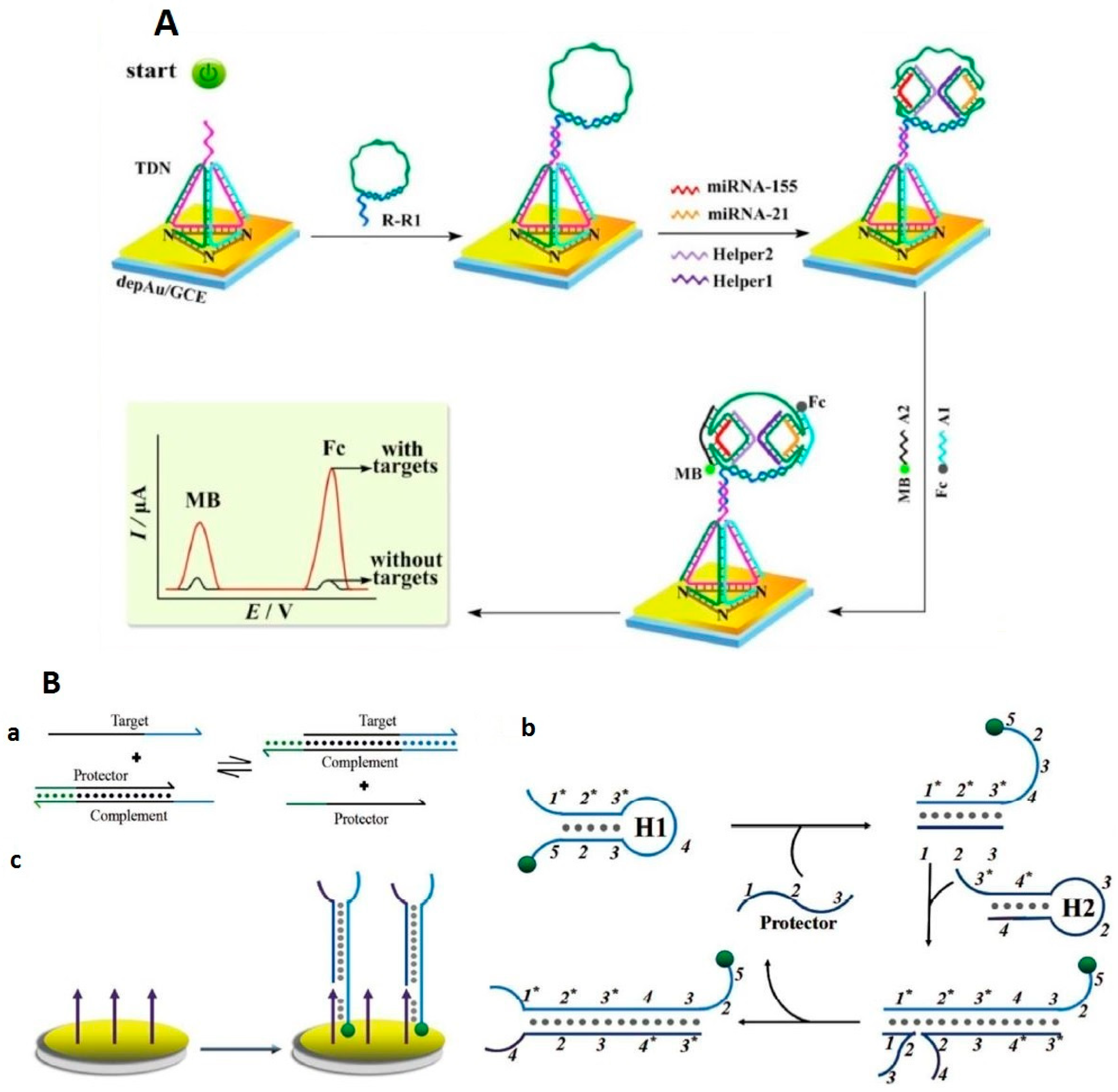
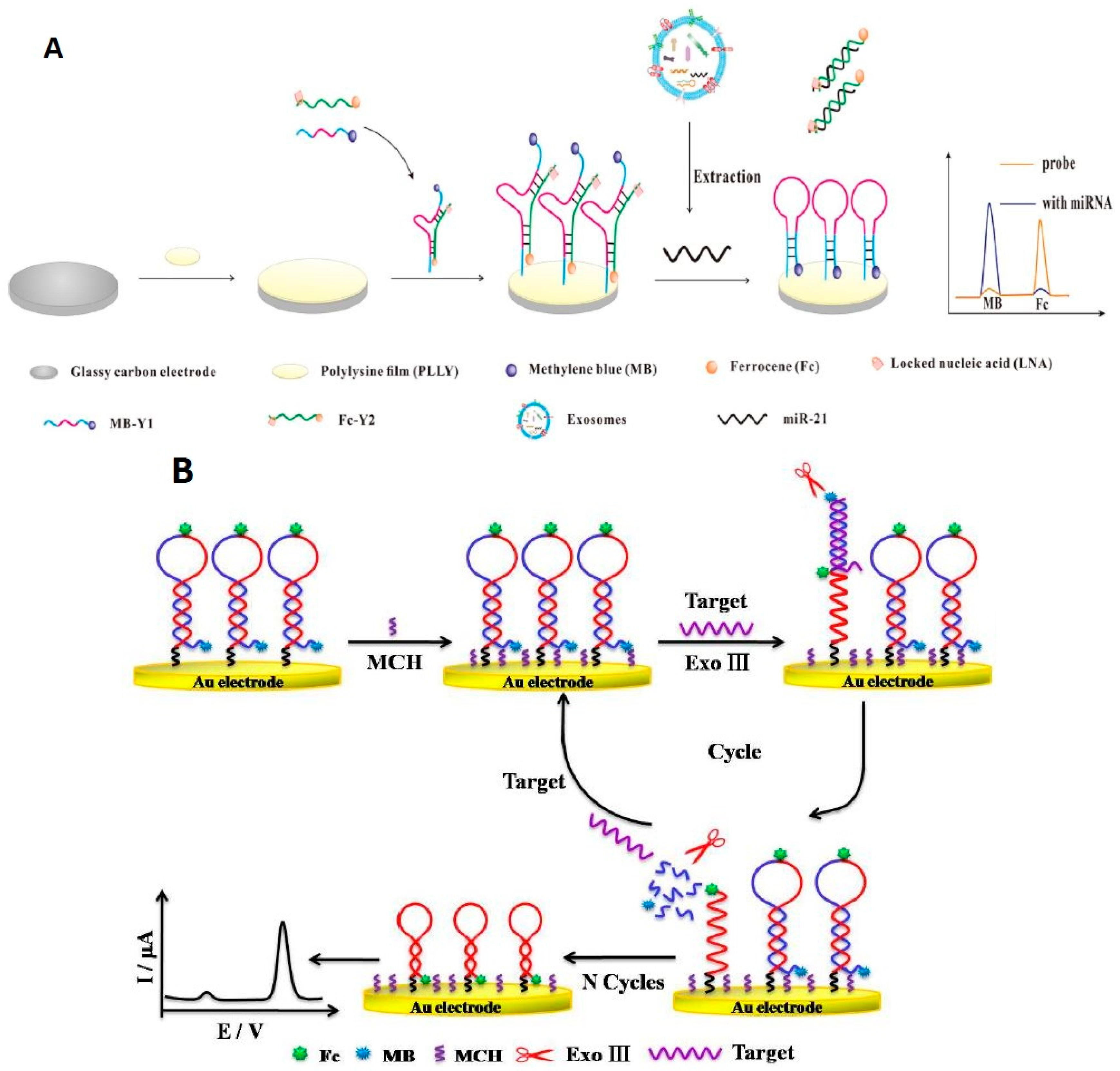
- Miao, P.; Wang, B.; Chen, X.; Li, X.; Tang, Y. Tetrahedral DNA Nanostructure-Based MicroRNA Biosensor Coupled with Catalytic Recycling of the Analyte. ACS Appl. Mater. Interfaces 2015, 7, 6238–6243. [Google Scholar] [CrossRef]
- Miao, P.; Wang, B.; Meng, F.; Yin, J.; Tang, Y. Ultrasensitive Detection of MicroRNA through Rolling Circle Amplification on a DNA Tetrahedron Decorated Electrode. Bioconjugate Chem. 2015, 26, 602–607. [Google Scholar] [CrossRef]
- Liu, H.; Bei, X.; Xia, Q.; Fu, Y.; Zhang, S.; Liu, M.; Fan, K.; Zhang, M.; Yang, Y. Enzyme-free electrochemical detection of microRNA-21 using immobilized hairpin probes and a target-triggered hybridization chain reaction amplification strategy. Microchim. Acta 2015, 183, 297–304. [Google Scholar] [CrossRef]
- Xu, S.; Chang, Y.; Wu, Z.; Li, Y.; Yuan, R.; Chai, Y.-Q. One DNA circle capture probe with multiple target recognition domains for simultaneous electrochemical detection of miRNA-21 and miRNA-155. Biosens. Bioelectron. 2020, 149, 111848. [Google Scholar] [CrossRef]
- Yao, J.; Zhang, Z.; Deng, Z.; Wang, Y.; Guo, Y. An enzyme free electrochemical biosensor for sensitive detection of miRNA with a high discrimination factor by coupling the strand displacement reaction and catalytic hairpin assembly recycling. Analyst 2017, 142, 4116–4123. [Google Scholar] [CrossRef] [PubMed]
- Zhou, L.; Wang, J.; Chen, Z.; Li, J.; Wang, T.; Zhang, Z.; Xie, G. A universal electrochemical biosensor for the highly sensitive determination of microRNAs based on isothermal target recycling amplification and a DNA signal transducer triggered reaction. Microchim. Acta 2017, 184, 1305–1313. [Google Scholar] [CrossRef]
- Xiong, E.; Zhang, X.; Liu, Y.; Zhou, J.; Yu, P.; Li, X.; Chen, J. Ultrasensitive Electrochemical Detection of Nucleic Acids Based on the Dual-Signaling Electrochemical Ratiometric Method and Exonuclease III-Assisted Target Recycling Amplification Strategy. Anal. Chem. 2015, 87, 7291–7296. [Google Scholar] [CrossRef] [PubMed]
- Zhang, J.; Wang, L.-L.; Hou, M.-F.; Xia, Y.-K.; He, W.-H.; Yan, A.; Weng, Y.-P.; Zeng, L.-P.; Chen, J. A ratiometric electrochemical biosensor for the exosomal microRNAs detection based on bipedal DNA walkers propelled by locked nucleic acid modified toehold mediate strand displacement reaction. Biosens. Bioelectron. 2018, 102, 33–40. [Google Scholar] [CrossRef]
- Li, X.; Dou, B.; Yuan, R.; Xiang, Y. Mismatched catalytic hairpin assembly and ratiometric strategy for highly sensitive electrochemical detection of microRNA from tumor cells. Sens. Actuators B Chem. 2019, 286, 191–197. [Google Scholar] [CrossRef]
- Cheng, F.-F.; He, T.-T.; Miao, H.-T.; Shi, J.-J.; Jiang, L.-P.; Zhu, J. Electron Transfer Mediated Electrochemical Biosensor for MicroRNAs Detection Based on Metal Ion Functionalized Titanium Phosphate Nanospheres at Attomole Level. ACS Appl. Mater. Interfaces 2015, 7, 2979–2985. [Google Scholar] [CrossRef]
- Li, X.-M.; Wang, L.-L.; Luo, J.; Wei, Q.-L. A dual-amplified electrochemical detection of mRNA based on duplex-specific nuclease and bio-bar-code conjugates. Biosens. Bioelectron. 2015, 65, 245–250. [Google Scholar] [CrossRef]
- Luo, L.; Wang, L.; Zeng, L.; Wang, Y.; Weng, Y.; Liao, Y.; Chen, T.; Xia, Y.; Zhang, J.; Chen, J. A ratiometric electrochemical DNA biosensor for detection of exosomal MicroRNA. Talanta 2020, 207, 120298. [Google Scholar] [CrossRef]
- Tao, Y.; Yin, D.; Jin, M.; Fang, J.; Dai, T.; Li, Y.; Li, Y.; Pu, Q.; Xie, G. Double-loop hairpin probe and doxorubicin-loaded gold nanoparticles for the ultrasensitive electrochemical sensing of microRNA. Biosens. Bioelectron. 2017, 96, 99–105. [Google Scholar] [CrossRef]
- Yuan, Y.-H.; Chi, B.-Z.; Wen, S.-H.; Liang, R.-P.; Li, Z.-M.; Qiu, J.-D. Ratiometric electrochemical assay for sensitive detecting microRNA based on dual-amplification mechanism of duplex-specific nuclease and hybridization chain reaction. Biosens. Bioelectron. 2018, 102, 211–216. [Google Scholar] [CrossRef]
- Zouari, M.; Campuzano, S.; Pingarrón, J.; Raouafi, N. Femtomolar direct voltammetric determination of circulating miRNAs in sera of cancer patients using an enzymeless biosensor. Anal. Chim. Acta 2020, 1104, 188–198. [Google Scholar] [CrossRef] [PubMed]
- Yuan, Y.-H.; Wu, Y.-D.; Chi, B.-Z.; Wen, S.-H.; Liang, R.-P.; Qiu, J.-D. Simultaneously electrochemical detection of microRNAs based on multifunctional magnetic nanoparticles probe coupling with hybridization chain reaction. Biosens. Bioelectron. 2017, 97, 325–331. [Google Scholar] [CrossRef] [PubMed]
- Mohammadniaeia, M.; Goa, A.; Chavan, S.G.; Koyappayila, A.; Kimb, S.-E.; Yoo, H.J.; Min, J.; Lee, M.-H. Relay-race RNA/barcode gold nanoflower hybrid for wide and sensitive detection of microRNA in total patient serum. Biosens. Bioelectron. 2019, 141, 111468. [Google Scholar] [CrossRef] [PubMed]
- Zhang, J.; Hun, X. Electrochemical determination of miRNA-155 using molybdenum carbide nanosheets and colloidal gold modified electrode coupled with mismatched catalytic hairpin assembly strategy. Microchem. J. 2019, 150, 104095. [Google Scholar] [CrossRef]
- Tian, L.; Qi, J.; Ma, X.; Wang, X.; Yao, C.; Song, W.; Wang, Y. A facile DNA strand displacement reaction sensing strategy of electrochemical biosensor based on N-carboxymethyl chitosan/molybdenum carbide nanocomposite for microRNA-21 detection. Biosens. Bioelectron. 2018, 122, 43–50. [Google Scholar] [CrossRef]
- Miao, P.; Tang, Y.; Zhang, Q.; Bo, B.; Wang, J. Identification of Cellular MicroRNA Coupling Strand Displacement Polymerization and Nicking-Endonuclease-Based Cleavage. ChemPlusChem 2015, 80, 1712–1715. [Google Scholar] [CrossRef]
- Miao, P.; Tang, Y.; Yin, J. MicroRNA detection based on analyte triggered nanoparticle localization on a tetrahedral DNA modified electrode followed by hybridization chain reaction dual amplification. Chem. Commun. 2015, 51, 15629–15632. [Google Scholar] [CrossRef]
- Que, H.; Zhang, D.; Guoa, B.; Wanga, T.; Wua, H.; Hana, D.; Yan, Y. Label-free and ultrasensitive electrochemical biosensor for the detection of EBV-related DNA based on AgDNCs@DNA/AgNCs nanocomposites and lambda exonuclease-assisted target recycling. Biosens. Bioelectron. 2019, 143, 111610. [Google Scholar] [CrossRef]
- Cheng, W.; Ma, J.; Cao, P.; Zhang, Y.; Xu, C.; Yi, Y.; Li, J. Enzyme-free electrochemical biosensor based on double signal amplification strategy for the ultra-sensitive detection of exosomal microRNAs in biological samples. Talanta 2020, 219, 121242. [Google Scholar] [CrossRef]
- Hakimian, F.; Ghourchian, H. Ultrasensitive electrochemical biosensor for detection of microRNA-155 as a breast cancer risk factor. Anal. Chim. Acta 2020, 1136, 1–8. [Google Scholar] [CrossRef]
- Deng, K.; Liu, X.; Li, C.; Huang, H. Sensitive electrochemical sensing platform for microRNAs detection based on shortened multi-walled carbon nanotubes with high-loaded thionin. Biosens. Bioelectron. 2018, 117, 168–174. [Google Scholar] [CrossRef] [PubMed]
- Yu, N.; Wang, Z.; Wang, C.; Han, J.; Bu, H. Combining padlock exponential rolling circle amplification with CoFe2O4 magnetic nanoparticles for microRNA detection by nanoelectrocatalysis without a substrate. Anal. Chim. Acta 2017, 962, 24–31. [Google Scholar] [CrossRef] [PubMed]
- Meng, T.; Jia, H.; An, S.; Wang, H.; Yang, X.; Zhang, Y. Pd nanoparticles-DNA layered nanoreticulation biosensor based on target-catalytic hairpin assembly for ultrasensitive and selective biosensing of microRNA-21. Sens. Actuators B Chem. 2020, 323, 128621. [Google Scholar] [CrossRef]
- Tang, H.; Zhu, J.; Wang, D.; Li, Y. Dual-signal amplification strategy for miRNA sensing with high sensitivity and selectivity by use of single Au nanowire electrodes. Biosens. Bioelectron. 2019, 131, 88–94. [Google Scholar] [CrossRef] [PubMed]
- Ge, L.; Wang, W.; Li, F. Electro-Grafted Electrode with Graphene-Oxide-Like DNA Affinity for Ratiometric Homogeneous Electrochemical Biosensing of MicroRNA. Anal. Chem. 2017, 89, 11560–11567. [Google Scholar] [CrossRef]
- Li, B.; Liu, F.; Peng, Y.; Yin, H.; Fan, W.; Yin, H.; Ai, S.; Zhang, X.-S. Two-stage cyclic enzymatic amplification method for ultrasensitive electrochemical assay of microRNA-21 in the blood serum of gastric cancer patients. Biosens. Bioelectron. 2016, 79, 307–312. [Google Scholar] [CrossRef]
- Shen, Z.; He, L.; Wang, W.; Tan, L.; Gan, N. Highly sensitive and simultaneous detection of microRNAs in serum using stir-bar assisted magnetic DNA nanospheres-encoded probes. Biosens. Bioelectron. 2019, 148, 111831. [Google Scholar] [CrossRef]
- Fu, P.; Xing, S.; Xu, M.; Zhao, Y.; Zhao, C. Peptide nucleic acid-based electrochemical biosensor for simultaneous detection of multiple microRNAs from cancer cells with catalytic hairpin assembly amplification. Sens. Actuators B Chem. 2020, 305, 127545. [Google Scholar] [CrossRef]
- Azzouzi, S.; Fredj, Z.; Turner, A.P.F.; Ben Ali, M.; Mak, W.C. Generic Neutravidin Biosensor for Simultaneous Multiplex Detection of MicroRNAs via Electrochemically Encoded Responsive Nanolabels. ACS Sens. 2019, 4, 326–334. [Google Scholar] [CrossRef]
- Mohammadniaei, M.; Koyappayil, A.; Sun, Y.; Min, J.; Lee, M.-H. Gold nanoparticle/MXene for multiple and sensitive detection of oncomiRs based on synergetic signal amplification. Biosens. Bioelectron. 2020, 159, 112208. [Google Scholar] [CrossRef]
- Kuah, E.; Toh, S.; Yee, J.; Ma, Q.; Gao, Z. Enzyme Mimics: Advances and Applications. Chem. A Eur. J. 2016, 22, 8404–8430. [Google Scholar] [CrossRef] [PubMed]
- Kosman, J.; Juskowiak, B. Peroxidase-mimicking DNAzymes for biosensing applications: A review. Anal. Chim. Acta 2011, 707, 7–17. [Google Scholar] [CrossRef] [PubMed]
- Wang, M.; Shen, B.; Yuan, R.; Cheng, W.; Xu, H.; Ding, S. An electrochemical biosensor for highly sensitive determination of microRNA based on enzymatic and molecular beacon mediated strand displacement amplification. J. Electroanal. Chem. 2015, 756, 147–152. [Google Scholar] [CrossRef]
- Ma, W.; Situ, B.; Lv, W.; Li, B.; Yin, X.; Vadgama, P.; Zheng, L.; Wang, W. Electrochemical determination of microRNAs based on isothermal strand-displacement polymerase reaction coupled with multienzyme functionalized magnetic micro-carriers. Biosens. Bioelectron. 2016, 80, 344–351. [Google Scholar] [CrossRef] [PubMed]
- Yang, J.; Tang, M.; Diao, W.; Cheng, W.; Zhang, Y.; Yan, Y. Electrochemical strategy for ultrasensitive detection of microRNA based on MNAzyme-mediated rolling circle amplification on a gold electrode. Microchim. Acta 2016, 183, 3061–3067. [Google Scholar] [CrossRef]
- Wang, H.; Zuo, Z.; Ren, L.; Yuan, R.; Li, Q.; Ding, S.; Luo, R. Ultrasensitive electrochemical biosensing strategy for microRNA-21 detection based on homogeneous target-initiated transcription amplification. J. Electroanal. Chem. 2016, 783, 22–27. [Google Scholar] [CrossRef]
- Xia, N.; Zhang, Y.; Wei, X.; Huang, Y.; Liu, L. An electrochemical microRNAs biosensor with the signal amplification of alkaline phosphatase and electrochemical–chemical–chemical redox cycling. Anal. Chim. Acta 2015, 878, 95–101. [Google Scholar] [CrossRef]
- Shuai, H.-L.; Huang, K.-J.; Zhang, W.-J.; Cao, X.; Jia, M.-P. Sandwich-type microRNA biosensor based on magnesium oxide nanoflower and graphene oxide–gold nanoparticles hybrids coupling with enzyme signal amplification. Sens. Actuators B Chem. 2017, 243, 403–411. [Google Scholar] [CrossRef]
- Mandli, J.; Mohammadi, H.; Amine, A. Electrochemical DNA sandwich biosensor based on enzyme amplified microRNA-21 detection and gold nanoparticles. Bioelectrochemistry 2017, 116, 17–23. [Google Scholar] [CrossRef]
- Zouari, M.; Campuzano, S.; Pingarrón, J.M.; Raouafi, N.; Campuzano, S. Competitive RNA-RNA hybridization-based integrated nanostructured-disposable electrode for highly sensitive determination of miRNAs in cancer cells. Biosens. Bioelectron. 2017, 91, 40–45. [Google Scholar] [CrossRef]
- Chen, Y.-X.; Zhang, W.-J.; Huang, K.-J.; Zheng, M.; Mao, Y.-C. An electrochemical microRNA sensing platform based on tungsten diselenide nanosheets and competitive RNA–RNA hybridization. Analyst 2017, 142, 4843–4851. [Google Scholar] [CrossRef] [PubMed]
- Torrente-Rodríguez, R.M.; Montiel, V.R.-V.; Campuzano, S.; Farchado-Dinia, M.; Barderas, R.; Segundo-Acosta, P.S.; Montoya, J.J.; Pingarron, J.M. Fast Electrochemical miRNAs Determination in Cancer Cells and Tumor Tissues with Antibody-Functionalized Magnetic Microcarriers. ACS Sens. 2016, 1, 896–903. [Google Scholar] [CrossRef]
- Zeng, D.; Wang, Z.; Meng, Z.; Wang, P.; San, L.; Wang, W.; Aldalbahi, A.; Lili, S.; Shen, J.; Mi, X. DNA Tetrahedral Nanostructure-Based Electrochemical miRNA Biosensor for Simultaneous Detection of Multiple miRNAs in Pancreatic Carcinoma. ACS Appl. Mater. Interfaces 2017, 9, 24118–24125. [Google Scholar] [CrossRef] [PubMed]
- Zhai, Q.; He, Y.; Li, X.; Guo, J.; Li, S.; Yi, G. A simple and ultrasensitive electrochemical biosensor for detection of microRNA based on hybridization chain reaction amplification. J. Electroanal. Chem. 2015, 758, 20–25. [Google Scholar] [CrossRef]
- Torrente-Rodríguez, R.; Campuzano, S.; Montiel, V.R.-V.; Montoya, J.J.; Pingarrón, J.M.; Campuzano, S. Sensitive electrochemical determination of miRNAs based on a sandwich assay onto magnetic microcarriers and hybridization chain reaction amplification. Biosens. Bioelectron. 2016, 86, 516–521. [Google Scholar] [CrossRef] [PubMed]
- Liu, L.; Gao, Y.; Liu, H.; Xia, N. An ultrasensitive electrochemical miRNAs sensor based on miRNAs-initiated cleavage of DNA by duplex-specific nuclease and signal amplification of enzyme plus redox cycling reaction. Sens. Actuators B Chem. 2015, 208, 137–142. [Google Scholar] [CrossRef]
- Shuai, H.-L.; Huang, K.-J.; Chen, Y.-X.; Fang, L.-X.; Jia, M.-P. Au nanoparticles/hollow molybdenum disulfide microcubes based biosensor for microRNA-21 detection coupled with duplex-specific nuclease and enzyme signal amplification. Biosens. Bioelectron. 2017, 89, 989–997. [Google Scholar] [CrossRef]
- Wang, J.; Lu, J.; Dong, S.; Zhu, N.; Gyimah, E.; Wang, K.; Li, Y.; Zhang, Z. An ultrasensitive electrochemical biosensor for detection of microRNA-21 based on redox reaction of ascorbic acid/iodine and duplex-specific nuclease assisted target recycling. Biosens. Bioelectron. 2019, 130, 81–87. [Google Scholar] [CrossRef]
- Chen, Y.-X.; Wu, X.; Huang, K.-J. A sandwich-type electrochemical biosensing platform for microRNA-21 detection using carbon sphere-MoS2 and catalyzed hairpin assembly for signal amplification. Sens. Actuators B Chem. 2018, 270, 179–186. [Google Scholar] [CrossRef]
- Zhang, Y.; Yan, Y.; Chen, W.; Cheng, W.; Li, S.; Ding, X.; Li, D.; Wang, H.; Ju, H.; Ding, S. A simple electrochemical biosensor for highly sensitive and specific detection of microRNA based on mismatched catalytic hairpin assembly. Biosens. Bioelectron. 2015, 68, 343–349. [Google Scholar] [CrossRef]
- Shuai, H.-L.; Huang, K.-J.; Xing, L.-L.; Chen, Y.-X. Ultrasensitive electrochemical sensing platform for microRNA based on tungsten oxide-graphene composites coupling with catalyzed hairpin assembly target recycling and enzyme signal amplification. Biosens. Bioelectron. 2016, 86, 337–345. [Google Scholar] [CrossRef] [PubMed]
- Li, Q.; Zeng, F.; Lyu, N.; Liang, J. Highly sensitive and specific electrochemical biosensor for microRNA-21 detection by coupling catalytic hairpin assembly with rolling circle amplification. Analyst 2018, 143, 2304–2309. [Google Scholar] [CrossRef] [PubMed]
- Zhang, H.; Fan, M.; Jiang, J.; Shen, Q.; Cai, C.; Shen, J. Sensitive electrochemical biosensor for MicroRNAs based on duplex-specific nuclease-assisted target recycling followed with gold nanoparticles and enzymatic signal amplification. Anal. Chim. Acta 2019, 1064, 33–39. [Google Scholar] [CrossRef] [PubMed]
- Fang, C.S.; Kim, K.-S.; Yu, B.; Jon, S.; Kim, M.-S.; Yang, H. Ultrasensitive Electrochemical Detection of miRNA-21 Using a Zinc Finger Protein Specific to DNA–RNA Hybrids. Anal. Chem. 2017, 89, 2024–2031. [Google Scholar] [CrossRef]
- Zouari, M.; Campuzano, S.; Pingarrón, J.M.; Raouafi, N. Amperometric Biosensing of miRNA-21 in Serum and Cancer Cells at Nanostructured Platforms Using Anti-DNA-RNA Hybrid Antibodies. ACS Omega 2018, 3, 8923–8931. [Google Scholar] [CrossRef]
- Vargas, E.; Torrente-Rodríguez, R.; Montiel, V.R.-V.; Povedano, E.; Pedrero, M.; Montoya, J.J.; Campuzano, S.; Pingarrón, J.M. Magnetic Beads-Based Sensor with Tailored Sensitivity for Rapid and Single-Step Amperometric Determination of miRNAs. Int. J. Mol. Sci. 2017, 18, 2151. [Google Scholar] [CrossRef]
- Zouari, M.; Campuzano, S.; Pingarrón, J.; Raouafi, N. Ultrasensitive determination of microribonucleic acids in cancer cells with nanostructured-disposable electrodes using the viral protein p19 for recognition of ribonucleic acid/microribonucleic acid homoduplexes. Electrochim. Acta 2018, 262, 39–47. [Google Scholar] [CrossRef]
- Hu, T.; Zhang, L.; Wen, W.; Zhang, X.; Wang, S. Enzyme catalytic amplification of miRNA-155 detection with graphene quantum dot-based electrochemical biosensor. Biosens. Bioelectron. 2016, 77, 451–456. [Google Scholar] [CrossRef]
- Zhang, H.; Wang, Q.; Yang, X.; Wang, K.; Li, Q.; Li, Z.; Gao, L.; Nie, W.; Zheng, Y. An isothermal electrochemical biosensor for the sensitive detection of microRNA based on a catalytic hairpin assembly and supersandwich amplification. Analyst 2017, 142, 389–396. [Google Scholar] [CrossRef]
- Wang, H.; Jian, Y.; Kong, Q.; Liu, H.; Lan, F.; Liang, L.; Ge, S.; Yu, J. Ultrasensitive electrochemical paper-based biosensor for microRNA via strand displacement reaction and metal-organic frameworks. Sens. Actuators B Chem. 2018, 257, 561–569. [Google Scholar] [CrossRef]
- Wang, Z.; Si, L.; Bao, J.; Dai, Z. A reusable microRNA sensor based on the electrocatalytic property of heteroduplex-templated copper nanoclusters. Chem. Commun. 2015, 51, 6305–6307. [Google Scholar] [CrossRef] [PubMed]
- Zhang, K.; Dong, H.; Dai, W.; Meng, X.; Lu, H.; Wu, T.; Zhang, X. Fabricating Pt/Sn–In2O3 Nanoflower with Advanced Oxygen Reduction Reaction Performance for High-Sensitivity MicroRNA Electrochemical Detection. Anal. Chem. 2016, 89, 648–655. [Google Scholar] [CrossRef] [PubMed]
- Guo, W.-J.; Wu, Z.; Yang, X.-Y.; Pang, D.-W.; Zhang, Z.-L. Ultrasensitive electrochemical detection of microRNA-21 with wide linear dynamic range based on dual signal amplification. Biosens. Bioelectron. 2019, 131, 267–273. [Google Scholar] [CrossRef] [PubMed]
- Sun, X.; Wang, H.; Jian, Y.; Lan, F.; Zhang, L.; Liu, H.; Ge, S.; Yu, J. Ultrasensitive microfluidic paper-based electrochemical/visual biosensor based on spherical-like cerium dioxide catalyst for miR-21 detection. Biosens. Bioelectron. 2018, 105, 218–225. [Google Scholar] [CrossRef] [PubMed]
- Lianga, Z.; Oua, D.; Sunab, D.; Tongc, Y.; Luo, H.-B.; Chen, Z. Ultrasensitive biosensor for microRNA-155 using synergistically catalytic nanoprobe coupled with improved cascade strand displacement reaction. Biosens. Bioelectron. 2019, 146, 111744. [Google Scholar] [CrossRef] [PubMed]
- Wu, Y.; Sheng, K.; Liu, Y.; Yu, Q.; Ye, B. Enzyme spheres as novel tracing tags coupled with target-induced DNAzyme assembly for ultrasensitive electrochemical microRNA assay. Anal. Chim. Acta 2016, 948, 1–8. [Google Scholar] [CrossRef]
- Zhou, L.; Wang, T.; Bai, Y.; Li, Y.; Qiu, J.; Yu, W.; Zhang, S. Dual-amplified strategy for ultrasensitive electrochemical biosensor based on click chemistry-mediated enzyme-assisted target recycling and functionalized fullerene nanoparticles in the detection of microRNA-141. Biosens. Bioelectron. 2020, 150, 111964. [Google Scholar] [CrossRef]
- Lu, J.; Wang, J.; Hu, X.; Gyimah, E.; Yakubu, S.; Wang, K.; Wu, X.; Zhanga, Z. Electrochemical Biosensor Based on Tetrahedral DNA Nanostructures and G-Quadruplex–Hemin Conformation for the Ultrasensitive Detection of MicroRNA-21 in Serum. Anal. Chem. 2019, 91, 7353–7359. [Google Scholar] [CrossRef]
- Liu, L.; Song, C.; Zhang, Z.; Yang, J.; Zhou, L.; Zhang, X.; Xie, G. Ultrasensitive electrochemical detection of microRNA-21 combining layered nanostructure of oxidized single-walled carbon nanotubes and nanodiamonds by hybridization chain reaction. Biosens. Bioelectron. 2015, 70, 351–357. [Google Scholar] [CrossRef]
- Huang, Y.L.; Mo, S.; Gao, Z.F.; Chen, J.R.; Lei, J.L.; Luo, H.Q.; Li, N.B. Amperometric biosensor for microRNA based on the use of tetrahedral DNA nanostructure probes and guanine nanowire amplification. Microchim. Acta 2017, 184, 2597–2604. [Google Scholar] [CrossRef]
- Cai, W.; Xie, S.; Tang, Y.; Chai, Y.; Yuan, R.; Zhang, J. A label-free electrochemical biosensor for microRNA detection based on catalytic hairpin assembly and in situ formation of molybdophosphate. Talanta 2017, 163, 65–71. [Google Scholar] [CrossRef] [PubMed]
- Mohammadi, H.; Amine, A. Spectrophotometric and Electrochemical Determination of MicroRNA-155 Using Sandwich Hybridization Magnetic Beads. Anal. Lett. 2018, 51, 411–423. [Google Scholar] [CrossRef]
- Jirakova, L.; Hrstka, R.; Campuzano, S.; Pingarrón, J.M.; Bartosik, M. Multiplexed Immunosensing Platform Coupled to Hybridization Chain Reaction for Electrochemical Determination of MicroRNAs in Clinical Samples. Electroanalysis 2018, 31, 293–302. [Google Scholar] [CrossRef]
- Povedano, E.; Montiel, V.R.-V.; Gamella, M.; Serafín, V.; Pedrero, M.; Moranova, L.; Bartosik, M.; Montoya, J.J.; Yáñez-Sedeño, P.; Campuzano, S.; et al. A novel zinc finger protein–based amperometric biosensor for miRNA determination. Anal. Bioanal. Chem. 2020, 412, 5031–5041. [Google Scholar] [CrossRef]
- Tian, L.; Qi, J.; Oderinde, O.; Yao, C.; Song, W.; Wang, Y. Planar intercalated copper (II) complex molecule as small molecule enzyme mimic combined with Fe3O4 nanozyme for bienzyme synergistic catalysis applied to the microRNA biosensor. Biosens. Bioelectron. 2018, 110, 110–117. [Google Scholar] [CrossRef]
- Wang, J.; Hui, N.; Hui, N. Electrochemical functionalization of polypyrrole nanowires for the development of ultrasensitive biosensors for detecting microRNA. Sens. Actuators B Chem. 2019, 281, 478–485. [Google Scholar] [CrossRef]
- Azimzadeh, M.; Rahaie, M.; Nasirizadeh, N.; Naderi-Manesh, H. Application of Oracet Blue in a novel and sensitive electrochemical biosensor for the detection of microRNA. Anal. Methods 2015, 7, 9495–9503. [Google Scholar] [CrossRef]
- Tian, L.; Qian, K.; Qi, J.; Liu, Q.; Yao, C.; Song, W.; Wang, Y. Gold nanoparticles superlattices assembly for electrochemical biosensor detection of microRNA-21. Biosens. Bioelectron. 2018, 99, 564–570. [Google Scholar] [CrossRef]
- Moccia, M.; Caratelli, V.; Cinti, S.; Pede, B.; Avitabile, C.; Saviano, M.; Imbriani, A.L.; Moscone, D.; Arduini, F. Paper-based electrochemical peptide nucleic acid (PNA) biosensor for detection of miRNA-492: A pancreatic ductal adenocarcinoma biomarker. Biosens. Bioelectron. 2020, 165, 112371. [Google Scholar] [CrossRef]
- Zhang, C.; Li, D.; Li, D.; Wen, K.; Yang, X.; Zhu, Y. Rolling circle amplification-mediated in situ synthesis of palladium nanoparticles for the ultrasensitive electrochemical detection of microRNA. Analyst 2019, 144, 3817–3825. [Google Scholar] [CrossRef]
- Ren, R.; Bi, Q.; Yuan, R.; Xiang, Y. An efficient, label-free and sensitive electrochemical microRNA sensor based on target-initiated catalytic hairpin assembly of trivalent DNAzyme junctions. Sens. Actuators B Chem. 2020, 304, 127068. [Google Scholar] [CrossRef]
- Yang, C.; Shi, K.; Dou, B.; Xiang, Y.; Chai, Y.; Yuan, R. In Situ DNA-Templated Synthesis of Silver Nanoclusters for Ultrasensitive and Label-Free Electrochemical Detection of MicroRNA. ACS Appl. Mater. Interfaces 2015, 7, 1188–1193. [Google Scholar] [CrossRef] [PubMed]
- Li, F.; Peng, J.; Zheng, Q.; Guo, X.; Tang, H.; Yao, S. Carbon Nanotube-Polyamidoamine Dendrimer Hybrid-Modified Electrodes for Highly Sensitive Electrochemical Detection of MicroRNA24. Anal. Chem. 2015, 87, 4806–4813. [Google Scholar] [CrossRef] [PubMed]
- Bao, J.; Hou, C.; Zhao, Y.; Geng, X.; Samalo, M.; Yang, H.; Bian, M.; Huoa, D. An enzyme-free sensitive electrochemical microRNA-16 biosensor by applying a multiple signal amplification strategy based on Au/PPy–rGO nanocomposite as a substrate. Talanta 2019, 196, 329–336. [Google Scholar] [CrossRef] [PubMed]
- Zhang, W.; Xu, H.; Zhao, X.; Tang, X.; Yang, S.; Yu, L.; Zhao, S.; Chang, K.; Chen, M. 3D DNA nanonet structure coupled with target-catalyzed hairpin assembly for dual-signal synergistically amplified electrochemical sensing of circulating microRNA. Anal. Chim. Acta 2020, 1122, 39–47. [Google Scholar] [CrossRef] [PubMed]
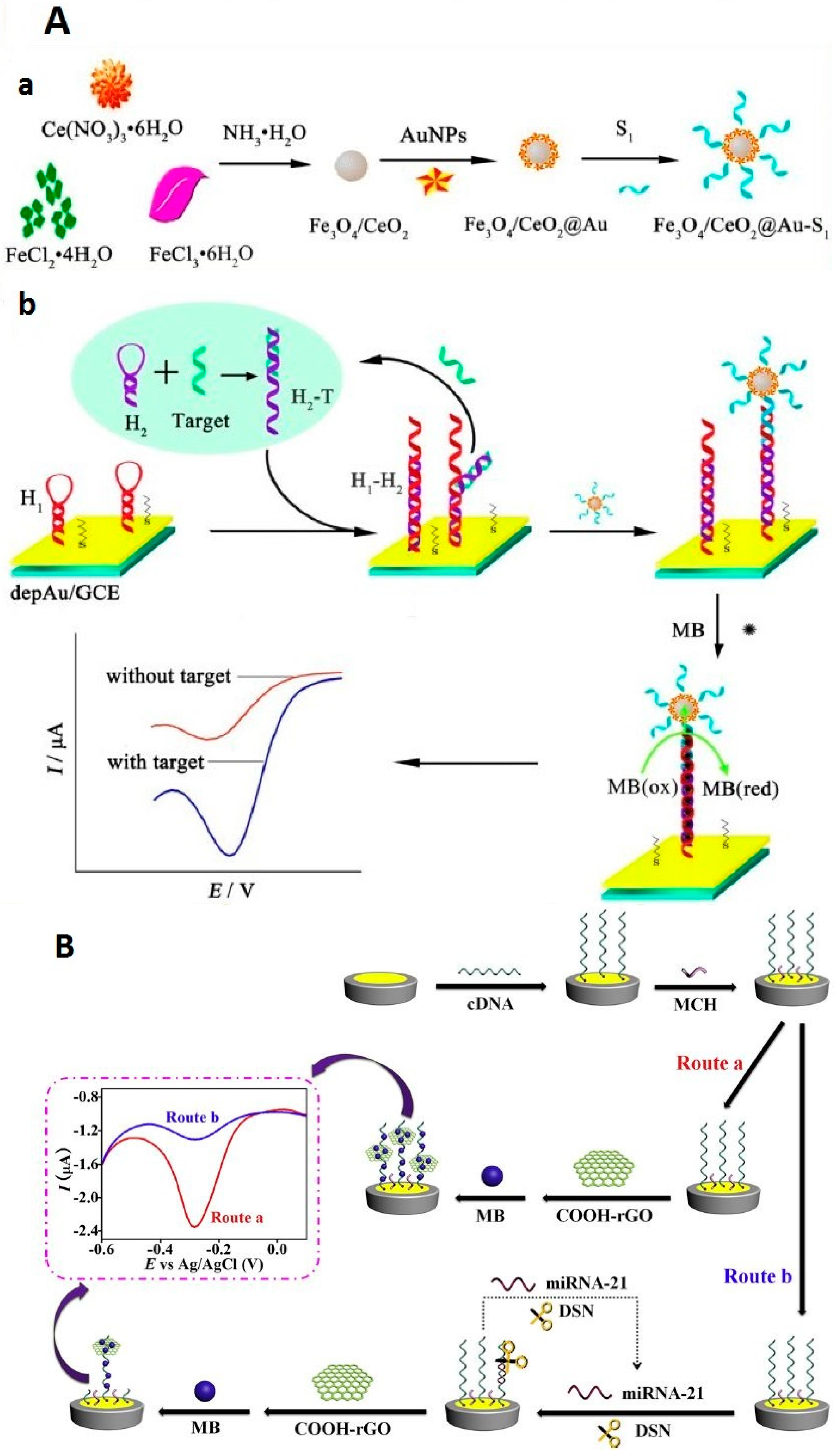
- Liu, S.; Yang, Z.; Chang, Y.; Chai, Y.; Yuan, R. An enzyme-free electrochemical biosensor combining target recycling with Fe3O4/CeO2@Au nanocatalysts for microRNA-21 detection. Biosens. Bioelectron. 2018, 119, 170–175. [Google Scholar] [CrossRef]
- Huang, S.; Li, J.; Jin, X.; Liu, Y.; Huang, S. Ultrasensitive electrochemical microRNA-21 biosensor coupling with carboxylate-reduced graphene oxide-based signal-enhancing and duplex-specific nuclease-assisted target recycling. Sens. Actuators B Chem. 2019, 297, 126740. [Google Scholar] [CrossRef]
- Guo, J.; Yuan, C.; Yan, Q.; Duan, Q.; Li, X.; Yi, G. An electrochemical biosensor for microRNA-196a detection based on cyclic enzymatic signal amplification and template-free DNA extension reaction with the adsorption of methylene blue. Biosens. Bioelectron. 2018, 105, 103–108. [Google Scholar] [CrossRef]
- Su, S.; Wu, Y.; Zhu, D.; Chao, J.; Liu, X.; Wan, Y.; Su, Y.; Zuo, X.; Fan, C.; Wang, L. On-Electrode Synthesis of Shape-Controlled Hierarchical Flower-Like Gold Nanostructures for Efficient Interfacial DNA Assembly and Sensitive Electrochemical Sensing of MicroRNA. Small 2016, 12, 3794–3801. [Google Scholar] [CrossRef]
- Yu, S.; Wang, Y.; Jiang, L.-P.; Bi, S.; Zhu, J. Cascade Amplification-Mediated In Situ Hot-Spot Assembly for MicroRNA Detection and Molecular Logic Gate Operations. Anal. Chem. 2018, 90, 4544–4551. [Google Scholar] [CrossRef]
- Chen, X.; Yao, L.; Wang, Y.-C.; Chen, Q.; Deng, H.; Lin, Z.; Yang, H.-H. Novel electrochemical nanoswitch biosensor based on self-assembled pH-sensitive continuous circular DNA. Biosens. Bioelectron. 2019, 131, 274–279. [Google Scholar] [CrossRef] [PubMed]
- Wang, S.; Lu, S.; Zhao, J.; Ye, J.; Huang, J.; Yang, X. An electric potential modulated cascade of catalyzed hairpin assembly and rolling chain amplification for microRNA detection. Biosens. Bioelectron. 2019, 126, 565–571. [Google Scholar] [CrossRef] [PubMed]
- Wang, Y.-H.; Huang, K.-J.; Wu, X.; Ma, Y.-Y.; Song, D.-L.; Du, C.-Y.; Chang, S.-H. Ultrasensitive supersandwich-type biosensor for enzyme-free amplified microRNA detection based on N-doped graphene/Au nanoparticles and hemin/G-quadruplexes. J. Mater. Chem. B 2018, 6, 2134–2142. [Google Scholar] [CrossRef]
- Asadzadeh-Firouzabadi, A.; Zare, H.R. Preparation and application of AgNPs/SWCNTs nanohybrid as an electroactive label for sensitive detection of miRNA related to lung cancer. Sens. Actuators B Chem. 2018, 260, 824–831. [Google Scholar] [CrossRef]
- Wang, F.; Chu, Y.; Ai, Y.; Chen, L.; Gao, F. Graphene oxide with in-situ grown Prussian Blue as an electrochemical probe for microRNA-122. Microchim. Acta 2019, 186, 116. [Google Scholar] [CrossRef]
- Liu, L.; Chang, Y.; Xia, N.; Peng, P.; Zhang, L.; Jiang, M.; Zhang, J.; Liu, L. Simple, sensitive and label–free electrochemical detection of microRNAs based on the in situ formation of silver nanoparticles aggregates for signal amplification. Biosens. Bioelectron. 2017, 94, 235–242. [Google Scholar] [CrossRef]
- Azimzadeh, M.; Rahaie, M.; Nasirizadeh, N.; Ashtari, K.; Naderi-Manesh, H. An electrochemical nanobiosensor for plasma miRNA-155, based on graphene oxide and gold nanorod, for early detection of breast cancer. Biosens. Bioelectron. 2016, 77, 99–106. [Google Scholar] [CrossRef]
- Cui, L.; Wang, M.; Sun, B.; Ai, S.; Wang, S.; Zhang, C.-Y. Substrate-free and label-free electrocatalysis-assisted biosensor for sensitive detection of microRNA in lung cancer cells. Chem. Commun. 2019, 55, 1172–1175. [Google Scholar] [CrossRef]
- Bo, B.; Zhang, T.; Jiang, Y.; Cui, H.; Miao, P. Triple Signal Amplification Strategy for Ultrasensitive Determination of miRNA Based on Duplex Specific Nuclease and Bridge DNA–Gold Nanoparticles. Anal. Chem. 2018, 90, 2395–2400. [Google Scholar] [CrossRef]
- Su, S.; Cao, W.; Liu, W.; Lu, Z.; Zhu, D.; Chao, J.; Weng, L.; Wang, L.; Fan, C.; Wang, L. Dual-mode electrochemical analysis of microRNA-21 using gold nanoparticle-decorated MoS2 nanosheet. Biosens. Bioelectron. 2017, 94, 552–559. [Google Scholar] [CrossRef]
- Feng, K.; Liu, J.; Deng, L.; Yu, H.; Yang, M. Amperometric detection of microRNA based on DNA-controlled current of a molybdophosphate redox probe and amplification via hybridization chain reaction. Microchim. Acta 2017, 185, 28. [Google Scholar] [CrossRef] [PubMed]
- Wang, Y.; Zhang, X.; Zhao, L.; Bao, T.; Wen, W.; Zhang, X.; Wang, S.-F. Integrated amplified aptasensor with in-situ precise preparation of copper nanoclusters for ultrasensitive electrochemical detection of microRNA 21. Biosens. Bioelectron. 2017, 98, 386–391. [Google Scholar] [CrossRef] [PubMed]
- Smith, D.A.; Newbury, L.J.; Drago, G.; Bowen, T.; Redman, J.E. Electrochemical detection of urinary microRNAs via sulfonamide-bound antisense hybridisation. Sens. Actuators B Chem. 2017, 253, 335–341. [Google Scholar] [CrossRef] [PubMed]
- Ebrahimi, A.; Nikokar, I.; Zokaei, M.; Bozorgzadeh, E. Design, development and evaluation of microRNA-199a-5p detecting electrochemical nanobiosensor with diagnostic application in Triple Negative Breast Cancer. Talanta 2018, 189, 592–598. [Google Scholar] [CrossRef] [PubMed]
- Duan, F.; Guo, C.; Hu, M.; Song, Y.; Wang, M.; He, L.; Zhang, Z.; Pettinari, R.; Zhou, L. Construction of the 0D/2D heterojunction of Ti3C2Tx MXene nanosheets and iron phthalocyanine quantum dots for the impedimetric aptasensing of microRNA-155. Sens. Actuators B Chem. 2020, 310, 127844. [Google Scholar] [CrossRef]
- Yaman, Y.T.; Vural, O.A.; Bolat, G.; Abaci, S. One-pot synthesized gold nanoparticle-peptide nanotube modified disposable sensor for impedimetric recognition of miRNA 410. Sens. Actuators B Chem. 2020, 320, 128343. [Google Scholar] [CrossRef]
- Yammouri, G.; Mandli, J.; Mohammadi, H.; Amine, A. Development of an electrochemical label-free biosensor for microRNA-125a detection using pencil graphite electrode modified with different carbon nanomaterials. J. Electroanal. Chem. 2017, 806, 75–81. [Google Scholar] [CrossRef]
- Voccia, D.; Sosnowska, M.; Bettazzi, F.; Roscigno, G.; Fratini, E.; De Franciscis, V.; Condorelli, G.; Chitta, R.; D’Souza, F.; Kutner, W.; et al. Direct determination of small RNAs using a biotinylated polythiophene impedimetric genosensor. Biosens. Bioelectron. 2017, 87, 1012–1019. [Google Scholar] [CrossRef]
- Mandli, J.; Amine, A. Impedimetric genosensor for miRNA-34a detection in cell lysates using polypyrrole. J. Solid State Electrochem. 2017, 22, 1007–1014. [Google Scholar] [CrossRef]
- Meng, T.; Zhao, D.; Ye, H.; Feng, Y.; Wang, H.; Zhanga, Y. Construction of an ultrasensitive electrochemical sensing platform for microRNA-21 based on interface impedance spectroscopy. J. Colloid Interface Sci. 2020, 578, 164–170. [Google Scholar] [CrossRef]
- Zhang, J.; Wu, D.-Z.; Cai, S.-X.; Chen, M.; Xia, Y.-K.; Wu, F.; Chen, J. An immobilization-free electrochemical impedance biosensor based on duplex-specific nuclease assisted target recycling for amplified detection of microRNA. Biosens. Bioelectron. 2016, 75, 452–457. [Google Scholar] [CrossRef] [PubMed]
- Salimi, A.; Kavosi, B.; Navaee, A. Amine-functionalized graphene as an effective electrochemical platform toward easily miRNA hybridization detection. Meas. J. Int. Meas. Confed. 2019, 143, 191–198. [Google Scholar] [CrossRef]
- Hu, F.; Zhang, W.; Zhang, J.; Zhang, Q.; Sheng, T.; Gu, Y. An electrochemical biosensor for sensitive detection of microRNAs based on target-recycled non-enzymatic amplification. Sens. Actuators B Chem. 2018, 271, 15–23. [Google Scholar] [CrossRef]
- Bharti, A.; Agnihotri, N.; Prabhakar, N. A voltammetric hybridization assay for microRNA-21 using carboxylated graphene oxide decorated with gold-platinum bimetallic nanoparticles. Microchim. Acta 2019, 186, 185. [Google Scholar] [CrossRef] [PubMed]
- Salahandish, R.; Ghaffarinejad, A.; Omidinia, E.; Zargartalebi, H.; Majidzadeh-A, K.; Naghib, S.M.; Sanati-Nezhad, A. Label-free ultrasensitive detection of breast cancer miRNA-21 biomarker employing electrochemical nano-genosensor based on sandwiched AgNPs in PANI and N-doped graphene. Biosens. Bioelectron. 2018, 120, 129–136. [Google Scholar] [CrossRef] [PubMed]
- Boriachek, K.; Umer, M.; Islam, N.; Gopalan, V.; Lam, A.K.-Y.; Nguyen, N.-T.; Shiddiky, M.J. An amplification-free electrochemical detection of exosomal miRNA-21 in serum samples. Analyst 2018, 143, 1662–1669. [Google Scholar] [CrossRef]
- Wan, Z.; Umer, M.; Lobino, M.; Thiel, D.; Nguyen, N.-T.; Trinchi, A.; Shiddiky, M.J.; Gao, Y.; Li, Q. Laser induced self-N-doped porous graphene as an electrochemical biosensor for femtomolar miRNA detection. Carbon 2020, 163, 385–394. [Google Scholar] [CrossRef]
- Koo, K.M.; Carrascosa, L.G.; Shiddiky, M.J.A.; Trau, M. Poly(A) Extensions of miRNAs for Amplification-Free Electrochemical Detection on Screen-Printed Gold Electrodes. Anal. Chem. 2016, 88, 2000–2005. [Google Scholar] [CrossRef]
- Kannan, P.; Maiyalagan, T.; Lin, B.; Lei, W.; Jie, C.; Guo, L.; Jiang, Z.; Mao, S.; Subramanian, P. Nickel-phosphate pompon flowers nanostructured network enables the sensitive detection of microRNA. Talanta 2020, 209, 120511. [Google Scholar] [CrossRef]
- Gai, P.; Gu, C.; Li, H.; Sun, X.; Li, F. Ultrasensitive Ratiometric Homogeneous Electrochemical MicroRNA Biosensing via Target-Triggered Ru(III) Release and Redox Recycling. Anal. Chem. 2017, 89, 12293–12298. [Google Scholar] [CrossRef]
- La, M.; Zhang, Y.; Gao, Y.; Li, M.; Liu, L.; Chang, Y. Impedimetric Detection of Micro RNAs by the SignalAmplification of Streptavidin Induced In Situ Formation ofBiotin Phenylalanine Nanoparticle Networks. J. Electrochem. Soc. 2020, 167. [Google Scholar] [CrossRef]
- Azzouzi, S.; Mak, W.C.; Kor, K.; Turner, A.P.; Ben Ali, M.; Beni, V. An integrated dual functional recognition/amplification bio-label for the one-step impedimetric detection of Micro-RNA-21. Biosens. Bioelectron. 2017, 92, 154–161. [Google Scholar] [CrossRef] [PubMed]
- Low, S.S.; Pan, Y.; Ji, D.; Li, Y.; Lu, Y.; He, Y.; Chen, Q.; Liu, Q. Smartphone-based portable electrochemical biosensing system for detection of circulating microRNA-21 in saliva as a proof-of-concept. Sens. Actuators B Chem. 2020, 308, 127718. [Google Scholar] [CrossRef]
- Yin, H.; Wang, M.; Yang, Z.; Lu, L.; Yin, H.; Ai, S. Electrochemical biosensor for microRNA detection based on hybridization protection against nuclease S1 digestion. J. Solid State Electrochem. 2015, 20, 413–419. [Google Scholar] [CrossRef]
- Jeong, B.; Kim, Y.J.; Jeong, J.-Y. Label-free electrochemical quantification of microRNA-375 in prostate cancer cells. J. Electroanal. Chem. 2019, 846, 113127. [Google Scholar] [CrossRef]
- Li, J.; Wang, Y.; Deng, R.; Lin, L.; Liu, Y.; Li, J. Carbon nanotube enhanced label-free detection of microRNAs based on hairpin probe triggered solid-phase rolling-circle amplification. Nanoscale 2015, 7, 987–993. [Google Scholar] [CrossRef]
- Akbarnia, A.; Zare, H.R.; Moshtaghioun, S.M.; Benvidi, A. Highly selective sensing and measurement of microRNA-541 based on its sequence-specific digestion by the restriction enzyme Hinf1. Colloids Surf. B Biointerfaces 2019, 182, 110360. [Google Scholar] [CrossRef]
- Azab, S.M.; Elhakim, H.K.; Fekry, A.M. The strategy of nanoparticles and the flavone chrysin to quantify miRNA-let 7a in zepto-molar level: Its application as tumor marker. J. Mol. Struct. 2019, 1196, 647–652. [Google Scholar] [CrossRef]
- Kangkamano, T.; Numnuam, A.; Limbut, W.; Kanatharana, P.; Vilaivan, T.; Thavarungkul, P. Pyrrolidinyl PNA polypyrrole/silver nanofoam electrode as a novel label-free electrochemical miRNA-21 biosensor. Biosens. Bioelectron. 2018, 102, 217–225. [Google Scholar] [CrossRef]
- Yang, L.; Wang, H.; Lü, H.; Hui, N. Phytic acid functionalized antifouling conducting polymer hydrogel for electrochemical detection of microRNA. Anal. Chim. Acta 2020, 1124, 104–112. [Google Scholar] [CrossRef]
- Zhu, D.; Liu, W.; Zhao, D.; Hao, Q.; Lianhui, W.; Huang, J.; Shi, J.; Chao, J.; Su, S.; Wang, L. Label-Free Electrochemical Sensing Platform for MicroRNA-21 Detection Using Thionine and Gold Nanoparticles Co-Functionalized MoS2 Nanosheet. ACS Appl. Mater. Interfaces 2017, 9, 35597–35603. [Google Scholar] [CrossRef] [PubMed]
- Zouari, M.; Campuzano, S.; Pingarrón, J.M.; Raouafi, N. Determination of miRNAs in serum of cancer patients with a label- and enzyme-free voltammetric biosensor in a single 30-min step. Microchim. Acta 2020, 187, 1–11. [Google Scholar] [CrossRef] [PubMed]
- Sabahi, A.; Salahandish, R.; Ghaffarinejad, A.; Omidinia, E. Electrochemical nano-genosensor for highly sensitive detection of miR-21 biomarker based on SWCNT-grafted dendritic Au nanostructure for early detection of prostate cancer. Talanta 2020, 209, 120595. [Google Scholar] [CrossRef] [PubMed]


| Cancer | Biomarkers | Reference |
|---|---|---|
| Breast | BRCA1, BRCA2, MUC1, CEA, CA 15-3, CA 27, CA29, EGFR, EpCAM, HER2; miRNA-21, miRNA-373, miRNA-182, miRNA-1246 and miRNA-105 | [7,8,9,10] |
| Prostate | PSA, Sarcosine; TEMPRSS2; miRNA-21, miRNA-141, miRNA-375 | [11,12,13] |
| Brain | MDM2; | [14] |
| Pancreas | CA 19-9, PAM4; miRNA-21, miRNA-155, miRNA-196 | [15,16] |
| Gastric/Stomach | CA72-4, CA19-9, CEA, IL-6; PVT1; miRNA-21, miRNA-331, miRNA-421 | [17,18] |
| Liver | AFP, DCP, GP73; miRNA-21, miRNA-122, miRNA-16 | [19] |
| Ovarian | CA 125 (MUC-16), CEA, Claudin-4; | [20,21] |
| Lung | ANXA2, CEA, Chromogranin A, CA 19-9, CYFRA 21-1 (CA-19 fragment), NSE, SCC, SAA1, HER1; P53, P16, Ras genes, Telomere length and telomere-related genes, EGFR gene (c-ErbB-1 and c-ErbB-2); miRNA-21 (in sputum) | [22,23,24,25,26] |
| Neck | MGMT gene | [27] |
| miRNA | labeled RedOx Molecule | Platform | Signal Amplification | Tech | Linear Range | LOD | Real-Samples Application | Ref |
|---|---|---|---|---|---|---|---|---|
| miRNA-141 | MB | AuE | SDR | SWV | 0.1 fM–0.2 pM | 23 aM | Human bladder cancer T24 cells | [53] |
| - | MB | GCE/PDDA/AuNPs | DSN | SWV | 100 aM–1 nM | 30 aM | human serum samples | [51] |
| miRNA-21 | MB | GCE/CNNS/AuNPs | DSN | SWV | 10 fM–1 nM | 2.9 fM | Spiked human serum | [52] |
| MiRNA-21 | MB | PtE/AuNFs | MB/barcode AuNPs | DPV | 500 aM–50 pM | 135 aM | spiked human serum | [73] |
| miRNA 21 | Fc | AuE | SWCNTs | DPV | 100 pM–3.5 fM | 0.01 fM | Human serum | [46] |
| miRNA-155 | Fc | GCE/Mo2C/AuNPs | CHA | DPV | 0.1 fM–1.0 nM | 0.033 fM | Spiked human serum | [74] |
| miRNA-21 | Fc | GCE/Mo2C/NCS | SDR | Amperometric | 1.0 fM–1.0 nM | 0.34 fM | Spiked human serum | [75] |
| miRNA-21 | AgNPs | GCE/GO | - | LSV | 100 fM–1 nM | 60 fM | serum samples from breast cancer patients | [42] |
| miRNA-155 | AgNPs | AuE | SDR and nicking endonuclease | LSV | 1 fM–1 pM | 70 aM | HeLa cells, A549, human renal cubularepithelial | [76] |
| Hsa-miR-17-5p | AgNPs | AuE | AuNPs HCR | LSV | 100 aM–0.1 nM | 2 aM | HUVEC, HK-2, HeLa, MCF-7 cells | [77] |
| - | AgNCs | AuE | pDNA-AgDNCs@DNA/AgNCs | DPV | 1 fM–1 nM | 0.38 fM | spiked human serum | [78] |
| miRNA-21 | AgNPs | AuE | SDR | SWV | 200 pM–1 fM | 0.4 fM | Blood sample | [79] |
| miRNA-155 | PEIAgNPs | AuE | - | CV | 200 zM–2 pM | 20 zM | cancerous humann serum | [80] |
| miRNA-21 | Thi | GCE/AuNPs | MWCNTs | DPV | 0.1–12000 pM | 0.032 pM | spiked human serum | [81] |
| miRNA-155 | Thi | 3D N-doped rGO/AuNPs | AuAgNR | DPV | 10 pM–100 µM | 1 pM | Spiked serum samples | [45] |
| miRNA-21 | TB | GCE/DpAu | - | SWV | 1 fM–2 nM | 0.3 fM | MCF-7, human breast cancer cell line | [82] |
| miRNA-21 | Pd NPs | GCE/GO | Pd/NPs-DNALNR and CHA | DPV | 1 fM–50 pM | 63.1 aM | Spiked human serum | [83] |
| miRNA-21 | Cd | GCE/Au-RGO | TPSs Ru(NH3)63+ | SWV | 1.0 aM–10.0 pM | 0.76 aM | spiked human serum | [69] |
| miRNA | CdTe/QDs | AuE | CESA 3-QD@DNA NC | DPV | 5 aM–5 fM | 1.2 aM | Spiked human serum | [43] |
| miRNA-21 | MB and Fc | AuE | - | SWV | 5 fM–0.1 nM | 1.1 fM | MCF-7 and HeLa | [65] |
| miRNA-16 | MB and Fc | AuE | - | SWV | 0.1 pM–100 nM | 16 fM | MCF-7 cells | [84] |
| miRNA-21 | MB and Fc | GCE/PLLy | LNA/structure“Y”shape | DPV | 10–70 fM | 2.3 fM | MCF-7 cells | [68] |
| let-7a | MB and Fc | NS-grafted ITO | - | DPV | 80 aM–300 fM | 25 aM | Spiked human serum | [85] |
| miRNA-21 | MB and Fc | AuE | - | DPV | 0.1–100.0 fM | 67 aM | breast cancer cell line MCF-7 | [64] |
| miRNA 21 | Fc | AuE/AuNPs | - | DPV | 100 pM to 1 fM | 0.36 fM | serum | [86] |
| - | CdSNPs | GCE | AuNPS and DSN | ASV | 1 fM–100 pM | 0.48 fM | HeLa | [67] |
| miRNA-21 and miRNA-155 | MB and Fc | SPCE | Fe3O4@Au@HHCR | SWV | 5 fM–2 nM | 1.5 fM 1.8 fM | Spiked human serum | [87] |
| miRNA-21 and miRNA-155 | MB and Fc | AuE | - | SWV | 10 fM–5 nM 50 fM–5 nM | 2.49 fM 11.63 fM | HeLa, MCF-7 and MDA-MB-231 cells | [88] |
| miRNA-21 and miRNA-141 | Au ion and Ag ion | GCE/Neutravidin | - | SSWV | 0.5−1000 pM 50−1000 pM | 0.3 pM 10 pM | Spiked Serum Sample | [89] |
| miRNA-21 and miRNA-141 | MB and Fc | SPGE/MXene/AuNPs | - | DPV | 500 aM–50 nM | 204 aM 138 aM | human plasma cancer patients | [90] |
| miRNA-1246 and miRNA-4521 | Pb (II) and Cd (II) | GCE/AuNPs | PbS@ZIF-8 CdS@ZIF-8 | DPV | 1 fM–1 mM | 0.19 fM 0.28 fM | spiked human blood | [44] |
| MicroRNA | Catalysts/Amplification Agents | Platform | Substrat or Reagent | Technique | Linear Range | LOD | Real-Samples Application | References |
|---|---|---|---|---|---|---|---|---|
| miRNA-21 | ALP/DNA-linked GO–AuNPs | GCE/MgO/AuNPs | AAP | DPV | 0.1–100 fM | 50 aM | Spiked human serum | [98] |
| miRNA -21 | ALP/HCR | AuE | α-NP | SWV | 1 fM–100 pM | 0.56 fM | Spiked HEK293T cells | [104] |
| miRNA-21 | ALP/CHA-WO3-Gr | GCE/WO3-Gr/AuNPs | Ascorbic acid 2-phosphate | DPV | 0.1 fM–100 pM | 0.05 fM | Serum samples from breast cancer patients | [111] |
| miRNA -21 | HRP/AuNPs | AuE | H2O2 | Amperometry | 0.1 fM–100 pM | 43.3 aM | A549 tumor cells | [113] |
| miRNA-155 | ALP/CHA | GCE/MWCNTs PtNPs | Phosphate ion Molybdophosphate anion | DPV | 10 fM–1 nM | 1.64 fM | Cervical cancer cells and human breast cancer cell lines | [131] |
| miRNA-155 | ALP/- | SCPE/Fe3O4 | AAP | DPV | 0.6–9 ng/mL | 29 pM | Spiked human serum | [132] |
| miRNA-21 | HRP/- | SCPE/Fe3O4 | H2O2 | Amperometry | 1.0–100 pM | 10 aM | MCF-7 cells | [116] |
| miRNA -155 | HRP/GQDs | AuE | TMB | Amperometry | 1fM–00 pM | 0.14 fM | Spiked human serum | [118] |
| miRNA-21, let-7a & miRNA-31 | HRP/HCR | SPCE/Fe3O4 | H2O2 | Amperometry | 1.2–100 pM | 0.66 pM | MCF-7 cells | [133] |
| miRNA-21 | HRP/- | SPCE/Fe3O4 | H2O2 | Amperometry | 3.0 to 100 nM | 0.91 nM | MCF-7 cells | [134] |
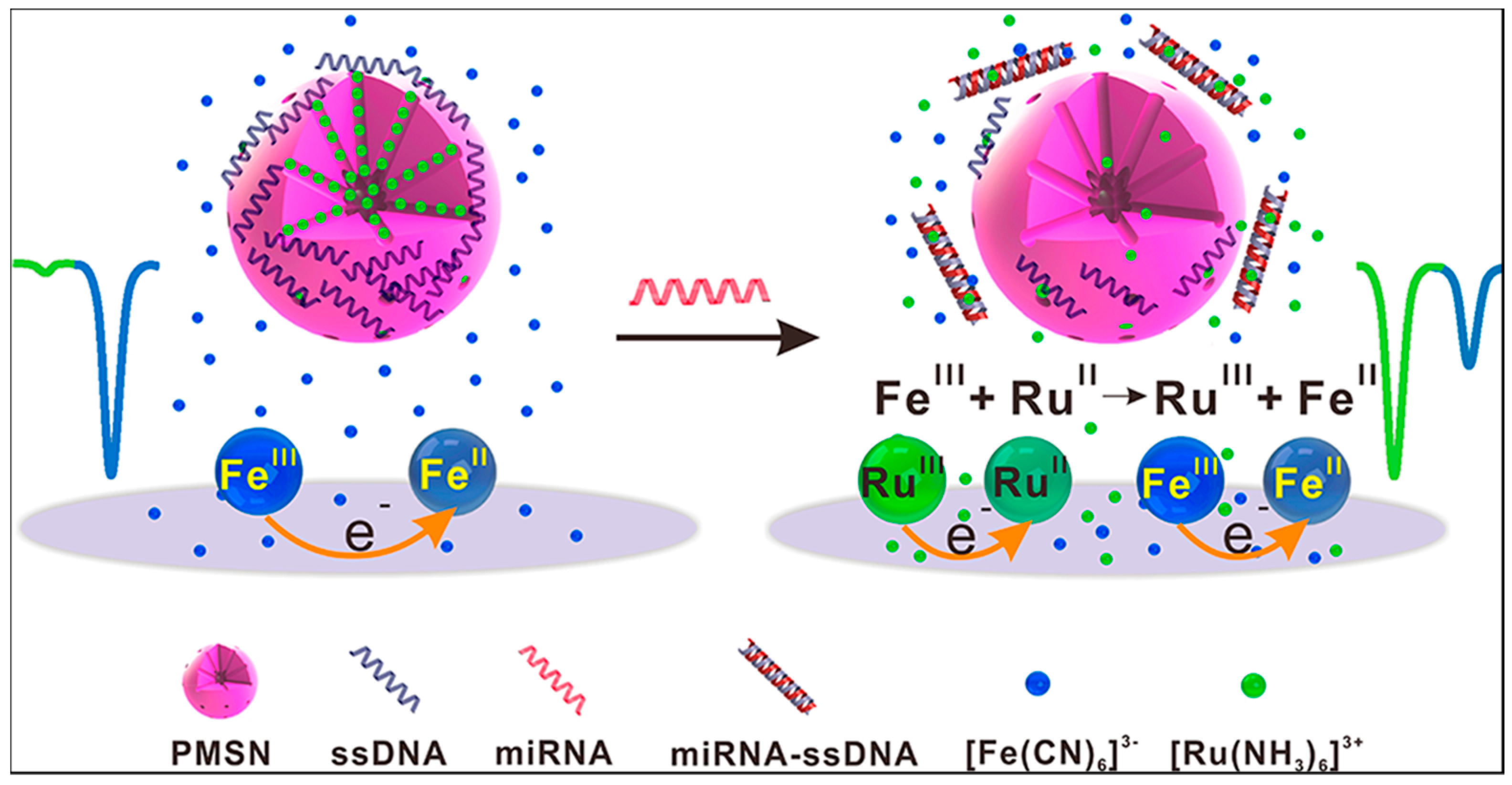
| miRNA-21 | Copper (II) complex/HCR | GCME/Fe3O4 | TMB | DPV | 100 aM–100 nM | 33 aM | human serum samples from breast cancer | [135] |
| miRNA-155 | Cu-MOFs/AuNPs | Au-PWE | Glucose | DPV | 1 fM–10 nM | 0.35 fM | Spiked human serum | [120] |
| miRNA-21 | Sn-In2O3/- | AuE | O2 | DPV | 5 pM–0.5 fM | 1.92 fM | A549 and HeLa cell lines | [122] |
| miRNA-21 | G-quadruplex−hemin/- | AuE | H2O2 | DPV | 0.1 fM–0.1 pM | 0.04 fM | Human serum samples from breast cancer | [128] |
| miRNA-21 | G-quadruplex−hemin/HCR | AuE/SWCNT-ox/NDs/SWCNTs-ox/AuNPs | H2O2 | DPV | 10 fM–1.0 nM | 1.95 fM | Spiked human serum | [129] |
| MicroRNA | Intercalent Agent | Platform | Amplification of Signal Elements | Tech | Linear Range | LOD | Real-Samples Application | Ref |
|---|---|---|---|---|---|---|---|---|
| miRNA-155 | OB | GCE/GO/GNR | - | DPV | 2.0 fM–8.0 pM | 0.6 fM | Spiked human plasma | [157] |
| miRNA-21 | TB | GCE/AuNPs-Ppy | - | DPV | 100 aM–1 nM | 78 aM | Spiked human serum. | [138] |
| miRNA-196a | MB | AuE | DNA extension reaction | DPV | 0.05 fM–50 pM | 15 aM | Spiked plasma | [148] |
| miRNA-486-5p | Thinonine | GCE/FeCN/AuNPs | FeCN | DPV | 1 fM–1000 pM | 8.53 fM | Human lung A549 cells | [158] |
| miRNA-141 | RuHex | AuE | HP-AuNPs | DPV | 0–10 nM | 25.1 aM | Human breast cancer cells MDA-MB-231 | [150] |
| miRNA-21 | RuHex | AuE | AuNPs enrichment by bridge DNA, | Chronocoulometry | 0.1fM–0.1nM | 68 aM | Serum samples from lung cancer patients | [159] |
| miRNA-21 | RuHex | GCE/AuNPs@MoS2 | AuNPs@MoS2 | DPV | 10 fM–1 nM | 0.78 fM | Spiked human serum | [160] |
| miRNA-21 | Molybdophosphate | AuE | HCR | SWV | 1 fM–1 nM | 0.78 fM | Spiked human serum | [161] |
| miRNA-21 | CuNCs | AuE | HCR | DPSV | 10 pM–0.1 fM | 10 aM | Spiked blood sample | [162] |
| miRNA-21 | Hemin | AuE | HCR | DPV | 15 fM–250 pM | 13.5 fM | Spiked human serum | [152] |
| miRNA-199 a | AgNPs | AuE | C-rich loop DNA templates | DPV | 1.0 fM–0.1 nM | 0.64 fM | Spiked human serum | [142] |
| MicroRNA | Free RedOx Indicator | Platform | Amplification Agent | Technique | Linear Range/LOD | LOD | Real-Samples Application | References |
|---|---|---|---|---|---|---|---|---|
| miRNA-21 | Fe(CN)63−/4- | AuE | biotin-FNPs | EIS | 0.1–250 fM | 0.1 fM | - | [181] |
| miRNA-21 | Fe(CN)63−/4- | GCE | AuNPs | EIS | 1–1000 pM | 0.3 pM | Spiked serum sample | [182] |
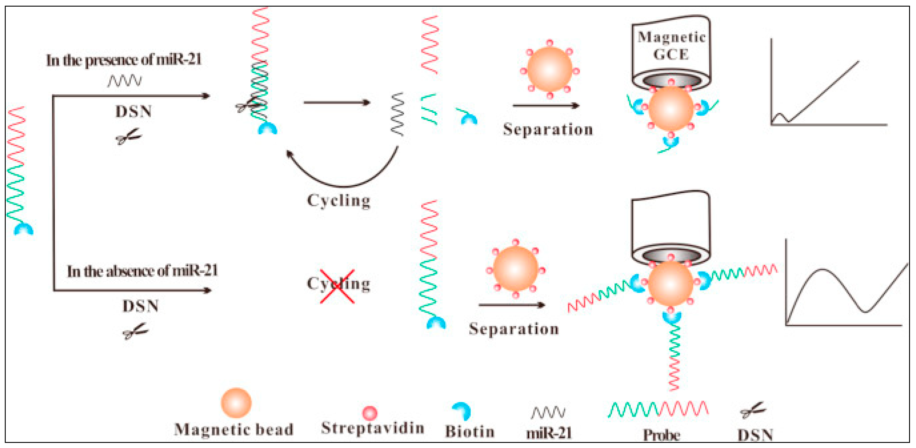
| miRNA-21 | Fe(CN)63−/4- | Magnetic GCE | DSN | EIS | - | 60 aM | Human serum from breast cancer patients | [171] |
| miRNA-199a-5p | Fe(CN)63−/4- | GCE/GO/GNR | GO and GNR | EIS | 148 pM–15 fM | 4.5 fM | Spiked human blood serum | [164] |
| miRNA-155 | Fe(CN)63−/4- | Pt wire/Ti3C2Tx@FePcQDs | Ti3C2Tx@FePcQDs | EIS | 0.01 fM–10 pM | 4.3 aM | Spiked human serum samples | [165] |
| miRNA-21 | Fe(CN)63−/4- | AuE | HCR | EIS | 10 fM–50 pM | 4.63 fM | A549, HeLa, MCF-7, RAW 264.7, and HUVEC cancer cells | [170] |
| miRNA-21 | Fe(CN)63−/4- | SPE/rGO-Au | - | DPV | 1 µM–1 pM | 1pM | Spiked artificial saliva | [183] |
| miRNA-319a | Fe(CN)63−/4- | GCE/AuNPs | nuclease S1 | DPV | 1000–5 pM | 1.8 pM | - | [184] |
| miRNA-21 | Fe(CN)63−/4- | FTO/NFG/AgNPs/PANI | - | DPV | 10 fM–10 μM | 0.2 fM | Spiked blood samples | [175] |
| miRNA-21 | Fe(CN)63−/4- | FTO/CGO/Au-PtBNPs/SA | - | DPV | 1 fM–1 μM | 1 fM | spiked human serum | [174] |
| miRNA-21 | Fe(CN)63−/4- | GCE/MWCNTs | TRNEAS | DPV | 0.1 fM–5 pM | 56.7 aM | MDA-MB-231, MCF-7, HeLa, and L02 cell | [173] |
| hsa-miR-486-5p | Fe(CN)63−/4- | Laser induced graphene | - | DPV | 10 fM | - | [177] | |
| miRNA-375 | Fe(CN)63−/4- | AuE | - | SWV | 10–30 fM | 11.7 aM | CaP cells (PC-3, DU145, and LNCaP) | [185] |
| miRNA | [Ru(NH3)6]3+/2+ | GCE/Ni PFNs | - | EIS | 0.1–2500 pM | 0.034 pM | A549 cancer cells | [179] |
| let-7a | [Ru(NH3)6]3+/2+ | GCE/CNTs | CNT based solid-phase RCA | DPV | 1.2 fM | HeLa cells | [186] | |
| miRNA-21 | Fe(CN)63−/4−/[Ru(NH3)6]3+/2+ | ITO | - | DPV | 0.1–1500 fM | 33 aM | HeLa, A549, MCF-7 cancer cells | [180] |
Publisher’s Note: MDPI stays neutral with regard to jurisdictional claims in published maps and institutional affiliations. |
© 2020 by the authors. Licensee MDPI, Basel, Switzerland. This article is an open access article distributed under the terms and conditions of the Creative Commons Attribution (CC BY) license (http://creativecommons.org/licenses/by/4.0/).
Share and Cite
El Aamri, M.; Yammouri, G.; Mohammadi, H.; Amine, A.; Korri-Youssoufi, H. Electrochemical Biosensors for Detection of MicroRNA as a Cancer Biomarker: Pros and Cons. Biosensors 2020, 10, 186. https://doi.org/10.3390/bios10110186
El Aamri M, Yammouri G, Mohammadi H, Amine A, Korri-Youssoufi H. Electrochemical Biosensors for Detection of MicroRNA as a Cancer Biomarker: Pros and Cons. Biosensors. 2020; 10(11):186. https://doi.org/10.3390/bios10110186
Chicago/Turabian StyleEl Aamri, Maliana, Ghita Yammouri, Hasna Mohammadi, Aziz Amine, and Hafsa Korri-Youssoufi. 2020. "Electrochemical Biosensors for Detection of MicroRNA as a Cancer Biomarker: Pros and Cons" Biosensors 10, no. 11: 186. https://doi.org/10.3390/bios10110186
APA StyleEl Aamri, M., Yammouri, G., Mohammadi, H., Amine, A., & Korri-Youssoufi, H. (2020). Electrochemical Biosensors for Detection of MicroRNA as a Cancer Biomarker: Pros and Cons. Biosensors, 10(11), 186. https://doi.org/10.3390/bios10110186