A Colorimetric Enzyme-Linked Immunosorbent Assay with CuO Nanoparticles as Signal Labels Based on the Growth of Gold Nanoparticles In Situ


Abstract
1. Introduction
2. Materials and Methods
2.1. Chemicals and Materials
2.2. Labeling of Antibody with CuO NPs
2.3. Procedure for PSA Detection
2.4. Assay of PSA with ELISA Kits
3. Results and Discussion
3.1. Detection Principle
3.2. Optimization of Experimental Conditions
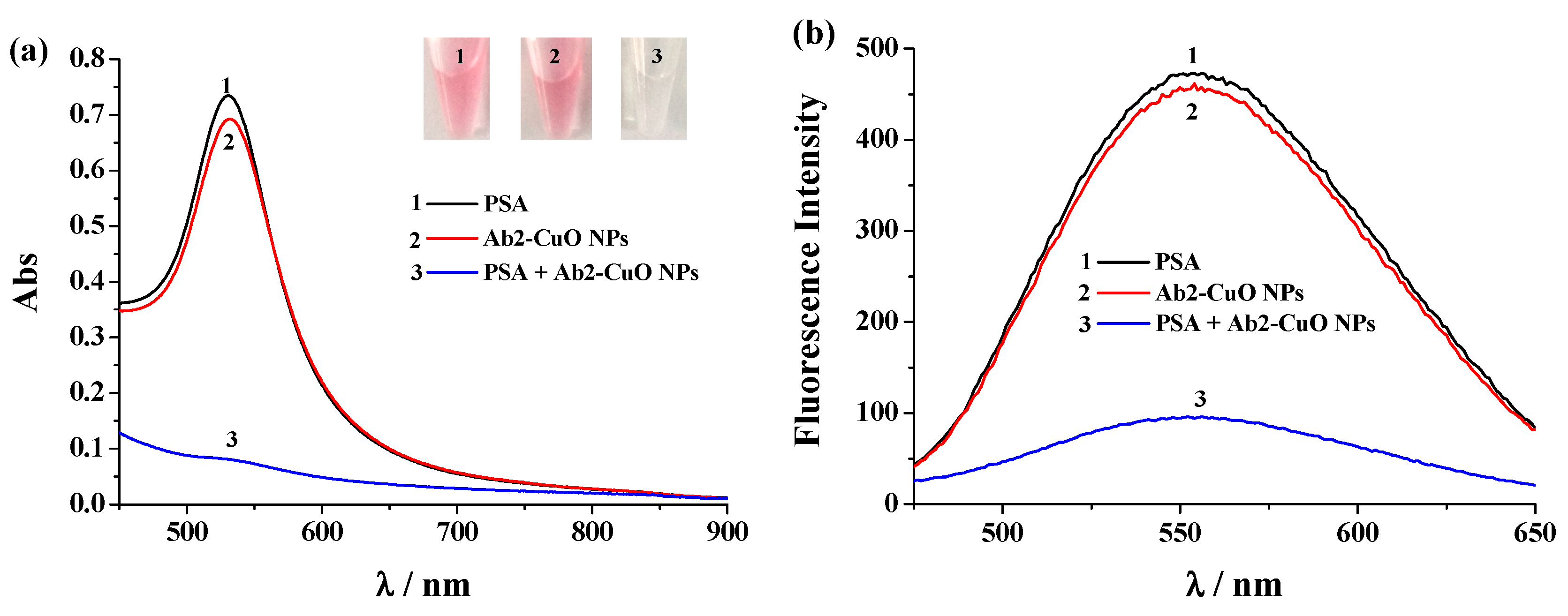
3.3. Feasibility
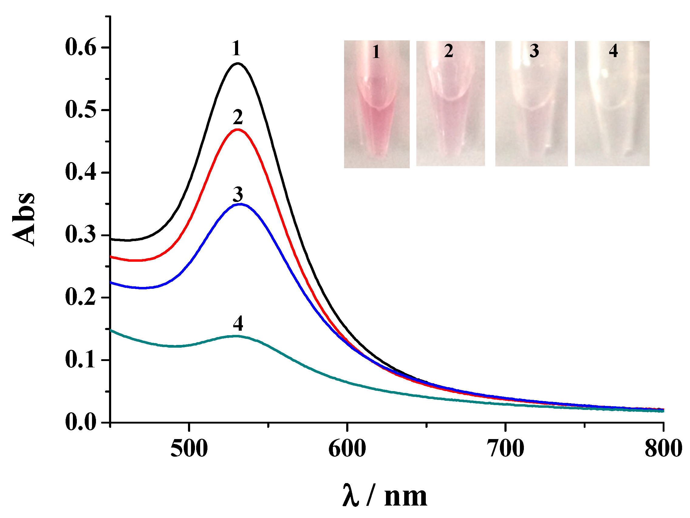
3.4. Sensitivity
3.5. Selectivity
3.6. Evaluation of Serum Samples
4. Conclusions
Author Contributions
Funding
Conflicts of Interest
References
- Farka, Z.; Juřík, T.; Kovář, D.; Trnková, L.; Skládal, P. Nanoparticle-based immunochemical biosensors and assays: Recent advances and challenges. Chem. Rev. 2017, 117, 9973–10042. [Google Scholar] [CrossRef] [PubMed]
- Patris, S.; Vandeput, M.; Kauffmann, J.-M. Antibodies as target for affinity biosensors. TrAC-Trend. Anal. Chem. 2016, 79, 239–246. [Google Scholar] [CrossRef]
- Li, Z.Y.; Chen, G.Y. Current conjugation methods for immunosensors. Nanomaterials 2018, 8, 278. [Google Scholar] [CrossRef]
- Zheng, W.; Jiang, X. Integration of nanomaterials for colorimetric immunoassays with improved performance: A functional perspective. Analyst 2016, 141, 1196–1208. [Google Scholar] [CrossRef]
- Rick, J.; Tsai, M.-C.; Hwang, B.J. Biosensors incorporating bimetallic nanoparticles. Nanomaterials 2016, 6, 5. [Google Scholar] [CrossRef] [PubMed]
- Zhang, S.; Geryak, R.; Geldmeier, J.; Kim, S.; Tsukruk, V.V. Synthesis, assembly, and applications of hybrid nanostructures for biosensing. Chem. Rev. 2017, 17, 12942–13038. [Google Scholar] [CrossRef]
- Fenzl, C.; Hirsch, T.; Baeumner, A.J. Nanomaterials as versatile tools for signal amplification in (bio)analytical applications. TrAC-Trend. Anal. Chem. 2016, 79, 306–316. [Google Scholar] [CrossRef]
- Saha, K.; Agasti, S.S.; Kim, C.; Li, X.; Rotello, V.M. Gold Nanoparticles in Chemical and Biological Sensing. Chem. Rev. 2012, 112, 2739–2779. [Google Scholar] [CrossRef]
- Kokkinos, C.; Economou, A. Emerging trends in biosensing using stripping voltammetric detection of metal-containing nanolabels—A review. Anal. Chim. Acta 2017, 961, 12–32. [Google Scholar] [CrossRef]
- Tian, S.; Zeng, K.; Yang, A.; Wang, Q.; Yang, M. A copper based enzyme-free fluorescence ELISA for HER2 detection. J. Immunol. Method. 2017, 451, 78–82. [Google Scholar] [CrossRef]
- Xie, Q.; Weng, X.; Lu, L.; Lin, Z.; Xu, X.; Fu, C. A sensitive fluorescent sensor for quantification of alpha-fetoprotein based on immunosorbent assay and click chemistry. Biosens. Bioelectron. 2016, 77, 46–50. [Google Scholar] [CrossRef] [PubMed]
- Yang, T.; Li, C.M.; He, J.H.; Chen, B.; Li, Y.F.; Huang, C.Z. Ratiometrically fluorescent electrospun nanofibrous film as a Cu2+-mediated solid-phase immunoassay platform for biomarkers. Anal. Chem. 2018, 90, 9966–9974. [Google Scholar] [CrossRef] [PubMed]
- Ge, S.; Ge, L.; Yan, M.; Song, X.; Yu, J.; Liu, S. A disposable immunosensor device for point-of-care test of tumor marker based on copper-mediated amplification. Biosens. Bioelectron. 2013, 43, 425–431. [Google Scholar] [CrossRef] [PubMed]
- Wang, Y.-W.; Chen, L.; Liang, M.; Xu, H.; Tang, S.; Yang, H.-H.; Song, H. Sensitive fluorescence immunoassay of alpha-fetoprotein throughcopper ions modulated growth of quantum dots in-situ. Sensor. Actuator B Chem. 2017, 247, 408–413. [Google Scholar] [CrossRef]
- Xu, Y.; Gao, Y.; Zhao, X.; Xu, X.; Zhou, W.; Liu, Y.; Li, C.; Liu, R. A sensitive atomic absorption spectrometric metalloimmunoassay with copper nanoparticles labeling. Microchem. J. 2016, 126, 1–6. [Google Scholar] [CrossRef]
- He, F.; Wang, J.; Yin, B.-C.; Ye, B.-C. Quantification of exosome based on a copper-mediated signal amplification strategy. Anal. Chem. 2018, 90, 8072–8079. [Google Scholar] [CrossRef] [PubMed]
- Li, B.; Lai, G.; Zhang, H.; Hu, S.; Yu, A. Copper chromogenic reaction based colorimetric immunoassay for rapid and sensitive detection of a tumor biomarker. Anal. Chim. Acta 2017, 963, 106–111. [Google Scholar] [CrossRef] [PubMed]
- Qu, W.; Liu, Y.; Liu, D.; Wang, Z.; Jiang, X. Copper-Mediated Amplification Allows Readout of Immunoassays by the Naked Eye. Angew. Chem. Int. Ed. 2011, 50, 3442–3445. [Google Scholar] [CrossRef] [PubMed]
- Zheng, A.; Zhang, X.; Gao, J.; Liu, X.; Liu, J. Peroxidase-like catalytic activity of copper ions and its application for highly sensitive detection of glypican-3. Anal. Chim. Acta 2016, 941, 87–93. [Google Scholar] [CrossRef] [PubMed]
- Dekker, A.O.; Dickinson, R.G. Oxidation of ascorbic acid by oxygen with cupric ion as catalyst. J. Am. Chem. Soc. 1941, 63, 3549. [Google Scholar] [CrossRef]
- Rastogi, L.; Dash, K.; Ballalb, A. Selective colorimetric/visual detection of Al3+ in ground water usingascorbic acid capped gold nanoparticles. Sensor. Actuator B Chem. 2017, 248, 124–132. [Google Scholar] [CrossRef]
- Liu, L.; Deng, D.; Wang, Y.; Song, K.; Shang, Z.; Wang, Q.; Xia, N.; Zhang, B. A colorimetric strategy for assay of protease activity based on gold nanoparticle growth controlled by ascorbic acid and Cu(II)-coordinated peptide. Sensor. Actuator B Chem. 2018, 266, 246–254. [Google Scholar] [CrossRef]
- Zheng, Y.; Cao, X.; Orbulescu, J.; Konka, V.; Andreopoulos, F.M.; Pham, S.M.; Leblanc, R.M. Peptidyl fluorescent chemosensors for the detection of divalent copper. Anal. Chem. 2003, 75, 1706–1712. [Google Scholar] [CrossRef] [PubMed]
- Wang, P.; Wu, J.; Di, C.; Zhou, R.; Zhang, H.; Su, P.; Xu, C.; Zhou, P.; Ge, Y.; Liu, D.; et al. A novel peptide-based fluorescence chemosensor for selective imaging of hydrogen sulfide both in living cells and zebrafish. Biosens. Bioelectron. 2017, 92, 602–609. [Google Scholar] [CrossRef] [PubMed]
- Wang, P.; Liu, L.; Zhou, P.; Wu, W.; Wu, J.; Liu, W.; Tang, Y. A peptide-based fluorescent chemosensor for multianalyte detection. Biosens. Bioelectron. 2015, 72, 80–86. [Google Scholar] [CrossRef]
- Li, Y.; Hong, M.; Lin, Y.; Bin, Q.; Lin, Z.; Cai, Z.; Chen, G. Highly sensitive electrochemical immunoassay for H1N1 influenza virus based on copper-mediated amplification. Chem. Commun. 2012, 48, 6562–6564. [Google Scholar] [CrossRef]


| Method | Target | Probe for Cu2+ Detection | Detection Limit | Linear Range | Ref. |
|---|---|---|---|---|---|
| Colorimetry | HIV | AuNPs | Not reported | Not reported | [18] |
| Colorimetry | CEA | DPHE | 26 pg/mL | 0.05~100 ng/mL. | [17] |
| Colorimetry | Glypican-3 | TMB/H2O2 | 0.26 pg/mL | 0.2~200 pg/mL | [19] |
| Fluorescence | HER2 | Quinoxaline derivative | 9.65 pg/mL | 5~25 pg/mL | [10] |
| Fluorescence | AFP | triazole complex | 0.012 ng/mL | 0.025~5.0 ng/mL | [11] |
| Fluorescence | AFP | CdTe QDs | 0.3 pg/mL | 0.001~100 ng/mL | [13] |
| CA125 | 0.061 mU/mL | 0.0002~100 U/mL | |||
| CA 153 | 0.29 mU/mL | 0.0001~200 U/mL | |||
| CEA | 1.4 pg/mL | 0.005~200 ng/mL | |||
| Fluorescence | AFP | ENFFs | 8.3 pg/mL | 0.01~200 ng/mL | [12] |
| Fluorescence | AFP | CdS QDs | 0.45 ng/mL, | 1~80 ng/mL | [14] |
| Fluorescence | Exosome | CuNPs | 4.8 × 10⁴ particles/μL | 7.5 × 10⁴~1.5 × 10⁷ particles/μL | [16] |
| DPV | H1N1 influenza virus | GCE | 10−12 g/mL | 10−11~10−5 g/mL | [26] |
| AAS | IgG | 0.19 ng/mL | 1~104 g/mL | [15] | |
| Colorimetry | PSA | AuNPs | 0.05 ng/mL | 0.1~10 ng/mL | This work |
| Fluorescence | PSA | SGHK-Dns | 0.1 ng/mL | 0.1~10 ng/mL | This work |
| Sample No. | Added (ng/mL) | Found (ng/mL) | ELISA (ng/mL) |
|---|---|---|---|
| 1 | 0 | 1.86 ± 0.12 | undetectable |
| 2 | 2 | 3.74 ± 0.23 | 4.14 ± 0.21 |
| 3 | 4 | 5.66 ± 0.42 | 5.72 ± 0.52 |
| 4 | 8 | 9.78 ± 0.74 | 10.46 ± 0.83 |
© 2018 by the authors. Licensee MDPI, Basel, Switzerland. This article is an open access article distributed under the terms and conditions of the Creative Commons Attribution (CC BY) license (http://creativecommons.org/licenses/by/4.0/).
Share and Cite
Deng, D.; Hao, Y.; Xue, J.; Liu, X.; Xu, X.; Liu, L. A Colorimetric Enzyme-Linked Immunosorbent Assay with CuO Nanoparticles as Signal Labels Based on the Growth of Gold Nanoparticles In Situ. Nanomaterials 2019, 9, 4. https://doi.org/10.3390/nano9010004
Deng D, Hao Y, Xue J, Liu X, Xu X, Liu L. A Colorimetric Enzyme-Linked Immunosorbent Assay with CuO Nanoparticles as Signal Labels Based on the Growth of Gold Nanoparticles In Situ. Nanomaterials. 2019; 9(1):4. https://doi.org/10.3390/nano9010004
Chicago/Turabian StyleDeng, Dehua, Yuanqiang Hao, Jiajia Xue, Xiuhua Liu, Xinyue Xu, and Lin Liu. 2019. "A Colorimetric Enzyme-Linked Immunosorbent Assay with CuO Nanoparticles as Signal Labels Based on the Growth of Gold Nanoparticles In Situ" Nanomaterials 9, no. 1: 4. https://doi.org/10.3390/nano9010004
APA StyleDeng, D., Hao, Y., Xue, J., Liu, X., Xu, X., & Liu, L. (2019). A Colorimetric Enzyme-Linked Immunosorbent Assay with CuO Nanoparticles as Signal Labels Based on the Growth of Gold Nanoparticles In Situ. Nanomaterials, 9(1), 4. https://doi.org/10.3390/nano9010004