
XPS Study of Grafting Paramagnetic Ions onto the Surface of Detonation Nanodiamonds


Abstract
1. Introduction
2. Materials and Methods
2.1. Samples
2.2. XPS Measurements
3. Results and Discussion
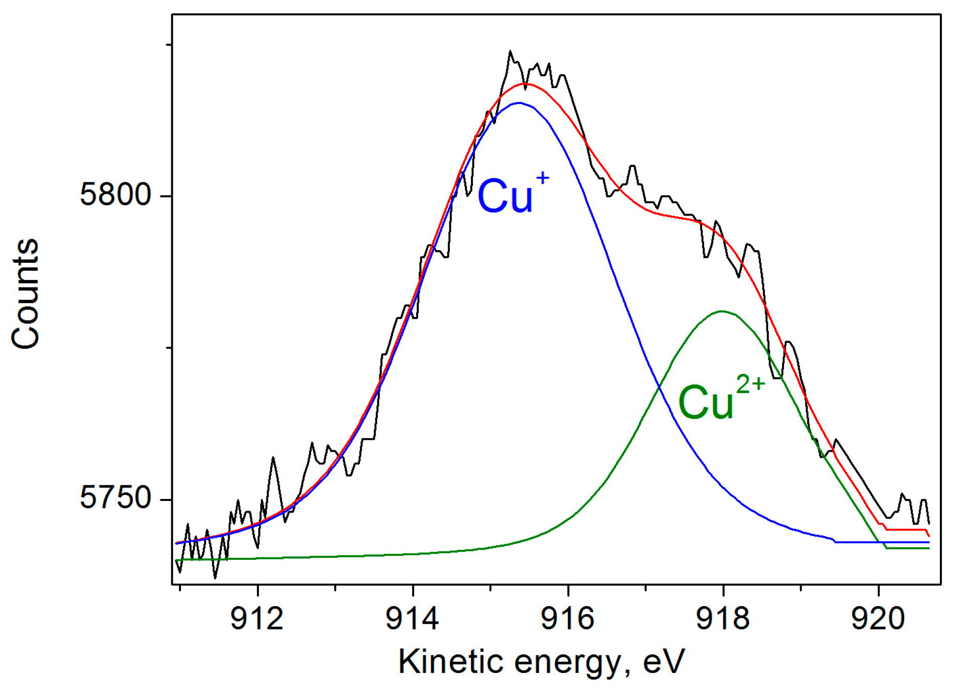
3.1. XPS of Cu-DND Powder
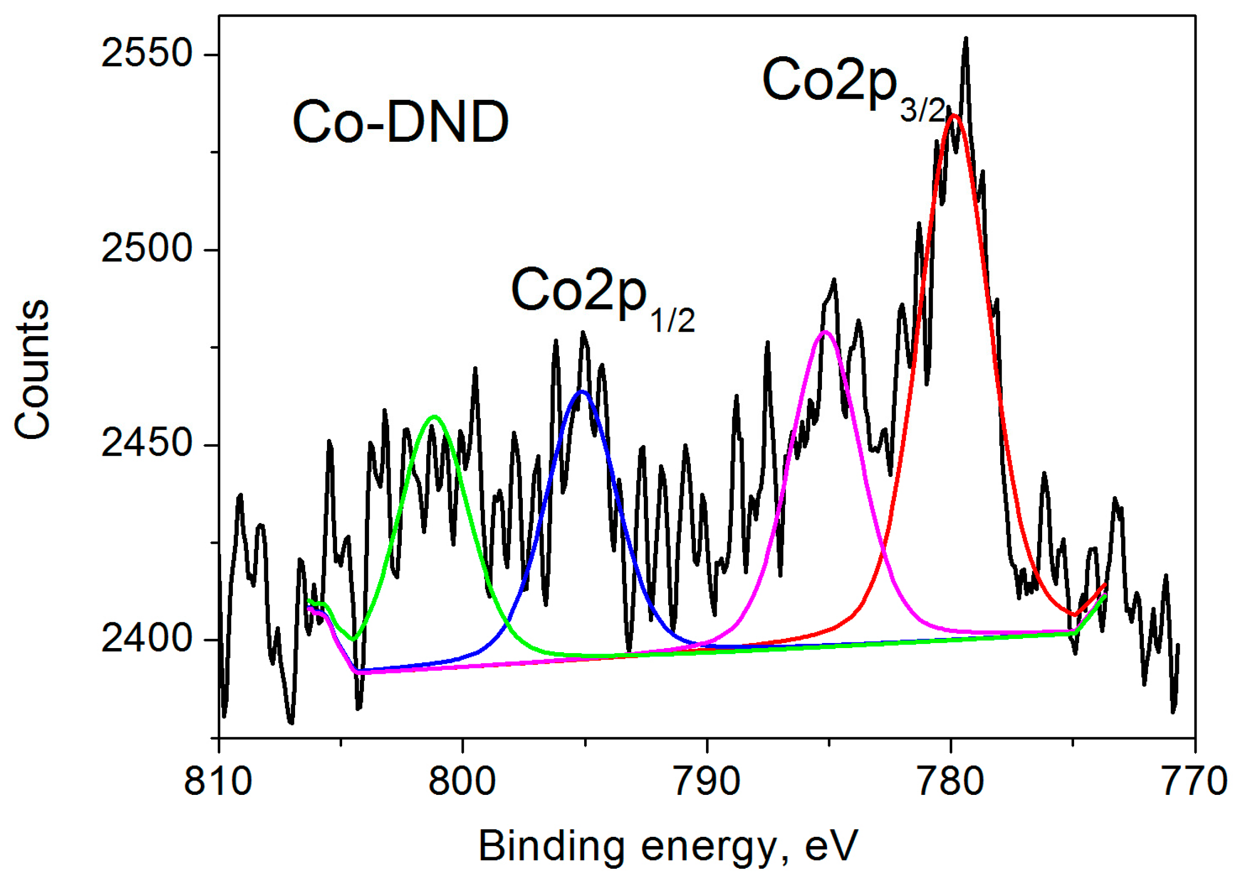
3.2. XPS of Co-DND Powder
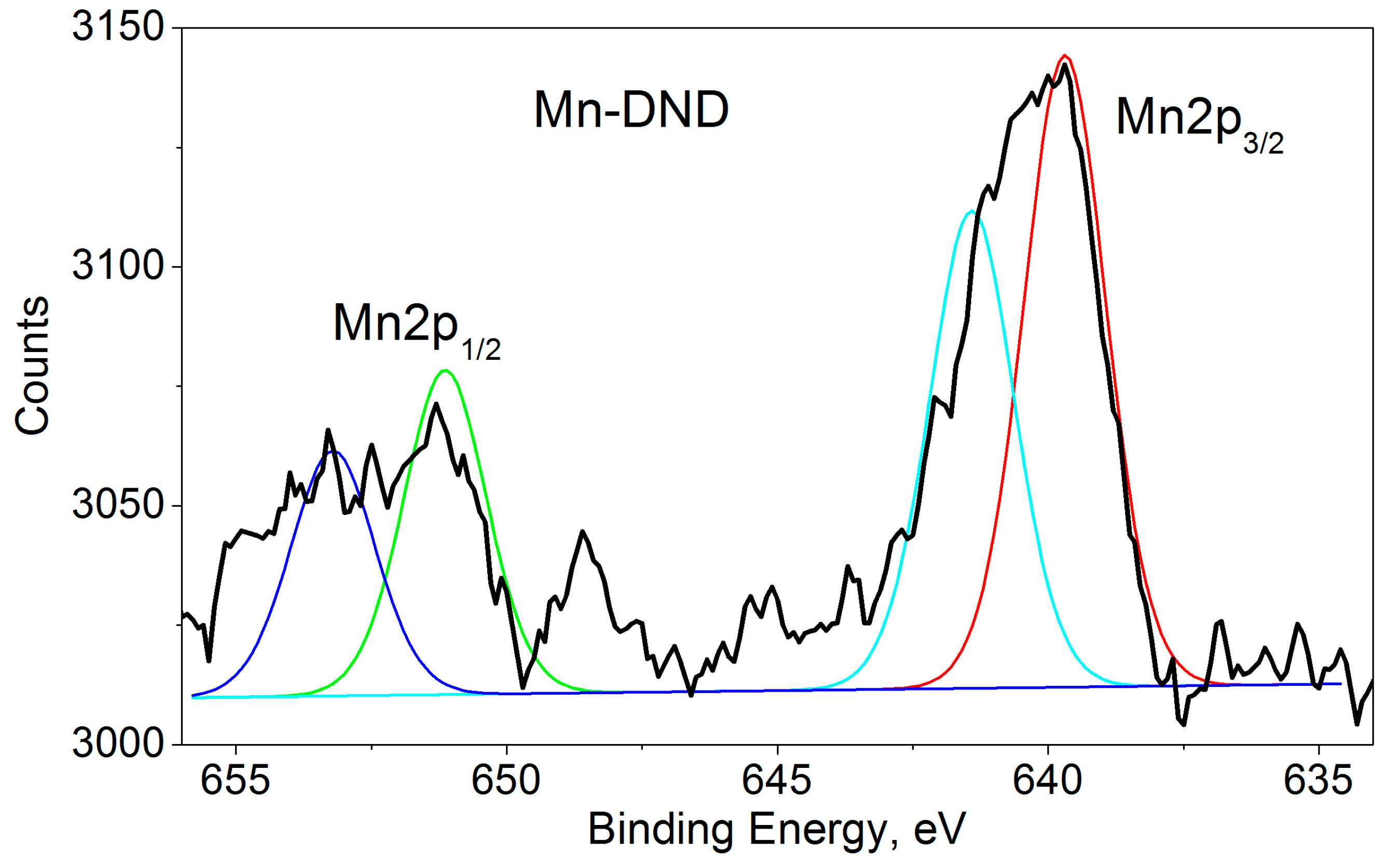
3.3. XPS of Mn-DND Powder
3.4. XPS of Gd-DND Powder
3.5. C1s, N1s and O1s XPS
4. Conclusions
Supplementary Materials
Author Contributions
Funding
Data Availability Statement
Acknowledgments
Conflicts of Interest
References
- Dolmatov, V.Y.; Ozerin, A.N.; Kulakova, I.I.; Bochechka, O.O.; Lapchuk, N.M.; Myllymaki, V.; Vehanen, A. Detonation nanodiamonds: New aspects in the theory and practice of synthesis, properties and applications. Russ. Chem. Rev. 2020, 89, 1428–1462. [Google Scholar] [CrossRef]
- Vul’, A.Y.; Shenderova, O.A. (Eds.) Detonation Nanodiamonds: Science and Applications; Pan Stanford Publishing: Singapore, 2014; pp. 181–204. [Google Scholar]
- Shenderova, O.A.; McGuire, G.E. Science and Engineering of Nanodiamond Particle Surfaces for Biological Applications (Review). Biointerphases 2015, 10, 030802. [Google Scholar] [CrossRef]
- Arnault, J.-C. (Ed.) Nanodiamonds. Advanced Material Analysis, Properties and Applications; Elsevier: Amsterdam, The Netherlands, 2017; 488p, Available online: https://www.elsevier.com/books/nanodiamonds/arnault/978-0-323-43029-6 (accessed on 17 April 2024).
- Aharonovich, I.; Neu, E. Diamond Nanophotonics. Adv. Opt. Mater. 2014, 2, 911–928. [Google Scholar] [CrossRef]
- Tegafaw, T.; Liu, S.; Ahmad, M.Y.; Saidi, A.K.A.A.; Zhao, D.; Liu, Y.; Yue, H.; Nam, S.W.; Chang, Y.; Lee, G.H. Production, surface modification, physicochemical properties, biocompatibility, and bioimaging applications of nanodiamonds. RSC Adv. 2023, 13, 32381. [Google Scholar] [CrossRef] [PubMed]
- Chang, S.L.Y.; Reineck, P.; Krueger, A.; Mochalin, V.N. Ultrasmall Nanodiamonds: Perspectives and Questions. ACS Nano 2022, 16, 8513–8524. [Google Scholar] [CrossRef] [PubMed]
- Panich, A.M.; Shames, A.I.; Mogilyansky, D.; Goren, S.D.; Dolmatov, V.Y. Detonation Nanodiamonds Fabricated from Tetryl: Synthesis, NMR, EPR and XRD study. Diam. Relat. Mater. 2020, 108, 107918. [Google Scholar] [CrossRef]
- Shames, A.I.; Panich, A.M.; Friedlander, L.; Dolmatov, V.Y. Magnetic resonance study of novel detonation nanodiamonds originated from non-conventional explosives. Diam. Relat. Mater. 2023, 136, 110059. [Google Scholar] [CrossRef]
- Wrachtrup, J.; Jelezko, F. Processing Quantum Information in Diamond. J. Phys. Condens. Matter 2006, 18, S807–S824. [Google Scholar] [CrossRef]
- Doherty, M.W.; Manson, N.B.; Delaney, P.; Jelezko, F.; Wrachtrupe, J.; Hollenberg, L.C.L. The Nitrogen-Vacancy Color Centre in Diamond. Phys. Rep. 2013, 528, 1–45. [Google Scholar] [CrossRef]
- Nunn, N.; d’Amora, M.; Prabhakar, N.; Panich, A.M.; Froumin, N.; Torelli, M.D.; Vlasov, I.; Reineck, P.; Gibson, B.; Rosenholm, J.M.; et al. Fluorescent Single-Digit Detonation Nanodiamond for Biomedical Applications. Methods Appl. Fluoresc. 2018, 6, 035010. [Google Scholar] [CrossRef]
- Shenderova, O.A.; Shames, A.I.; Nunn, N.A.; Torelli, M.D.; Vlasov, I.; Zaitsev, A. Review Article: Synthesis, Properties, and Applications of Fluorescent Diamond Particles. J. Vac. Sci. Technol. B 2019, 37, 030802. [Google Scholar] [CrossRef] [PubMed]
- Pezzagna, S.; Meijer, J. Quantum Computer Based on Color Centers in Diamond. Appl. Phys. Rev. 2021, 8, 011308. [Google Scholar] [CrossRef]
- Nunn, N.; Shames, A.I.; Torelli, M.; Smirnov, A.I.; Shenderova, O. Luminescent diamond: A platform for next generation nanoscale optically driven quantum sensors. In Luminescent Nanomaterials; Jenny Stanford Publishing: Singapore, 2022; pp. 1–95. [Google Scholar] [CrossRef]
- Shames, A.; Panich, A.; Friedlander, L.; Cohen, H.; Butler, J.; Moreh, R. Magnetic Resonance Study of Bulky CVD Diamond Disc. Materials 2024, 17, 1871. [Google Scholar] [CrossRef] [PubMed]
- Shames, A.I.; Panich, A.M.; Kempiński, W.; Alexenskii, A.E.; Baidakova, M.V.; Dideikin, A.T.; Osipov, V.Y.; Siklitski, V.I.; Osawa, E.; Ozawa, M.; et al. Defects and impurities in nanodiamonds: EPR, NMR and TEM study. J. Phys. Chem. Solids 2002, 63, 1993–2001. [Google Scholar] [CrossRef]
- Dey, T.; Ghosh, A.; Sanya, A.; Charles, C.J.S.; Pokharel, S.; Nair, L.; Singh, M.; Kaity, S.; Ravichandiran, V.; Kaur, K.; et al. Surface engineered nanodiamonds: Mechanistic intervention in biomedical applications for diagnosis and treatment of cancer. Biomed. Mater. 2024, 19, 032003. [Google Scholar] [CrossRef] [PubMed]
- Francis, S.J.; Torelli, M.D.; Nunn, N.A.; Arepally, G.M.; Shenderova, O.A. Clot Imaging Using Photostable Nanodiamond. Nanomaterials 2023, 13, 961. [Google Scholar] [CrossRef] [PubMed]
- Bogdanowicz, R. Functionalized nanodiamonds as a perspective green carbo-catalyst for removal of emerging organic pollutants. Curr. Opin. Solid State Mater. Sci. 2022, 26, 100991. [Google Scholar] [CrossRef]
- Navalon, S.; Dhakshinamoorthy, A.; Álvaro, M.; García, H. Diamond Nanoparticles in Heterogeneous Catalysis. Chem. Mater. 2020, 32, 4116–4143. [Google Scholar] [CrossRef]
- Gawande, M.B.; Fornasiero, P.; Zbořil, R. Carbon-Based Single-Atom Catalysts for Advanced Applications. ACS Catal. 2020, 10, 2231–2259. [Google Scholar] [CrossRef]
- Manus, L.M.; Mastarone, D.J.; Waters, E.A.; Zhang, X.-Q.; Schultz-Sikma, E.A.; MacRenaris, K.W.; Ho, D.; Meade, T.J. Gd(III)-Nanodiamond Conjugates for MRI Contrast Enhancement. Nano Lett. 2010, 10, 484–489. [Google Scholar] [CrossRef] [PubMed]
- Rammohan, N.; MacRenaris, K.W.; Moore, L.K.; Parigi, G.; Mastarone, D.J.; Manus, L.M.; Lilley, L.M.; Preslar, A.T.; Waters, E.A.; Filicko, A.; et al. Nanodiamond-Gadolinium(III) Aggregates for Tracking Cancer Growth In Vivo at High Field. Nano Lett. 2016, 16, 7551–7564. [Google Scholar] [CrossRef]
- Nakamura, T.; Ohana, T.; Yabuno, H.; Kasai, R.; Suzuki, T.; Hasebe, T. Simple Fabrication of Gd(III)-DTPA-Nanodiamond Particles by Chemical Modification for Use as Magnetic Resonance Imaging (MRI) Contrast Agent. Appl. Phys. Express 2013, 6, 015001. [Google Scholar] [CrossRef]
- Zhao, L.; Shiino, A.; Qin, H.; Kimura, T.; Komatsu, N. Synthesis, Characterization, and Magnetic Resonance Evaluation of Polyglycerol-Functionalized Detonation Nanodiamond Conjugated with Gadolinium(III) Complex. J. Nanosci. Nanotechnol. 2015, 15, 1076–1082. [Google Scholar] [CrossRef] [PubMed]
- Hou, W.; Toh, T.B.; Abdullah, L.N.; Yvonne, T.W.Z.; Lee, K.J.; Guenther, I.; Chow, E.K.H. Nanodiamond–Manganese dual mode MRI contrast agents for enhanced liver tumor detection. Nanomed. Nanotech. Biol. Med. 2017, 13, 783–793. [Google Scholar] [CrossRef] [PubMed]
- Panich, A.M.; Shames, A.I.; Medvedev, O.; Osipov, V.Y.; Alexenskii, A.E.; Vul’, A.Y. Magnetic Resonance Study of Detonation Nanodiamonds with Surface Chemically Modified by Transition Metal Ions. Appl. Magn. Reson. 2009, 36, 317–329. [Google Scholar] [CrossRef]
- Panich, A.M.; Altman, A.; Shames, A.I.; Osipov, V.Y.; Alexenskiy, A.E.; Vul’, A.Y. Proton magnetic resonance study of diamond nanoparticles decorated by transition metal ions. J. Phys. D Appl. Phys. 2011, 44, 125303. [Google Scholar] [CrossRef]
- Shames, A.I.; Panich, A.M.; Osipov, V.Y.; Aleksenskiy, A.E.; Vul’, A.Y.; Enoki, T.; Takai, K. Structure and Magnetic Properties of Detonation Nanodiamond Chemically Modified by Copper. J. Appl. Phys. 2010, 107, 014318. [Google Scholar] [CrossRef]
- Panich, A.M.; Shames, A.I.; Sergeev, N.A.; Osipov, V.Y.; Alexenskiy, A.E.; Vul’, A.Y. Magnetic Resonance Study of Gadolinium-Grafted Nanodiamonds. J. Phys. Chem. C 2016, 120, 19804–19811. [Google Scholar] [CrossRef]
- Panich, A.M.; Shames, A.I.; Aleksenskii, A.E.; Yudina, E.B.; Vul’, A.Y. Manganese-grafted detonation nanodiamond, a novel potential MRI contrast agent. Diam. Relat. Mater. 2021, 119, 108590. [Google Scholar] [CrossRef]
- Panich, A.M. Nuclear Magnetic Resonance Studies of Nanodiamond Surface Modification. Diam. Relat. Mater. 2017, 79, 21–31. [Google Scholar] [CrossRef]
- Panich, A.M.; Aleksenskii, A.E.; Yudina, E.B.; Vul’, A.Y. Spatially Resolved Spin−Lattice Relaxation Times and Line Widths in Manganese-Grafted Detonation Nanodiamonds. J. Phys. Chem. C 2022, 126, 1489–1495. [Google Scholar] [CrossRef]
- Panich, A.M.; Salti, M.; Goren, S.D.; Yudina, E.B.; Alexenskii, A.E.; Vul’, A.Y.; Shames, A.I. Gd(III)-Grafted Detonation Nanodiamonds for MRI Contrast Enhancement. J. Phys. Chem. C 2019, 123, 2627–2631. [Google Scholar] [CrossRef]
- Panich, A.M.; Salti, M.; Prager, O.; Swissa, E.; Kulvelis, Y.V.; Yudina, E.B.; Aleksenskii, A.E.; Goren, S.D.; Vul’, A.Y.; Alexander, I. Shames, PVP-coated Gd-grafted nanodiamonds as a novel and potentially safer contrast agent for in-vivo MRI. Magn. Reson. Med. 2021, 86, 935–942. [Google Scholar] [CrossRef] [PubMed]
- Panich, A.M.; Salti, M.; Aleksenskii, A.E.; Kulvelis, Y.V.; Chizhikova, A.; Vul’, A.Y.; Shames, A.I. Suspensions of manganese-grafted nanodiamonds: Preparation, NMR, and MRI study. Diam. Relat. Mater. 2023, 131, 109591. [Google Scholar] [CrossRef]
- Comet, M.; Pichot, V.; Siegert, B.; Britz, F.; Spitzer, D. Detonation Nanodiamonds for Doping Kevlar. J. Nanosci. Nanotechnol. 2010, 10, 4286–4292. [Google Scholar] [CrossRef] [PubMed]
- Osipov, V.Y.; Aleksenskiy, A.E.; Takai, K.; Vul’, A.Y. Magnetic Studies of a Detonation Nanodiamond with the Surface Modified by Gadolinium Ions. Phys. Solid State 2015, 57, 2314–2319. [Google Scholar] [CrossRef]
- Osipov, V.Y.; Boukhvalov, D.W.; Takai, K. Gadolinium ion bonding on the surface of carboxylated detonation nanodiamond in terms of magnetochemistry and density functional theory. Mendeleev Commun. 2020, 30, 436–438. [Google Scholar] [CrossRef]
- Osipov, V.Y.; Romanov, N.M.; Suvorkova, I.E.; Osipova, E.V.; Tsuji, T.; Ishiguro, Y.; Takai, K. Magnetic resonance tracking of copper ion fixation on the surface of carboxylated nanodiamonds from viewpoint of changes in carbon-inherited paramagnetism. Mendeleev Commun. 2022, 32, 132–135. [Google Scholar] [CrossRef]
- Gridnev, I.D.; Osipov, V.Y. Transition metal atoms grafted on the nanodiamonds surface: Identification and guest–host spin–spin interactions. Mendeleev Commun. 2022, 32, 143–151. [Google Scholar] [CrossRef]
- Osipov, V.Y.; Shames, A.I.; Enoki, T.; Takai, K.; Baidakova, M.V.; Vul’, A.Y. Paramagnetic defects and exchange coupled spins in pristine ultrananocrystalline diamonds. Diam. Relat. Mater. 2007, 16, 2035–2038. [Google Scholar] [CrossRef]
- Aleksenski, A.E.; Yagovkina, M.A.; Vul’, A.Y. Intercalation of Ultrafine-Dispersed Diamond in Aqueous Suspensions. Phys. Solid State 2004, 46, 685–686. [Google Scholar] [CrossRef]
- Yudina, E.B.; Aleksenskii, A.E.; Fomina, I.G.; Shvidchenko, A.V.; Danilovich, D.P.; Eremenko, I.L.; Vul, A.Y. Interaction of Carboxyl Groups with Rare Metal Ions on the Surface of Detonation Nanodiamonds. Eur. J. Inorg. Chem. 2019, 2019, 4345–4349. [Google Scholar] [CrossRef]
- Biesinger, M.C.; Lau, L.W.M.; Gerson, A.R.; Smart, R.S.C. Resolving surface chemical states in XPS analysis of first row transition metals, oxides and hydroxides: Sc, Ti, V, Cu and Zn. Appl. Surf. Sci. 2010, 257, 887–898. [Google Scholar] [CrossRef]
- Naumkin, A.V.; Kraut-Vass, A.; Gaarenstroom, S.W.; Powell, C.J. NIST X-ray Photoelectron Spectroscopy Database, Version 5.0 (Last Update to Data Content: 2023). Available online: https://srdata.nist.gov/xps/ (accessed on 23 December 2024).
- Bui, N.T.; Kang, H.; Teat, S.J.; Su, G.M.; Pao, C.W.; Liu, Y.S.; Zaia, E.W.; Guo, J.; Chen, J.L.; Meihaus, K.R.; et al. A nature-inspired hydrogen-bonded supramolecular complex for selective copper ion removal from water. Nat. Commun. 2020, 11, 3947. [Google Scholar] [CrossRef] [PubMed]
- de Sousa, P.V.F.; de Oliveira, A.F.; da Silva, A.A.; Lopes, R.P. Environmental remediation processes by zero valence copper: Reaction mechanisms. Environ. Sci. Pollut. Res. 2019, 26, 14883–14903. [Google Scholar] [CrossRef] [PubMed]
- Lázaro-Martínez, J.M.; Lupano, L.V.L.; Piehl, L.L.; Rodríguez-Castellón, E.; Orto, V.C.D. New Insights about the Selectivity in the Activation of Hydrogen Peroxide by Cobalt or Copper Hydrogel Heterogeneous Catalysts in the Generation of Reactive Oxygen Species. J. Phys. Chem. C 2016, 120, 29332–29347. [Google Scholar] [CrossRef]
- Li, X.; Ai, S.; Huang, Y.; Huang, C.; Yu, W.; Mao, Z. Fast and reversible adsorption for dibenzothiophene in fuel oils with metallic nano-copper supported on mesoporous silica. Environ. Sci. Pollut. Res. 2021, 28, 2741–2752. [Google Scholar] [CrossRef] [PubMed]
- Varma, K.S.; Shukla, A.D.; Tayade, R.J.; Joshi, P.A.; Das, A.K.; Modi, K.B.; Gandhi, V.G. Photocatalytic performance and interaction mechanism of reverse micelle synthesized Cu-TiO2 nanomaterials towards levofloxacin under visible LED light. Photochem. Photobiol. Sci. 2022, 21, 77–89. [Google Scholar] [CrossRef]
- Thermo Fisher Scientific. Cobalt X-ray Photoelectron Spectra, Cobalt Electron Configuration, and Other Elemental Information. Available online: https://www.thermofisher.com/il/en/home/materials-science/learning-center/periodic-table/transition-metal/cobalt.html (accessed on 23 December 2024).
- Wei, W.F.; Chen, W.X.; Ivey, D.G. Rock. Salt-Spinel Structural Transformation in Anodically Electrodeposited Mn-Co-O Nanocrystals. Chem. Mater. 2008, 20, 1941–1947. [Google Scholar] [CrossRef]
- Zhou, W.; Wu, J.; Ouyang, C.; Gao, Y.; Xu, X.; Huang, Z. Optical properties of Mn-Co-Ni-O thin films prepared by radio frequency sputtering deposition. J. Appl. Phys. 2014, 115, 093512. [Google Scholar] [CrossRef]
- Murray, J.W.; Dillard, J.G.; Giovanoly, R.; Moers, H.; Stumm, W. Oxidation of Mn(I1): Initial mineralogy, oxidation state and ageing. Geochim. Cosmochim. Acta Vol. 1985, 49, 463–470. [Google Scholar] [CrossRef]
- Nesbitt, H.W.; Banerjee, D. Interpretation of XPS Mn(2p) spectra of Mn oxyhydroxides and constraints on the mechanism of MnO2 precipitation. Am. Mineral. 1998, 83, 305–315. [Google Scholar] [CrossRef]
- Biesinger, M.C.; Payne, B.P.; Grosvenor, A.P.; Lau, L.W.M.; Gerson, A.R.; Smart, R.S.C. Resolving surface chemical states in XPS analysis of first row transition metals, oxides and hydroxides: Cr, Mn, Fe, Co and Ni. Appl. Surf. Sci. 2011, 257, 2717–2730. [Google Scholar] [CrossRef]
- Ilton, E.S.; Post, J.E.; Heaney, P.J.; Ling, F.T.; Kerisit, S.N. XPS determination of Mn oxidation states in Mn (hydr)oxides. Appl. Surf. Sci. 2016, 366, 475–485. [Google Scholar] [CrossRef]
- Moulder, J.F.; Stickle, W.F.; Sobol, P.E.; Bomben, K.D. Handbook of X-ray Photoelectron Spectroscopy. A Reference Book of Standard Spectra for Identification and Interpretation of XPS Data; Chastain, J., Ed.; Perkin-Elmer Corporation: Eden Prairie, MN, USA, 1992. [Google Scholar]
- Brunckova, H.; Kolev, H.; Rocha, L.A. XPS characterization and luminescent properties of GdNbO4 and GdTaO4 thin Films. Appl. Surf. Sci. 2020, 504, 144358. [Google Scholar] [CrossRef]
- Barreca, D.; Gasparotto, A.; Milanov, A.; Tondello, E.; Devi, A.; Fischer, R.A. Gd2O3 Nanostructured Thin Films Analyzed by XPS. Surf. Sci. Spectr. 2007, 14, 60–67. [Google Scholar] [CrossRef]
- Haunsbhavi, K.; Kumar, A.; Ubaidullah, M.; Shaikh, S.F.; Venkatesh, R.; Alagarasan, D.; Murahari, P.; Angadi, B. The effect of rare-earth element (Gd, Nd, La) doping of NiO films on UV photodetector. Phys. Scr. 2022, 97, 055815. [Google Scholar] [CrossRef]
- Khan, S.A.; Gambhir, S.; Ahmad, A. Extracellular biosynthesis of gadolinium oxide (Gd2O3) nanoparticles, their biodistribution and bioconjugation with the chemically modified anticancer drug taxol. Beilstein J. Nanotechnol. 2014, 5, 249–257. [Google Scholar] [CrossRef] [PubMed]
- Milanov, A.P.; Toader, T.; Parala, H.; Barreca, D.; Gasparotto, A.; Bock, C.; Becker, H.W.; Ngwashi, D.K.; Cross, R.; Paul, S.; et al. Lanthanide Oxide Thin Films by Metalorganic Chemical Vapor Deposition Employing Volatile Guanidinate Precursors. Chem. Mater. 2009, 21, 5443–5455. [Google Scholar] [CrossRef]
- Vijayaprasath, G.; Habibulla, I.; Dharuman, V.; Balasubramanian, S.; Ganesan, R. Fabrication of Gd2O3 Nanosheet-Modified Glassy Carbon Electrode for Nonenzymatic Highly Selective Electrochemical Detection of Vitamin B2. ACS Omega 2020, 5, 17892–17899. [Google Scholar] [CrossRef]
- Raiser, D.; Deville, J.P. Study of XPS photoemission of some gadolinium compounds. J. Electron Spectrosc. Relat. Phenom. 1991, 57, 91–97. [Google Scholar] [CrossRef]
- Majeed, S.; Shivashankar, S.A. Rapid, microwave-assisted synthesis of Gd2O3 and Eu:Gd2O3 nanocrystals: Characterization, magnetic, optical and biological studies. J. Mater. Chem. B 2014, 2, 5585–5593. [Google Scholar] [CrossRef]
- Datta, P.; Majewski, P.; Aldinger, F. Study of gadolinia-doped ceria solid electrolyte surface by XPS. Mater. Charact. 2009, 60, 138–143. [Google Scholar] [CrossRef]
- Osipov, V.Y.; Boukhvalov, D.W.; Takai, K. Isolated Spin-7/2 Species of Gadolinium (III) Chelate Complexes on the Surface of 5-nm Diamond Particles. Nanomaterials 2023, 13, 1995. [Google Scholar] [CrossRef] [PubMed]
- Boukhvalov, D.W.; Osipov, V.Y.; Serikkanov, A.; Takai, K. Unveiling the Structure of Metal–Nanodiamonds Bonds: Experiment and Theory. C J. Carbon Res. 2024, 10, 63. [Google Scholar] [CrossRef]


| Compound | Ion | S | NA, wt% | N | NS, spin/g | Ref. |
|---|---|---|---|---|---|---|
| Cu-DND | Cu2+ (3d9) | 1/2 | 0.6 | 4 | 1.67 × 10¹⁹ | [28] |
| Co-DND | Co2+ (3d7) | 1/2 | 0.6 | - | - | [28] |
| Mn-DND | Mn2+ (3d5) | 5/2 | 0.12 | 8–9 | 4.1 × 10¹⁹ | [32] |
| Gd-DND | Gd3+ (4f7) | 7/2 | 3.28 | 18 | 7.85 ×10¹⁹ | [31] |
| Compound | at%—XPS * | BE, eV | at%, XPS Components | at%—EPR *, SQUID |
|---|---|---|---|---|
| Cu-DND Cu2p3 | 0.14 | 932.63 | 100 | |
| Cu+ | 0.098 | 932.09 | 70 | |
| Cu2+ | 0.042 | 933.94 | 30 | 0.034 |
| Co-DND Co2p3 | 0.13 | 780.9 | 52.9 | - |
| Co-DND Co2p1 | 796.4 | 52.9 | - | |
| 801.9 | 47.1 | |||
| Mn-DND Mn2p3 | 0.07 | 639.7 | 56.3 | 0.085 |
| 641.4 | 43.7 | |||
| Mn-DND Mn2p1 | 651.1 | 56.3 | ||
| 653.2 | 43.7 | |||
| Gd-DND Gd3d5 | 0.19 | 1185.4 | 54.4 | 0.164 |
| 1187.9 | 45.6 | |||
| Gd-DND Gd4d | 0.14 | 139.9 | 70. 5 | 0.164 |
| 142.2 | 29.55 | |||
| 145.2 | 70.45 | |||
| 147.5 | 29.6 |
Disclaimer/Publisher’s Note: The statements, opinions and data contained in all publications are solely those of the individual author(s) and contributor(s) and not of MDPI and/or the editor(s). MDPI and/or the editor(s) disclaim responsibility for any injury to people or property resulting from any ideas, methods, instructions or products referred to in the content. |
© 2025 by the authors. Licensee MDPI, Basel, Switzerland. This article is an open access article distributed under the terms and conditions of the Creative Commons Attribution (CC BY) license (https://creativecommons.org/licenses/by/4.0/).
Share and Cite
Panich, A.; Froumin, N.; Aleksenskii, A.; Chizhikova, A. XPS Study of Grafting Paramagnetic Ions onto the Surface of Detonation Nanodiamonds. Nanomaterials 2025, 15, 260. https://doi.org/10.3390/nano15040260
Panich A, Froumin N, Aleksenskii A, Chizhikova A. XPS Study of Grafting Paramagnetic Ions onto the Surface of Detonation Nanodiamonds. Nanomaterials. 2025; 15(4):260. https://doi.org/10.3390/nano15040260
Chicago/Turabian StylePanich, Alexander, Natalya Froumin, Aleksandr Aleksenskii, and Anastasiya Chizhikova. 2025. "XPS Study of Grafting Paramagnetic Ions onto the Surface of Detonation Nanodiamonds" Nanomaterials 15, no. 4: 260. https://doi.org/10.3390/nano15040260
APA StylePanich, A., Froumin, N., Aleksenskii, A., & Chizhikova, A. (2025). XPS Study of Grafting Paramagnetic Ions onto the Surface of Detonation Nanodiamonds. Nanomaterials, 15(4), 260. https://doi.org/10.3390/nano15040260