You are currently viewing a new version of our website. To view the old version click .
Information
  • Article
  • Open Access

21 February 2022

Automatic Hemiplegia Type Detection (Right or Left) Using the Levenberg-Marquardt Backpropagation Method

,
,
,
,
,
,
,
,
and
1
Department of Informatics and Telecommunications, University of Ioannina, GR47100 Arta, Greece
2
Department of Physical Medicine and Rehabilitation, University of Ioannina, S. Niarchos Ave, GR45110 Ioannina, Greece
3
Department of Electrical and Computer Engineering, University of Western Macedonia, GR50100 Kozani, Greece
*
Authors to whom correspondence should be addressed.
This article belongs to the Special Issue Design Automation, Computer Engineering, Computer Networks and Social Media (SEEDA-CECNSM 2021)

Abstract

Hemiplegia affects a significant portion of the human population. It is a condition that causes motor impairment and severely reduces the patient’s quality of life. This paper presents an automatic system for identifying the hemiplegia type (right or left part of the body is affected). The proposed system utilizes the data taken from patients and healthy subjects using the accelerometer sensor from the RehaGait mobile gait analysis system. The collected data undergo a pre-processing procedure followed by a feature extraction stage. The extracted features are then sent to a neural network trained by the Levenberg-Marquardt backpropagation (LM-BP) algorithm. The experimental part of this research involved creating a custom-created dataset containing entries taken from ten healthy and twenty non-healthy subjects. The data were taken from seven different sensors placed in specific areas of the subjects’ bodies. These sensors can capture a three-dimensional (3D) signal using the accelerometer, magnetometer, and gyroscope device types. The proposed system used the signals taken from the accelerometers, which were split into 2-sec windows. The proposed system achieved a classification accuracy of 95.12% and was compared with fourteen commonly used machine learning approaches.

1. Introduction

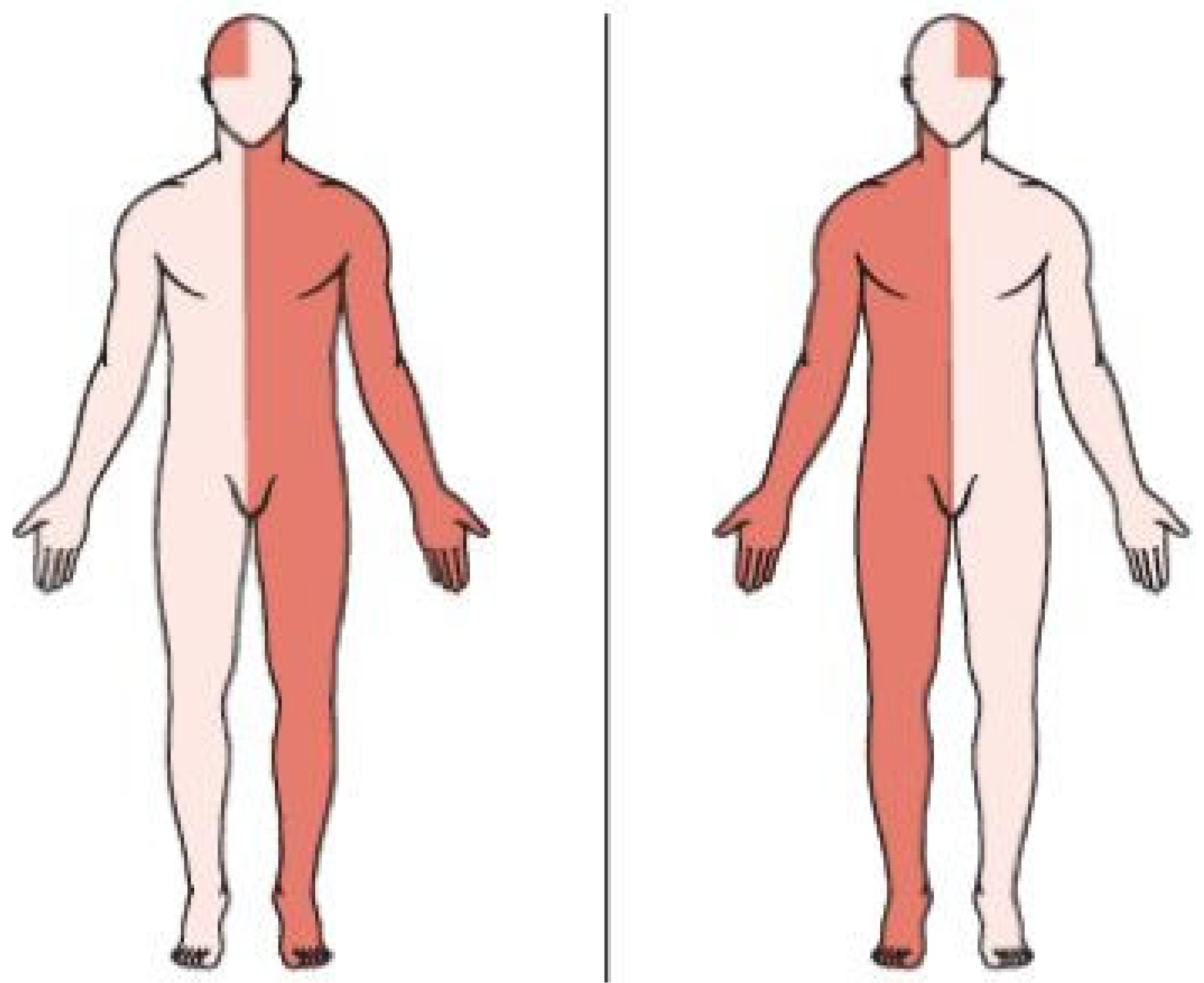
Hemiplegia is a disease that causes the patient to lose motor control in one part of the body. The hemiplegia symptoms include the patient’s inability to move its right or left body parts (arm and leg) and the creation of spastic mass patterns. The paralysis in half of the patient’s body is usually caused by a stroke and has severe consequences on their health and quality of life. It is an effect caused by damage in one of the two cerebral hemispheres. The problems caused by a brain lesion in one area are spread to other areas since the human brain is an extensive network of interconnected neurons participating in two different types of communication (feed-forward and feedback). The unaffected areas of the brain face problems due to the lack of information or misinformation created from the affected areas. The stroke patient will have to deal with severe problems in both body sides, with side effects spreading to all brain functions causing motor impairment in the whole body. These impairments will affect sensory perception, memory, and behavior, causing a challenging situation to the patient’s rehabilitation process. Hemiplegia types can be right or left, as seen in Figure 1. In the former type, the left body side is paralyzed, while in the latter type, the right body side is paralyzed. An example causing either type is an incomplete spinal cord injury [,].
Figure 1. Hemiplegia types. The left picture depicts brain damage in the left part of the brain (all the affected body parts are marked with red color), which causes paralysis to the right part of the body. The right picture depicts brain damage in the right part of the brain, which causes paralysis to the right part of the body.
The novelty behind the proposed system is creating a tool capable of detecting the hemiplegia category (right or left) between patients and healthy individuals using a low-cost system that doesn’t require the use of an expensive three-dimensional (3D) camera. The proposed method can be used as a supplementary tool for diagnosing the hemiplegia types mentioned above amongst patients and healthy subjects. The system works by using seven sensors to the subject’s body which record Spatio-temporal specific parameters during walking. One sensor is placed on the hip, and one is placed at each patient’s foot, shank, and thigh. All the sensors are provided from the RehaGait mobile gait analysis system [,,].
The mobile gait analysis system offers significant advantages, including monitoring the user’s status using a built-in video capture feature. It can recognize the damaged areas, estimate the gait pattern and find asymmetries in the patient’s lower limbs. The rest of its advantages include movement freedom, mobility since it does not require a gait lab, and the ability to represent the captured data graphically [,].
The sensors can capture 3-dimensional (3D) signals using an accelerometer that records the linear acceleration, a magnetometer that records the earth’s magnetic, and a gyroscope for recording the angular velocity []. The proposed method used the captured accelerometer data, divided into widows with a 2-s length. Then, they underwent a pre-processing and feature extraction stage before being sent to a single-layer neural network trained by the Levenberg-Marquardt backpropagation (LM-BP) algorithm. The training procedure of the neural network involved using a custom dataset that contained data taken from thirty participants (twenty non-healthy individuals having right or left hemiplegia and ten healthy individuals).
The article is structured in seven sections. It begins with the “Introduction” section, which includes the problem’s description and the motivation, followed by the “Related Work” section. “The LM-BP Algorithm” section describes the architecture of the Levenberg-Marquardt backpropagation variant utilized for the classification task of the proposed method. The “System Architecture” section describes an analysis of the system. The “Experimental Results” section contains the outcome from comparing the proposed approach with fourteen machine learning algorithms. Finally, the last two sections contain the “Discussion” and “Conclusion.”

3. The LM-BP Algorithm

This section represents a detailed explanation of the LM algorithm. The LM algorithm was initially developed by Kenneth Levenberg [] and reinvented by Donald Marquardt [] to minimize a non-linear function.
The LM Algorithm has O ( N 3 ) complexity and is illustrated in Algorithm 1 []. The algorithm trains a neural network by adapting the network’s weights and thresholds according to the weight update function depicted in (1).
w k + 1 = w k ( J k T J k + μ I ) 1 J k e k
The first five lines of the algorithm initialize its parameters. These initializations include:
  • Randomizing weights and thresholds.
  • Setting the maximum number of steps, the initial value of the learning coefficient, and the maximum allowed sum of squares network error.
Algorithm 1: The Levenberg-Marquardt Algorithm
1:   w 1 w N ,   N
2:   b 1 b N ,   N
3:   m m a x
4:   μ
5:   E m a x
6:   x = [ x 11 x 1 n x N 1 x N n ] N × n ,   n , N
7: do
8:     m = 1
9:     y = n e t ( w b k , x )
10:   E k = i = 1 n ( t i y i ) 2
11:     J =   [ e 1 , 1 w 1 e 1 , 1 w 2 e 1 , 1 w N e 1 , 1 b N e 1 , 2 w 1 e 1 , 2 w 2 e 1 , 2 w N e 1 , 2 b N e 1 , M w 1 e 1 , M w 2 e 1 , M w N e 1 , M b N e P , 1 w 1 e P , 1 w 2 e P , 1 w N e P , 1 b N e P , 2 w 1 e P , 2 w 2 e P , 2 w N e P , 2 b N e P , M w 1 e P , M w 2 e P , M w N e P , M b N ] P ,   M
12:     w b k + 1 = w b k ( J k T J k + μ I ) 1 J k e k
13:     y = n e t ( w b k + 1 , x )
14:     E k + 1 = i = 1 n ( t i y i ) 2
15:     if E k + 1 > E k
16:              if m m m a x
17:                       m = m + 1
18:                       μ = 10 μ
19:                       w b k + 1 = w b k ( J k T J k + μ I ) 1 J k e k
20:              else
21:                       w b k = w b k + 1
22:              end if
23:     else
24:              μ = μ 10
25:              w b k = w b k + 1
26:     end if
27:     while E k + 1 > E m a x  
28:     return y
Line 6 declares the neural network inputs. Line 7 starts the algorithm’s epochs, and line 8 resets the current step number iterator for each epoch. The next step calculates the neural network output, while the formula in line 10 defines the sum of squares error for the whole network. The next line involves calculating the Jacobian matrix J, used in the weight and threshold update function (line 12). Line 13 calculates the neural network output using the updated weights and thresholds. The next step calculates the updated sum of squares error for the whole network, used as a stopping criterion for the algorithm. If the error is larger than the previous error, the LM method checks if m m m a x (lines 15, 16). If the latter condition is satisfied, then m is increased by one, and the learning coefficient is multiplied by a factor of 10 (lines 17, 18). Then, the previous weight and threshold updates are retracted, and a new update is calculated considering the new learning coefficient (line 19). On the other hand, if the maximum allowed number of steps has been exceeded ( m > m m a x ) the weight and threshold update procedure is accepted, and a new training epoch begins (line 21). If the error is smaller than the previous error, μ is reduced by a factor of 10, the weight and threshold update procedure is accepted, and a new training epoch begins (lines 24, 25). Finally, the training epochs continue until the stopping criterion is satisfied (line 27) and the neural network output ( y ) is returned (line 28).

4. System Architecture

The architectural structure of the system utilizes the RehaGait mobile gait analysis system [,], containing seven sensors that can be placed at various patient body parts. Every sensor contains an accelerometer, a magnetometer, and a gyroscope. The proposed system utilizes the accelerometer signals, which are windowed into 2-s windows and undergo a pre-processing and feature extraction stage. The signals are transmitted wirelessly from the RehaGait mobile gait analysis system to a laptop, responsible for the pre-processing, feature extraction, and classification tasks. The pre-processing stage involves using a low-pass filter to smooth the signal.
The feature extraction stage involves using four time-domain features and two frequency-domain features. The first from the time-domain features is the mean ( μ ) seen below where N denotes the number of scalar observations and A is a random variable vector.
μ = 1 N i = 1 N A i
The second feature was the standard deviation shown in Formula (3), where N is the number of scalar observations, A is a random variable vector, and μ is the mean of A .
S = 1 N 1 i = 1 N ( A i μ ) 2  
The third feature is the kurtosis of a distribution depicted in Equation (4) where E ( t ) denotes the expected value of quantity t ,   μ is the mean of x and σ is the standard deviation of x .
k = E ( x μ ) 4 σ 4
The fourth feature is the peak-magnitude-to-RMS ratio depicted in Equation (5).
RMS = A   1 N i = 1 N | A i | 2
Regarding the frequency-domain features, the acceleration energy and the acceleration signal energy are selected. The first one is described in Equation (6) with A i defining the i t h spectral line of the acceleration signal and N the total lines.
E n g =   i = 1 N A i 2 N
The last feature is the acceleration signal entropy seen below with p i defining the probability of the A i value occurring in the amplitude spectrum.
E n t = i = 1 N p i log 2 p i
The above six features formed the feature vector sent as input to a neural network trained with the LM-BP algorithm. The motivation behind selecting those features was the reduction of the initial large dataset to a smaller dataset that can be easily transferred and processed with a low-cost credit card type computer like Raspberry Pi Zero W. Moreover, the above feature combination achieved the highest classification accuracy over alternative ones containing different feature sets. The neural network classified the input data into three classes (healthy, left, or right hemiplegia). The data were taken from a custom-created dataset divided into training, validation, and a separate test set. The k-fold cross-validation method was utilized in the training and validation sets. A visualization of the proposed system can be seen in Figure 2.
Figure 2. The system architecture. The system receives the accelerometer signals from seven sensors placed in various body parts, which undergo a pre-processing and feature extraction procedure. Then, the extracted features are sent as input to a neural network trained with the LM-BP algorithm responsible for their classification into three classes (healthy, left, or right hemiplegia).

5. Experimental Results

The proposed LM-BP-based system was tested using data from ten healthy, eight non-healthy subjects having left hemiplegia and twelve non-healthy subjects having right hemiplegia. The study was conducted according to the guidelines of the Declaration of Helsinki and approved by the Ethics Committee of the University of Ioannina. Informed consent was obtained from all subjects involved in the study, and written informed consent was obtained from the patients to publish this paper. The signals from the seven accelerometers were sent wirelessly to a laptop and were divided into 2-s windows. The windowed signals underwent a pre-processing phase using a low pass filter and a feature extraction process according to the procedure described in the previous section. These features were sent to a neural network trained with the LM-BP algorithm. The architecture of the neural network involved the creation of one hidden layer with 30 neurons having the sigmoid ( y = 1 1 + e x ) transfer function and three output layer neurons with the identity ( y = x ) transfer function with each input vector containing 126 values (6 features × 7 accelerometers × 3 dimensions).
The proposed system was tested with the following fourteen machine learning approaches:
  • SCG-BP
  • FPCG-BP
  • BFGS-BP
  • OSS-BP
  • GD-BP
  • RPROP
  • BR-BP
  • RMSProp
  • Adam
  • AdaMax
  • AMSGrad
  • ELM
  • OP-ELM
  • SVM
The motivation behind comparing the LM-BP algorithm with other BP variants was to evaluate its accuracy compared to other alternative BP-based solutions. The accuracy level achieved by LM-BP was also higher compared to other non-BP-based solutions like SVM, ELM, and OP-ELM. ELM and OP-ELM are relatively new SLNN training methods that can train SLNNs faster than other iterative-based methods.
The experiments were run ten times for all neural network-based approaches to avoid any bias due to the random initialization of the hidden weights and thresholds. In the RMSProp, Adam, AdaMax, AMSGrad, ELM-based, and SVM methods, 20% of the dataset was kept as a test set, and the rest was used as a training set. In the other approaches, 20% of the dataset was kept as a test set, and the rest was further divided into training and validation sets using 10-fold cross-validation. The RMSProp, Adam, AdaMax, and AMSGrad approaches were run for 500 epochs. The dataset contained 2036 entries, with 1630 rows forming the training/validation sets and 406 entries the test set. All neural networks had one hidden layer with thirty nodes. The parameters for all the experiments are summarized below. The heterogenous OP-ELM algorithm was used with three different types of kernels (linear, Gaussian, and sigmoid). The parameters for the execution of the experiments can be seen in Table 1.
Table 1. Parameter Values.
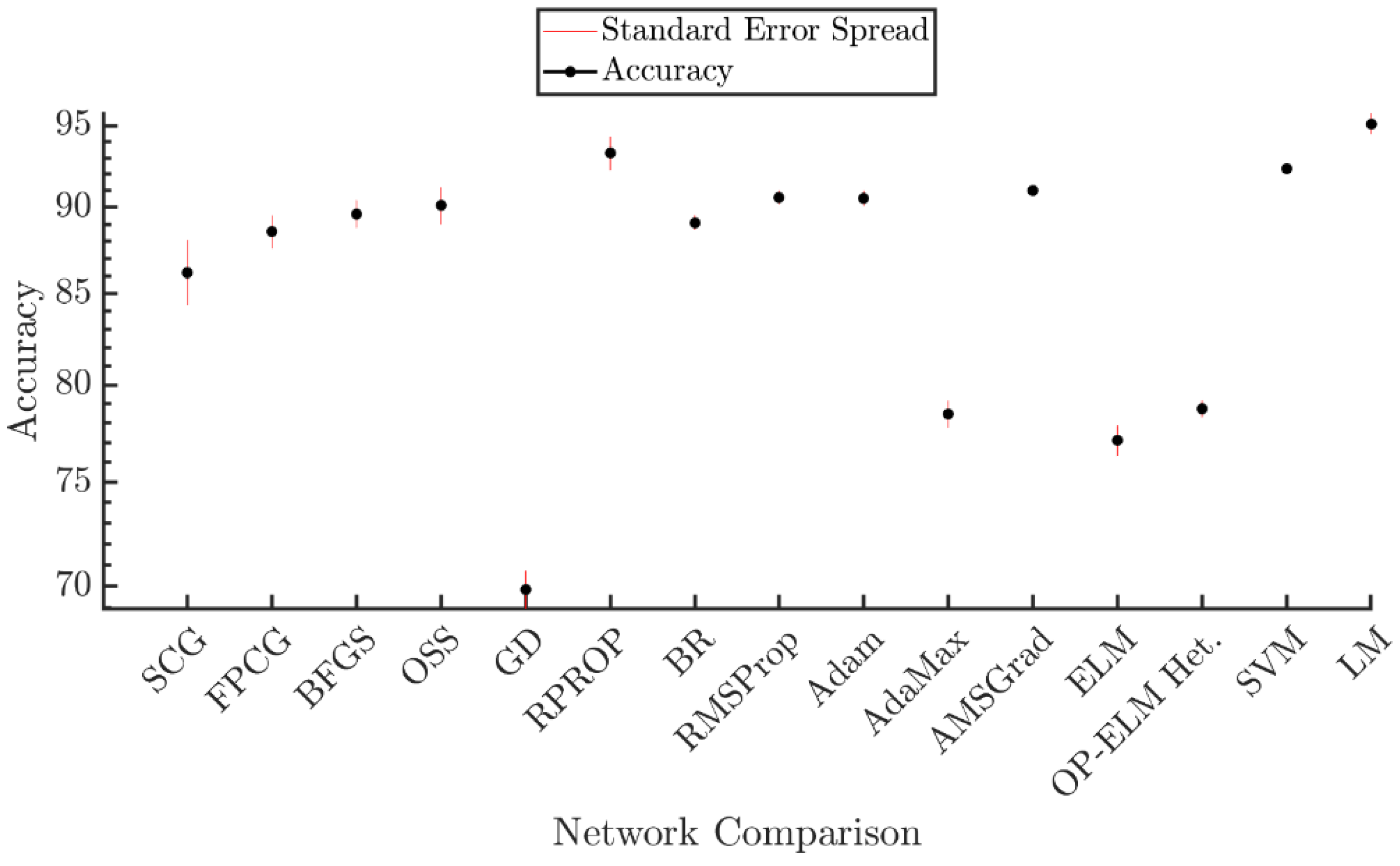
The proposed LM-BP-based method achieved the highest average accuracy over all experiment runs compared to the other fourteen machine learning methods. The results are visualized in Figure 3 and summarized in Table 2.
Figure 3. Experimental results plot. The proposed LM-BP-based system achieved the highest accuracy compared to fourteen existing machine learning algorithms.
Table 2. Comparison Results.
Two statistical tests were conducted to investigate the proposed method’s significance compared to the other fourteen methods. The first test involved creating an 80% confidence interval (CI) in each approach presented in Table 2, while the second test involved the creation of a 95% CI for each method. Then, the confidence intervals of the compared approaches were checked for overlaps with the CI from the LP-BP algorithm. If they do not overlap, it is a strong indication that the results from the LM-BP are statistically significant. The creation of the CI followed formula (8) where x ¯ is the samples average, Z is the standardized score ( Z = 1.282 for an 80% CI while Z = 1.96 for a 95% CI), s is the sample’s standard deviation and s m p is the sample size.
CI = x ¯ ± Z s   s m p
The results from the first statistical test can be seen in Figure 4, where it is shown that there is no overlap between the CI from the LM-BP algorithm and the other compared methods, which is interpreted as a strong indication that the results are statistically significant.
Figure 4. Statistical analysis of the results using an 80% CI. It can be seen from this figure that there is no overlap between the LM-BP algorithm and the other approaches, which is a strong indication that the results from the proposed method are statistically significant.
The results from the second statistical test can be seen in Figure 5. It is shown that there is no overlap between the CIs from the LM-BP algorithm and the other compared methods, which is interpreted as a strong indication that the results are statistically significant. One exception is the RPROP method which contains an overlap with the LM-BP algorithm. This overlap doesn’t allow any conclusion on whether the results between these methods are statistically significant or not.
Figure 5. Statistical analysis of the results using a 95% CI. It can be seen from this figure that there is no overlap between the LM-BP algorithm and the other approaches, which is a strong indication that the results from the proposed method are statistically significant. One exception is the RPROP method, which overlaps with the LM-BP algorithm, which doesn’t allow any conclusion regarding the statistical significance between the two methods.

6. Discussion

This article presents a system for automatically classifying the hemiplegia type between patients and healthy individuals. The system receives data from RehaGait’s mobile gait analysis system accelerometer, which underwent a pre-processing and feature extraction procedure. Then, the extracted features create the input vector to an LM-BP-based classification algorithm, classifying them into three classes (healthy, left, or right hemiplegia). The experimental results from comparing the LM-BP-based method with fourteen different machine learning methods experimentally verified the proposed method’s advantage over existing ones in terms of classification accuracy.
The proposed method was run one additional time. The results from this run were used to create the confusion matrix shown in Figure 6.
Figure 6. The confusion matrix. The green boxes show the correctly classified samples in this figure, while the red boxes show the misclassified ones. The vertical white boxes depict the percentages of all the samples predicted to belong to each class that is correctly (green color) and incorrectly (red color) classified. The horizontal white boxes depict the percentages of all the examples belonging to each class correctly (green color) and incorrectly (red color) classified. Finally, the grey box depicts the overall accuracy.
The LM-BP algorithm achieved a very high accuracy (95.8%). The number of test samples introduced to the network was 406 (132 for left hemiplegia, 219 for right hemiplegia, and 55 for non-patients). Each row defined the classification output of the network where the green boxes corresponded to correctly classified samples while the red boxes corresponded to incorrectly classified ones. The white box at the end of each row defined the percentage of all the samples predicted to belong to each class that was correctly (green color) and incorrectly (red color) classified. Each column defined the target class where the green boxes corresponded to correctly classified samples while the red boxes corresponded to incorrectly classified ones. At the end of each column, the white box defined the percentage of all the samples that belonged to each class and was correctly (green color) or incorrectly (red color) classified.
The true positive (TP), true negative (TN), false positive (FP), and false negative (FN) metrics can be calculated using the data from the confusion matrix. TP is the number of correctly classified positive class samples as positive. TN is the number of correctly classified negative class samples as negative. FP is the number of incorrectly classified negative class samples as positive and FN is the number of incorrectly classified positive class samples.as negative. The TP, TN, FP, and FN are calculated for each class in a multi-class classification problem.
The above metrics were used to calculate the precision, recall, specificity, and F-score performance metrics. Precision shows what fraction of predictions as a positive class were actually positive and is calculated using the formula TP TP + FP . Recall shows what fraction of all positive samples were correctly predicted as positive by the classifier and is defined using the equation TP TP + FN . Specificity shows what fraction of all negative samples are correctly predicted as negative by the classifier and is calculated using the formula TN TN + FP . Finally, F-score is a measure of a test’s accuracy and combines the precision and recall metrics ( Fscore = 2 precision × recall precision + recall ) . The precision, recall, specificity, and F-score performance metrics are summarized in Table 3. They show that the proposed method managed to get high scores in each class for all metrics.
Table 3. Performance Metrics.
The next figure depicts each class’s receiver operating characteristic (ROC) curve. A ROC curve is a graphical plot describing the trade-off between true positive rate (sensitivity) and false positive rate (1–specificity). If the curve is very close to the top left corner, it is an indication that the classifier has very good performance. On the other hand, if the curve is very close to the 45-degree diagonal of the ROC space, it indicates poor classification performance. All the curves in Figure 7 are very close to the upper left corner, and they have the area under the curve (AUC) values 0.9851 (left hemiplegia), 0.9772 (right hemiplegia), and 0.9919 (normal). Both observations show that the classifier has excellent classification performance (AUC values between 0.9 and 1 indicate an excellent performing classifier).
Figure 7. The ROC curves for each class. This figure depicts the ROC curves for left hemiplegic (blue color), right hemiplegic (green color), and normal subjects. All three curves are near the top left corner of the plot, which indicates a good-performing classifier.
The proposed system’s main characteristic is its ability to distinguish between the two hemiplegic types (left or right) and achieve a high classification accuracy (95.12%). It utilizes the gait data from only ten healthy and twenty hemiplegic individuals. One limitation of the proposed method is the small number of participants, resulting in small dataset size. Another restriction of the proposed system is the limitation in one type of motor disability.

7. Conclusions

This study proposed an automated tool for classifying hemiplegic from non-hemiplegic patients and can also diagnose the hemiplegia type (right or left). The proposed automatic classification method achieved a high accuracy classification rate using the LM-BP algorithm. The obtained accuracy results were tested for their significance using 80% and 95% CIs. In the first test, they were found statistically significant compared to fourteen other popular machine learning methods. At the same time, it was statistically significant in thirteen out of fourteen cases in the second test. The CI of the LM-BP algorithm had an overlap with the CI of the RPROP algorithm, which didn’t allow any conclusion on whether the results between these two algorithms are statistically significant.
Further plans for this project involve using a larger dataset containing more data from healthy and hemiplegic individuals. Including additional data would make the LM-BP classification algorithm more accurate and achieve an even higher classification accuracy. Future work involves expanding the dataset with more motor disability diseases like diplegia and Parkinson’s disease. The addition of more diseases will allow the system to recognize more motor disabilities, resulting in a complete system. This system can be used as a supplementary diagnostic tool by physiotherapists, allowing them to adapt their patient treatments automatically.

Author Contributions

Conceptualization, I.T., E.G. and N.G.; methodology, V.C., A.A. and I.T.; software, I.T. and A.A.; validation, A.T.T. and M.G.T.; investigation, C.G. and A.T.T.; data curation, D.V., D.D. and A.P.; writing—original draft preparation, V.C., A.A., D.D., D.V. and I.T.; writing—review and editing, I.T., A.T.T., C.G., M.G.T., E.G., A.P. and N.G.; visualization, V.C.; supervision, A.P. and N.G.; funding acquisition, A.P. and N.G. All authors have read and agreed to the published version of the manuscript.

Funding

We acknowledge support of this work by the project “MEGATRON” (MIS 5047227) which is implemented under the Action “Reinforcement of the Research and Innovation Infrastructure”, funded by the Operational Programme “Competitiveness, Entrepreneurship and Innovation” (NSRF 2014-2020) and co-financed by Greece and the European Union (European Regional Development Fund).

Institutional Review Board Statement

The study was conducted according to the guidelines of the Declaration of Helsinki, and approved by Ethics Committee of University of Ioannina.

Data Availability Statement

New data were created and analyzed in this study. Data sharing not applicable.

Conflicts of Interest

The authors declare no conflict of interest.

References

  1. Davies, P.M. Steps to Follow: The Comprehensive Treatment of Patients with Hemiplegia; Springer Science & Business Media: Berlin/Heidelberg, Germany, 2000. [Google Scholar]
  2. Ruskin, A.P. Understanding stroke and its rehabilitation. Stroke 1983, 14, 438–442. [Google Scholar] [CrossRef] [PubMed][Green Version]
  3. Donath, L.; Faude, O.; Lichtenstein, E.; Pagenstert, G.; Nüesch, C.; Mündermann, A. Mobile inertial sensor based gait analysis: Validity and reliability of spatiotemporal gait characteristics in healthy seniors. Gait Posture 2016, 49, 371–374. [Google Scholar] [CrossRef] [PubMed]
  4. HASOMED. RehaGait—Mobile Gait Analysis. Available online: https://hasomed.de/en/products/rehagait/ (accessed on 3 September 2021).
  5. Schwesig, R.; Fischer, D.; Lauenroth, A.; Becker, S.; Leuchte, S. Can falls be predicted with gait analytical and posturographic measurement systems? A prospective follow-up study in a nursing home population. Clin. Rehabilit. 2013, 27, 183–190. [Google Scholar] [CrossRef] [PubMed]
  6. Lee, J.; Park, S.; Shin, H. Detection of Hemiplegic Walking Using a Wearable Inertia Sensing Device. Sensors 2018, 18, 1736. [Google Scholar] [CrossRef] [PubMed]
  7. Ji, N.; Zhou, H.; Guo, K.; Samuel, O.W.; Huang, Z.; Xu, L.; Li, G. Appropriate mother wavelets for continuous gait event detection based on time-frequency analysis for hemiplegic and healthy individuals. Sensors 2019, 19, 3462. [Google Scholar] [CrossRef]
  8. Pauk, J.; Minta-Bielecka, K. Gait patterns classification based on cluster and bicluster analysis. Biocybern. Biomed. Eng. 2016, 36, 391–396. [Google Scholar] [CrossRef]
  9. Patil, S.; Shah, A.; Dalvi, S.; Sisodia, J. Early Detection of Hemiplegia by Analyzing the Gait Characteristics and Walking Patterns Using. In Proceedings of the Soft Computing and Signal Processing, Proceedings of the 2nd ICSCSP 2019, Hyderabad, India, 21–22 June 2019; Springer: Singapore, 2019; Volume 1118, p. 39. [Google Scholar]
  10. Padilla, U. Fuzzy Classification of Hemiplegic Gait Using Kinematic Indicators in Knee. In Proceedings of the VI Latin American Congress on Biomedical Engineering CLAIB 2014 Paraná, Argentina, 29–31 October 2014; Springer: Cham, Switzerland, 2014; pp. 596–599. [Google Scholar]
  11. Manca, M.; Ferraresi, G.; Cosma, M.; Cavazzuti, L.; Morelli, M.; Benedetti, M.G. Gait Patterns in Hemiplegic Patients with Equinus Foot Deformity. BioMed Res. Int. 2014, 2014, 1–7. [Google Scholar] [CrossRef]
  12. Kim, J.; Oh, S.-I.; Cho, H.; Kim, H.S.; Chon, J.; Lee, W.J.; Shin, J.H.; Ahn, J.Y.; Kim, T.; Han, J.-S.; et al. Gait patterns of chronic ambulatory hemiplegic elderly compared with normal Age-Matched elderly. Int. J. Precis. Eng. Manuf. 2015, 16, 385–392. [Google Scholar] [CrossRef]
  13. LeMoyne, R.; Kerr, W.; Mastroianni, T.; Hessel, A. Implementation of machine learning for classifying hemiplegic gait disparity through use of a force plate. In Proceedings of the 2014 13th International Conference on Machine Learning and Applications, Detroit, MI, USA, 3–6 December 2014; pp. 379–382. [Google Scholar]
  14. Jung, S.; Bong, J.; Kim, S.-J.; Park, S. DNN-Based FES Control for Gait Rehabilitation of Hemiplegic Patients. Appl. Sci. 2021, 11, 3163. [Google Scholar] [CrossRef]
  15. Yardimci, A. Fuzzy Logic Based Gait Classification for Hemiplegic Patients. In International Symposium on Intelligent Data Analysis; Springer: Berlin/Heidelberg, Germany, 2007; pp. 344–354. [Google Scholar]
  16. Luo, H.; Luo, J. Evaluating the Intra-limb Coordination during Gait in Hemiplegia. In Proceedings of the 2018 IEEE International Conference on Cyborg and Bionic Systems (CBS), Shenzhen, China, 25–27 October 2018; pp. 612–615. [Google Scholar]
  17. Wong, A.M.; Pei, Y.-C.; Hong, W.-H.; Chung, C.-Y.; Lau, Y.-C.; Chen, C.P. Foot contact pattern analysis in hemiplegic stroke patients: An implication for neurologic status determination. Arch. Phys. Med. Rehabilit. 2004, 85, 1625–1630. [Google Scholar] [CrossRef]
  18. LeMoyne, R.; Mastroianni, T. Implementation of a smartphone as a wearable and wireless gyroscope platform for machine learning classification of hemiplegic gait through a multi-layer perceptron neural network. In Proceedings of the 2018 17th IEEE International Conference on Machine Learning and Applications (ICMLA), Orlando, FL, USA, 17–20 December 2018; pp. 946–950. [Google Scholar]
  19. Aguilera, A.; Subero, A. Automatic gait classification patterns in spastic hemiplegia. Adv. Data Anal. Classif. 2020, 14, 897–925. [Google Scholar] [CrossRef]
  20. Morbidoni, C.; Cucchiarelli, A.; Agostini, V.; Knaflitz, M.; Fioretti, S.; Di Nardo, F. Machine-learning-based prediction of gait events from EMG in cerebral palsy children. IEEE Trans. Neural Syst. Rehabilit. Eng. 2021, 29, 819–830. [Google Scholar] [CrossRef] [PubMed]
  21. Agostini, V.; Knaflitz, M.; Nascimberi, A.; Gaffuri, A. Gait measurements in hemiplegic children: An automatic analysis of foot-floor contact sequences and electromyographic patterns. In Proceedings of the 2014 IEEE International Symposium on Medical Measurements and Applications (MeMeA), Lisboa, Portugal, 11–12 June 2014; pp. 1–4. [Google Scholar]
  22. Di Nardo, F. EMG-based characterization of walking asymmetry in children with mild hemiplegic cerebral palsy. Biosensors 2019, 9, 82. [Google Scholar] [CrossRef]
  23. McAloon, M.T.; Hutchins, S.; Twiste, M.; Jones, R.; Forchtner, S. Validation of the activPAL activity monitor in children with hemiplegic gait patterns resultant from cerebral palsy. Prosthet. Orthot. Int. 2014, 38, 393–399. [Google Scholar] [CrossRef] [PubMed]
  24. Krzak, J.J.; Corcos, D.M.; Graf, A.; Smith, P.; Harris, G.F. Effect of fine wire electrode insertion on gait patterns in children with hemiplegic cerebral palsy. Gait Posture 2013, 37, 251–257. [Google Scholar] [CrossRef] [PubMed]
  25. Wang, X.; Wang, Y. Gait analysis of children with spastic hemiplegic cerebral palsy. Neural Regen. Res. 2012, 7, 1578–1584. [Google Scholar]
  26. Aguilera, A.; Subero, A.; Mata-Toledo, R. Application of Data Mining Techniques on EMG Registers of Hemiplegic Patients. In Industrial Conference on Data Mining; Springer: Berlin/Heidelberg, Germany, 2013; pp. 254–265. [Google Scholar]
  27. Abaid, N.; Cappa, P.; Palermo, E.; Petrarca, M.; Porfiri, M. Gait detection in children with and without hemiplegia using single-axis wearable gyroscopes. PLoS ONE 2013, 8, e73152. [Google Scholar] [CrossRef]
  28. Watanabe, T.; Miyazawa, T.A. Validation Test of a Simple Method of Stride Length Measurement Only with Inertial Sensors and a Preliminary Test in FES-assisted Hemiplegic Gait. In World Congress on Medical Physics and Biomedical Engineering Toronto, Ontario, Canada; Springer: Cham, Switzerland, 2015; pp. 1111–1114. [Google Scholar]
  29. Granat, M.; Maxwell, D.; Bosch, C.; Ferguson, A.; Lees, K.; Barbenel, J. A body-worn gait analysis system for evaluating hemiplegic gait. Med. Eng. Phys. 1995, 17, 390–394. [Google Scholar] [CrossRef]
  30. Ohnishi, T.; Iwasaki, T.; Tanaka, M. Evaluation of hemiplegia caused by stroke by using joint detection of depth sensors-case of SIAS. Electr. Eng. Jpn. 2019, 206, 33–43. [Google Scholar] [CrossRef]
  31. Kumari, P.; Cooney, N.J.; Kim, T.-S.; Minhas, A.S. Gait analysis in Spastic Hemiplegia and Diplegia cerebral palsy using a wearable activity tracking device-a data quality analysis for deep convolutional neural networks. In Proceedings of the 2018 5th Asia-Pacific World Congress on Computer Science and Engineering (APWC on CSE), Nadi, Fiji, 10–12 December 2018; pp. 1–4. [Google Scholar]
  32. Li, Q.; Wang, Y.; Sharf, A.; Cao, Y.; Tu, C.; Chen, B.; Yu, S. Classification of gait anomalies from kinect. Vis. Comput. 2018, 34, 229–241. [Google Scholar] [CrossRef]
  33. Pandit, T.; Nahane, H.; Lade, D.; Rao, V. Abnormal gait detection by classifying inertial sensor data using transfer learning. In Proceedings of the 18th IEEE International Conference On Machine Learning And Applications (ICMLA), Boca Raton, FL, USA, 16–19 December 2019; pp. 1444–1447. [Google Scholar]
  34. Azlan, W.N.W.; Zakaria, W.N.W.; Othman, N.; Mohd, M.N.H.; Ghani, M.N.A. Evaluation of Leap Motion Controller Usability in Development of Hand Gesture Recognition for Hemiplegia Patients. In Proceedings of the 11th National Technical Seminar on Unmanned System Technology 2019; Springer: Berlin/Heidelberg, Germany, 2021; pp. 671–682. [Google Scholar]
  35. Cai, S.; Li, G.; Huang, S.; Zheng, H.; Xie, L. Automatic detection of compensatory movement patterns by a pressure distribution mattress using machine learning methods: A pilot study. IEEE Access 2019, 7, 80300–80309. [Google Scholar] [CrossRef]
  36. Christou, V. Neural network-based approach for hemiplegia detection via accelerometer signals. In Proceedings of the 6th South-East Europe Design Automation, Computer Engineering, Computer Networks and Social Media Conference, Preveza, Greece, 24–26 September 2021. [Google Scholar]
  37. Priya, S.J.; Rani, A.J.; Subathra, M.; Mohammed, M.A.; Damaševičius, R.; Ubendran, N. Local pattern transformation based feature extraction for recognition of Parkinson’s disease based on gait signals. Diagnostics 2021, 11, 1395. [Google Scholar] [CrossRef] [PubMed]
  38. Møller, M.F. A scaled conjugate gradient algorithm for fast supervised learning. Neural Netw. 1993, 6, 525–533. [Google Scholar] [CrossRef]
  39. Levenberg, K. A method for the solution of certain non-linear problems in least squares. Q. Appl. Math. 1944, 2, 164–168. [Google Scholar] [CrossRef]
  40. Marquardt, D.W. An Algorithm for Least-Squares Estimation of Nonlinear Parameters. J. Soc. Ind. Appl. Math. 1963, 11, 431–441. [Google Scholar] [CrossRef]
  41. Scales, L. Introduction to Non-Linear Optimization; Macmillan International Higher Education: London, UK, 1985. [Google Scholar]
  42. Hery, M.A.; Ibrahim, M.; June, L. BFGS method: A new search direction. Sains Malays. 2014, 43, 1591–1597. [Google Scholar]
  43. Battiti, R. First-and second-order methods for learning: Between steepest descent and Newton’s method. Neural Comput. 1992, 4, 141–166. [Google Scholar] [CrossRef]
  44. Lemaréchal, C. Cauchy and the gradient method. Doc. Math. Extra. 2012, 251, 10. [Google Scholar]
  45. Riedmiller, M.; Braun, H. A direct adaptive method for faster backpropagation learning: The RPROP algorithm. In Proceedings of the IEEE International Conference on Neural Networks, San Francisco, CA, USA, 28 March–1 April 1993; pp. 586–591. [Google Scholar]
  46. MacKay, D.J. Bayesian interpolation. Neural Comput. 1992, 4, 415–447. [Google Scholar] [CrossRef]
  47. Foresee, F.D.; Hagan, M.T. Gauss-Newton approximation to Bayesian learning. In Proceedings of the International Conference on Neural Networks (ICNN’97), Houston, TX, USA, 12 June 1997; Volume 3, pp. 1930–1935. [Google Scholar]
  48. Tieleman, T.; Hinton, G. Coursera: Neural Networks for Machine Learning-Lecture 6.5: RMSprop; University of Toronto: Toronto, ON, Canada, 2012. [Google Scholar]
  49. Kingma, D.P.; Adam, J.B. Adam: A method for stochastic optimizatio. In Proceedings of the International Conference on Learning Representations, San Diego, CA, USA, 7–9 May 2015. [Google Scholar]
  50. Duchi, J.; Hazan, E.; Singer, Y. Adaptive subgradient methods for online learning and stochastic optimization. J. Mach. Learn. Res. 2011, 12, 7. [Google Scholar]
  51. Huang, G.-B.; Zhu, Q.-Y.; Siew, C.-K. Extreme learning machine: Theory and applications. Neurocomputing 2006, 70, 489–501. [Google Scholar] [CrossRef]
  52. Miche, Y.; Sorjamaa, A.; Bas, P.; Simula, O.; Jutten, C.; Lendasse, A. OP-ELM: Optimally pruned extreme learning machine. IEEE Trans. Neural Netw. 2009, 21, 158–162. [Google Scholar] [CrossRef] [PubMed]
  53. Zahir, N.; Mahdi, H. Snow depth estimation using time series passive microwave imagery via genetically support vector regression (case study urmia lake basin). ISPRS—Int. Arch. Photogramm. Remote Sens. Spat. Inf. Sci. 2015, 40, 555–558. [Google Scholar] [CrossRef]
  54. Farias, F.S., Jr.; Azevedo, R.A.; Rivera, E.C.; Herrera, W.E.; Rubens, F.M.; Lima, L.P., Jr. Product Quality Monitoring Using Extreme Learning Machines and Bat Algorithms: A Case Study in Second-Generation Ethanol Production. In Computer Aided Chemical Engineering; Elsevier: Amsterdam, The Netherlands, 2014; Volume 33, pp. 955–960. [Google Scholar]
Publisher’s Note: MDPI stays neutral with regard to jurisdictional claims in published maps and institutional affiliations.

Article Metrics

Citations

Article Access Statistics

Multiple requests from the same IP address are counted as one view.