
Clinical Efficacy of Er,Cr:YSGG Laser for Deepithelialization of Free Gingival Grafts in Gingival Recession Treatment: A Randomized, Split-Mouth Clinical Trial

 , , ,
, , ,  ,
, 
Abstract
1. Introduction
2. Materials and Methods
2.1. Study Design
2.2. Sample Size
- age over 18;
- presence of at least two teeth with gingival recession RT1 [11] in the jaw in incisors, canines, premolars;
- buccal gingival recession defects ≥2–4 mm in depth;
- no periodontal inflammation (CPITN < 2);
- proper oral hygiene API < 15;
- probing depths < 3 mm;
- detectable cementoenamel junction (CEJ);
- no history of previous periodontal plastic surgery at the affected sites.
- smoker, also e-cigarettes;
- systemic diseases affecting periodontal status or healing (e.g., diabetes mellitus, immunodeficiency, osteoporosis, cardiovascular disease);
- intake of medications;
- presence of caries lesions or restorations in the cervical area;
- pregnancy and lactation.
2.3. Initial Therapy and Clinical Measurements
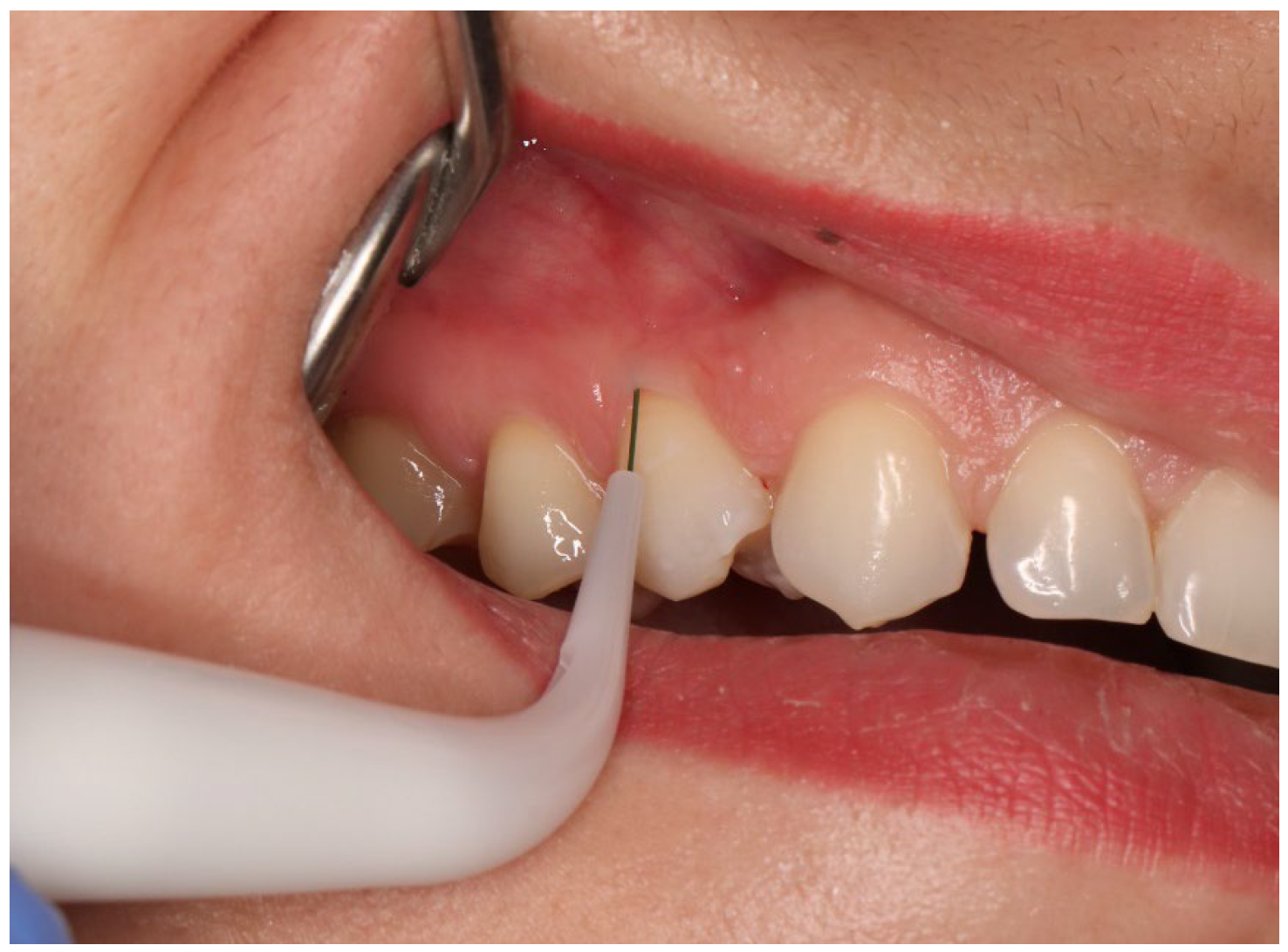
- recession depth (RD) measured at the mid-buccal aspect of the study tooth from the CEJ to the most apical extension of gingival margin using electronic periodontal probe PA-ON (Orangedental, Biberach, Germany); Figure 2;
- keratinized tissue (KT) measured from the most apical point of gingival margin to the muco-gingival junction (MGJ); the MGJ was identified by means of Lugol staining using a manual PCP-UNC 15 periodontal probe (Hu-Friedy, Chicago, IL, USA);
- gingival thickness (GT) measured at the mid-buccal aspect of the study tooth on a long axis, 2 mm apically from the gingival margin on CBCT slide (Kodak 8100 3D, Carestream/Trophy, Marne-la-Vallée, France), with a Field of View (FOV) of 5 cm × 4 cm, nominal beam of 73 kV, 12 mA and a voxel size of 150 µm. The measurement was valued by using Carestream 3D Suite (Carestream Health, Inc, Rochester, NY, USA);
- probing depth (PD) measured at the mesial, distal side, and mid-buccal aspect of the study tooth from the gingival margin to the bottom of the sulcus, PA-ON probe (Orangedental, Biberach, Germany); Figure 2;
- clinical attachment level (CAL) measured at the mid-buccal aspect of the study tooth from the CEJ to the bottom of the sulcus using electronic periodontal probe PA-ON (Orangedental, Biberach, Germany);
- approximal plaque index (API);
- community periodontal index of treatment needs (CPITN);
- mean root coverage (MRC), calculated as [(Baseline RD) − (6 months RD)/Baseline RD] × 100% [50];
- complete root coverage (CRC), calculated as the percentage of the teeth with recession defects having complete coverage achieved [(Teeth with CRC)/(All treated teeth)] × 100% [50];
- the graft area (GA), measured using the ImageJ (version 2.3.0/1.53t) Ver. G*Power (version 3.1.9.7) 1.53k computer program (https://imagej.nih.gov/ij accessed on 2 November 2024, NIH, Bethesda, MD, USA). Based on a 90-degree photo (Canon Eos 200, 135 mm + Yongnuo YN24EX flash ring) of the graft taken with a periodontal probe. Before the measurements, a calibration was performed, which consisted in marking a 15 mm reference section on periodontal probe PCP-UNC 15 (Hu-Friedy, Chicago, IL, USA);
- the pink aesthetics score (PES) developed by Furhauser et al. [57], assessed in the 3rd and 6th post-surgical month. It rates seven variables on a scale of 0 to 2 (mesial papilla, distal papilla, level of the gingival margin, marginal tissue contour, alveolar process contour, soft tissue color, soft tissue texture), yielding a maximum score of 14 points.
2.4. Randomization
2.5. Surgery
2.6. Post-Surgical Protocol
2.7. Statistical Analyses
3. Results
3.1. Recession Depth (RD)
3.2. Probing Depth (PD)
3.3. Keratinized Tissue (KT)
3.4. Gingival Thickness (GT)
3.5. Clinical Attachment Level (CAL)
3.6. Mean (MRC) and Complete (CMC) Root Coverage
3.7. Pink Aesthetic Score (PES)
3.8. Graft Area (GA)
3.9. Histopathological Analysis
4. Discussion
5. Conclusions
Author Contributions
Funding
Institutional Review Board Statement
Informed Consent Statement
Data Availability Statement
Conflicts of Interest
References
- Imber, J.C.; Kasaj, A. Treatment of Gingival Recession: When and How? Int. Dent. J. 2021, 71, 178–187. [Google Scholar] [CrossRef] [PubMed] [PubMed Central]
- Zhan, Y.; Wang, M.; Cao, X.; Liu, F. Effectiveness of acellular dermal matrix graft with a coronally advanced flap for the treatment of Miller Class I/II single gingival recession with thin gingival phenotype: Study protocol for a split-mouth randomised controlled trial. BMJ Open 2022, 12, e047703. [Google Scholar] [CrossRef] [PubMed] [PubMed Central]
- Sarhan, S.; Ahmed, E.; Hussein, R.R.; Abou-Bakr, A. Prevalence, etiology and clinical characteristics of gingival recession in a sample of adult Egyptian dental patients: A cross sectional study. BMC Oral Health 2025, 25, 691. [Google Scholar] [CrossRef] [PubMed] [PubMed Central]
- Mythri, S.; Arunkumar, S.M.; Hegde, S.; Rajesh, S.K.; Munaz, M.; Ashwin, D. Etiology and occurrence of gingival recession—An epidemiological study. J. Indian Soc. Periodontol. 2015, 19, 671–675. [Google Scholar] [CrossRef]
- Fageeh, H.I.; Fageeh, H.N.; Bhati, A.K.; Thubab, A.Y.; Sharrahi, H.M.H.; Aljabri, Y.S.; Alotaibi, F.I. Assessing the Reliability of Miller’s Classification and Cairo’s Classification in Classifying Gingival Recession Defects: A Comparison Study. Medicina 2024, 60, 205. [Google Scholar] [CrossRef] [PubMed] [PubMed Central]
- Rathore, P.; Manjunath, S.; Singh, R. Evaluating and comparing the efficacy of the microsurgical approach and the conventional approach for the periodontal flap surgical procedure: A randomized controlled trial. Dent. Med. Probl. 2024, 61, 23–28. [Google Scholar] [CrossRef] [PubMed]
- Sarlati, F.; Moghaddas, O.; Shabahangfar, R.; Safari, S.; Valaei, N. Inter- and intra-examiner agreement of three classification systems of gingival recession. J. Adv. Periodontol. Implant Dent. 2019, 11, 1–6. [Google Scholar] [CrossRef] [PubMed] [PubMed Central]
- Golob Deeb, J.; Reddy, N.; Kitten, T.; Carrico, C.K.; Grzech-Leśniak, K. Viability of bacteria associated with root caries after Nd:YAG laser application in combination with various antimicrobial agents: An in vitro study. Dent. Med. Probl. 2023, 60, 649–655. [Google Scholar] [CrossRef]
- Golob Deeb, J.; Smith, J.; Belvin, B.R.; Lewis, J.; Grzech-Leśniak, K. Er:YAG Laser Irradiation Reduces Microbial Viability When Used in Combination with Irrigation with Sodium Hypochlorite, Chlorhexidine, and Hydrogen Peroxide. Microorganisms 2019, 7, 612. [Google Scholar] [CrossRef]
- Sterczała, B.; Grzech-Leśniak, K.; Michel, O.; Trzeciakowski, W.; Dominiak, M.; Jurczyszyn, K. Assessment of Human Gingival Fibroblast Proliferation after Laser Stimulation In Vitro Using Different Laser Types and Wavelengths (1064, 980, 635, 450, and 405 nm)-Preliminary Report. J. Pers. Med. 2021, 11, 98. [Google Scholar] [CrossRef]
- Grzech-Leśniak, K.; Belvin, B.R.; Lewis, J.P.; Golob Deeb, J. Treatment with Nd:YAG Laser Irradiation Combined with Sodium Hypochlorite or Hydrogen Peroxide Irrigation on Periodontal Pathogens: An In Vitro Study. Photobiomodul. Photomed. Laser Surg. 2021, 39, 46–52. [Google Scholar] [CrossRef]
- Grzech-Leśniak, Z.; Pyrkosz, J.; Szwach, J.; Lelonkiewicz, M.; Pajączkowska, M.; Nowicka, J.; Matys, J.; Grzech-Leśniak, K. In vitro evaluation of the effect of Er:YAG laser with a fractional PS04 handpiece on microbial biofilm survival. Dent. Med. Probl. 2025. ahead of print. [Google Scholar] [CrossRef]
- Sanavi, F.; Weisgold, A.S.; Rose, L.F. Biologic width and its relation to periodontal biotypes. J. Esthetor. Dent. 1998, 10, 157–163. [Google Scholar] [CrossRef] [PubMed]
- Zucchelli, G.; Mounssif, I. Periodontal plastic surgery. Periodontol. 2000 2015, 68, 333–368. [Google Scholar] [CrossRef]
- Mahajan, A. Mahajan’s modification of Miller’s classification for gingival recession. Dent. Hypotheses 2010, 1, 45–50. [Google Scholar] [CrossRef]
- Cairo, F.; Nieri, M.; Cincinelli, S.; Mervelt, J.; Pagliaro, U. The interproximal clinical attachment level to classify gingival recessions and predict root coverage outcomes: An explorative and reliability study. J. Clin. Periodontol. 2011, 38, 661–666. [Google Scholar] [CrossRef] [PubMed]
- Miller, P.D., Jr. A classification of marginal tissue recession. Int. J. Periodontics Restor. Dent. 1985, 5, 8–13. [Google Scholar]
- Darzi, A.; Smith, S.; Taffinder, N. Assessing operative skill. Needs to become more objective. BMJ 1999, 318, 887–888. [Google Scholar] [CrossRef]
- Burkhardt, R.; Lang, N.P. Coverage of localized gingival recessions: Comparison of micro- and macrosurgical techniques. J. Clin. Periodontol. 2005, 32, 287–293. [Google Scholar] [CrossRef]
- Dembicka-Mączka, D.; Kępa, M.; Fiegler-Rudol, J.; Grzech-Leśniak, Z.; Matys, J.; Grzech-Leśniak, K.; Wiench, R. Evaluation of the Disinfection Efficacy of Er: YAG Laser Light on Single-Species Candida Biofilms—An In Vitro Study. Dent. J. 2025, 13, 88. [Google Scholar] [CrossRef]
- Crespi, R.; Barone, A.; Covani, U. Er: YAG laser scaling of diseased root surfaces: A histologic study. J. Periodontol. 2006, 77, 218–222. [Google Scholar] [CrossRef] [PubMed]
- Theodoro, L.H.; Haypek, P.; Bachmann, L.; Garcia, V.G.; Sampaio, J.E.; Zezell, D.M.; Eduardo, C.d.P. Effect of Er:YAG and diode laser irradiation on the root surface: Morphological and thermal analysis. J. Periodontol. 2003, 74, 838–843. [Google Scholar] [CrossRef] [PubMed]
- Crespi Romanos, G.E.; Cassinelli, C.; Gherlone, E. Effects of Er:YAG laser and ultrasonic treatment on fibroblast attachment to root surfaces: An in vitro study. J. Periodontol. 2006, 77, 1217–1222. [Google Scholar] [CrossRef]
- Kensy, J.; Dobrzyński, M.; Wiench, R.; Grzech-Leśniak, K.; Matys, J. Fibroblasts Adhesion to Laser-Modified Titanium Surfaces-A Systematic Review. Materials 2021, 14, 7305. [Google Scholar] [CrossRef] [PubMed]
- Gursoy, H.; Yarimoglu, E.; Kuru, B.; Ozkan Karaca, E.; Ince Kuka, G. Evaluation of the Effects of Er:YAG Laser for the De-Epithelialization of the Palatal Graft in the Treatment of Multiple Gingival Recessions: A Randomized Clinical Trial. Photobiomodul. Photomed. Laser Surg. 2019, 37, 715–721. [Google Scholar] [CrossRef]
- Ebrahimi, P.; Hadilou, M.; Naserneysari, F.; Dolatabadi, A.; Tarzemany, R.; Vahed, N.; Nikniaz, L.; Fekrazad, R.; Gholami, L. Effect of photobiomodulation in secondary intention gingival wound healing-a systematic review and meta-analysis. BMC Oral Health 2021, 21, 258. [Google Scholar] [CrossRef]
- Lafzi, A.; Kadkhodazadeh, M.; Mojahedi, S.M.; Amid, R.; Shidfar, S.; Baghani, M.T. The Clinical Evaluation of the Effects of Low-Level Laser Therapy on the Donor and Recipient Sites of the Free Gingival Graft: A Case Series. J. Lasers Med. Sci. 2019, 10, 355–360. [Google Scholar] [CrossRef]
- Michalak, F.; Dominiak, M.; Grzech-Leśniak, Z.; Kiryk, J.; Grzech-Leśniak, K. Photobiomodulation in Medication-Related Osteonecrosis of the Jaw: Outcomes in Stage I and Its Adjunctive Role in Advanced Cases. Biomedicines 2025, 13, 1042. [Google Scholar] [CrossRef]
- El Mobadder, M.; Grzech-Lesniak, Z.; El Mobadder, W.; Rifai, M.; Ghandour, M.; Nammour, S. Management of Medication-Related Osteonecrosis of the Jaw with Photobiomodulation and Minimal Surgical Intervention. Dent. J. 2023, 11, 127. [Google Scholar] [CrossRef]
- Pini-Prato, G.P.; Cairo, F.; Nieri, M.; Franceschi, D.; Rotundo, R.; Cortellini, P. Coronally advanced flap versus connective tissue graft in the treatment of multiple gingival recessions: A split-mouth study with a 5-year follow-up. J. Clin. Periodontol. 2010, 37, 644–650. [Google Scholar] [CrossRef]
- Naziker, Y.; Ertugrul, A.S. Aesthetic evaluation of free gingival graft applied by partial de-epithelialization and free gingival graft applied by conventional method: A randomized controlled clinical study. Clin. Oral Investig. 2023, 27, 4029–4038. [Google Scholar] [CrossRef] [PubMed]
- Sullivan, H.C.; Atkins, J.H. Freeutogenous gingival grafts. 1. Principles of successful grafting. Periodontics 1968, 6, 5–13. [Google Scholar] [PubMed]
- Bernimoulin, J.P.; Lüscher, B.; Mühlemann, H.R. Coronally repositioned periodontal flap. Clinical evaluation after one year. J. Clin. Periodontol. 1975, 2, 1–13. [Google Scholar] [CrossRef] [PubMed]
- Miller, P.D., Jr. Root coverage using the free soft tissue autograft following citric acid application. III. A successful and predictalbe procedure in areas of deep-wide recession. Int. J. Periodontics Restor. Dent. 1985, 3, 14–36. [Google Scholar]
- Paolantonio, M.; Murro, C.D.; Cattabriga, A.; Cattabriga, M. Subpedicle connective tissue graft versus free gingival graft in the coverage of exposed root surfaces A 5-year clinical study. J. Clin. Periodontol. 1997, 24, 51–56. [Google Scholar] [CrossRef]
- Langer, B.; Langer, L. Subepithelial connective tissue graft technique for root coverage. J. Periodontol. 1985, 56, 715–720. [Google Scholar] [CrossRef]
- Zucchelli, G.; De Sanctis, M. Treatment of multiple recession-type defects in patients with esthetic demands. J. Periodontol. 2000, 71, 1506–1514. [Google Scholar] [CrossRef]
- Allen, A.L. Use of the supraperiosteal envelope in soft tissue grafting for root coverage. II. Clinical results. Int. J. Periodontics Restor. Dent. 1994, 14, 302–315. [Google Scholar]
- Azzi, R.; Etienne, D.; Carranza, F. Surgical reconstruction of the interdental papilla. Int. J. Periodontics Restor. Dent. 1998, 18, 466–473. [Google Scholar]
- Zabalegui, I.; Sicilia, A.; Cambra, J.; Gil, J.; Sanz, M. Treatment of multiple adjacent gingival recessions with the tunnel subepithelial connective tissue graft: A clinical report. Int. J. Periodontics Restor. Dent. 1999, 19, 199–206. [Google Scholar]
- Tatakis, D.N.; Chambrone, L.; Allen, E.P.; Langer, B.; McGuire, M.K.; Richardson, C.R.; Zabalegui, I.; Zadeh, H.H. Periodontal soft tissue root coverage procedures: A consensus report from the AAP Regeneration Workshop. J. Periodontol. 2015, 86 (Suppl. S2), S52–S55. [Google Scholar] [CrossRef] [PubMed]
- Tavelli, L.; Barootchi, S.; Nguyen, T.V.; Tattan, M.; Ravidà, A.; Wang, H.L. Efficacy of tunnel technique in the treatment of localized and multiple gingival recessions: A systematic review and me-ta-analysis. J. Periodontol. 2018, 89, 1075–1090. [Google Scholar] [CrossRef] [PubMed]
- Aroca, S.; Molnár, B.; Windisch, P.; Gera, I.; Salvi, G.E.; Nikolidakis, D.; Sculean, A. Treatment of multiple adjacent Miller class I and II gingival recessions with a Modified Coronally Advanced Tunnel (MCAT) technique and a collagen matrix or palatal connective tissue graft: A randomized, controlled clinical trial. J. Clin. Periodontol. 2013, 40, 713–720. [Google Scholar] [CrossRef] [PubMed]
- Bertl, K.; Pifl, M.; Hirtler, L.; Rendl, B.; Nürnberger, S.; Stavropoulos, A.; Ulm, C. Relative Composition of Fibrous Connective and Fatty/Glandular Tissue in Connective Tissue Grafts Depends on the Harvesting Technique but not the Donor Site of the Hard Palate. J. Periodontol. 2015, 86, 1331–1339. [Google Scholar] [CrossRef]
- Reiser, G.M.; Bruno, J.F.; EMahan, P.; Larkin, L.H. The subepithelial connective tissue graft palatal donor site: Anatomic considerations for surgeons. Int. J. Periodontics Restor. Dent. 1996, 16, 130–137. [Google Scholar]
- Kuriakose, A.; Raju, S. Assessment of thickness of palatal mucosal donor site and its association with age and gender. J. Indian Soc. Periodontol. 2012, 16, 370–374. [Google Scholar] [CrossRef]
- Fiegler-Rudol, J.; Kapłon, K.; Kotucha, K.; Moś, M.; Skaba, D.; Kawczyk-Krupka, A.; Wiench, R. Hypocrellin-Mediated PDT: A Systematic Review of Its Efficacy, Applications, and Outcomes. Int. J. Mol. Sci. 2025, 26, 4038. [Google Scholar] [CrossRef]
- Edel, A. Clinical evaluation of free connective tissue grafts used to increase the width of keratinised gingiva. J. Clin. Periodontal. 1974, 1, 185–196. [Google Scholar] [CrossRef]
- Hürzeler, M.B.; Weng, D. A single-incision technique to harvest subepithelial connective tissue grafts from the palate. Int. J. Periodontics Restor. Dent. 1999, 19, 279–287. [Google Scholar]
- Zucchelli, G.; Mele, M.; Stefanini, M.; Mazzotti, C.; Marzadori, M.; Montebugnoli, L.; De Sanctis, M. Patient morbidity and root coverage outcome after subepithelial connective tissue and de—Epithelialized grafts: A comparative randomized—Controlled clinical trial. J. Clin. Periodontol. 2010, 37, 728–738. [Google Scholar] [CrossRef]
- Zuhr, O.; Fickl, S.; Wachtel, H.; Bolz, W.; Hürzeler, M.B. Covering of gingival recessions with a modified microsurgical tunnel technique: Case report. Int. J. Periodontics Restor. Dent. 2007, 27, 457–463. [Google Scholar]
- de Mattos, P.M.; Papalexiou, V.; Tramontina, V.A.; Kim, S.H.; Luczyszyn, S.M.; Bettega, P.V.C.; Johann, A.C.B.R. Evaluation of 2 techniques of epithelial removal in subepithelial connective tissue graft surgery: A comparative histological study. J. Periodontal Implant Sci. 2020, 50, 2–13. [Google Scholar] [CrossRef]
- Harris, R. Histologic evaluation of connective tissue grafts in humans. Int. J. Periodontics Restor. Dent. 2003, 23, 575–583. [Google Scholar]
- Novaes, A.B., Jr.; Grisi, D.C.; Molina, G.O.; Souza, S.L.; Taba, M., Jr.; Grisi, M.F. Comparative 6-month clinical study of a subepithelial connective tissue graft and acellular dermal matrix graft for the treatment of gingival recession. J. Periodontol. 2001, 72, 1477–1484. [Google Scholar] [CrossRef] [PubMed]
- Tal, H.; Moses, O.; Zohar, R.; Meir, H.; Nemcovsky, C. Root coverage of advanced gingival recession: A comparative study between acellular dermal matrix allograft and subepithelial connective tissue grafts. J. Periodontol. 2002, 73, 1405–1411. [Google Scholar] [CrossRef] [PubMed]
- Paolantonio, M.; Dolci, M.; Esposito, P.; D’Archivio, D.; Lisanti, L.; Di Luccio, A.; Perinetti, G. Subpedicle acellular dermal matrix graft and autogenous connective tissue graft in the treatment of gingival recessions: A comparative 1-year clinical study. J. Periodontol. 2002, 73, 1299–1307. [Google Scholar] [CrossRef]
- Fürhauser, R.; Florescu, D.; Benesch, T.; Haas, R.; Mailath, G.; Watzek, G. Evaluation of soft tissue around single-tooth implant crowns: The pink esthetic score. Clin. Oral Implants Res. 2005, 16, 639–644. [Google Scholar] [CrossRef]
- Aroca, S.; Keglevich, T.; Nikolidakis, D.; Gera, I.; Nagy, K.; Azzi, R.; Etienne, D. Treatment of class III multiple gingival recessions: A randomized-clinical trial. J. Clin. Periodontol. 2010, 37, 88–97. [Google Scholar] [CrossRef]
- D’Arcangelo, C.; Di Maio, F.D.N.; Prosperi, G.D.; Conte, E.; Baldi, M.; Caputi, S. A preliminary study of healing of diode laser versus scalpel incisions in rat oral tissue: A comparison of clinical, histological, and immunohistochemical results. Oral Surg. Oral Med. Oral Pathol. Oral Radiol. Endodontol. 2007, 103, 764–773. [Google Scholar] [CrossRef]
- Wang, X.; Zhang, C. Matsumoto K In vivo study of thehealing processes that occur in the jaws of rabbits following perforation by an Er,Cr:YSGG laser. Lasers Med. Sci. 2005, 20, 21–27. [Google Scholar] [CrossRef]
- Schwarz, F.; Sculean, A.; Georg, T.; Reich, E. Periodontal treatment with an Er:YAG laser compare to scaling and root planing. A controlled clinical study. J. Periodontol. 2001, 72, 361–367. [Google Scholar] [CrossRef]
- Folwaczny, M.; Mehl, A.; Aggstaller, H.; Hickel, R. Antimicrobial effects of 2,94 micron Er:YAG laser radiation on root surfaces: An in vitro study. J. Clin. Periodontol. 2002, 29, 73–78. [Google Scholar] [CrossRef] [PubMed]
- Wang, X.; Ishizaki, N.; Suzuki, N.; Kimura, Y.; Matsumoto, K. Morphological changes of bovine mandibular bone irradiated by Er,Cr:YSGG laser: An in vitro study. J. Clin. Laser Med. Surg. 2002, 20, 245–250. [Google Scholar] [CrossRef] [PubMed]
- Arashiro, D.S.; Rapley, J.W.; Cobb, C.M.; Killoy, W.J. Histologic evaluation of porcine skin incisions produced by CO2 laser electrosurgery and scalpel. Int. J. Periodontics Restor. Dent. 1996, 16, 479–491. [Google Scholar]
- Lewandrowski, K.U.; Lorente, C.; Schomacker, K.T.; Fiotte, T.J.; Wilkes, J.W.; Deutsch, T.F. Use of the Er:YAG laser for improved plating in maxillofacial surgery: Comparison of bone healing in laser and drill osteotomies. Lasers Surg. Med. 1996, 19, 40–45. [Google Scholar] [CrossRef]
- Aoki, A.; Sasaki, K.M.; Watanabe, H.; Ishikawa, I. Lasers in nonsurgical periodontal therapy. Periodontol. 2000 2004, 36, 59–97. [Google Scholar] [CrossRef]
- Cercadillo-Ibarguren, I.; España Tost, A.J.; Arnabat Domínguez, J.; Valmaseda Castellón, E.; Berini Aytés, L.; Gay Escoda, C. Histologic evaluation of thermal damage produced on soft tissues by CO2, Er,Cr:YSGG and diode lasers. Med. Oral Patol. Oral Cir. Bucal 2010, 15, 912–918. [Google Scholar] [CrossRef]
- Monteiro, L.; Delgado, M.L.; Garcês, F.; Machado, M.; Ferreira, F.; Martins, M.; Salazar, F.; Pacheco, J.J. A histological evaluation of the surgical margins from human oral fibrous-epithelial lesions excised with CO2 laser, Diode laser, Er:YAG laser, Nd: Laser, electrosurgical scalpel and cold scalpel. Med. Oral Patol. Oral Cir. Bucal 2019, 24, e271–e280. [Google Scholar] [CrossRef]
- Grzech-Leśniak, K.; Matys, J.; Jurczyszyn, K.; Ziółkowski, P.; Dominiak, M.; Junior, A.B.; Romeo, U. Histological and Thermometric Examination of Soft Tissue De-Epithelialization Using Digitally Controlled Er:YAG Laser Handpiece: An Ex Vivo Study. Photomed Laser Surg. 2018, 36, 313–319. [Google Scholar] [CrossRef]
- Lin, J.C.; Nevins, M.; Kim, D.M. Laser de-epithelialization of autogenous gingival graft for root coverage and soft tissue augmentation procedures. Int. J. Periodontics Restor. Dent. 2018, 38, 405–411. [Google Scholar] [CrossRef]
- Fekrazad, R.; Chiniforush, N.; Kalhori, K. All done procedure by laser in free gingival graft treatment: A case series study. J. Cosmet. Laser Ther. 2019, 21, 4–10. [Google Scholar] [CrossRef]
- Kawamura, R.; Mizutani, K.; Lin, T.; Kakizaki, S.; Mimata, A.; Watanabe, K.; Saito, N.; Meinzer, W.; Iwata, T.; Izumi, Y.; et al. Ex Vivo Evaluation of Gingival Ablation with Various Laser Systems and Electroscalpel. Photobiomodul. Photomed Laser Surg. 2020, 38, 364–373. [Google Scholar] [CrossRef] [PubMed]
- Ozcelik, O.; Seydaoglu, G.; Haytac, C.M. Diode laser for harvesting de—Epithelialized palatal graft in the treatment of gingival recession defects: A randomized clinical trial. J. Clin. Periodontol. 2016, 43, 63–71. [Google Scholar] [CrossRef] [PubMed]
- Yoshino, H.; Hasuike, A.; Sanjo, N.; Sato, D.; Kubota, T.; Nagashima, H.; Sato, S. CO2 Laser De-epithelization Technique for Subepithelial Connective Tissue Graft: A Study of 21 Recessions. In Vivo 2020, 34, 869–875. [Google Scholar] [CrossRef] [PubMed]
- Harris, R.J. A comparison of two techniques for obtaining a connective tissue graft from the palate. Int. J. Periodontics Restor. Dent. 1997, 17, 260–271. [Google Scholar]
- Bakhishov, H.; Isler, S.C.; Bozyel, B.; Yıldırım, B.; Tekindal, M.A.; Ozdemir, B. De-epithelialized gingival graft versus subepithelial connective tissue graft in the treatment of multiple adjacent gingival recessions using the tunnel technique: 1-year results of a randomized clinical trial. J. Clin. Periodontol. 2021, 48, 970–983. [Google Scholar] [CrossRef]
- Cortellini, P.; Tonetti, M.S. Improved wound stability with a modified minimally invasive surgical technique in the regenerative treatment of isolated interdental intrabony defects. J. Clin. Periodontol. 2009, 36, 157–163. [Google Scholar] [CrossRef]
- Tonetti, M.S.; Jepsen, S.; Bouchard, P.; Cairo, F.; Eickholz, P.; Graziani, F.; Herrera, D.; Jepsen, S.; Jung, R.; Machtei, E.; et al. Clinical efficacy of periodontal plastic surgery procedures: Consensus report of Group 2 of the 10th European Workshop on Periodontology. J. Clin. Periodontal. 2014, 41, S36–S43. [Google Scholar] [CrossRef]
- Cairo, F.; Rotundo, R.; Miller, P.D., Jr.; Pini Prato, G.P. Root coverage esthetic score: A system to evaluate the esthetic outcome of the treatment of gingival recession through evaluation of clinical cases. J. Periodontal. 2009, 80, 705–710. [Google Scholar] [CrossRef]
- Parker, S.; Grzech-Leśniak, K.; Cronshaw, M.; Matys, J.; Jr, A.B.; Nammour, S. Full operating parameter recording as an essential component of the reproducibility of laser-tissue interaction and treatments. Adv. Clin. Exp. Med. 2024, 33, 653–656. [Google Scholar] [CrossRef]
- Matys, J.; Grzech-Lesniak, K.; Dominiak, M. Assessment of an Impact of a Diode Laser Mode with Wavelength of 980 nm on a Temperature Rise Measured by Means of k-02 Thermocouple: Preliminary Results. Dent. Med. Probl. 2016, 53, 345–351. [Google Scholar] [CrossRef]
- Kolberg-Babrzyńska, I.; Grzech-Leśniak, K.; Kiryk, J.; Dominiak, M.; Matys, J. Effects of endodontic retreatment by conventional therapy compared to combined therapy with an Er:YAG laser and photobiomodulation: A randomized clinical trial. Dent. Med. Probl. 2025. [Google Scholar] [CrossRef] [PubMed]
- Grzech-Leśniak, Z.; Pyrkosz, J.; Szwach, J.; Kosidło, P.; Matys, J.; Wiench, R.; Pajączkowska, M.; Nowicka, J.; Dominiak, M.; Grzech-Leśniak, K. Antibacterial Effects of Er:YAG Laser Irradiation on Candida-Streptococcal Biofilms. Life 2025, 15, 474. [Google Scholar] [CrossRef] [PubMed]
- Grzech-Leśniak, Z.; Szwach, J.; Lelonkiewicz, M.; Migas, K.; Pyrkosz, J.; Szwajkowski, M.; Kosidło, P.; Pajączkowska, M.; Wiench, R.; Matys, J.; et al. Effect of Nd:YAG Laser Irradiation on the Growth of Oral Biofilm. Microorganisms 2024, 12, 2231. [Google Scholar] [CrossRef]
- Deeb, J.G.; Grzech-Leśniak, K.; Weaver, C.; Matys, J.; Bencharit, S. Retrieval of Glass Fiber Post Using Er:YAG Laser and Conventional Endodontic Ultrasonic Method: An In Vitro Study. J. Prosthodont. 2019, 28, 1024–1028. [Google Scholar] [CrossRef]
- Alfawal, A.M.H.; Hajeer, M.Y.; Ajaj, M.A.; Hamadah, O.; Brad, B.; Latifeh, Y. Evaluation of patient-centered outcomes associated with the acceleration of canine retraction by using minimally invasive surgical procedures: A randomized clinical controlled trial. Dent. Med. Probl. 2020, 57, 285–293. [Google Scholar] [CrossRef]
- Gonçalves, R.C.G.; Cardoso, R.B.; Bauer, J.; Santos, V.M.D.; Jabur, R.O.; Bortoluzzi, M.C. Exploring the relationship between anxiety, patient characteristics and pain outcomes in oral surgery under local anesthesia: The measurement problem. Dent. Med. Probl. 2024, 61, 515–523. [Google Scholar] [CrossRef]
- Kumar, G.; Jena, S.; Manila, N.; Fareed, M.; Karobari, M.I. Incidence of postoperative pain after single-visit and multiple-visit root canal therapy: A systematic review. BMC Oral Health 2025, 25, 47. [Google Scholar] [CrossRef]
- Elmsmari, F.; Shujaie, H.; Alzaabi, R.; González, J.A.; Aljafarawi, T.; Olivieri, J.G.; Jurado, C.A.; Afrashtehfar, K.I. Lasers efficacy in pain management after primary and secondary endodontic treatment: A systematic review and meta-analysis of randomized clinical trials. Sci. Rep. 2024, 14, 26028. [Google Scholar] [CrossRef]


| LDGG | DGG | |
|---|---|---|
| Patient | 9 (100%) | |
| Males | 2 (2.2%) | |
| Females | 7 (77.7%) | |
| Age (range) | 36.1 ± 8.46 (19–51) | |
| Total teeth | 23 (100%) | 23 (100%) |
| Mesial incisors | 0 (0%) | 0 (0%) |
| Lateral incisors | 2 (8.7%) | 1 (4,3%) |
| Canines | 9 (39.1%) | 7 (30.4%) |
| First premolar | 9 (39.1%) | 9 (39.1%) |
| Second premolar | 3 (13%) | 6 (26.1%) |
| API | ||
| Baseline | 11.61 ± 1.76 | |
| 3 months post-op. | 11.95 ± 2.27 | |
| 6 months post-op | 12.2 ± 2.12 | |
| CPITN | 1.44 ± 0.53 |
| Baseline (T0) | 3 Months (T1) | 6 Months (T2) | p-Value | |
|---|---|---|---|---|
| RD | ||||
| LDEP | 2.60 ± 0.72 | 0.08 ± 0.28 | 0.13 ± 0.30 | p < 0.001 |
| DEP | 2.83 ± 0.90 | 0.11 ± 0.29 | 0.15 ± 0.35 | p < 0.001 |
| p-value | NS | NS | NS | |
| PD | ||||
| LDEP | 1.66 ± 0.34 | 1.38 ± 0.44 | 1.47 ± 0.43 | p < 0.001 |
| DEP | 1.51 ± 0.36 | 1.18 ± 0.33 | 1.28 ± 0.40 | p < 0.001 |
| p-value | NS | NS | NS | |
| KT | ||||
| LDEP | 2.19 ± 1.18 | 3.73 ± 0,72 | 3.98 ± 0.76 | p < 0.001 |
| DEP | 2.25 ± 0.71 | 3.21 ± 0.61 | 3.44 ± 0.74 | p < 0.001 |
| p-value | NS | p < 0.05 | p < 0.05 | |
| GT | ||||
| LDEP | 0.83 ± 0.17 | 2.17 ± 0.31 | 2.09 ± 0.28 | p < 0.001 |
| DEP | 0.86 ± 0.28 | 2.19 ± 0.41 | 1.98 ± 0.35 | p < 0.001 |
| p-value | NS | NS | NS | |
| CAL | ||||
| LDEP | 4.26 ± 0.66 | 1.12 ± 0.47 | 1.24 ± 0.58 | p < 0.001 |
| DEP | 4.34 ± 0.91 | 1.04 ± 0.65 | 1.15 ± 0.76 | p < 0.001 |
| p-value | NS | NS | NS |
| △T0-T1 | △T0-T2 | |
|---|---|---|
| RD | ||
| LDEP | 2.52 ± 0.67 | 2.47 ± 0.67 |
| DEP | 2.72 ± 0.79 | 2.68 ± 0.83 |
| p-value | NS | NS |
| PD | ||
| LDEP | 0.28 ± 0.33 | 0.19 ± 0.36 |
| DEP | 0.33 ± 0.41 | 0.23 ± 0.44 |
| p-value | NS | NS |
| KT | ||
| LDEP | 1.54 ± 1.16 | 1.79 ± 1.19 |
| DEP | 0.96 ± 1.08 | 1.19 ± 1.27 |
| p-value | p < 0.05 | p < 0.05 |
| GT | ||
| LDEP | 1.34 ± 0.22 | 1.26 ± 0.22 |
| DEP | 1.33 ± 0.37 | 1.12 ± 0.33 |
| p-value | NS | NS |
| CAL | ||
| LDEP | 3.14 ± 0.7 | 3.02 ± 0.66 |
| DEP | 3.3 ± 0.92 | 3.19 ± 0.99 |
| p-value | NS | NS |
| MRC | CRC | GA [mm2] (Range) | PES | ||
|---|---|---|---|---|---|
| T2 | T2 | T0 | T1 | T2 | |
| LDEP | 95% | 82.6% | 103.88 ± 18.61 (77.18–120.24) | 12.5 ± 0.73 | 13.2 ± 0.8 |
| DEP | 94.8% | 82.6% | 103.45 ± 14.02 (75.5–127.31) | 12.5 ± 0.9 | 13.3 ± 0.64 |
| p-value | NS | NS | NS | NS | NS |
Disclaimer/Publisher’s Note: The statements, opinions and data contained in all publications are solely those of the individual author(s) and contributor(s) and not of MDPI and/or the editor(s). MDPI and/or the editor(s) disclaim responsibility for any injury to people or property resulting from any ideas, methods, instructions or products referred to in the content. |
© 2025 by the authors. Licensee MDPI, Basel, Switzerland. This article is an open access article distributed under the terms and conditions of the Creative Commons Attribution (CC BY) license (https://creativecommons.org/licenses/by/4.0/).
Share and Cite
Banyś, A.; Fiegler-Rudol, J.; Grzech-Leśniak, Z.; Wiench, R.; Matys, J.; Shibli, J.A.; Grzech-Leśniak, K. Clinical Efficacy of Er,Cr:YSGG Laser for Deepithelialization of Free Gingival Grafts in Gingival Recession Treatment: A Randomized, Split-Mouth Clinical Trial. J. Clin. Med. 2025, 14, 5335. https://doi.org/10.3390/jcm14155335
Banyś A, Fiegler-Rudol J, Grzech-Leśniak Z, Wiench R, Matys J, Shibli JA, Grzech-Leśniak K. Clinical Efficacy of Er,Cr:YSGG Laser for Deepithelialization of Free Gingival Grafts in Gingival Recession Treatment: A Randomized, Split-Mouth Clinical Trial. Journal of Clinical Medicine. 2025; 14(15):5335. https://doi.org/10.3390/jcm14155335
Chicago/Turabian StyleBanyś, Artur, Jakub Fiegler-Rudol, Zuzanna Grzech-Leśniak, Rafał Wiench, Jacek Matys, Jamil A. Shibli, and Kinga Grzech-Leśniak. 2025. "Clinical Efficacy of Er,Cr:YSGG Laser for Deepithelialization of Free Gingival Grafts in Gingival Recession Treatment: A Randomized, Split-Mouth Clinical Trial" Journal of Clinical Medicine 14, no. 15: 5335. https://doi.org/10.3390/jcm14155335
APA StyleBanyś, A., Fiegler-Rudol, J., Grzech-Leśniak, Z., Wiench, R., Matys, J., Shibli, J. A., & Grzech-Leśniak, K. (2025). Clinical Efficacy of Er,Cr:YSGG Laser for Deepithelialization of Free Gingival Grafts in Gingival Recession Treatment: A Randomized, Split-Mouth Clinical Trial. Journal of Clinical Medicine, 14(15), 5335. https://doi.org/10.3390/jcm14155335