
Longitudinal Three-Dimensional Stereophotogrammetric Growth Analysis in Infants with Unilateral Cleft Lip and Palate from 3 to 12 Months of Age


 , ,
, ,  and
and
Abstract
:1. Introduction
2. Materials and Methods
2.1. Participants
2.2. Surgical Protocol
2.3. Data Acquisition
- (1)
- A predefined, evenly spread template was roughly aligned on the facial model of the patient using a procrustus technique with both endocanthia, cheilon, and the pronasale landmark as reference points.
- (2)
- A rigid registration technique further aligned the template.
- (3)
- A non-rigid registration technique described by Matthews et al. [38] was used to morph every individual point on the template towards the contours of the face.
- (4)
- Finally, each point of the template was projected onto the facial mesh. This results in a standardized, uniform indexed mesh in which individual datapoints correspond with those of other standardized meshes.
2.4. Statistical Analysis
3. Results
3.1. Sample and Image Selection
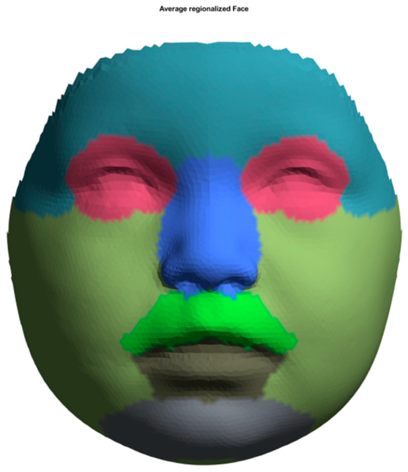
3.2. Average Faces of CUCLAP
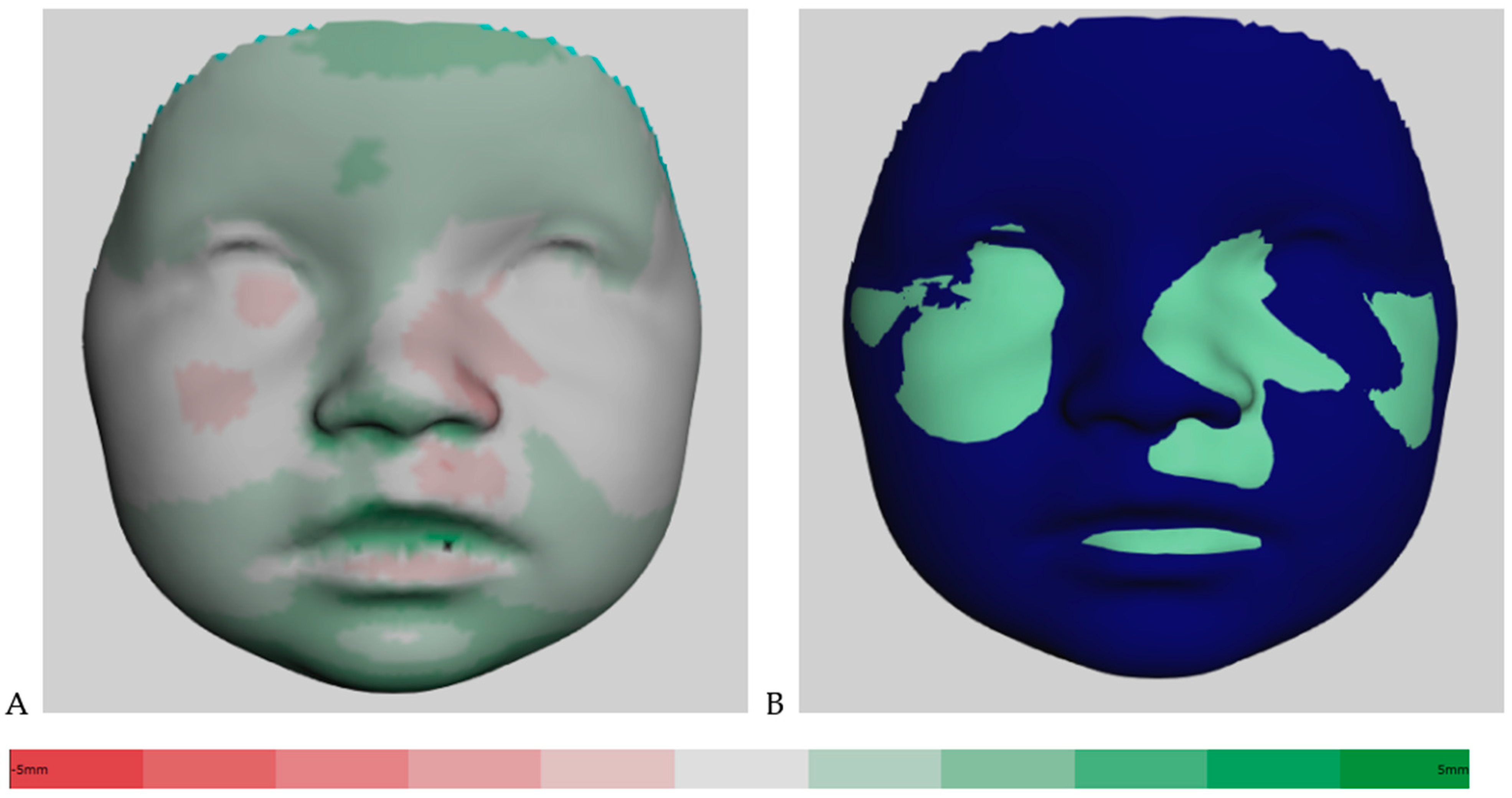
3.3. Assessment of Facial Growth
3.4. Growth of the Face in Total and Facial Areas from 3 to 9 Months of Age
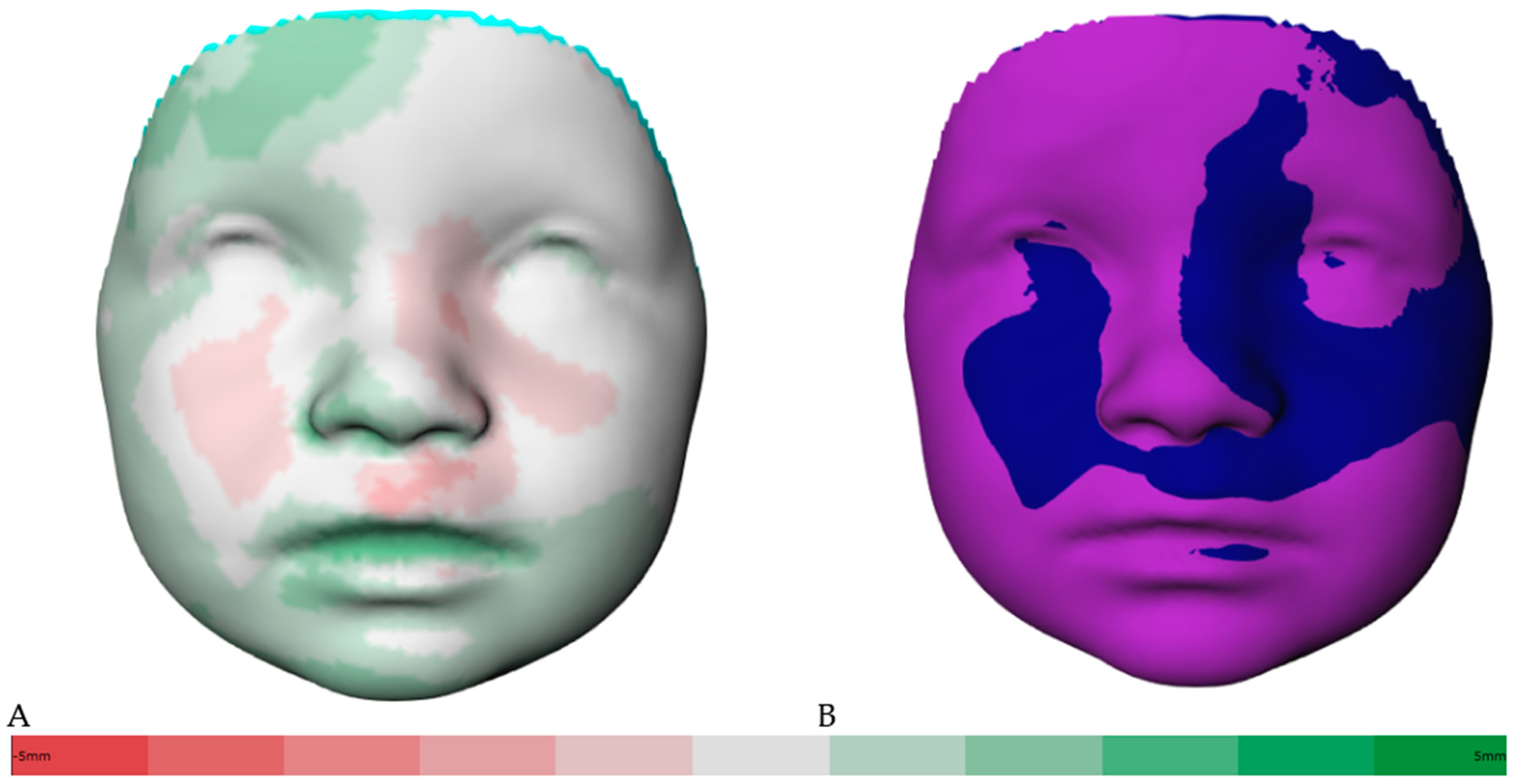
3.5. Growth of the Face in Total and Facial Areas from 9 to 12 Months of Age
4. Discussion
5. Conclusions
Author Contributions
Funding
Institutional Review Board Statement
Informed Consent Statement
Data Availability Statement
Acknowledgments
Conflicts of Interest
References
- Vanderas, A.P. Prevalence of craniomandibular dysfunction in children and adolescents: A review. Pediatr. Dent. 1987, 9, 312–316. [Google Scholar]
- Mossey, P.; Modell, B. Epidemiology of oral clefts 2012: An international perspective. In Cleft Lip and Palate; Karger: Wellesley, MA, USA, 2012; Volume 16, pp. 1–18. [Google Scholar] [CrossRef]
- Phan, M.; Conte, F.; Khandelwal, K.D.; Ockeloen, C.W.; Bartzela, T.; Kleefstra, T.; van Bokhoven, H.; Rubini, M.; Zhou, H.; Carels, C.E. Tooth agenesis and orofacial clefting: Genetic brothers in arms? Hum. Genet. 2016, 135, 1299–1327. [Google Scholar] [CrossRef] [PubMed]
- Mangold, E.; Kreiß, M.; Nöthen, M.M. Syndromale und nichtsyndromale orofaziale Spalten. Med. Genet. 2017, 29, 397–412. [Google Scholar] [CrossRef]
- Howe, L.J.; Lee, M.K.; Sharp, G.C.; Smith, G.D.; Pourcain, B.S.; Shaffer, J.R.; Ludwig, K.U.; Mangold, E.; Marazita, M.L.; Feingold, E.; et al. Investigating the shared genetics of non-syndromic cleft lip/palate and facial morphology. PLoS Genet. 2018, 14, e1007501. [Google Scholar] [CrossRef]
- Korolenkova, M.V.; Starikova, N.V.; Udalova, N.V. The role of external aetiological factors in dental anomalies in non-syndromic cleft lip and palate patients. Eur. Arch. Paediatr. Dent. 2019, 20, 105–111. [Google Scholar] [CrossRef]
- Hilton, E.N.; Manson, F.D.; Urquhart, J.E.; Johnston, J.J.; Slavotinek, A.M.; Hedera, P.; Stattin, E.-L.; Nordgren, A.; Biesecker, L.G.; Black, G.C. Left-sided embryonic expression of the BCL-6 corepressor, BCOR, is required for vertebrate laterality determination. Hum. Mol. Genet. 2007, 16, 1773–1782. [Google Scholar] [CrossRef]
- Hilton, E.; Johnston, J.; Whalen, S.; Okamoto, N.; Hatsukawa, Y.; Nishio, J.; Kohara, H.; Hirano, Y.; Mizuno, S.; Torii, C.; et al. BCOR analysis in patients with OFCD and Lenz microphthalmia syndromes, mental retardation with ocular anomalies, and cardiac laterality defects. Eur. J. Hum. Genet. 2009, 17, 1325–1335. [Google Scholar] [CrossRef] [PubMed]
- Bartzela, T. Cleft Lip and Palate: A Variable Clinical Spectrum. Medical and Dental Considerations; Charité—Universitätsmedizin Berlin: Berlin, Germany, 2021; pp. 7–9. [Google Scholar]
- Fraser, F.C. Thoughts on the etiology of clefts of the palate and lip. Acta Genet. Stat. Med. 1955, 5, 358–369. [Google Scholar] [CrossRef]
- Sivertsen, Å.; Wilcox, A.; Johnson, G.E.; Åbyholm, F.; Vindenes, H.A.; Lie, R.T. Prevalence of major anatomic variations in oral clefts. Plast. Reconstr. Surg. 2008, 121, 587–595. [Google Scholar] [CrossRef]
- Rahimov, F.; Jugessur, A.; Murray, J.C. Genetics of nonsyndromic orofacial clefts. Cleft Palate-Craniofac. J. 2012, 49, 73–91. [Google Scholar] [CrossRef]
- Dixon, M.J.; Marazita, M.L.; Beaty, T.H.; Murray, J.C. Cleft lip and palate: Understanding genetic and environmental influences. Nat. Rev. Genet. 2011, 12, 167–178. [Google Scholar] [CrossRef] [PubMed]
- Kouwenberg, M.; Draaisma, J.; Kuijpers-Jagtman, A.M.; Bartzela, T. Associated congenital malformations in patients with complete bilateral cleft lip and palate. Tijdschr. Kindergeneeskd. 2011, 78, 13–17. [Google Scholar]
- Bartzela, T.; Theuerkauf, B.; Reichardt, E.; Spielmann, M.; Opitz, C. Clinical characterization of 266 patients and family members with cleft lip and/or palate with associated malformations and syndromes. Clin. Oral Investig. 2021, 25, 5531–5540. [Google Scholar] [CrossRef]
- Mangold, E.; Ludwig, K.U.; Nöthen, M.M. Breakthroughs in the genetics of orofacial clefting. Trends Mol. Med. 2011, 17, 725–733. [Google Scholar] [CrossRef] [PubMed]
- Brons, S.; Meulstee, J.W.; Loonen, T.G.; Nada, R.M.; Kuijpers, M.A.; Bronkhorst, E.M.; Bergé, S.J.; Maal, T.J.; Kuijpers-Jagtman, A.M. Three-dimensional facial development of children with unilateral cleft lip and palate during the first year of life in comparison with normative average faces. PeerJ 2019, 7, e7302. [Google Scholar] [CrossRef]
- Linden, O.E.; Taylor, H.O.; Vasudavan, S.; Byrne, M.E.; Deutsch, C.K.; Mulliken, J.B.; Sullivan, S.R. Three-Dimensional analysis of nasal symmetry following primary correction of unilateral cleft lip nasal deformity. Cleft Palate-Craniofac. J. 2017, 54, 715–719. [Google Scholar] [CrossRef]
- Schwenzer-Zimmerer, K.; Chaitidis, D.; Berg-Boerner, I.; Krol, Z.; Kovacs, L.; Schwenzer, N.F.; Zimmerer, S.; Holberg, C.; Zeilhofer, H.-F. Quantitative 3D soft tissue analysis of symmetry prior to and after unilateral cleft lip repair compared with non-cleft persons (performed in Cambodia). J. Cranio-Maxillofac. Surg. 2008, 36, 431–438. [Google Scholar] [CrossRef] [PubMed]
- Manyama, M.; Larson, J.R.; Liberton, D.K.; Rolian, C.; Smith, F.J.; Kimwaga, E.; Gilyoma, J.; Lukowiak, K.D.; Spritz, R.A.; Hallgrimsson, B. Facial morphometrics of children with non-syndromic orofacial clefts in Tanzania. BMC Oral Health 2014, 14, 93. [Google Scholar] [CrossRef]
- Cakan, D.G.; Ulkur, F.; Taner, T.U. The genetic basis of facial skeletal characteristics and its relation with orthodontics. Eur. J. Dent. 2012, 6, 340–345. [Google Scholar] [CrossRef]
- Bruggink, R.; Baan, F.; Kramer, G.; Kuijpers-Jagtman, A.; Bergé, S.; Maal, T.; Ongkosuwito, E. Symmetry of palatal shape during the first year of life in healthy infants. Clin. Oral Investig. 2021, 25, 1069–1076. [Google Scholar] [CrossRef]
- Naros, A.; Brocks, A.; Kluba, S.; Reinert, S.; Krimmel, M. Health-related quality of life in cleft lip and/or palate patients—A cross-sectional study from preschool age until adolescence. J. Cranio-Maxillofac. Surg. 2018, 46, 1758–1763. [Google Scholar] [CrossRef] [PubMed]
- Bartzela, T.; Katsaros, C.; Shaw, W.C.; Ronning, E.; Rizell, S.; Bronkhorst, E.; Okada, T.O.; De, S.L. Pinheiro, F.H.; Dominguez-Gonzalez, S.; et al. A longitudinal three-center study of dental arch relationship in patients with bilateral cleft lip and palate. Cleft Palate Craniofac. J. 2010, 47, 167–174. [Google Scholar] [CrossRef] [PubMed]
- Bartzela, T.; Katsaros, C.; Rønning, E.; Rizell, S.; Semb, G.; Bronkhorst, E.; Halazonetis, D.; Kuijpers-Jagtman, A.M. A longitudinal three-center study of craniofacial morphology at 6 and 12 years of age in patients with complete bilateral cleft lip and palate. Clin. Oral Investig. 2012, 16, 1313–1324. [Google Scholar] [CrossRef] [PubMed]
- Brons, S.; van Beusichem, M.E.; Bronkhorst, E.M.; Draaisma, J.; Bergé, S.J.; Maal, T.J.; Kuijpers-Jagtman, A.M. Methods to quantify soft-tissue based facial growth and treatment outcomes in children: A systematic review. PLoS ONE 2012, 7, e41898. [Google Scholar] [CrossRef]
- Brons, S.; van Beusichem, M.E.; Bronkhorst, E.M.; Draaisma, J.M.; Bergé, S.J.; Schols, J.G.; Kuijpers-Jagtman, A.M. Methods to quantify soft tissue–based cranial growth and treatment outcomes in children: A systematic review. PLoS ONE 2014, 9, e89602. [Google Scholar] [CrossRef]
- Hood, C.A.; Bock, M.; Hosey, M.T.; Bowman, A.; Ayoub, A.F. Facial asymmetry—3D assessment of infants with cleft lip & palate. Int. J. Paediatr. Dent. 2003, 13, 404–410. [Google Scholar] [CrossRef]
- Mancini, L.; Gibson, T.L.; Grayson, B.H.; Flores, R.L.; Staffenberg, D.; Shetye, P.R. Three-dimensional soft tissue nasal changes after nasoalveolar molding and primary ceilorhinoplasty in infants with unilateral cleft lip and palate. Cleft Palate Craniofac. J. 2019, 56, 31–38. [Google Scholar] [CrossRef]
- Antonacci, D.; Caponio, V.C.A.; Troiano, G.; Pompeo, M.G.; Gianfreda, F.; Canullo, L. Facial scanning technologies in the era of digital workflow: A systematic review and network meta-analysis. J. Prosthodont. Res. 2022, 67, 321–336. [Google Scholar] [CrossRef]
- Bugaighis, I.; Mattick, C.; Tiddeman, B.; Hobson, R.; Bugaighis, B.I.; Hermann, N.; Darvann, T.; Larsen, P.; Lindholm, P.; Andersen, M.; et al. 3D Facial morphometry in children with oral clefts. Cleft Palate-Craniofac. J. 2014, 51, 452–461. [Google Scholar] [CrossRef]
- Wong, K.W.F.; Keeling, A.; Achal, K.; Khambay, B. Using three-dimensional average facial meshes to determine nasolabial soft tissue deformity in adult UCLP patients. Surgeon 2019, 17, 19–27. [Google Scholar] [CrossRef]
- Kornreich, D.; Mitchell, A.A.; Webb, B.D.; Cristian, I.; Jabs, E.W. Quantitative assessment of facial asymmetry using three-dimensional surface imaging in adults: Validating the precision and reliability of a global approach. Cleft Palate Craniofac. J. 2016, 53, 126–131. [Google Scholar] [CrossRef] [PubMed]
- Hall, N.J.; Eaton, S.; Pierro, A. The evidence base for neonatal surgery. Early Hum. Dev. 2009, 85, 713–718. [Google Scholar] [CrossRef] [PubMed]
- Shaw, W.C.; Semb, G.; Nelson, P.; Brattström, V.; Mølsted, K.; Prahl-Andersen, B.; Gundlach, K.K. The Eurocleft Project 1996–2000: Overview. J. Cranio-Maxillofac. Surg. 2001, 29, 131–140. [Google Scholar] [CrossRef] [PubMed]
- Brons, S.; Meulstee, J.W.; Nada, R.M.; Kuijpers, M.A.R.; Bronkhorst, E.M.; Bergé, S.J.; Maal, T.J.J.; Kuijpers-Jagtman, A.M. Uniform 3D meshes to establish normative facial averages of healthy infants during the first year of life. PLoS ONE 2019, 14, e0217267. [Google Scholar] [CrossRef]
- Brons, S.; van Beusichem, M.; Maal, T.; Plooij, J.; Bronkhorst, E.; Bergé, S.; Kuijpers-Jagtman, A. Development and reproducibility of a 3D stereophotogrammetric reference frame for facial soft tissue growth of babies and young children with and without orofacial clefts. Int. J. Oral Maxillofac. Surg. 2013, 42, 2–8. [Google Scholar] [CrossRef]
- Matthews, H.S.; Palmer, R.L.; Baynam, G.S.; Quarrell, O.W.; Klein, O.D.; Spritz, R.A.; Hennekam, R.C.; Walsh, S.; Shriver, M.; Weinberg, S.M.; et al. Large-scale open-source three-dimensional growth curves for clinical facial assessment and objective description of facial dysmorphism. Sci. Rep. 2021, 11, 12175. [Google Scholar] [CrossRef]
- Bruggink, R.; Baan, F.; Brons, S.; Loonen, T.G.; Kuijpers-Jagtman, A.M.; Maal, T.J.; Ongkosuwito, E.M. A semi-automatic three-dimensional technique using a regionalized facial template enables facial growth assessment in healthy children from 1.5 to 5.0 years of age. PeerJ 2022, 10, e13281. [Google Scholar] [CrossRef]
- Al-Rudainy, D.; Ju, X.; Mehendale, F.V.; Ayoub, A. Longitudinal 3D assessment of facial asymmetry in unilateral cleft lip and palate. Cleft Palate-Craniofac. J. 2019, 56, 495–501. [Google Scholar] [CrossRef]
- Djordjevic, J.; Lewis, B.M.; Donaghy, C.E.; Zhurov, A.I.; Knox, J.; Hunter, L.; Richmond, S. Facial shape and asymmetry in 5-year-old children with repaired unilateral cleft lip and/or palate: An exploratory study using laser scanning. Eur. J. Orthod. 2012, 36, 497–505. [Google Scholar] [CrossRef]
- Ubaya, T.; Sherriff, A.; Ayoub, A.; Khambay, B. Soft tissue morphology of the naso-maxillary complex following surgical correction of maxillary hypoplasia. Int. J. Oral Maxillofac. Surg. 2012, 41, 727–732. [Google Scholar] [CrossRef]
- Parveen, S.M.; Husain, A.M.; Johns, G.M.; Mascarenhas, R.M.; Reddy, S.G.F. Three-dimensional analysis of craniofacial structures of individuals with nonsyndromic unilateral complete cleft lip and palate. J. Craniofac. Surg. 2020, 32, e65–e69. [Google Scholar] [CrossRef]
- Alazzawi, O.; Morioka, D.; Miyabe, M.; Tosa, Y.; Ohkubo, F.; Yoshimoto, S. Nasolabial growth in individuals with unilateral cleft lip and palate: A preliminary study of longitudinal observation using three-dimensional stereophotogrammetry. J. Craniofac. Surg. 2017, 28, e449–e451. [Google Scholar] [CrossRef]
- Morioka, D.; Mandrano, N.; Fujimoto, H.; Koga, Y.; Sato, N.; Tosa, Y.; Ohkubo, F.; Yoshimoto, S. Longitudinal follow-up of individuals with cleft lip using three-dimensional stereophotogrammetry. J. Craniofac. Surg. 2018, 29, 1261–1265. [Google Scholar] [CrossRef] [PubMed]
- Kuijpers, M.; Maal, T.; Meulstee, J.; Carels, C.; Bronkhorst, E.; Bergé, S.; Fudalej, P. Nasolabial shape and aesthetics in unilateral cleft lip and palate: An analysis of nasolabial shape using a mean 3D facial template. Int. J. Oral Maxillofac. Surg. 2021, 50, 267–272. [Google Scholar] [CrossRef] [PubMed]
- Ambrosio, E.C.P.; Sforza, C.; De Menezes, M.; Gibelli, D.; Codari, M.; Carrara, C.F.C.; Machado, M.A.A.M.; Oliveira, T.M. Longitudinal morphometric analysis of dental arch of children with cleft lip and palate: 3D stereophotogrammetry study. Oral Surg. Oral Med. Oral Pathol. Oral Radiol. 2018, 126, 463–468. [Google Scholar] [CrossRef] [PubMed]
- Sakoda, K.L.; Jorge, P.K.; Carrara, C.F.C.; Machado, M.A.D.A.M.; Valarelli, F.P.; Pinzan, A.; Oliveira, T.M. 3D analysis of effects of primary surgeries in cleft lip/palate children during the first two years of life. Braz. Oral Res. 2017, 31, e46. [Google Scholar] [CrossRef] [PubMed]
- White, J.E.; Ayoub, A.F.; Hosey, M.T.; Bock, M.; Bowman, A.; Bowman, J.; Siebert, J.P.; Ray, A. Three-dimensional facial characteristics of caucasian infants without cleft and correlation with body measurements. Cleft Palate Craniofac. J. 2004, 41, 593–602. [Google Scholar] [CrossRef]
- Kesterke, M.J.; Raffensperger, Z.D.; Heike, C.L.; Cunningham, M.L.; Hecht, J.T.; Kau, C.H.; Nidey, N.L.; Moreno, L.M.; Wehby, G.L.; Marazita, M.L.; et al. Using the 3D facial norms database to investigate craniofacial sexual dimorphism in healthy children, adolescents, and adults. Biol. Sex Differ. 2016, 7, 7–23. [Google Scholar] [CrossRef]
- Ritschl, L.M.; Grill, F.D.; Mittermeier, F.; Lonic, D.; Wolff, K.-D.; Roth, M.; Loeffelbein, D.J. Evaluation of a portable low-budget three-dimensional stereophotogrammetry system for nasal analysis. J. Cranio-Maxillofac. Surg. 2018, 46, 2008–2016. [Google Scholar] [CrossRef]
- Ritschl, L.M.; Roth, M.; Fichter, A.M.; Mittermeier, F.; Kuschel, B.; Wolff, K.-D.; Grill, F.D.; Loeffelbein, D.J. The possibilities of a portable low-budget three-dimensional stereophotogrammetry system in neonates: A prospective growth analysis and analysis of accuracy. Head Face Med. 2018, 14, 11. [Google Scholar] [CrossRef]
- Kimura, N.; Nozoe, E.; Okawachi, T.; Ishihata, K.; Fuchigami, T.; Nakamura, N. Three-dimensional analyses of nasolabial forms and upper lip surface symmetry after primary lip repair in patients with complete unilateral cleft lip and palate. J. Cranio-Maxillofac. Surg. 2019, 47, 245–254. [Google Scholar] [CrossRef] [PubMed]
- Mai, H.-N.; Lee, D.-H. Effects of artificial extraoral markers on accuracy of three-dimensional dentofacial image integration: Smartphone face scan versus stereophotogrammetry. J. Pers. Med. 2022, 12, 490. [Google Scholar] [CrossRef]
- Gattani, S.; Ju, X.; Gillgrass, T.; Bell, A.; Ayoub, A. An Innovative assessment of the dynamics of facial movements in surgically managed unilateral cleft lip and palate using 4D imaging. Cleft Palate-Craniofac. J. 2020, 57, 1125–1133. [Google Scholar] [CrossRef]
- Bartzela, T.; Leenarts, C.; Bronkhorst, E.; Borstlap, W.; Katsaros, C.; Kuijpers-Jagtman, A.M. Comparison of two scoring systems for evaluation of treatment outcome in patients with complete bilateral cleft lip and palate. Cleft Palate-Craniofac. J. 2011, 48, 455–461. [Google Scholar] [CrossRef]
- Liu, Y.P.; Behrents, R.G.; Buschang, P.H. Mandibular growth, remodeling, and maturation during infancy and early childhood. Angle Orthod. 2010, 80, 97–105. [Google Scholar] [CrossRef]
- Brons, S.; Darroudi, A.; Nada, R.; Bronkhorst, E.M.; Vreeken, R.; Berge, S.J.; Maal, T.; Kuijpers-Jagtman, A.M. Influence of involuntary facial expressions on reproducibility of 3D stereophotogrammetry in children with and without complete unilateral cleft lip and palate from 3 to 18 months of age. Clin. Oral Investig. 2019, 23, 1041–1050. [Google Scholar] [CrossRef] [PubMed]
- Kuijpers, M.A.R.; Brons, S.; Kuijpers-Jagtman, A.M. Three-dimensional surface imaging of the face in patients with cleft lip and palate. In Cleft Orthodontics: A Holistic and Interdisciplinary Approach; Kharbanda, O.P., Ed.; Thieme Medical and Scientific Publishers: Stuttgart, Germany, 2021; Chapter 20; pp. 226–233. [Google Scholar]
- Matthews, H.; Penington, T.; Saey, I.; Halliday, J.; Muggli, E.; Claes, P. Spatially dense morphometrics of craniofacial sexual dimorphism in 1-year-olds. J. Anat. 2016, 229, 549–559. [Google Scholar] [CrossRef] [PubMed]

| Age | Treatment Protocol |
|---|---|
| Prenatal to 3 months | Parent counseling Neonatal presurgical infant orthopedics |
| 2 months | Nasoalveolar molding, Latham’s appliance (for jaw segment relocation, four weeks before surgery) |
| 3–4 months | Lip adhesion, soft palate closure (Kriens’ technique), gingivoperiosteoplasty |
| 9 months | Hard palate closure, lip-nose-plasty (Millard’s technique) |
| 12 months | Lip-nose-plasty (Millard’s technique), if not performed previously |
| Age | 0 Months, T0 | 3 Months, T1 | 9 Months, T2 | 12 Months, T3 | Total |
|---|---|---|---|---|---|
| Available infants with CL/P | 40 | 40 | 38 | 36 | 26 |
| Included infants with CUCLAP | 22 | 22 | 22 | 22 | 22 |
| Included images after a detailed assessment | 9 | 22 | 15 | 19 | 83 |
| Region | Min (mm) | Mean Growth Difference Range (mm) | Median (50%) | Max (mm) | 5% CI (mm) | 95% CI (mm) | Std (mm) | Abs Mean Growth (mm) | Abs Std (mm) | p-Value |
|---|---|---|---|---|---|---|---|---|---|---|
| Full face | −3.50 | 1.89 | 2.23 | 4.09 | −2.16 | 3.36 | 1.48 | 2.31 | 0.65 | <0.001 ** |
| Nose | −3.12 | 1.05 | 2.24 | 3.14 | −2.57 | 3.06 | 2.28 | 2.48 | 0.38 | 0.0416 * |
| Forehead | −1.05 | 1.65 | 1.74 | 2.26 | 0.89 | 2.12 | 0.43 | 1.66 | 0.38 | <0.001 ** |
| Eyes | −1.98 | 1.05 | 1.36 | 1.98 | −1.85 | 1.91 | 1.15 | 1.53 | 0.32 | <0.001 ** |
| Upper lip | −3.47 | 1.92 | 2.82 | 4.09 | −3.28 | 3.79 | 2.45 | 3.07 | 0.45 | 0.0014 * |
| Lower lip | −3.50 | 2.30 | 2.85 | 4.04 | −3.20 | 3.29 | 1.76 | 2.88 | 0.27 | <0.001 ** |
| Chin | 2.54 | 3.17 | 3.13 | 3.81 | 2.77 | 3.71 | 0.29 | 3.17 | 0.29 | <0.001 ** |
| Cheeks | −2.91 | 1.94 | 2.35 | 3.98 | −2.11 | 3.06 | 1.39 | 2.33 | 0.49 | <0.001 ** |
| Region | Min (mm) | Mean Growth Difference Range (mm) | Median (50%) | Max (mm) | 5% CI (mm) | 95% CI (mm) | Std (mm) | Abs Mean Growth (mm) | Abs Std (mm) | p-Value |
|---|---|---|---|---|---|---|---|---|---|---|
| Full face | −3.45 | 1.20 | 1.96 | 3.48 | −2.15 | 2.68 | 1.64 | 1.98 | 0.48 | 0.0025 * |
| Nose | −2.79 | 0.67 | 1.85 | 2.90 | −1.87 | 2.64 | 1.96 | 2.05 | 0.32 | 0.1221 |
| Forehead | −1.64 | 1.35 | 1.86 | 2.45 | −1.06 | 2.30 | 1.11 | 1.67 | 0.52 | <0.001 ** |
| Eyes | −1.97 | 1.04 | 1.54 | 2.13 | −1.72 | 2.07 | 1.24 | 1.57 | 0.40 | 0.0007 ** |
| Upper lip | −3.45 | 0.65 | 2.40 | 3.41 | −3.36 | 3.22 | 2.77 | 2.82 | 0.34 | 0.2834 |
| Lower lip | −2.53 | 2.05 | 2.05 | 3.39 | 1.60 | 2.90 | 0.64 | 2.11 | 0.41 | <0.001 ** |
| Chin | 1.65 | 2.30 | 2.25 | 3.29 | 1.80 | 2.97 | 0.34 | 2.30 | 0.34 | <0.001 ** |
| Cheeks | −2.73 | 0.97 | 1.93 | 3.48 | −2.41 | 2.48 | 1.78 | 1.99 | 0.38 | 0.0185 * |
Disclaimer/Publisher’s Note: The statements, opinions and data contained in all publications are solely those of the individual author(s) and contributor(s) and not of MDPI and/or the editor(s). MDPI and/or the editor(s) disclaim responsibility for any injury to people or property resulting from any ideas, methods, instructions or products referred to in the content. |
© 2023 by the authors. Licensee MDPI, Basel, Switzerland. This article is an open access article distributed under the terms and conditions of the Creative Commons Attribution (CC BY) license (https://creativecommons.org/licenses/by/4.0/).
Share and Cite
Kluge, J.; Bruggink, R.; Pandis, N.; Unkovskiy, A.; Jost-Brinkmann, P.-G.; Kuijpers-Jagtman, A.M.; Bartzela, T. Longitudinal Three-Dimensional Stereophotogrammetric Growth Analysis in Infants with Unilateral Cleft Lip and Palate from 3 to 12 Months of Age. J. Clin. Med. 2023, 12, 6432. https://doi.org/10.3390/jcm12206432
Kluge J, Bruggink R, Pandis N, Unkovskiy A, Jost-Brinkmann P-G, Kuijpers-Jagtman AM, Bartzela T. Longitudinal Three-Dimensional Stereophotogrammetric Growth Analysis in Infants with Unilateral Cleft Lip and Palate from 3 to 12 Months of Age. Journal of Clinical Medicine. 2023; 12(20):6432. https://doi.org/10.3390/jcm12206432
Chicago/Turabian StyleKluge, Jennifer, Robin Bruggink, Nikolaos Pandis, Alexey Unkovskiy, Paul-Georg Jost-Brinkmann, Anne Marie Kuijpers-Jagtman, and Theodosia Bartzela. 2023. "Longitudinal Three-Dimensional Stereophotogrammetric Growth Analysis in Infants with Unilateral Cleft Lip and Palate from 3 to 12 Months of Age" Journal of Clinical Medicine 12, no. 20: 6432. https://doi.org/10.3390/jcm12206432
APA StyleKluge, J., Bruggink, R., Pandis, N., Unkovskiy, A., Jost-Brinkmann, P.-G., Kuijpers-Jagtman, A. M., & Bartzela, T. (2023). Longitudinal Three-Dimensional Stereophotogrammetric Growth Analysis in Infants with Unilateral Cleft Lip and Palate from 3 to 12 Months of Age. Journal of Clinical Medicine, 12(20), 6432. https://doi.org/10.3390/jcm12206432