
Gynecological Diagnosis and Treatment of Ectopic Ureter Insertion into Vagina: Analysis of Five Cases and a Literature Review

 ,
,
Abstract
1. Introduction
2. Case Reports
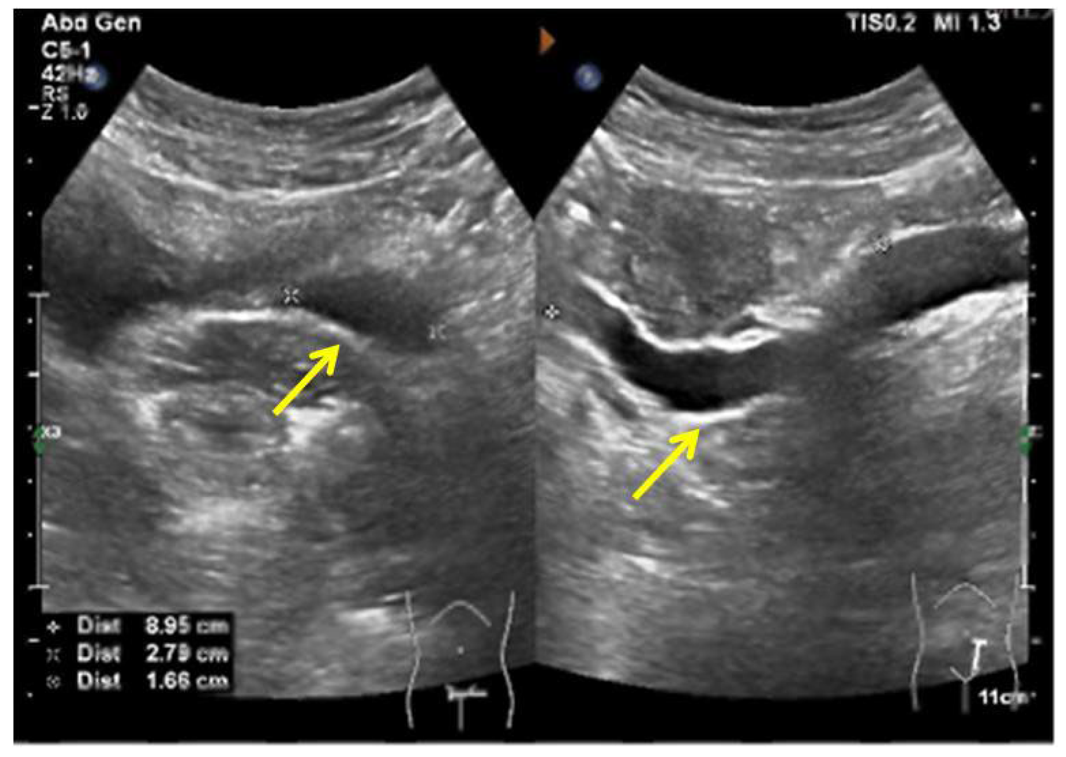
2.1. Case 1
2.2. Case 2
2.3. Case 3
2.4. Case 4
2.5. Case 5
2.6. Clinical Characteristics of the Five Cases
3. Discussion
4. Conclusions
Author Contributions
Funding
Institutional Review Board Statement
Informed Consent Statement
Data Availability Statement
Conflicts of Interest
References
- Baskin Laurence, S. Ectopic Ureter. Up to Date. Available online: http://www.uptodate.com/contents/ectopic-ureter (accessed on 13 July 2022).
- Duicu, C.; Kiss, E.; Simu, I.; Aldea, C. A rare case of double-system with ectopic ureteral openings into vagina. Front. Pediatr. 2018, 6, 176. [Google Scholar] [CrossRef] [PubMed]
- Roy Choudhury, S.; Chadha, R.; Bagga, D.; Puri, A.; Debnath, P.R. Spectrum of ectopic ureters in children. Pediatr. Surg. Int. 2008, 24, 819–823. [Google Scholar] [CrossRef] [PubMed]
- Toia, B.; Pakzad, M.; Hamid, R.; Wood, D.; Greenwell, T.; Ockrim, J. Diagnosis and surgical outcomes of ectopic ureters in adults: A case series and literature review. Neurourol. Urodyn. 2019, 38, 1745–1750. [Google Scholar] [CrossRef] [PubMed]
- Zhang, J.; Wang, L. Dysplastic kidney and ectopic ureter in association with obstructed hemivagina and ipsilateral renal anomaly. Int. Urogynecol. J. 2020, 31, 1707–1709. [Google Scholar] [CrossRef] [PubMed]
- Yuan, P.; Qi, L.; Wang, L. Incontinence after vaginal septum resection for a missed diagnosis of ectopic dysplastic kidney and ureter. Int. Urogynecol. J. 2017, 28, 645–646. [Google Scholar] [CrossRef] [PubMed]
- Kuliniec, I.; Mitura, P.; Plaza, P.; Widz, D.; Sudol, D.; Godzisz, M.; Kolodynska, A.; Monist, M.; Wisz, A.; Bar, K. Urinary incontinence in adulthood in a course of ectopic ureter-description of two clinical cases with review of literature. Int. J. Environ. Res. Public Health 2021, 18, 7084. [Google Scholar] [CrossRef] [PubMed]
- Chowdhary, S.K.; Lander, A.; Parashar, K.; Corkery, J.J. Single-system ectopic ureter: A 15-year review. Pediatr. Surg. Int. 2001, 17, 638–641. [Google Scholar] [CrossRef] [PubMed]
- Bohnenpoll, T.; Feraric, S.; Nattkemper, M.; Weiss, A.C.; Rudat, C.; Meuser, M.; Trowe, M.O.; Kispert, A. Diversification of cell lineages in ureter development. J. Am. Soc. Nephrol. 2017, 28, 1792–1801. [Google Scholar] [CrossRef] [PubMed]
- Chen, S.; Zhu, L.; Yang, S.; Tan, J. Y-type partial duplication of a vaginal ectopic ureter with ipsilateral hypoplastic pelvic kidney and bicornuate uterus. Singapore Med. J. 2013, 54, e135–e137. [Google Scholar] [CrossRef] [PubMed]
- Amenu, D.; Asmare, A.; Siraj, A. Congenital ureterovaginal fistula: A rare case of single-system ectopic ureter with ipsilateral ectopic kidney managed by vaginal approach: A case report. J. Med. Case Rep. 2021, 15, 617. [Google Scholar] [CrossRef] [PubMed]
- Patel, M.; Parikh, U.; Shrotriya, R.; Kadam, S.; Shah, J.; Chandna, S. Bilateral single system ectopic ureters with vaginal insertion in a female child, a rare variant. Urology 2021, 149, e37–e39. [Google Scholar] [CrossRef] [PubMed]
- Figueroa, V.H.; Chavhan, G.B.; Oudjhane, K.; Farhat, W. Utility of MR urography in children suspected of having ectopic ureter. Pediatr. Radiol. 2014, 44, 956–962. [Google Scholar] [CrossRef] [PubMed]
- Jain, P.; Sarkar, D.; Maiti, K.; Gupta, S.; Pal, D.K. Rare cases of ectopic ureter: Analysis from a single centre with review of the literature. Turk. J. Urol. 2019, 45, S92–S97. [Google Scholar] [CrossRef] [PubMed]
- Woolf, A.S.; Davies, J.A. Cell biology of ureter development. J. Am. Soc. Nephrol. 2013, 24, 19–25. [Google Scholar] [CrossRef] [PubMed]
- Magro-Lopez, E.; Munoz-Fernandez, M.A. The Role of BMP Signaling in Female Reproductive System Development and Function. Int. J. Mol. Sci. 2021, 22, 11927. [Google Scholar] [CrossRef] [PubMed]
- Wang, S.; Lang, J.H.; Zhu, L.; Zhou, H.M. Duplicated uterus and hemivaginal or hemicervical atresia with ipsilateral renal agenesis: An institutional clinical series of 52 cases. Eur. J. Obstet. Gynecol. Reprod. Biol. 2013, 170, 507–511. [Google Scholar] [CrossRef] [PubMed]
- Acien, P.; Acien, M. Unilateral renal agenesis and female genital tract pathologies. Acta Obstet. Gynecol. Scand. 2010, 89, 1424–1431. [Google Scholar] [CrossRef] [PubMed]


| Case 1 | Case 2 | Case 3 | Case 4 | Case 5 | |
|---|---|---|---|---|---|
| Admission date | 2022.03 | 2018.06 | 2015.07 | 2019.12 | 2020.11 |
| Age (years) | 46 | 14 | 37 | 29 | 33 |
| Gender | female | female | female | female | female |
| Chief complaint | dysmenorrhea and heavy menstrual bleeding | cyclic pelvic pain | vaginal discharge | urinary incontinence and ovarian cyst | endometrial polyps |
| Gynecological- related conditions | adenomyosis and menorrhagia | imperforate hymen with vaginal abscess | persistent vaginal discharge | ovarian endometrioma, right hydrosalpinx | recurrent pregnancy loss, endometrial polyps, reproductive duct anomalies |
| Imaging test | pelvic ultrasound, urinary ultrasound, pelvic MRI, CTU | pelvic ultrasound, urinary ultrasound, pelvic MRI, CTU, ascending urography | pelvic ultrasound, MRU, CTU, pelvic CT, abdominal CT | pelvic ultrasound, urinary ultrasound, pelvic MRI, CTU | pelvic ultrasound, pelvic MRI, CTU |
| Imaging test demonstrating ectopic ureter with vaginal insertion | pelvic ultrasound, urinary ultrasound | pelvic MRI, ascending urography | MRU | pelvic MRI, CTU | pelvic ultrasound, pelvic MRI, CTU |
| Congenital malformations of the female genital tract | uterine septum, oblique vaginal septum | double primordial uterus, imperforate hymen | none | none | oblique vaginal septum, double cervix, complete septate uterus |
| Urinary tract malformation | absence of right kidney, right ureteral dysplasia, ipsilateral ectopic ureter opening into the vagina | left kidney atrophy, left ureteral dilatation, left ureteral ectopic opening into the vagina | right duplex kidney, right ureteral ectopic opening into the vagina | left ureteral ectopic opening into the vagina | left renal agenesis and left dilated ureter with ectopic opening |
| The orifice site of ectopic ureter | vaginal wall | inside a sac of the vaginal wall | the right anterior wall of vaginal fornix | upper region of vagina | vagina |
| Surgical treatment of ectopic ureter | ureterectomy | ureterectomy | ureteral reimplantation | ureteral reimplantation | conservative observation |
| Karyotype | / | 46, XX | / | / | 46, XX |
Publisher’s Note: MDPI stays neutral with regard to jurisdictional claims in published maps and institutional affiliations. |
© 2022 by the authors. Licensee MDPI, Basel, Switzerland. This article is an open access article distributed under the terms and conditions of the Creative Commons Attribution (CC BY) license (https://creativecommons.org/licenses/by/4.0/).
Share and Cite
Wang, Q.; Wu, Z.; Zhang, F.; Akbar, R.; Lou, Y.; Zhou, J.; Ruan, F. Gynecological Diagnosis and Treatment of Ectopic Ureter Insertion into Vagina: Analysis of Five Cases and a Literature Review. J. Clin. Med. 2022, 11, 6267. https://doi.org/10.3390/jcm11216267
Wang Q, Wu Z, Zhang F, Akbar R, Lou Y, Zhou J, Ruan F. Gynecological Diagnosis and Treatment of Ectopic Ureter Insertion into Vagina: Analysis of Five Cases and a Literature Review. Journal of Clinical Medicine. 2022; 11(21):6267. https://doi.org/10.3390/jcm11216267
Chicago/Turabian StyleWang, Qijing, Zaigui Wu, Fengbin Zhang, Rubab Akbar, Yiyun Lou, Jianhong Zhou, and Fei Ruan. 2022. "Gynecological Diagnosis and Treatment of Ectopic Ureter Insertion into Vagina: Analysis of Five Cases and a Literature Review" Journal of Clinical Medicine 11, no. 21: 6267. https://doi.org/10.3390/jcm11216267
APA StyleWang, Q., Wu, Z., Zhang, F., Akbar, R., Lou, Y., Zhou, J., & Ruan, F. (2022). Gynecological Diagnosis and Treatment of Ectopic Ureter Insertion into Vagina: Analysis of Five Cases and a Literature Review. Journal of Clinical Medicine, 11(21), 6267. https://doi.org/10.3390/jcm11216267