
Hypofractionated Radiotherapy in Intermediate-Risk Prostate Cancer Patients: Long-Term Results

 , ,
, , 
 and
and
Abstract
1. Introduction
2. Materials and Methods
2.1. Patients’ Characteristics
2.2. Toxicity and Follow-Up
2.3. Statistical Analysis
3. Results
4. Toxicities
5. Discussions
6. Conclusions
7. Patents
Author Contributions
Funding
Institutional Review Board Statement
Informed Consent Statement
Data Availability Statement
Conflicts of Interest
References
- Mottet, N.; van den Bergh, R.C.N.; Briers, E.; van den Broeck, T.; Cumberbatch, M.G.; De Santis, M.; Fanti, S.; Fossati, N.; Gandaglia, G.; Gillessen, S.; et al. EAU-EANM-ESTRO-ESUR-SIOG Guidelines on Prostate Cancer-2020 Update. Part 1: Screening, Diagnosis, and Local Treatment with Curative Intent. Eur. Urol. 2021, 79, 243–262. [Google Scholar] [CrossRef] [PubMed]
- Viani, G.A.; Stefano, E.J.; Afonso, S.L. Higher-than-conventional radiation doses in localized prostate cancer treatment: A meta-analysis of randomized, controlled trials. Int. J. Radiat. Oncol. Biol. Phys. 2009, 74, 1405–1418. [Google Scholar] [CrossRef] [PubMed]
- Fowler, J.F. The radiobiology of prostate cancer including new aspects of fractionated radiotherapy. Acta Oncol. 2005, 44, 265–276. [Google Scholar] [CrossRef]
- Fowler, J.F.; Ritter, M.A.; Chappell, R.J.; Brenner, D.J. What hypofractionated protocols should be tested for prostate cancer? Int. J. Radiat. Oncol. Biol. Phys. 2003, 56, 1093–1104. [Google Scholar] [CrossRef]
- Nahum, A.E. The radiobiology of hypofractionation. Clin. Oncol. R. Coll. Radiol. 2015, 27, 260–269. [Google Scholar] [CrossRef]
- De Bari, B.; Arcangeli, S.; Ciardo, D.; Mazzola, R.; Alongi, F.; Russi, E.G.; Santoni, R.; Magrini, S.M.; Jereczek-Fossa, B.A. On the behalf of the Italian Association of Radiation Oncology (AIRO). Extreme hypofractionation for early prostate cancer: Biology meets technology. Cancer Treat. Rev. 2016, 50, 48–60. [Google Scholar] [CrossRef]
- Dearnaley, D.; Syndikus, I.; Mossop, H.; Khoo, V.; Birtle, A.; Bloomfield, D.; Graham, J.; Kirkbride, P.; Logue, J.; Malik, Z.; et al. CHHiP Investigators. Conventional versus hypofractionated high-dose intensity-modulated radiotherapy for prostate cancer: 5-year outcomes of the randomised, non-inferiority, phase 3 CHHiP trial. Lancet Oncol. 2016, 17, 1047–1060, Erratum in Lancet Oncol. 2016, 17, e321. [Google Scholar] [CrossRef]
- Lee, W.R.; Dignam, J.J.; Amin, M.B.; Bruner, D.W.; Low, D.; Swanson, G.P.; Shah, A.B.; D’Souza, D.P.; Michalski, J.M.; Dayes, I.S.; et al. Randomized Phase III Noninferiority Study Comparing Two Radiotherapy Fractionation Schedules in Patients with Low-Risk Prostate Cancer. J. Clin. Oncol. 2016, 34, 2325–2332. [Google Scholar] [CrossRef] [PubMed]
- Catton, C.N.; Lukka, H.; Gu, C.-S.; Martin, J.M.; Supiot, S.; Chung, P.W.M.; Bauman, G.S.; Bahary, J.-P.; Ahmed, S.; Cheung, P.; et al. Randomized Trial of a Hypofractionated Radiation Regimen for the Treatment of Localized Prostate Cancer. J. Clin. Oncol. 2017, 35, 1884–1890. [Google Scholar] [CrossRef]
- Jones, C.U.; Hunt, D.; McGowan, D.G.; Amin, M.B.; Chetner, M.P.; Bruner, D.W.; Leibenhaut, M.H.; Husain, S.M.; Rotman, M.; Souhami, L.; et al. Radiotherapy and short-term androgen deprivation for localized prostate cancer. N. Engl. J. Med. 2011, 365, 107–118. [Google Scholar] [CrossRef]
- D’Amico, A.V.; Chen, M.H.; Renshaw, A.A.; Loffredo, M.; Kantoff, P.W. Androgen suppression and radiation vs radiation alone for prostate cancer: A randomized trial. JAMA 2008, 299, 289–295. [Google Scholar] [CrossRef] [PubMed]
- Denham, J.W.; Steigler, A.; Lamb, D.S.; Joseph, D.; Turner, S.; Matthews, J.; Atkinson, C.; North, J.; Christie, D.; Spry, N.A.; et al. Short-term neoadjuvant androgen deprivation and radiotherapy for locally advanced prostate cancer: 10-year data from the TROG 96.01 randomised trial. Lancet Oncol. 2011, 12, 451–459. [Google Scholar] [CrossRef]
- Horwitz, E.M.; Bae, K.; Hanks, G.E.; Porter, A.; Grignon, D.J.; Brereton, H.D.; Venkatesan, V.; Lawton, C.A.; Rosenthal, S.A.; Sandler, H.M.; et al. Ten-year follow-up of radiation therapy oncology group protocol 92-02: A phase III trial of the duration of elective androgen deprivation in locally advanced prostate cancer. J. Clin. Oncol. 2008, 26, 2497–2504. [Google Scholar] [CrossRef] [PubMed]
- Zumsteg, Z.S.; Spratt, D.; Pei, I.; Zhang, Z.; Yamada, Y.; Kollmeier, M.; Zelefsky, M. A new risk classification system for therapeutic decision making with intermediate-risk prostate cancer patients undergoing dose-escalated external-beam radiation therapy. Eur. Urol. 2013, 64, 895–902. [Google Scholar] [CrossRef] [PubMed]
- D’Amico, A.V.; Renshaw, A.A.; Cote, K.; Hurwitz, M.; Beard, C.; Loffredo, M.; Chen, M.-H. Impact of the percentage of positive prostate cores on prostate cancer-specific mortality for patients with low or favorable intermediate-risk disease. J. Clin. Oncol. 2004, 22, 3726–3732. [Google Scholar] [CrossRef]
- Cox, J.D.; Stetz, J.; Pajak, T.F. Toxicity criteria of the Radiation Therapy Oncology Group (RTOG) and the European Organization for Research and Treatment of Cancer (EORTC). Int. J. Radiat. Oncol. Biol. Phys. 1995, 31, 1341–1346. [Google Scholar] [CrossRef]
- Roach, M., 3rd; Hanks, G.; Thames, H., Jr.; Schellhammer, P.; Shipley, W.U.; Sokol, G.H.; Sandler, H. Defining biochemical failure following radiotherapy with or without hormonal therapy in men with clinically localized prostate cancer: Recommendations of the RTOG-ASTRO Phoenix Consensus Conference. Int. J. Radiat. Oncol. Biol. Phys. 2006, 65, 965–974. [Google Scholar] [CrossRef]
- Schemper, M.; Smith, T.L. A note on quantifying follow-up in studies of failure time. Control Clin. Trials 1996, 17, 343–346. [Google Scholar] [CrossRef]
- Arcangeli, G.; Saracino, B.; Arcangeli, S.; Gomellini, S.; Petrongari, M.G.; Sanguineti, G.; Strigari, L. Moderate Hypofractionation in High-Risk, Organ-Confined Prostate Cancer: Final Results of a Phase III Randomized Trial. J. Clin. Oncol. 2017, 35, 1891–1897. [Google Scholar] [CrossRef]
- Avkshtol, V.; Ruth, K.J.; Ross, E.A.; Hallman, M.A.; Greenberg, R.E.; Price, R.A.P., Jr.; Leachman, B.; Uzzo, R.G.; Ma, C.; Chen, D.; et al. Ten-Year Update of a Randomized, Prospective Trial of Conventional Fractionated Versus Moderate Hypofractionated Radiation Therapy for Localized Prostate Cancer. J. Clin. Oncol. 2020, 38, 1676–1684, Erratum in J. Clin. Oncol. 2021, 39, 258. [Google Scholar] [CrossRef]
- De Vries, K.C.; Wortel, R.C.; Hoop, E.O.-D.; Heemsbergen, W.D.; Pos, F.J.; Incrocci, L. Hyprofractionated Versus Conventionally Fractionated Radiation Therapy for Patients with Intermediate- or High-Risk, Localized, Prostate Cancer: 7-Year Outcomes from the Randomized, Multicenter, Open-Label, Phase 3 HYPRO Trial. Int. J. Radiat. Oncol. Biol. Phys. 2020, 106, 108–115. [Google Scholar] [CrossRef] [PubMed]
- Aluwini, S.; Pos, F.; Schimmel, E.; Krol, S.; van der Toorn, P.P.; de Jager, H.; Alemayehu, W.G.; Heemsbergen, W.; Heijmen, B.; Incrocci, L. Hypofractionated versus conventionally fractionated radiotherapy for patients with prostate cancer (HYPRO): Late toxicity results from a randomised, non-inferiority, phase 3 trial. Lancet Oncol. 2016, 17, 464–474. [Google Scholar] [CrossRef]
- Staffurth, J.N.; Haviland, J.S.; Wilkins, A.; Syndikus, I.; Khoo, V.; Bloomfield, D.; Parker, C.; Logue, J.; Scrase, C.; Birtle, A.; et al. CHHiP Trial Management Group. Impact of Hypofractionated Radiotherapy on Patient-reported Outcomes in Prostate Cancer: Results up to 5 yr in the CHHiP trial (CRUK/06/016). Eur. Urol Oncol. 2021, 4, 980–992. [Google Scholar] [CrossRef] [PubMed]
- Zumsteg, Z.S.; Zelefsky, M.J. Short-term androgen deprivation therapy for patients with intermediate-risk prostate cancer undergoing dose-escalated radiotherapy: The standard of care? Lancet Oncol. 2012, 13, e259–e269. [Google Scholar] [CrossRef]
- Zumsteg, Z.S.; Zelefsky, M.J.; Woo, K.M.; Spratt, D.E.; Kollmeier, M.A.; McBride, S.; Pei, X.; Sandler, H.M.; Zhang, Z. Unification of favourable intermediate-, unfavourable intermediate-, and very high-risk stratification criteria for prostate cancer. BJU Int. 2017, 120, E87–E95. [Google Scholar] [CrossRef]
- Tom, M.C.; Reddy, C.A.; Smile, T.D.; Zhang, R.X.; Ciezki, J.P.; Stephans, K.L.; Mian, O.Y.; Klein, E.A.; Campbell, S.; Ulchaker, J.; et al. Validation of the NCCN prostate cancer favorable- and unfavorable-intermediate risk groups among men treated with I-125 low dose rate brachytherapy monotherapy. Brachytherapy 2020, 19, 43–50. [Google Scholar] [CrossRef]
- Bracci, S.; Osti, M.F.; Agolli, L.; Bertaccini, L.; De Sanctis, V.; Valeriani, M. Different outcomes among favourable and unfavourable intermediate-risk prostate cancer patients treated with hypofractionated radiotherapy and androgen deprivation therapy. Radiat. Oncol. 2016, 11, 78. [Google Scholar] [CrossRef][Green Version]
- De Crevoisier, R.; Bayar, M.A.; Pommier, P.; Muracciole, X.; Pêne, F.; Dudouet, P.; Latorzeff, I.; Beckendorf, V.; Bachaud, J.-M.; Laplanche, A.; et al. Daily Versus Weekly Prostate Cancer Image Guided Radiation Therapy: Phase 3 Multicenter Randomized Trial. Int. J. Radiat. Oncol. Biol. Phys. 2018, 102, 1420–1429. [Google Scholar] [CrossRef]


| Total Number of Patients: 313 | |
|---|---|
| Age | |
| Median (range) | 74 years (48–88) |
| ≤70 years | 58 pz (18.5%) |
| >70 years | 255 pz (81.5%) |
| PSA at diagnosis | |
| Median (range) | 8.15 ng/mL (2.6–19.9) |
| <10 ng/mL | 198 (63.2%) |
| 10–9.9 ng/mL | 115 (36.3%) |
| Tumor Stage | |
| T1c | 68 (21.8%) |
| T2a | 92 (29.4%) |
| T2b | 78 (24.9%) |
| T2c | 75 (23.9%) |
| Gleason Score | |
| 3 + 3 | 91 (29.1%) |
| 3 + 4 | 133 (42.5%) |
| 4 + 3 | 89 (28.4%) |
| Hormonal Therapy | |
| Antiandrogen | 97 (31.0%) |
| LHRH analogue | 216 (69.0%) |
| Risk Group | |
| Favorable Group | 189 (60.4%) |
| Unfavorable Group | 124 (39.6%) |
| Variables | Before Matching | After Matching | ||||
|---|---|---|---|---|---|---|
| Without IGRT 136 pz (43.5%) | With IGRT 177 pz (56.5%) | Chi-Square (p-value) | Without IGRT 112 pz | With IGRT 112 pz | Chi-Square (p-value) | |
| Age | 0.035 | 0.304 | ||||
| ≤70 years | 18 (5.8%) | 40 (12.8%) | 18 (8.0%) | 24 (10.7%) | ||
| >70 years | 118 (37.7%) | 137 (43.8%) | 94 (42.0%) | 88 (39.3%) | ||
| PSA at diagnosis | 0.158 | 0.211 | ||||
| <10 ng/mL | 92 (29.4%) | 106 (33.9%) | 76 (33.9%) | 67 (29.9%) | ||
| 10–19.9 ng/mL | 44 (14.1%) | 71 (22.7%) | 36 (16.1%) | 45 (20.1%) | ||
| Gleason Score | 0.059 | 0.698 | ||||
| 3 + 3 | 49 (15.7%) | 42 (13.4%) | 25 (11.2%) | 26 (11.6%) | ||
| 3 + 4 | 52 (16.6%) | 81 (25.9%) | 52 (23.2%) | 46 (20.5%) | ||
| 4 + 3 | 35 (11.2%) | 54 (17.3%) | 35 (15.6%) | 40 (17.9%) | ||
| Tumor Stage | 0.422 | 0.592 | ||||
| T1c–T2a | 66 (21.1%) | 94 (30.0%) | 58 (25.9%) | 62 (27.7%) | ||
| T2b–T2c | 70 (22.4%) | 83 (26.5%) | 54 (24.1%) | 50 (22.3%) | ||
| Hormonal Therapy | <0.001 | 0.426 | ||||
| Antiandrogen | 23 (7.3%) | 74 (23.6%) | 23 (10.3%) | 28 (12.5%) | ||
| LHRH Analogous | 113 (36.1%) | 103 (32.9%) | 89 (39.7%) | 84 (37.5%) |
| Variables | Before Matching | After Matching | Before Matching | After Matching | Before Matching | After Matching | ||||||
|---|---|---|---|---|---|---|---|---|---|---|---|---|
| 8-year OS (N. Patients) | (p-value) | 8-year OS (N. Patients) | (p-value) | 8-year CSS | (p-value) | 8-year CSS | (p-value) | 8-year bRFS | (p-value) | 8-year bRFS | (p-value) | |
| IGRT | 0.140 | 0.223 | 0.361 | 0.407 | 0.581 | 0.249 | ||||||
| Yes | 85.6% (177) | 82.9% (112) | 97.1% | 99.0% | 86.0% | 87.4% | ||||||
| No | 76.6% (136) | 77.1% (112) | 92.2% | 97.0% | 86.2% | 84.5% | ||||||
| Age | 0.557 | 0.923 | 0.187 | 96.2% | 0.128 | 0.129 | 0.125 | |||||
| ≤70 years | 84.6% (58) | 81.0% (42) | 97.3% | 98.6% | 81.4% | 76.7% | ||||||
| >70 years | 81.3% (255) | 79.7% (182) | 98.0% | 86.9% | 88.5% | |||||||
| Favorable Group | 84.6% (189) | 0.841 | 80.8% (127) | 0.808 | 99.1% | 0.962 | 98.6% | 0.676 | 88.4% | 0.068 | 83.3% | 0.715 |
| Unfavorable Group | 77.3% (124) | 78.7% (97) | 95.8% | 97.2% | 81.6% | 90.9% | ||||||
| Hormonal Therapy | 0.270 | 0.958 | 0.078 | 0.205 | 0.917 | 0.531 | ||||||
| Antiandrogen | 88.0% (97) | 81.1% (51) | 100.0% | 100.0% | 89.1% | 93.1% | ||||||
| LHRH Analogous | 77.0% (216) | 78.9% (173) | 96.3% | 97.3% | 84.2% | 83.6% | ||||||
| PSA before RT | 0.715 | 0.937 | 0.145 | 0.206 | 0.237 | 0.306 | ||||||
| ≤0.7 ng/mL | 77.6% (83) | 72.4% (66) | 97.9% | 97.2% | 87.8% | 88.3% | ||||||
| >0.7 ng/mL | 83.4% (230) | 82.8% (158) | 97.9% | 98.4% | 85.2% | 85.2% | ||||||
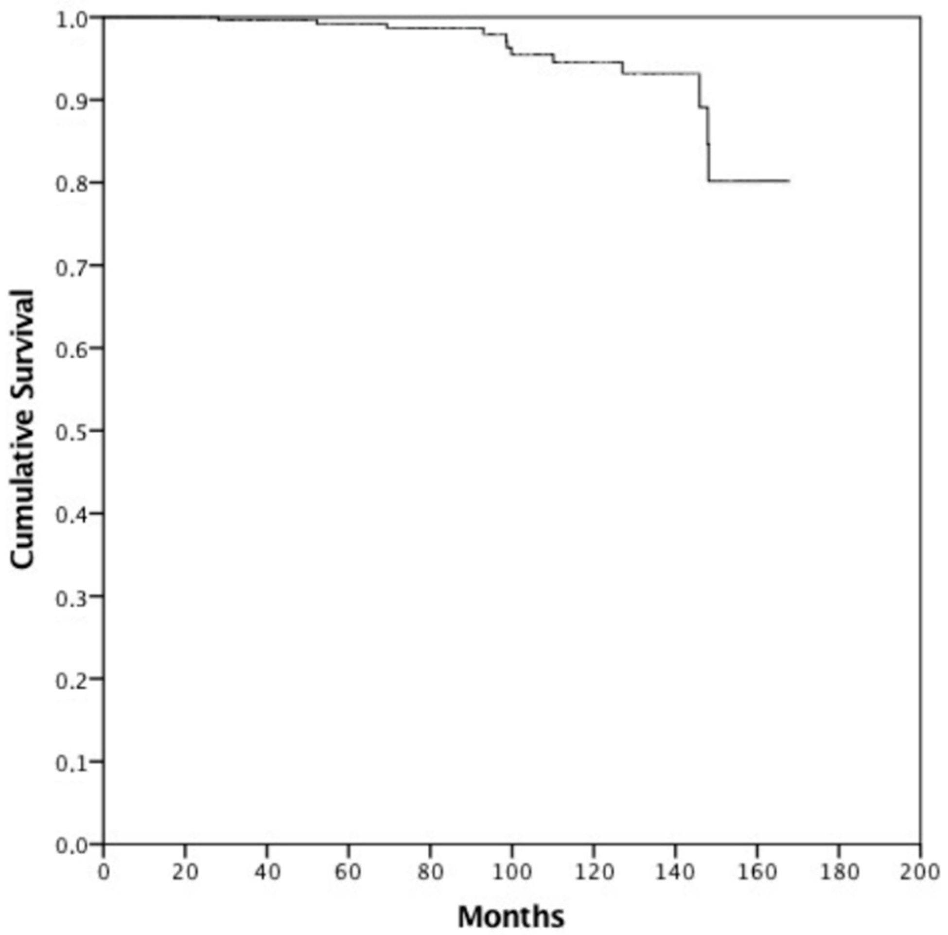
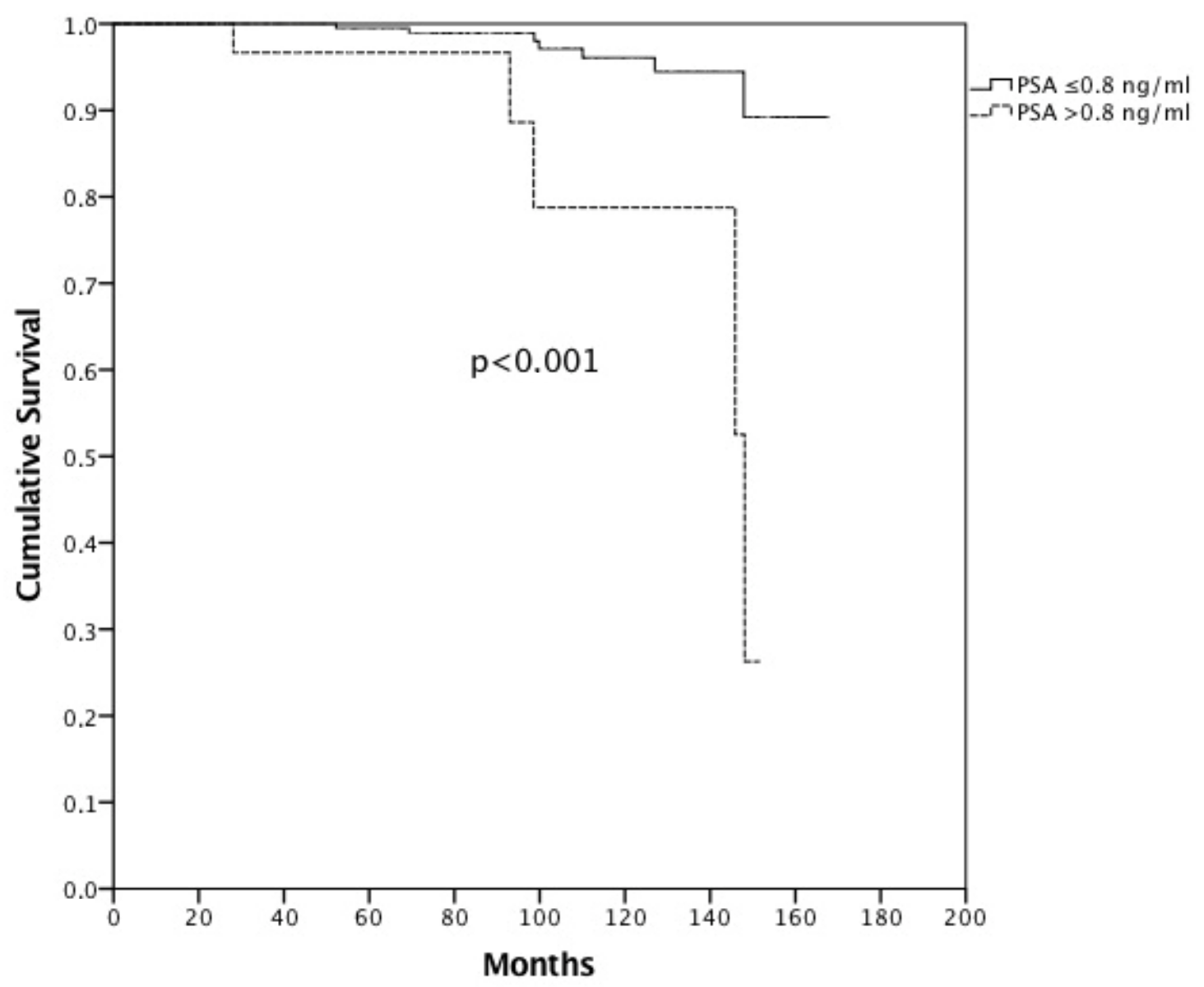
| PSA at first FU | 0.019 | 0.001 | <0.001 | 9 | 0.001 | <0.001 | 0.011 | |||||
| ≤0.8 ng/mL | 83.7% (282) | 82.2% (201) | 98.9% | 8.4% | 88.1% | 87.3% | ||||||
| >0.8 ng/mL | 65.6% (31) | 60.9% (23) | 88.6% | 95.7% | 65.9% | 77.3% | ||||||
| 8-year LRFS | (p-value) | 8-year LRFS | (p-value) | 8-year c-PFS | (p-value) | 8-year c-PFS | (p-value) | 8-year MFS | (p-value) | 8-year MFS | (p-value) | |
| IGRT | 0.275 | 0.467 | 0.443 | 0.835 | 0.693 | 0.831 | ||||||
| Yes | 88.9% | 87.7% | 86.2% | 97.7% | 93.7% | 94.4% | ||||||
| No | 95.9% | 95.1% | 94.4% | 93.3% | 97.7% | 97.2% | ||||||
| Age | 0.657 | 0.842 | 0.750 | 0.868 | 93. | 0.447 | 0.891 | |||||
| ≤70 years | 92.5% | 89.3% | 89.0% | 87.0% | 7% | 93.7% | ||||||
| >70 years | 89.8% | 92.0% | 89.4% | 91.5% | 95.5% | 96.4% | ||||||
| Favorable Group | 93.7% | 0.078 | 90.3% | 0.818 | 93.1% | 0.042 | 89.5% | 0.884 | 96.5% | 0.610 | 94.6% | 0.259 |
| Unfavorable Group | 84.7% | 94.0% | 83.1% | 93.0% | 93.1% | 97.9% | ||||||
| Hormonal Therapy | 0.673 | 0.935 | 0.659 | 0.868 | 0.862 | 0.605 | ||||||
| Antiandrogen | 90.0% | 92.9% | 88.9% | 92.9% | 97.5% | 93.1% | ||||||
| LHRH Analogous | 90.9% | 91.0% | 90.0% | 89.8% | 93.7% | 94.3% | ||||||
| PSA before RT | 0.948 | 0.798 | 0.665 | 0.940 | 0.691 | 0.735 | ||||||
| ≤0.7 ng/mL | 88.4% | 89.1% | 88.4% | 89.1% | 94.2% | 92.4% | ||||||
| >0.7 ng/mL | 91.2% | 92.6% | 89.8% | 91.3% | 95.6% | 97.3% | ||||||
| PSA at first FU | 0.014 | 0.103 | 0.005 | 0.026 | 0.004 | 0.003 | ||||||
| ≤0.8 ng/mL | 91.8% | 92.0% | 91.0% | 91.5% | 96.8% | 97.3% | ||||||
| >0.8 ng/mL | 76.3% | 83.7% | 73.7% | 79.9% | 78.8% | 79.9% |
| Before Matching | After Matching | |||||
|---|---|---|---|---|---|---|
| OS | HR | 95% CI | (p-Value) | HR | 95% CI | (p-Value) |
| IGRT (Yes vs. No) | 1.368 | 0.799–2.344 | 0.254 | 1.648 | 0.842–3.227 | 0.145 |
| Age (≤70 years vs. >70 years) | 0.923 | 0.479–1.777 | 0.810 | 1.178 | 0.556–2.494 | 0.669 |
| FG vs. UG | 0.973 | 0.587–1.615 | 0.917 | 1.032 | 0.561–1.898 | 0.920 |
| HT (A vs. LHRHA) | 0.842 | 0.484–1.465 | 0.544 | 1.112 | 0.584–2.119 | 0.746 |
| PSA before RT (≤0.7 vs. >0.7) | 1342 | 0.689–2.617 | 0.387 | 1.352 | 0.662–2.762 | 0.408 |
| PSA at first FU (≤0.8 vs. >0.8) | 0.468 | 0.243–0.900 | 0.023 | 0.307 | 0.147–0.641 | 0.002 |
| CSS | ||||||
| IGRT (Yes vs. No) | 1.108 | 0.248–4.944 | 0.893 | 1.356 | 0.236–7.781 | 0.732 |
| Age (≤70 years vs. >70 years) | 2.580 | 0.788–8.454 | 0.117 | 2.090 | 0.560–7.797 | 0.272 |
| FG vs. UG | 0.699 | 0.205–2.380 | 0.567 | 0.797 | 0.203–3.125 | 0.745 |
| HT (A vs. LHRHA) | 0.431 | 0.094–1.977 | 0.279 | 0.623 | 0.117–3.324 | 0.580 |
| PSA before RT (≤0.7 vs. >0.7) | 0.530 | 0.058–4.880 | 0.575 | |||
| PSA at first FU (≤0.8 vs. >0.8) | 0.155 | 0.046–0.530 | 0.003 | 0.195 | 0.046–0.833 | 0.027 |
| bPFS | ||||||
| IGRT (Yes vs. No) | 1.358 | 0.695–2.653 | 0.370 | 1.684 | 0.724–3.754 | 0.234 |
| Age (≤70 years vs. >70 years) | 2.154 | 1.073–4.326 | 0.031 | 2.192 | 0.966–4.971 | 0.060 |
| FG vs. UG | 0.541 | 0.291–1.004 | 0.052 | 0.986 | 0.461–2.111 | 0.972 |
| HT (A vs. LHRHA) | 0.929 | 0.466–1.853 | 0.835 | 0.730 | 0.304–1.753 | 0.481 |
| PSA before RT (≤0.7 vs. >0.7) | 1.158 | 0.488–2.745 | 0.740 | 0.902 | 0.335–2.432 | 0.839 |
| PSA at first FU (≤0.8 vs. >0.8) | 0.222 | 0.107–0.460 | <0.001 | 0.283 | 0.111–0.721 | 0.008 |
| LPFS | ||||||
| IGRT (Yes vs. No) | 0.624 | 0.241–1.612 | 0.330 | 0.729 | 0.252–2.113 | 0.561 |
| Age (≤70 years vs. >70 years) | 0.860 | 0.278–2.574 | 0.788 | 0.909 | 0.250–3.302 | 0.885 |
| FG vs. UG | 0.504 | 0.221–1.149 | 0.103 | 1.107 | 0.396–3.097 | 0.846 |
| HT (A vs. LHRHA) | 1.031 | 0.422–2.520 | 0.946 | 1.019 | 0.342–3.034 | 0.973 |
| PSA before RT (≤0.7 vs. >0.7) | 1.077 | 0.357–3.250 | 0.896 | 1.139 | 0.358–3.621 | 0.826 |
| PSA at first FU (≤0.8 vs. >0.8) | 0.328 | 0.120–0.894 | 0.029 | 0.368 | 0.100–1.354 | 0.113 |
| c-PFS | ||||||
| IGRT (Yes vs. No) | 0.781 | 0.328–1.863 | 0.578 | 0.949 | 0.348–2.596 | 0.918 |
| Age (≤70 years vs. >70 years) | 1.340 | 0.528–3.400 | 0.538 | 1.197 | 0.382–3.750 | 0.757 |
| FG vs. UG | 0.454 | 0.208–0.989 | 0.047 | 0.997 | 0.381–2.612 | 0.996 |
| HT (A vs. LHRHA) | 1.065 | 0.457–2.479 | 0.885 | 0.897 | 0.310–2.596 | 0.841 |
| PSA before RT (≤0.7 vs. >0.7) | 1.072 | 0.343–3.345 | 0.905 | |||
| PSA at first FU (≤0.8 vs. >0.8) | 0.299 | 0.119–0.752 | 0.010 | 0.293 | 0.093–0.928 | 0.037 |
| MFS | ||||||
| IGRT (Yes vs. No) | 0.776 | 0.236–2.551 | 0.676 | 1.502 | 0.332–6.793 | 0.597 |
| Age (≤70 years vs. >70 years) | 1.826 | 0.556–5.998 | 0.321 | 1.314 | 0.261–6.614 | 0.741 |
| FG vs. UG | 0.739 | 0.252–2.167 | 0.582 | 2.177 | 0.439–10.803 | 0.341 |
| HT (A vs. LHRHA) | 0.747 | 0.227–2.463 | 0.632 | 0.753 | 0.150–3.769 | 0.730 |
| PSA before RT (≤0.7 vs. >0.7) | 0.876 | 0.161–4.772 | 0.878 | 1.969 | 0.388–9.991 | 0.414 |
| PSA at first FU (≤0.8 vs. >0.8) | 0.204 | 0.063–0.661 | 0.008 | 0.149 | 0.034–0.641 | 0.011 |
Publisher’s Note: MDPI stays neutral with regard to jurisdictional claims in published maps and institutional affiliations. |
© 2022 by the authors. Licensee MDPI, Basel, Switzerland. This article is an open access article distributed under the terms and conditions of the Creative Commons Attribution (CC BY) license (https://creativecommons.org/licenses/by/4.0/).
Share and Cite
Valeriani, M.; Di Staso, M.; Facondo, G.; Vullo, G.; De Sanctis, V.; Gravina, G.L.; di Genesio Pagliuca, M.; Osti, M.F.; Bonfili, P. Hypofractionated Radiotherapy in Intermediate-Risk Prostate Cancer Patients: Long-Term Results. J. Clin. Med. 2022, 11, 4783. https://doi.org/10.3390/jcm11164783
Valeriani M, Di Staso M, Facondo G, Vullo G, De Sanctis V, Gravina GL, di Genesio Pagliuca M, Osti MF, Bonfili P. Hypofractionated Radiotherapy in Intermediate-Risk Prostate Cancer Patients: Long-Term Results. Journal of Clinical Medicine. 2022; 11(16):4783. https://doi.org/10.3390/jcm11164783
Chicago/Turabian StyleValeriani, Maurizio, Mario Di Staso, Giuseppe Facondo, Gianluca Vullo, Vitaliana De Sanctis, Giovanni Luca Gravina, Milena di Genesio Pagliuca, Mattia Falchetto Osti, and Pierluigi Bonfili. 2022. "Hypofractionated Radiotherapy in Intermediate-Risk Prostate Cancer Patients: Long-Term Results" Journal of Clinical Medicine 11, no. 16: 4783. https://doi.org/10.3390/jcm11164783
APA StyleValeriani, M., Di Staso, M., Facondo, G., Vullo, G., De Sanctis, V., Gravina, G. L., di Genesio Pagliuca, M., Osti, M. F., & Bonfili, P. (2022). Hypofractionated Radiotherapy in Intermediate-Risk Prostate Cancer Patients: Long-Term Results. Journal of Clinical Medicine, 11(16), 4783. https://doi.org/10.3390/jcm11164783