
Assessing the Placement of the Cochlear Slim Perimodiolar Electrode Array by Trans Impedance Matrix Analysis: A Temporal Bone Study

 , , , , and
Abstract
:1. Introduction
2. Material and Methods
- First: Correct insertion (according to the implant specifications, and with two white marks of the electrode array out of the Round window) (n = 8).
- Second: Intentional fold-over of the electrode array (n = 8).
- The loaded sheath is guided into the cochlea until the sheath stopper reaches the opening of the round window.
- The sheath handle is inserted and stabilized using straight forceps until the sheath stopper is flush with the opening.
- With the sheath stopper resting, the electrode is slowly advanced with forceps. When the two white markers align, the electrode is fully inserted.
- In both cases, the whitemarkers of the electrode arrays were visible after a ”pull back” maneuver was performed.
- The round window approach was used in both implantations.
2.1. Description of the Trans Impedance Matrix Patterns
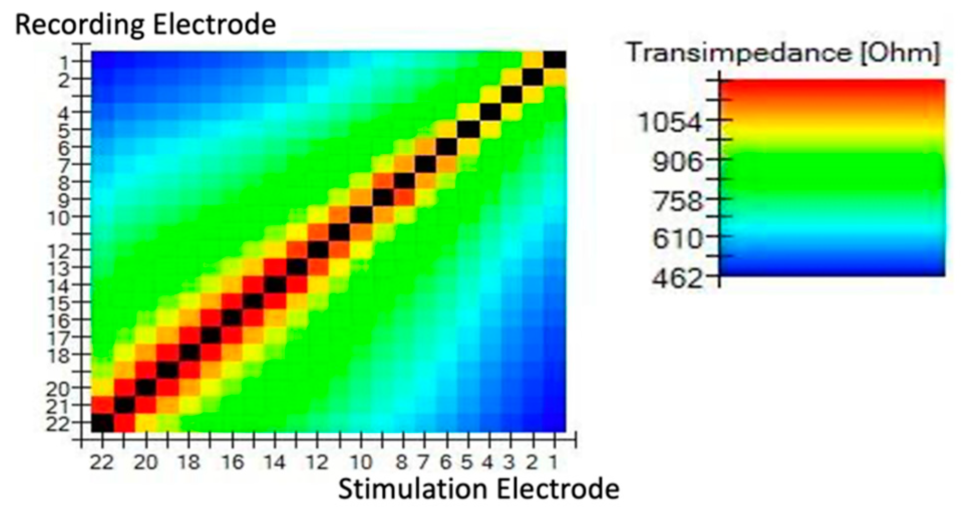
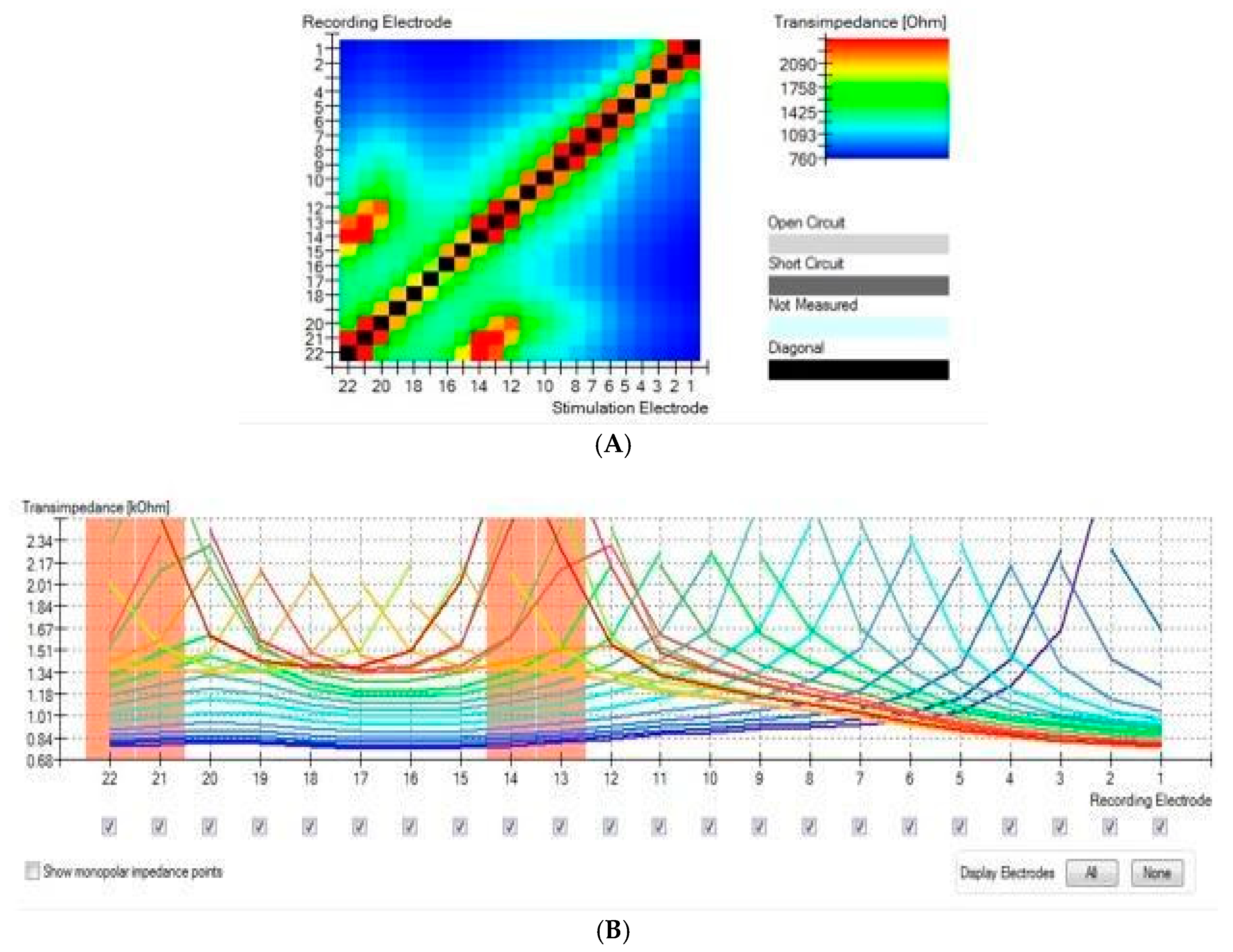
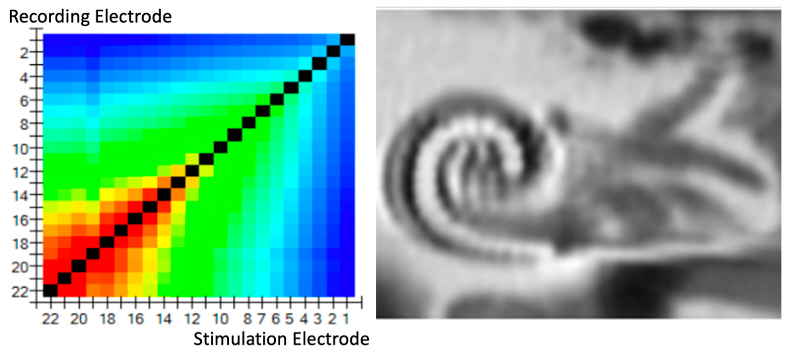
2.1.1. Normal Perimodiolar Position
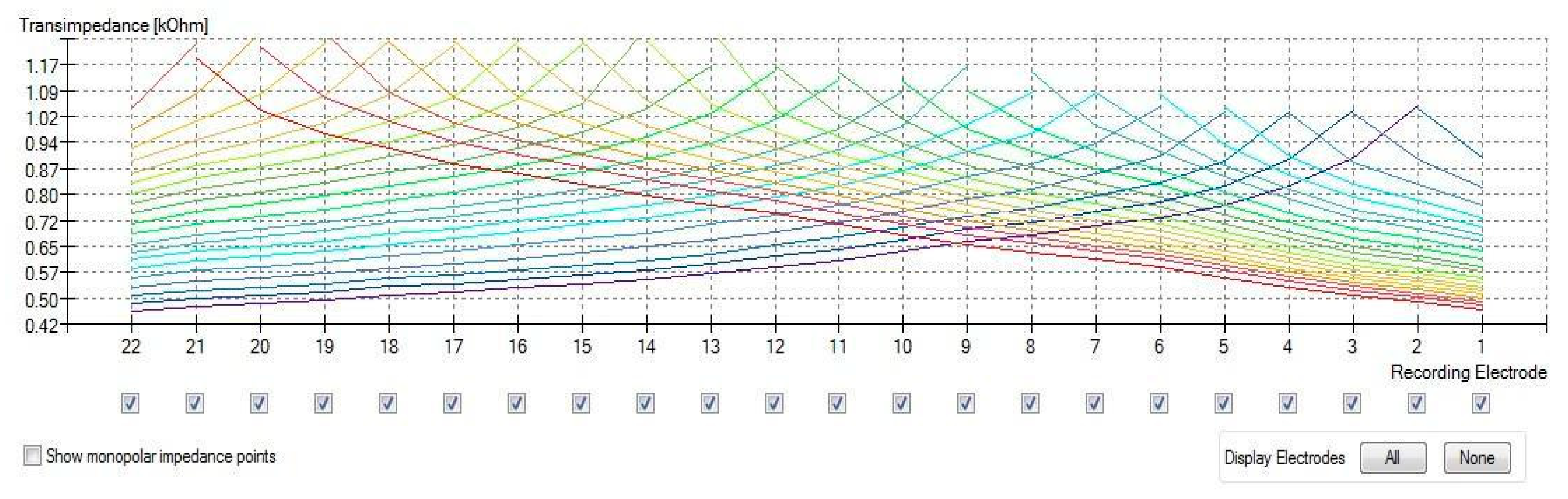
2.1.2. Electrode Fold-Over
3. Results
4. Discussion
- (a)
- Some of these studies did not achieve significantly different results in TIM conditions, and there was considerable variation among subjects; as such, large groups are required to detect differences among groups. On the other hand, in our research, as the conditions were the same for both insertions, the results were more consistent [27,28].
- (b)
- (c)
- It is also important to mention that CT scans (even ConeBeamCT) expose patients to x-rays and should be performed only in difficult situations or in cases where there are problems with fitting or hearing.
Limitations
5. Conclusions
Author Contributions
Funding
Institutional Review Board Statement
Informed Consent Statement
Acknowledgments
Conflicts of Interest
References
- Chang, A.; Eastwood, H.; Sly, D.; James, D.; Richardson, R.; O’Leary, S. Factors influencing the efficacy of round window dexamethasone protection of residual hearing post-cochlear implant surgery. Hear Res. 2009, 255, 67–72. [Google Scholar] [CrossRef] [PubMed]
- Briggs, R.J.; Tykocinski, M.; Xu, J.; Risi, F.; Svehla, M.; Cowan, R.; Stover, T.; Erfurt, P.; Lenarz, T. Comparison of round window and cochleostomy approaches with a prototype hearing preservation electrode. Audiol. Neurootol. 2006, 11 (Suppl. S1), 42–48. [Google Scholar] [CrossRef] [PubMed]
- Cuda, D.; Murri, A. Cochlear implantation with the nucleus slim modiolar electrode (CI532): A preliminary experience. Eur. Arch Otorhinolaryngol. 2017, 274, 4141–4148. [Google Scholar] [CrossRef] [PubMed]
- Aschendorff, A.; Briggs, R.; Brademann, G.; Helbig, S.; Hornung, J.; Lenarz, T.; Marx, M.; Ramos, A.; Stöver, T.; Escudé, B.; et al. Clinical investigation of the Nucleus Slim Modiolar Electrode. Audiol. Neurotol. 2017, 22, 169–179. [Google Scholar] [CrossRef] [Green Version]
- Franke-Trieger, A.; Mürbe, D. Estimation of insertion depth angle based on cochlea diameter and linear insertion depth: A prediction tool for the CI422. Eur. Arch. Otorhinolaryngol. 2015, 272, 3193–3199. [Google Scholar] [CrossRef]
- Erixon, E.; Högstorp, H.; Wadin, K.; Rask-Andersen, H. Variational anatomy of the human cochlea. Otol. Neurotol. 2009, 30, 14–22. [Google Scholar] [CrossRef]
- Helbig, S.; Settevendemie, C.; Mack, M.; Baumann, U.; Helbig, M.; Stöver, T. Evaluation of an electrode prototype for atraumatic cochlear implantation in hearing preservation candidates: Preliminary results from a temporal bone study. Otol. Neurotol. 2011, 32, 419–423. [Google Scholar] [CrossRef]
- Roland, J.T., Jr.; Fishman, A.J.; Alexiades, G.; Cohen, N.L. Electrode to modiolus proximity: A fluoroscopic and histologic analysis. Am. J. Otol. 2000, 21, 218–225. [Google Scholar] [CrossRef]
- Xu, J.; Stevenson, A.W.; Gao, D.; Tykocinski, M.; Lawrence, D.; Wilkins, S.W.; Cowan, R.S. The role of radiographic phase-contrast imaging in the development of intracochlear electrode arrays. Otol. Neurotol. 2001, 22, 862–868. [Google Scholar] [CrossRef] [Green Version]
- Schuman, T.A.; Noble, J.H.; Wright, C.G.; Wanna, G.B.; Dawant, B.; Labadie, R.F. Anatomic verification of a novel method for precise intrascalar localization of cochlear implant electrodes in adult temporal bones using clinically available computed tomography. Laryngoscope 2010, 120, 2277–2283. [Google Scholar] [CrossRef] [Green Version]
- Xu, J.; Xu, S.A.; Cohen, L.T.; Clark, G.M. Cochlear view: Postoperative radiography for cochlear implantation. Otol. Neurotol. 2000, 21, 49–56. [Google Scholar]
- Marx, M.; Risi, F.; Escudé, B.; Durmo, I.; James, C.; Lauwers, F.; Deguine, O.; Fraysse, B. Reliability of cone beam computed tomography in scalar localization of the electrode array: A radio histological study. Eur. Arch. Oto-Rhino-Laryngol. 2013, 271, 673–679. [Google Scholar] [CrossRef] [PubMed]
- Grolman, W.; Maat, A.; Verdam, F.; Simis, Y.; Carelsen, B.; Freling, N.; Tange, R.A. Spread of excitation measurements for the detection of electrode array foldovers: A prospective study comparing 3-dimensional rotational x-ray and intraoperative spread of excitation measurements. Otol. Neurotol. 2009, 30, 27–33. [Google Scholar] [CrossRef] [PubMed]
- Vanpoucke, F.J.; Boermans, P.P.B.; Frijns, J.H. Assessing the placement of a cochlear electrode array by multidimensional scaling. IEEE Trans. Biomed. Eng. 2012, 59, 307. [Google Scholar]
- De Rijk, S.R.; Tam, Y.C.; Carlyon, R.P.; Bance, M.L. Detection of extracochlear electrodes in cochlear implants with electric field imaging/transimpedance measurements. Ear Hear. 2020, 41, 1196–1207. [Google Scholar] [CrossRef]
- Ramos-Macias, A.; De Miguel, A.R.; Falcon-González, J.C. Mechanisms of electrode fold-over in cochlear implant surgery when using a flexible and slim perimodiolar electrode array. Acta Oto-Laryngol. 2017, 137, 1129–1135. [Google Scholar] [CrossRef]
- De Miguel, R.; Durmo, I.; González, J.C.F.; Barreiro, S.B.; Macías, A.R. Evaluation of intracochlear position of a slim modiolar electrode array, by using different radiological analyses. Otol. Neurotol. 2019, 40 (Suppl. S5), S10–S17. [Google Scholar] [CrossRef]
- Vanpoucke, F.V.; Zarowski, A.J.; Peeters, S.A. Identification of the impedance model of an implanted cochlear prosthesis from intracochlearpotential measurements. IEEE Trans. Biomed. Eng. 2004, 51, 2174–2183. [Google Scholar] [CrossRef]
- Van der Marel, K.S.; Briaire, J.J.; Verbist, B.M.; Muurling, T.J.; Frijns, J.H. The influence of cochlear implant electrode position on performance. Audiol. Neurotol. 2015, 20, 202–211. [Google Scholar] [CrossRef]
- Zelener, F.; Majdani, O.; Roemer, A.; Lexow, G.J.; Giesemann, A.; Lenarz, T.; Warnecke, A. Relations between scalar shift and insertion depth in human cochlear implantation. Otol. Neurotol. 2020, 41, 178–185. [Google Scholar] [CrossRef]
- Dong, Y.; Briaire, J.J.; Siebrecht, M.; Stronks, H.C.; Frijns, J.H.M. Detection of translocation of cochlear implant electrode arrays by intracochlear impedance measurements. Ear Hear. 2021, 42, 1397–1404. [Google Scholar] [CrossRef] [PubMed]
- Adunka, O.; Gstoettner, W.; Hambek, M.; Unkelbach, M.H.; Radeloff, A.; Kiefer, J. Preservation of Basal Inner Ear Structures in Cochlear Implantation. ORL 2004, 66, 306–312. [Google Scholar] [CrossRef] [PubMed]
- O’Connell, B.P.; Cakir, A.; Hunter, J.; Francis, D.O.; Noble, J.H.; Labadie, R.F.; Zuniga, M.G.; Dawant, B.M.; Rivas, A.; Wanna, G.B. Electrode location and angular insertion depth are predictors of audiologic outcomes in cochlear implantation. Otol. Neurotol. 2016, 37, 1016–1023. [Google Scholar] [CrossRef] [PubMed] [Green Version]
- Zierhofer, C.; Schatzer, R. Simultaneous intracochlear stimulation based on channel interaction compensation: Analysis and first results. IEEE Trans. Biomed. Eng. 2008, 55, 1907–1916. [Google Scholar] [CrossRef] [PubMed]
- Van den Honert, C.; Kelsall, D.C. Focused intracochlear electric stimulation with phased array channels. J. Am. Stat. Assoc. 2007, 121, 3703–3716. [Google Scholar] [CrossRef]
- Di Lella, F.A.; De Marco, D.; Fernández, F.; Parreño, M.; Boccio, C.M. In Vivo real-time remote cochlear implant capacitive impedance measurements: A glimpse into the implanted inner ear. Otol. Neurotol. 2019, 40 (5S Suppl. 1), S18–S22. [Google Scholar] [CrossRef]
- Klabbers, T.M.; Huinck, W.J.; Heutink, F.; Verbist, B.M.; Mylanus, E.A. Transimpedance matrix (TIM) measurement for the detection of intraoperative electrode tip foldover using the slim modiolar electrode: A proof of concept study. Otol. Neurotol. 2021, 42, e124–e129. [Google Scholar] [CrossRef]
- Hans, S.; Arweiler-Harbeck, D.; Kaster, F.; Ludwig, J.; Hagedorn, E.; Lang, S.; Meyer, M.; Holtmann, L.C. Transimpedance Matrix measurements reliably detect electrode tip fold-over in cochlear implantation. Otol. Neurotol. 2021, 42, e1494–e1502. [Google Scholar] [CrossRef]
- De Miguel, R.; Argudo, A.A.; Barreiro, S.A.B.; González, J.C.F.; Macías, A.R. Imaging evaluation of electrode placement and effect on electrode discrimination on different cochlear implant electrode arrays. Eur. Arch. Otorhinolaryngol. 2018, 275, 1385–1394. [Google Scholar] [CrossRef]
- Saunders, E.; Cohen, L.; Aschendorff, A.; Shapiro, W.; Knight, M.; Stecker, M.; Richter, B.; Waltzman, S.; Tykocinski, M.; Roland, T.; et al. Threshold, Comfortable Level and Impedance Changes as a Function of Electrode-Modiolar Distance. Ear Hear. 2002, 23, 28S–40S. [Google Scholar] [CrossRef]
- Aebischer, P.; Meyer, S.; Caversaccio, M.; Wimmer, W. Estimación basada en la impedancia intraoperatoria de la profundidad de inserción de la matriz de electrodos del implante coclear. IEEE Trans. Biomed. Eng. 2021, 68, 545–555. [Google Scholar] [CrossRef] [PubMed]
- Needham, K.; Stathopoulos, D.; Newbold, C.; Leavens, J.; Risi, F.; Manouchehri, S.; Durmo, I.; Cowan, R. Electrode impedance changes after implantation of a dexamethasone-eluting intracochlear array. Cochlea-Implant. Int. 2020, 21, 98–109. [Google Scholar] [CrossRef] [PubMed]


| A | B | 0° | 90° | 180° | Dif 0–180° | ||
|---|---|---|---|---|---|---|---|
| TB1 |  | 8.45 | 6.19 | 1.97 | 1.83 | 1.65 | 16.2% |
| TB2 |  | 8.54 | 6.19 | 2.04 | 1.59 | 1.92 | 5.8% |
| TB3 |  | 8.11 | 5.82 | 1.65 | 1.43 | 1.49 | 9.1% |
| TB4 |  | 9.58 | 7.08 | 2.33 | 2.20 | 1.86 | 20.1% |
| TB5 |  | 8.85 | 6.95 | 2.23 | 1-92 | 2.01 | 9.8% |
| TB6 |  | 9.28 | 6.80 | 2.36 | 1.92 | 1.85 | 21.6% |
| TB7 |  | 8.20 | 5.94 | 2.05 | 2.17 | 1.54 | 24.8% |
| TB8 |  | 8.59 | 6.00 | 2.11 | 1.81 | 1.61 | 23.6% |
| TB1 | Regular insertion | Good Perimodiolar position (ICPI = 0.12) | Normal transimpedance slope |
| TB2 | Regular insertion | Good Perimodiolar position (ICPI = 0.19) | Normal transimpedance slope |
| TB3 | Regular insertion | Good Perimodiolar position (ICPI = 0.23) | Normal transimpedance slope |
| TB4 | Regular insertion | Good Perimodiolar position (ICPI = 0.21) | Normal transimpedance slope |
| TB5 | Regular insertion | Mid and apex perimodiloar postion (ICPI = 0.32) | High impedance in tip TIM in apical/mid electrodes |
| TB6 | Regular insertion | Mid and apex perimodiloar postion (ICPI = 0.28) | High impedance in tip TIM in apical/mid electrodes |
| TB7 | Regular insertion | Good Perimodiolar position (ICPI = 0.15) | Normal transimpedance slope |
| TB8 | Regular insertion | Good Perimodiolar position (ICPI = 0.21) | Normal transimpedance slope |
| TFO Electrode | CT Correlation | |
|---|---|---|
| TFO-TB1 | 18 | Yes |
| TFO-TB2 | 17 | Yes |
| TFO-TB3 | 15 | Yes |
| TFO-TB4 | 19 | Yes |
| TFO-TB5 | 10 | Yes |
| TFO-TB6 | 12 | Yes |
| TFO-TB7 | 18 | Yes |
| TFO-TB8 | 17 | Yes |
Publisher’s Note: MDPI stays neutral with regard to jurisdictional claims in published maps and institutional affiliations. |
© 2022 by the authors. Licensee MDPI, Basel, Switzerland. This article is an open access article distributed under the terms and conditions of the Creative Commons Attribution (CC BY) license (https://creativecommons.org/licenses/by/4.0/).
Share and Cite
Ramos de Miguel, Á.; Riol Sancho, D.; Falcón-González, J.C.; Pavone, J.; Rodríguez Herrera, L.; Borkoski Barreiro, S.; Falcón Benitez, N.; Ramos Macias, Á. Assessing the Placement of the Cochlear Slim Perimodiolar Electrode Array by Trans Impedance Matrix Analysis: A Temporal Bone Study. J. Clin. Med. 2022, 11, 3930. https://doi.org/10.3390/jcm11143930
Ramos de Miguel Á, Riol Sancho D, Falcón-González JC, Pavone J, Rodríguez Herrera L, Borkoski Barreiro S, Falcón Benitez N, Ramos Macias Á. Assessing the Placement of the Cochlear Slim Perimodiolar Electrode Array by Trans Impedance Matrix Analysis: A Temporal Bone Study. Journal of Clinical Medicine. 2022; 11(14):3930. https://doi.org/10.3390/jcm11143930
Chicago/Turabian StyleRamos de Miguel, Ángel, Diego Riol Sancho, Juan Carlos Falcón-González, Joana Pavone, Leandro Rodríguez Herrera, Silvia Borkoski Barreiro, Nadia Falcón Benitez, and Ángel Ramos Macias. 2022. "Assessing the Placement of the Cochlear Slim Perimodiolar Electrode Array by Trans Impedance Matrix Analysis: A Temporal Bone Study" Journal of Clinical Medicine 11, no. 14: 3930. https://doi.org/10.3390/jcm11143930
APA StyleRamos de Miguel, Á., Riol Sancho, D., Falcón-González, J. C., Pavone, J., Rodríguez Herrera, L., Borkoski Barreiro, S., Falcón Benitez, N., & Ramos Macias, Á. (2022). Assessing the Placement of the Cochlear Slim Perimodiolar Electrode Array by Trans Impedance Matrix Analysis: A Temporal Bone Study. Journal of Clinical Medicine, 11(14), 3930. https://doi.org/10.3390/jcm11143930