
The Effects of Preterm Birth on Musculoskeletal Health-Related Disorders


Abstract
:1. Introduction
2. Preterm-Related Orthopedic Disorders
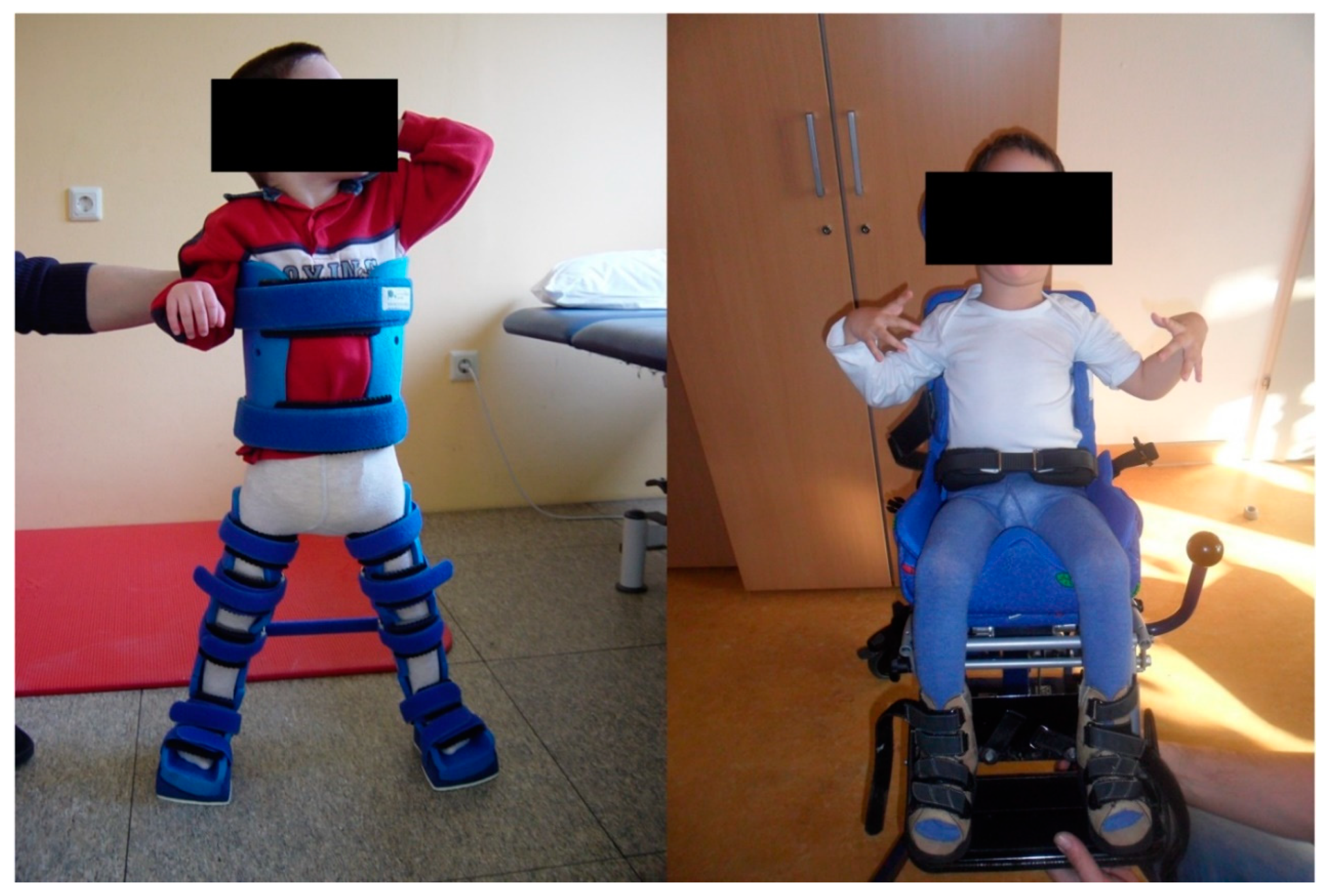
2.1. Cerebral Palsy
2.2. Fractures
2.3. Metabolic Bone Disease
2.4. Developmental Dysplasia of the Hip
3. Conclusions
Author Contributions
Funding
Data Availability Statement
Conflicts of Interest
Abbreviations
| CP | cerebral palsy |
| DDH | developmental dysplasia of the hip |
| GMFCS | Gross Motor Function Classification System |
| MBD | metabolic bone disease |
| PTB | preterm birth |
| ROM | range of motion |
References
- Trilla, C.C.; Medina, M.C.; Ginovart, G.; Betancourt, J.; Armengol, J.A.; Calaf, J. Maternal risk factors and obstetric complications in late preterm prematurity. Eur. J. Obstet. Gynecol. Reprod. Biol. 2014, 179, 105–109. [Google Scholar] [CrossRef]
- Cobo, T.; Kacerovsky, M.; Jacobsson, B. Risk factors for spontaneous preterm delivery. Int. J. Gynecol. Obstet. 2020, 50, 17–23. [Google Scholar] [CrossRef] [PubMed]
- Goldenberg, R.L.; Culhane, J.F.; Iams, J.D.; Romero, R. Epidemiology and causes of preterm birth. Lancet 2008, 371, 75–84. [Google Scholar] [CrossRef]
- Iams, J.D.; Goldenberg, R.L.; Meis, P.J.; Mercer, B.M.; Moawad, A.; Das, A.; Thom, E.; McNellis, D.; Copper, R.L.; Johnson, F.; et al. The length of the cervix and the risk of spontaneous premature delivery. National Institute of Child Health and Human Development Maternal Fetal Medicine Unit Network. N. Engl. J. Med. 1996, 334, 567–572. [Google Scholar] [CrossRef]
- Grobman, W.A.; Lai, Y.; Iams, J.D.; Reddy, U.M.; Mercer, B.M.; Saade, G.; Tita, A.T.; Rouse, D.J.; Sorokin, Y.; Wapner, R.J.; et al. Prediction of spontaneous preterm birth among nulliparous women with a short cervix. J. Ultrasound Med. 2016, 35, 1293–1297. [Google Scholar] [CrossRef] [Green Version]
- Phillips, C.; Velji, Z.; Hanly, C.; Metcalfe, A. Risk of recurrent spontaneous preterm birth: A systematic review and meta-analysis. BMJ Open 2017, 7, e015402. [Google Scholar] [CrossRef]
- Saigal, S.; Doyle, L.W. An overview of mortality and sequelae of preterm birth from infancy to adulthood. Lancet 2008, 371, 261–269. [Google Scholar] [CrossRef]
- Crump, C.; Sundquist, K.; Sundquist, J.; Winkleby, M.A. Gestational age at birth and mortality in young adulthood. JAMA 2011, 306, 1233–1240. [Google Scholar] [CrossRef]
- Rosenbaum, P.; Paneth, N.; Leviton, A.; Goldstein, M.; Bax, M. A report: The definition and classification of cerebral palsy April 2006. Dev. Med. Child Neurol. 2007, 49, 480. [Google Scholar]
- Paneth, N.; Hong, T.; Korzeniewski, S. The Descriptive Epidemiology of Cerebral Palsy. Clin. Perinatol. 2006, 33, 251–267. [Google Scholar] [CrossRef] [PubMed]
- Christensen, D.; Van Naarden Braun, K.; Doernberg, N.S.; Maenner, M.J.; Arneson, C.L.; Durkin, M.S.; Benedict, R.E.; Kirby, R.S.; Wingate, M.S.; Fitzgerald, R.; et al. Prevalence of cerebral palsy, co-occurring autism spectrum disorders, and motor functioning—Autism and Developmental Disabilities Monitoring Network, USA, 2008. Dev. Med. Child Neurol. 2014, 56, 59–65. [Google Scholar] [CrossRef] [PubMed]
- Little, W.J. On the influence of abnormal parturition, difficult labours, premature birth, and asphyxia neonatorum, on the mental and physical condition of the child, especially in relation to deformities. Clin. Orthop. Relat. Res. 1966, 46, 7–22. [Google Scholar] [CrossRef]
- Cans, C.; Guillem, P. Prevalence and characteristics of children with cerebral palsy in Europe. Dev. Med. Child Neurol. 2002, 44, 633–640. [Google Scholar]
- Moster, D.; Wilcox, A.J.; Vollset, S.E.; Markestad, T.; Lie, R.T. Cerebral palsy among term and postterm births. JAMA 2010, 304, 976–982. [Google Scholar] [CrossRef] [PubMed]
- Dammann, O.; Leviton, A. Maternal intrauterine infection, cytokines, and brain damage in the preterm newborn. Pediatr. Res. 1997, 42, 1–8. [Google Scholar] [CrossRef]
- Leviton, A.; Fichorova, R.N.; O’Shea, T.M.; Kuban, K.; Paneth, N.; Dammann, O.; Allred, E.N. Two-hit model of brain damage in the very preterm newborn: Small for gestational age and postnatal systemic inflammation. Pediatr. Res. 2013, 73, 362–370. [Google Scholar] [CrossRef] [PubMed] [Green Version]
- Kuban, K.C.K.; O’Shea, T.M.; Allred, E.N.; Fichorova, R.N.; Heeren, T.; Paneth, N.; Hirtz, D.; Dammann, O.; Leviton, A. The breadth and type of systemic inflammation and the risk of adverse neurological outcomes in extremely low gestation newborns. Pediatr. Neurol. 2015, 52, 42–48. [Google Scholar] [CrossRef] [Green Version]
- Thornton, C.; Rousset, C.I.; Kichev, A.; Miyakuni, Y.; Vontell, R.; Baburamani, A.A.; Fleiss, B.; Gressens, P.; Hagberg, H. Molecular mechanisms of neonatal brain injury. Neurol. Res. Int. 2012, 2012, 506320. [Google Scholar] [CrossRef]
- Prasad, J.D.; Gunn, K.C.; Davidson, J.O.; Galinsky, R.; Graham, S.E.; Berry, M.J.; Bennet, L.; Gunn, A.J.; Dean, J.M. Anti-inflammatory therapies for treatment of inflammation-related preterm brain injury. Int. J. Mol. Sci. 2021, 22, 4008. [Google Scholar] [CrossRef] [PubMed]
- Ellenberg, J.H.; Nelson, K.B. The association of cerebral palsy with birth asphyxia: A definitional quagmire. Dev. Med. Child Neurol. 2013, 55, 210–216. [Google Scholar] [CrossRef] [PubMed]
- Graham, H.K.; Rosenbaum, P.; Paneth, N.; Dan, B.; Lin, J.P.; Damiano, D.L.; Becher, J.G.; Gaebler-Spira, D.; Colver, A.; Reddihough, D.S.; et al. Cerebral palsy. Nat. Rev. Dis. Prim. 2016, 2, 15082. [Google Scholar] [CrossRef] [PubMed]
- Conde-Agudelo, A.; Romero, R. Antenatal magnesium sulfate for the prevention of cerebral palsy in preterm infants less than 34 weeks’ gestation: A systematic review and metaanalysis. Am. J. Obstet. Gynecol. 2009, 200, 595–609. [Google Scholar] [CrossRef] [Green Version]
- Shepherd, E.; Salam, R.A.; Middleton, P.; Makrides, M.; Mcintyre, S.; Badawi, N.; Crowther, C.A. Antenatal and intrapartum interventions for preventing cerebral palsy: An overview of Cochrane systematic reviews. Cochrane Database Syst. Rev. 2017, 8, CD012077. [Google Scholar] [CrossRef] [PubMed]
- Türkyilmaz, C.; Türkyilmaz, Z.; Atalay, Y.; Söylemezoglu, F.; Celasun, B. Magnesium pre-treatment reduces neuronal apoptosis in newborn rats in hypoxia-ischemia. Brain Res. 2002, 955, 133–137. [Google Scholar] [CrossRef]
- Burd, I.; Breen, K.; Friedman, A.; Chai, J.; Elovitz, M.A. Magnesium sulfate reduces inflammation-associated brain injury in fetal mice. Am. J. Obstet. Gynecol. 2010, 202, 292.e1–292.e29. [Google Scholar] [CrossRef] [PubMed] [Green Version]
- Cho, G.J.; Hong, H.R.; Hong, S.C.; Oh, M.J.; Kim, H.J. The neuroprotective effect of magnesium sulfate in preterm fetal mice. J. Perinat. Med. 2015, 43, 537–543. [Google Scholar] [CrossRef]
- Jacobs, S.E.; Berg, M.; Hunt, R.; Tarnow-Mordi, W.O.; Inder, T.E.; Davis, P.G. Cooling for newborns with hypoxic ischaemic encephalopathy. Cochrane Database Syst. Rev. 2013, 2013, CD003311. [Google Scholar] [CrossRef]
- Moreau, N.G.; Falvo, M.J.; Damiano, D.L. Rapid force generation is impaired in cerebral palsy and is related to decreased muscle size and functional mobility. Gait Posture 2012, 35, 154–158. [Google Scholar] [CrossRef] [Green Version]
- Ross, S.A.; Engsberg, J.R. Relationships Between Spasticity, Strength, Gait, and the GMFM-66 in Persons With Spastic Diplegia Cerebral Palsy. Arch. Phys. Med. Rehabil. 2007, 88, 1114–1120. [Google Scholar] [CrossRef]
- Damiano, D.L.; Martellotta, T.L.; Sullivan, D.J.; Granata, K.P.; Abel, M.F. Muscle force production and functional performance in spastic cerebral palsy: Relationship of cocontraction. Arch. Phys. Med. Rehabil. 2000, 81, 895–900. [Google Scholar] [CrossRef]
- Smith, L.R.; Lee, K.S.; Ward, S.R.; Chambers, H.G.; Lieber, R.L. Hamstring contractures in children with spastic cerebral palsy result from a stiffer extracellular matrix and increased in vivo sarcomere length. J. Physiol. 2011, 589, 2625–2639. [Google Scholar] [CrossRef]
- Beckung, E.; Hagberg, G. Neuroimpairments, activity limitations, and participation restrictions in children with cerebral palsy. Dev. Med. Child Neurol. 2002, 44, 309–316. [Google Scholar] [CrossRef] [PubMed]
- Gao, F.; Mei, X.; Chen, A.C.N. Delayed finger tapping and cognitive responses in preterm-born male teenagers with mild spastic diplegia. Pediatr. Neurol. 2015, 52, 206–213. [Google Scholar] [CrossRef]
- Ashwal, S.; Russman, B.S.; Blasco, P.A.; Miller, G.; Sandler, A.; Shevell, M.; Stevenson, R. Practice Parameter: Diagnostic assessment of the child with cerebral palsy: Report of the Quality Standards Subcommittee of the American Academy of Neurology and the Practice Committee of the Child Neurology Society. Neurology 2004, 62, 851–863. [Google Scholar] [CrossRef] [PubMed] [Green Version]
- Bosanquet, M.; Copeland, L.; Ware, R.; Boyd, R. A systematic review of tests to predict cerebral palsy in young children. Dev. Med. Child Neurol. 2013, 55, 418–426. [Google Scholar] [CrossRef]
- Palisano, R.; Rosenbaum, P.; Walter, S.; Russell, D.; Wood, E.; Galuppi, B. Development and reliability of a system to classify gross motor function in children with cerebral palsy. Dev. Med. Child Neurol. 1997, 39, 214–223. [Google Scholar] [CrossRef] [PubMed]
- Palisano, R.J.; Rosenbaum, P.; Bartlett, D.; Livingston, M.H. Content validity of the expanded and revised Gross Motor Function Classification System. Dev. Med. Child Neurol. 2008, 50, 744–750. [Google Scholar] [CrossRef]
- Rosenbaum, P.L.; Walter, S.D.; Hanna, S.E.; Palisano, R.J.; Russell, D.J.; Raina, P.; Wood, E.; Bartlett, D.J.; Galuppi, B.E. Prognosis for Gross Motor Function in Cerebral Palsy. JAMA 2002, 288, 1357–1363. [Google Scholar] [CrossRef] [Green Version]
- Nelson, K.B.; Ellenberg, J.H. Children who “outgrew” cerebral palsy. Pediatrics 1982, 69, 529–536. [Google Scholar] [CrossRef]
- Ferrari, F.; Cioni, G.; Einspieler, C.; Roversi, M.F.; Bos, A.F.; Paolicelli, P.B.; Ranzi, A.; Prechtl, H.F.R. Cramped synchronized general movements in preterm infants as an early marker for cerebral palsy. Arch. Pediatr. Adolesc. Med. 2002, 156, 460–467. [Google Scholar] [CrossRef]
- Cahill-Rowley, K.; Rose, J. Temporal-spatial gait parameters and neurodevelopment in very-low-birth-weight preterm toddlers at 18–22 months. Gait Posture 2016, 45, 83–89. [Google Scholar] [CrossRef] [Green Version]
- Galli, J.; Ambrosi, C.; Micheletti, S.; Merabet, L.B.; Pinardi, C.; Gasparotti, R.; Fazzi, E. White matter changes associated with cognitive visual dysfunctions in children with cerebral palsy: A diffusion tensor imaging study. J. Neurosci. Res. 2018, 96, 1766–1774. [Google Scholar] [CrossRef]
- Jiang, H.; Li, X.; Jin, C.; Wang, M.; Liu, C.; Chan, K.C.; Yang, J. Early diagnosis of spastic cerebral palsy in infants with periventricular white matter injury using diffusion tensor imaging. Am. J. Neuroradiol. 2019, 40, 162–168. [Google Scholar] [CrossRef] [Green Version]
- Tsimis, M.E.; Johnson, C.T.; Raghunathan, R.S.; Northington, F.J.; Burd, I.; Graham, E.M. Risk factors for periventricular white matter injury in very low birthweight neonates. Am. J. Obstet. Gynecol. 2016, 214, 380.e1–380.e6. [Google Scholar] [CrossRef] [Green Version]
- Khwaja, O.; Volpe, J.J. Pathogenesis of cerebral white matter injury of prematurity. Arch. Dis. Child. Fetal Neonatal Ed. 2008, 93, F153–F161. [Google Scholar] [CrossRef]
- Woodward, L.J.; Anderson, P.J.; Austin, N.C.; Howard, K.; Inder, T.E. Neonatal MRI to Predict Neurodevelopmental Outcomes in Preterm Infants. N. Engl. J. Med. 2006, 355, 685–694. [Google Scholar] [CrossRef] [PubMed] [Green Version]
- Cahill-Rowley, K.; Schadl, K.; Vassar, R.; Yeom, K.W.; Stevenson, D.K.; Rose, J. Prediction of Gait Impairment in Toddlers Born Preterm From Near-Term Brain Microstructure Assessed With DTI, Using Exhaustive Feature Selection and Cross-Validation. Front. Hum. Neurosci. 2019, 13, 305. [Google Scholar] [CrossRef] [PubMed]
- Schadl, K.; Vassar, R.; Cahill-Rowley, K.; Yeom, K.W.; Stevenson, D.K.; Rose, J. Prediction of cognitive and motor development in preterm children using exhaustive feature selection and cross-validation of near-term white matter microstructure. NeuroImage Clin. 2018, 17, 667–679. [Google Scholar] [CrossRef] [PubMed]
- Rose, J.; Cahill-Rowley, K.; Vassar, R.; Yeom, K.W.; Stecher, X.; Stevenson, D.K.; Hintz, S.R.; Barnea-Goraly, N. Neonatal brain microstructure correlates of neurodevelopment and gait in preterm children 18–22 mo of age: An MRI and DTI study. Pediatr. Res. 2015, 78, 700–708. [Google Scholar] [CrossRef]
- Blumetti, F.C.; Belloti, J.C.; Tamaoki, M.J.; Pinto, J.A. Botulinum toxin type A in the treatment of lower limb spasticity in children with cerebral palsy. Cochrane Database Syst. Rev. 2019, 10, CD001408. [Google Scholar] [CrossRef]
- Fonseca, P.R.; Calhes Franco De Moura, R.; Galli, M.; Santos Oliveira, C. Effect of physiotherapeutic intervention on the gait after the application of botulinum toxin in children with cerebral palsy: Systematic review. Eur. J. Phys. Rehabil. Med. 2018, 54, 757–765. [Google Scholar] [CrossRef]
- Bonouvrié, L.A.; Becher, J.G.; Vles, J.S.H.; Vermeulen, R.J.; Buizer, A.I. The Effect of Intrathecal Baclofen in Dyskinetic Cerebral Palsy: The IDYS Trial. Ann. Neurol. 2019, 86, 79–90. [Google Scholar] [CrossRef] [PubMed] [Green Version]
- Desailly, E.; Badina, A.; Khouri, N. Kinematics after unilateral femoral derotation osteotomy in children with diplegic cerebral palsy. Orthop. Traumatol. Surg. Res. 2020, 106, 1325–1331. [Google Scholar] [CrossRef] [PubMed]
- Braatz, F.; Poljuchow, J.; Klotz, M.C.; Heitzmann, D.W.W.; Wolf, S.I.; Dreher, T. Femoral derotation in children with cerebral palsy—Does the result depend on the age at operation and the kind of surgery? Z. Orthop. Unfall. 2015, 153, 636–642. [Google Scholar] [CrossRef] [PubMed]
- Huh, K.; Rethlefsen, S.A.; Wren, T.A.L.; Kay, R.M. Surgical management of hip subluxation and dislocation in children with cerebral palsy: Isolated VDRO or combined surgery? J. Pediatr. Orthop. 2011, 31, 858–863. [Google Scholar] [CrossRef] [Green Version]
- Al-Ghadir, M.; Masquijo, J.J.; Guerra, L.A.; Willis, B. Combined femoral and pelvic osteotomies versus femoral osteotomy alone in the treatment of hip dysplasia in children with cerebral palsy. J. Pediatr. Orthop. 2009, 29, 779–783. [Google Scholar] [CrossRef] [PubMed]
- Yun, A.G.; Severino, R.; Reinker, K. Varus derotational osteotomy for spastic hip instability: The roles of femoral shortening and obturator neurectomy. Am. J. Orthop. 2005, 34, 81–85. [Google Scholar]
- Carlson, E.J.; Carlson, M.G. Treatment of swan neck deformity in cerebral palsy. J. Hand Surg. Am. 2014, 39, 768–772. [Google Scholar] [CrossRef]
- Drefus, L.C.; Buckland, M.A.; Backus, S.I.; Root, L. The functional effect of a distal rectus femoris tenotomy in adults with cerebral palsy. Gait Posture 2014, 40, 145–149. [Google Scholar] [CrossRef]
- Wijesekera, M.P.C.; Wilson, N.C.; Trinca, D.; Holmes, G.; Bass, A.; Wright, D.M.; Walton, R. Pelvic Tilt Changes after Hamstring Lengthening in Children with Cerebral Palsy. J. Pediatr. Orthop. 2019, 39, e380–e385. [Google Scholar] [CrossRef]
- Morais Filho, M.C.; Blumetti, F.C.; Kawamura, C.M.; Fujino, M.H.; Matias, M.S.; Lopes, J.A.F. Comparison of the Results of Primary Versus Repeat Hamstring Surgical Lengthening in Cerebral Palsy. J. Pediatr. Orthop. 2020, 40, e380–e384. [Google Scholar] [CrossRef] [PubMed]
- Nazareth, A.; Rethlefsen, S.; Sousa, T.C.; Mueske, N.M.; Wren, T.A.L.; Kay, R.M. Percutaneous Hamstring Lengthening Surgery is as Effective as Open Lengthening in Children with Cerebral Palsy. J. Pediatr. Orthop. 2019, 39, 366–371. [Google Scholar] [CrossRef] [PubMed]
- Hsieh, H.C.; Wang, T.M.; Kuo, K.N.; Huang, S.C.; Wu, K.W. Guided Growth Improves Coxa Valga and Hip Subluxation in Children with Cerebral Palsy. Clin. Orthop. Relat. Res. 2019, 477, 2568–2576. [Google Scholar] [CrossRef] [PubMed]
- Portinaro, N.; Turati, M.; Cometto, M.; Bigoni, M.; Davids, J.R.; Panou, A. Guided Growth of the Proximal Femur for the Management of Hip Dysplasia in Children with Cerebral Palsy. J. Pediatr. Orthop. 2019, 39, e622–e628. [Google Scholar] [CrossRef]
- Lee, W.C.; Kao, H.K.; Yang, W.E.; Ho, P.C.; Chang, C.H. Guided growth of the proximal femur for hip displacement in children with cerebral palsy. J. Pediatr. Orthop. 2016, 36, 511–515. [Google Scholar] [CrossRef]
- Klatt, J.; Stevens, P.M. Guided growth for fixed knee flexion deformity. J. Pediatr. Orthop. 2008, 28, 626–631. [Google Scholar] [CrossRef]
- Schweizer, K.; Romkes, J.; Coslovsky, M.; Brunner, R. The influence of muscle strength on the gait profile score (GPS) across different patients. Gait Posture 2014, 39, 80–85. [Google Scholar] [CrossRef]
- Chinoy, A.; Mughal, M.Z.; Padidela, R. Metabolic bone disease of prematurity: Causes, recognition, prevention, treatment and long-term consequences. Arch. Dis. Child. Fetal Neonatal Ed. 2019, 104, F560–F566. [Google Scholar] [CrossRef] [PubMed]
- Wagner, K.; Wagner, S.; Susi, A.; Gorman, G.; Hisle-Gorman, E. Prematurity Does Not Increase Early Childhood Fracture Risk. J. Pediatr. 2019, 207, 148–153. [Google Scholar] [CrossRef]
- Michaud, J.; Luu, T.M.; LeBlanc, J.C.; Healy-Profitós, J.; Ayoub, A.; Auger, N. Preterm birth and the future risk of orthopedic fracture. Pediatr. Res. 2020, 88, 466–472. [Google Scholar] [CrossRef]
- Morgen, C.S.; Bjørk, C.; Andersen, P.K.; Mortensen, L.H.; Nybo Andersen, A.M. Socioeconomic position and the risk of preterm birth—A study within the Danish National Birth Cohort. Int. J. Epidemiol. 2008, 37, 1109–1120. [Google Scholar] [CrossRef] [Green Version]
- Shapiro, G.D.; Fraser, W.D.; Frasch, M.G.; Séguin, J.R. Psychosocial stress in pregnancy and preterm birth: Associations and mechanisms. J. Perinat. Med. 2013, 41, 631–645. [Google Scholar] [CrossRef]
- Buttazzoni, C.; Rosengren, B.; Tveit, M.; Landin, L.; Nilsson, J.Å.; Karlsson, M. Preterm Children Born Small for Gestational Age are at Risk for Low Adult Bone Mass. Calcif. Tissue Int. 2016, 98, 105–113. [Google Scholar] [CrossRef] [PubMed]
- Chen, W.; Yang, C.; Chen, H.; Zhang, B. Risk factors analysis and prevention of metabolic bone disease of prematurity. Medicine 2018, 97, e12861. [Google Scholar] [CrossRef] [PubMed]
- Loder, R.T.; Skopelja, E.N. The Epidemiology and Demographics of Hip Dysplasia. ISRN Orthop. 2011, 2011, 238607. [Google Scholar] [CrossRef] [PubMed] [Green Version]
- Kolb, A.; Schweiger, N.; Mailath-Pokorny, M.; Kaider, A.; Hobusch, G.; Chiari, C.; Windhager, R. Low incidence of early developmental dysplasia of the hip in universal ultrasonographic screening of newborns: Analysis and evaluation of risk factors. Int. Orthop. 2016, 40, 123–127. [Google Scholar] [CrossRef]
- Wilkinson, J.A. Etiologic factors in congenital displacement of the hip and myelodysplasia. Clin. Orthop. Relat. Res. 1992, 281, 75–83. [Google Scholar] [CrossRef]
- Manoukian, D.; Rehm, A. Oligohydramnios: Should it be considered a risk factor for developmental dysplasia of the hip? J. Pediatr. Orthop. Part B 2019, 28, 442–445. [Google Scholar] [CrossRef] [PubMed]
- Kenanidis, E.; Gkekas, N.K.; Karasmani, A.; Anagnostis, P.; Christofilopoulos, P.; Tsiridis, E. Genetic Predisposition to Developmental Dysplasia of the Hip. J. Arthroplast. 2020, 35, 291–300.e1. [Google Scholar] [CrossRef] [Green Version]
- Roposch, A.; Liu, L.Q.; Hefti, F.; Clarke, N.M.P.; Wedge, J.H. Standardized diagnostic criteria for developmental dysplasia of the hip in early infancy. Clin. Orthop. Relat. Res. 2011, 469, 3451–3461. [Google Scholar] [CrossRef] [Green Version]
- Barlow, T.G. Early Diagnosis and Treatment of Congenital Dislocation of the Hip. Proc. R. Soc. Med. 1963, 56, 804–806. [Google Scholar] [CrossRef] [PubMed]
- Ortolani, M. The classic: Congenital hip dysplasia in the light of early and very early diagnosis. Clin. Orthop. Relat. Res. 1976, 119, 6–10. [Google Scholar] [CrossRef]
- Graf, R. New possibilities for the diagnosis of congenital hip joint dislocation by ultrasonography. J. Pediatr. Orthop. 1983, 3, 354–359. [Google Scholar] [CrossRef]
- Graf, R. Fundamentals of sonographic diagnosis of infant hip dysplasia. J. Pediatr. Orthop. 1984, 4, 735–740. [Google Scholar] [CrossRef] [PubMed]
- Grill, F.; Müller, D. Results of hip ultrasonographic screening in Austria. Orthopade 1997, 26, 25–32. [Google Scholar] [CrossRef]
- Graf, R. Hip sonography. [Basic principles and current aspects]. Orthopade 1997, 26, 14–24. [Google Scholar] [CrossRef]
- Farr, S.; Grill, F.; Müller, D. When is the optimal time for hip ultrasound screening? Orthopade 2008, 37, 532–540. [Google Scholar] [CrossRef]
- Schaeffer, E.K.; Mulpuri, K. Developmental dysplasia of the hip: Addressing evidence gaps with a multicentre prospective international study. Med. J. Aust. 2018, 208, 359–364. [Google Scholar] [CrossRef] [PubMed]
- Tõnnis, D.; Storch, K.; Ulbrich, H. Results of newborn screening for CDH with and without sonography and correlation of risk factors. J. Pediatr. Orthop. 1990, 10, 145–152. [Google Scholar] [CrossRef]
- Sewell, M.D.; Rosendahl, K.; Eastwood, D.M. Developmental dysplasia of the hip. BMJ 2009, 339, b4454. [Google Scholar] [CrossRef] [Green Version]
- Hefti, F. Pediatric Orthopedics in Practice, 2nd ed.; Springer: Berlin/Heidelberg, Germany, 2015; ISBN 9783662468104. [Google Scholar]
- Dorn, U. Hip screening in newborn infants. Clinical and ultrasound results. Wien. Klin. Wochenschr. Suppl. 1990, 181, 3–22. [Google Scholar]
- Hinderaker, T.; Daltveit, A.K.; Irgens, L.M.; Udén, A.; Reikeräs, O. The impact of intra-uterine factors on neonatal hip instability. An analysis of 1,059,479 children in Norway. Acta Orthop. Scand. 1994, 65, 239–242. [Google Scholar] [CrossRef] [Green Version]
- Lange, A.E.; Lange, J.; Ittermann, T.; Napp, M.; Krueger, P.C.; Bahlmann, H.; Kasch, R.; Heckmann, M. Population-based study of the incidence of congenital hip dysplasia in preterm infants from the Survey of Neonates in Pomerania (SNiP). BMC Pediatr. 2017, 17, 78. [Google Scholar] [CrossRef] [Green Version]
- Verbruggen, S.W.; Kainz, B.; Shelmerdine, S.C.; Arthurs, O.J.; Hajnal, J.V.; Rutherford, M.A.; Phillips, A.T.M.; Nowlan, N.C. Altered biomechanical stimulation of the developing hip joint in presence of hip dysplasia risk factors. J. Biomech. 2018, 78, 1–9. [Google Scholar] [CrossRef] [PubMed]
- Koob, S.; Garbe, W.; Bornemann, R.; Ploeger, M.M.; Scheidt, S.; Gathen, M.; Placzek, R. Is Prematurity a Protective Factor against Developmental Dysplasia of the Hip? A Retrospective Analysis of 660 Newborns. Ultraschall Med. 2020. [Google Scholar] [CrossRef]
- Orak, M.M.; Onay, T.; Gümüştaş, S.A.; Gürsoy, T.; Muratlí, H.H. Is prematurity a risk factor for developmental dysplasia of the hip?: A prospective study. Bone Joint J. 2015, 97-B, 716–720. [Google Scholar] [CrossRef] [PubMed]
- Hussain, S.M.; Wang, Y.; Wluka, A.E.; Shaw, J.E.; Magliano, D.J.; Graves, S.; Cicuttini, F.M. Association of low birth weight and preterm birth with the incidence of knee and hip arthroplasty for osteoarthritis. Arthritis Care Res. 2015, 67, 502–508. [Google Scholar] [CrossRef] [PubMed]
- Novak, I.; Morgan, C.; Adde, L.; Blackman, J.; Boyd, R.N.; Brunstrom-Hernandez, J.; Cioni, G.; Damiano, D.; Darrah, J.; Eliasson, A.C.; et al. Early, accurate diagnosis and early intervention in cerebral palsy: Advances in diagnosis and treatment. JAMA Pediatr. 2017, 171, 897–907. [Google Scholar] [CrossRef]

Publisher’s Note: MDPI stays neutral with regard to jurisdictional claims in published maps and institutional affiliations. |
© 2021 by the authors. Licensee MDPI, Basel, Switzerland. This article is an open access article distributed under the terms and conditions of the Creative Commons Attribution (CC BY) license (https://creativecommons.org/licenses/by/4.0/).
Share and Cite
Schachinger, F.; Farr, S. The Effects of Preterm Birth on Musculoskeletal Health-Related Disorders. J. Clin. Med. 2021, 10, 5082. https://doi.org/10.3390/jcm10215082
Schachinger F, Farr S. The Effects of Preterm Birth on Musculoskeletal Health-Related Disorders. Journal of Clinical Medicine. 2021; 10(21):5082. https://doi.org/10.3390/jcm10215082
Chicago/Turabian StyleSchachinger, Florian, and Sebastian Farr. 2021. "The Effects of Preterm Birth on Musculoskeletal Health-Related Disorders" Journal of Clinical Medicine 10, no. 21: 5082. https://doi.org/10.3390/jcm10215082
APA StyleSchachinger, F., & Farr, S. (2021). The Effects of Preterm Birth on Musculoskeletal Health-Related Disorders. Journal of Clinical Medicine, 10(21), 5082. https://doi.org/10.3390/jcm10215082