Article

Rickettsia Vaccine Candidate pVAX1-OmpB24 Stimulates TCD4+INF-γ+ and TCD8+INF-γ+ Lymphocytes in Autologous Co-Culture of Human Cells

by
Karla Dzul-Rosado
1,*,
Luis Donis-Maturano
2,*,
Juan Arias-León
3,
Jesús Machado-Contreras
4,
Guillermo Valencia-Pacheco
1,
Candi Panti-Balam
1,
Javier Balam-Romero
1,
Angela Ku-González
5,
Gaspar Peniche-Lara
3,
Juan Mosqueda
6,
Oscar E. Zazueta
7,
Cesar Lugo-Caballero
1 and
Fernando Puerto-Manzano
1
1
Dr. Hideyo Noguchi Regional Research Center, Autonomous University of Yucatan, Mérida 97000, Mexico
2
Faculty of Higher Studies-Iztacala, National Autonomous University of Mexico, Tlalnepantla de Baz 54090, Mexico
3
Faculty of Medicine, Autonomous University of Yucatan, Mérida 97000, Mexico
4
Immunology Laboratory, Faculty of Medicine, Autonomous University of Baja California, Mexicali 21000, Mexico
5
Yucatan Scientific Research Center, Biochemistry and Plant Molecular Biology Unit, Mérida 97200, Mexico
6
Immunology and Vaccines Laboratory, College of Natural Sciences, Autonomous University of Queretaro, Queretaro 76010, Mexico
7
Ministry of Health of Baja California, Mexicali 21000, Mexico
*
Authors to whom correspondence should be addressed.
Vaccines 2023, 11(1), 173; https://doi.org/10.3390/vaccines11010173
Submission received: 15 December 2022 / Revised: 8 January 2023 / Accepted: 10 January 2023 / Published: 13 January 2023
(This article belongs to the Special Issue Immunity and Vaccination against Bacterial Infections)

Abstract

:
Background: In recent years, promising vaccination strategies against rickettsiosis have been described in experimental animal models and human cells. OmpB is considered an immunodominant antigen that is recognized by T and B cells. The aim of this study was to identify TCD4+INF-γ+ and TCD8+INF-γ+ lymphocytes in an autologous system with macrophages transfected with the vaccine candidate pVAX1-OmpB24. Lymphocytes and monocytes from 14 patients with Rickettsia were isolated from whole blood. Monocytes were differentiated into macrophages and transfected with the plasmid pVAX1-OmpB24 pVax1. Isolated lymphocytes were cultured with transfected macrophages. IFN-γ-producing TCD4+ and TCD8+ lymphocyte subpopulations were identified by flow cytometry, as was the percentage of macrophages expressing CD40+, CD80+, HLA-I and HLA-II. Also, we analyzed the exhausted condition of the T lymphocyte subpopulation by PD1 expression. Macrophages transfected with pVAX1-OmpB24 stimulated TCD4+INF-γ+ cells in healthy subjects and patients infected with R. typhi. Macrophages stimulated TCD8+INF-γ+ cells in healthy subjects and patients infected with R. rickettsii and R. felis. Cells from healthy donors stimulated with OmpB-24 showed a higher percentage of TCD4+PD1+. Cells from patients infected with R. rickettsii had a higher percentage of TCD8+PD-1+, and for those infected with R. typhi the larger number of cells corresponded to TCD4+PD1+. Human macrophages transfected with pVAX1-OmpB24 activated TCD4+IFN-γ+ and CD8+IFN-γ+ in patients infected with different Rickettsia species. However, PD1 expression played an important role in the inhibition of T lymphocytes with R. felis.

1. Introduction

Rickettsioses are worldwide febrile infectious diseases caused by small obligate intracellular bacteria of the genera Rickettsia: spotted fever (SFG) and typhus groups (TG) are responsible for rickettsiosis as emerging diseases [1]. In surveys of febrile illness, the proportion of fever attributed to rickettsia varies from 0.9% to 18% [2]. Oral doxycycline is effective as treatment, but in the case of severe life-threatening disease intravenous therapy is recommended. Unfortunately, doxycycline is not always available [3].
There have been concerted efforts to develop an effective vaccine against Rickettsia. Rickettsia proteins linked to cellular invasion (invA), division (fts), protein secretion (sec gene family) and virulence (OmpA, OmpB, virB, cap, Adr2, YbgF, TolC, tlyA and tlyC gene family), have been implicated [4,5,6,7]. Different vaccines containing non-viable R. rickettsii have been prepared from infected tick tissues [8], eggs [9], and embryonic chicken cells [10]. These vaccines have been tested on humans but have only been proven to be minimally effective in preventing Rocky Mountain Spotted Fever (RMSF) [11,12]. The generation of DNA vaccines against the rOmpA and rOmpB has led to a high immune response in murine models infected with R. conorii and R. rickettsia [13]. However, evaluation of rOmpB protein in murine models might be a limiting factor when compared with human models [14].
Because of the intracellular nature of R. rickettsii, an experimental strategy is to identify T lymphocytes that specifically recognize epitopes presented by HLA-I molecules. Naturally activated lymphocytes can be reactivated in vitro after a second exposure and their immune activity can be compared with that of the lymphocytes from healthy subjects [15]. Human ex vivo trials using macrophages or dendritic cells as antigen-presenting cells (APC), in co-culture with sensitized lymphocytes, have created high expectations, with higher predictive value than any preclinical strategy [16]. In preliminary studies, it has been shown that peripheral blood mononuclear cells (PBMC) on patients previously infected with rickettsia generated proliferative responses after stimulation with pVAX1-OmpB24, with production of IL-2, IFN-γ, IL-12p70, IL-6 and TNF-α in heterologous co-culture supernatants [17]. However, the extent of their recognition and processing by the human immune system and potential HLA restriction is still unclear. Our objective was to identify TCD4+IFN-γ+ and TCD8+IFN-γ+ lymphocytes induced in an autologous system by macrophages transfected with the vaccine candidate rickettsia pVAX1-OmpB24.

2. Materials and Methods

2.1. Study Population

It was included in the study two groups: one of patients and other one of healthy subjects, all of them needed to be over 18 years of age, born in Mexico, matched for age and sex, with no history of autoimmune, immunosuppressive, or infectious diseases, were included in the study. In the study were excluded patients with any acute or newly vac-cinated infections. Blood samples were obtained from patients and healthy subjects who signed the informed consent. The work was approved by Ethics Committee of Hideyo Noguchi Regional Investigation Research Center (Merida, Yucatan, Mexico) (CIE-05-2017).

2.2. Molecular Identification of Rickettsia in Patients

DNA extraction was performed using a QIAGEN QIAamp DNA kit according to the manufacturer’s instructions. PCR was performed to amplify a region of 17 kDa (htrA) and rOmpB [18,19]. Positive PCR amplicons were sequenced.

2.3. Isolation of Lymphocytes and Monocytes from Peripheral Blood

Blood from patients and controls was collected in heparinized tubes (Vacutainer, BD, Hombrechtikon, Switzerland) for purification of PBMC. The blood was diluted 1:1 with Hank’s balanced saline solution (HBSS) pH 7.4 (5.33 mM KCl, 0.44 mM KH2PO4, 137.93 mM NaCl, 0.33 mM Na2HPO4), then it was added to a Ficoll-histopaque solution at a ratio of 3:1 and centrifuged at 574× g max. RCF for 20 min at 4 °C. The separated blood had a ring of white blood cells between the ficoll phase and the plasma. The leukocytes were taken and placed into a 15 mL Falcon tube. 14 mL of PBS 1X was added to the Falcon tube and it was then centrifuged at 574× g max. RCF for 8 min at 24 °C for the first wash. After that, the supernatant was removed. 14 mL of PBS 1X was added again to the supernatant and it was then centrifuged at 380× g max. RCF for 8 min at 4 °C. This wash was repeated 2 times. One mL of sterile distilled water was added and combined with 14 mL of PBS 1X. It was again centrifuged at 380× g max. RCF for 8 min at 4 °C. The ring of PBMC was collected and washed twice with HBSS at pH 7.4 and then put in a complete medium [RPMI-1640 medium (Gibco, Grand Island, NE, USA), with 1000 μg/mL streptomycin, 2 mM L-glutamine and 10% fetal bovine serum (FBS)].
Monocyte purification was achieved by density centrifugation using hyperosmotic Percoll Plus® at a density adjusted to 1.064 g/mL and 1.05 g/mL. A 10 mL aliquot of Percoll® solution was added to a 15-mL centrifuge tube and the PBMC suspension layered on the Percoll® solution in 200 µL aliquots at a speed of approximately 200 µL/60 s using a gel loading tip of 1–200 µL. The preparation was centrifuged at 169× g max. RCF for 15 min at 25 °C; brake and the monocytes within a band located at the interface of the IMDM (Iscove’s Modified Dulbecco’s Medium) solution and Percoll® were collected and diluted with 4 mL of HBSS. The lymphocyte ring was cultured in cell culture flasks in a humidified incubator with 5% CO2 at 36 °C. The cells were sedimented by centrifugation at 169× g max. RCF for 10 min at 25 °C and the supernatant discarded. Monocytes were resuspended in 1 mL of IMDM, and the number of cells determined using the trypan blue exclusion method and Neubauer chamber count. All cell suspensions of T cells were stained with trypan blue to be counted and to assess their viability. In this way, all the trials had a feasibility greater than 95%.

2.4. Differentiation and Transfection of Monocyte-Derived Macrophages

Monocytes were seeded on 24-well culture plates (Costar, Corning, NY, USA) at a density of 3 × 10⁵ cells/well and a final volume 600 μL/well. The plates were transferred to a humidified incubator with 5% CO2 at 36 °C for a period of 2 h. Afterwards, the supernatant containing non-adherent cells was removed by a gentle rinse and the medium replaced with IMDM containing 100 ng/mL of GM-CSF. This was left for 6 days, with medium changes being made on days 1 and 4, removing the supernatant by a gentle rinsing of the cell, until finally macrophages were obtained.
Transfection using lipofectamine. Lipofectamine (LTX, 5 μL) and Plus reagent (5 μL, Opti-MEM, Invitrogen) were added to plasmid pVAX1-OmpB24 (1 μL) previously obtained [17]. The mixtures were incubated at 24 °C for 10 min to allow complexes of DNA-LTX, which were added directly to the macrophages (100 μL/well) for 24 h at 37 °C and 5% CO2. Empty plasmid was used as a negative control. To verify transfection, RNA extraction was carried out on transfected macrophages, recovery being by mechanical action: scraping and centrifugation after 12, 24, 48 and 78 h, using TRIzol reagent. Purified RNA was resuspended in DEPC water, quantified by spectrophotometry, and stored at −70 °C until use. The cDNA was obtained by RT-PCR using the ImProm II reverse transcriptase system kit (Promega, Madison, WI, USA) cunder the following conditions: 25 °C for 5 min for alignment, 42 °C for 60 min, and inactivation at 70 °C for 15 min, followed by 94 °C for 30 s for denaturation, 30 s at 62 °C for annealing and 3 min at 72 °C for extension, for 35 cycles. The primers used to amplify the OmpB-24 sequence were: forward 5-ATGGTCGGTGGACAGCAAGGTAATAAG-3 and reverse 5-GTAACGATAGCTCCAACAAAG-3.
Transfection was also validated by indirect immunofluorescence (IFI). For this, the transfected macrophages were fixed on glass coverslips, and detection of the expressed antigens was carried out using immune serum and preimmunized serum diluted 1:64 with PBS 1x, and 3% skimmed milk powder. The coverslips were incubated for 1 h at 37 °C in 5% CO2 and 2 washes were performed with 1 mL of PBS-0.05% Tween-20 for 5 min at room temperature. Subsequently, the FITC-conjugated anti-human IgG secondary antibody (Jakson Immuno R®, West Grove, PA, USA), was diluted in PBS 1X with 3% skimmed milk powder, and the anti-DAPI (CTR® scientific, Monterrey, Nuevo Leon, Mexico) antibodies were added, and incubated at 37 °C for 1 h in 5% CO2. A final wash was performed and incubated for 5 min at room temperature. Finally, the coverslips were removed from the culture box, allowed to dry, and drops of glycerol were added before they were sealed for observation under the confocal microscope, image was observed at 40× (LSMC FV-1000, Olympus, Tokyo, Japan).

2.5. Autologous Culture of Lymphocytes with Transfected Macrophages

Lymphocytes recovered by centrifugation gradient were cultured with transfected macrophages (1.5 × 10⁵ per well at 2:1 ratio) in an RPMI-1640 medium free of antibiotics, for 72 h at 37 °C in 5% CO2. Triplicate assays were performed using lymphocytes and non-transfected macrophages: lymphocytes from sensitized subjects with transfected macrophages, lymphocytes with macrophages transfected with empty pVAX1 plasmid as a negative control, and macrophages and lymphocytes stimulated with concanavalin (ConA) as a positive control. All cell suspensions of T cells were stained with trypan blue to be counted and to assess their viability. In this way, all the trials had a feasibility greater than 95%.

2.6. Flow Cytometric Analysis

Using the FACSverse flow cytometer (Becton Dickinson), we proceeded to identify the populations of LcT CD4+INF-γ+ and LcT CD8+IFN-γ+, as well as the antigenic presentation of transfected macrophages using the FlowJo 10.8.1 program and GraphPad 8.0.1 to process data obtained in the analysis and construction of graphs. Surface immunostaining was performed on the lymphocytes using monoclonal antibodies: anti-human CD3-PE, anti-human CD4-APC and anti-human CD8-PercP. Then, an intracellular staining using the monoclonal anti-human INF-γ-Alexa 488 was done. Antigenic presentation in macrophages was analyzed by expression of costimulatory molecules and MHC-I/MHC-II, with the following monoclonal antibodies: anti-human CD14-FITC, anti-human CD40-PE, anti-human CD80-APC, anti-human HLA-I-Pacific Blue, and anti-human HLA-II-PerCP. Additionally, the level of expression of PD-1 in T lymphocytes was determine, by means of the anti-human PD1 antibody, to establish the degree of cellular fatigue. All antibodies were from BD (San Jose, CA, USA). Mean Fluorescence Intensity (MFI) of costimulatory molecules and MHC-I/MHC-II was evaluated in the patient and control groups.

3. Results

3.1. Study Population

A total of 14 patients, 9 from Yucatan and 5 from Baja California, Mexico, were positive for R. felis, R. typhi, and R. rickettsii, confirmed by PCR. The healthy subjects were of the same age, region, and ancestry. None of the patients or healthy subjects were under treatment prior to enrollment in the study (Table 1).

3.2. Transfection of Macrophages with pVAX1-OmpB24

The expression of the mRNA from OmpB-24 in transfected macrophages was verified 12 to 72 h post-transfection by real time PCR, in which the expected 1.7 kb amplicon was detected. We chose 24 h as the standard time for the following experiments with the exogenous protein. (Figure 1).
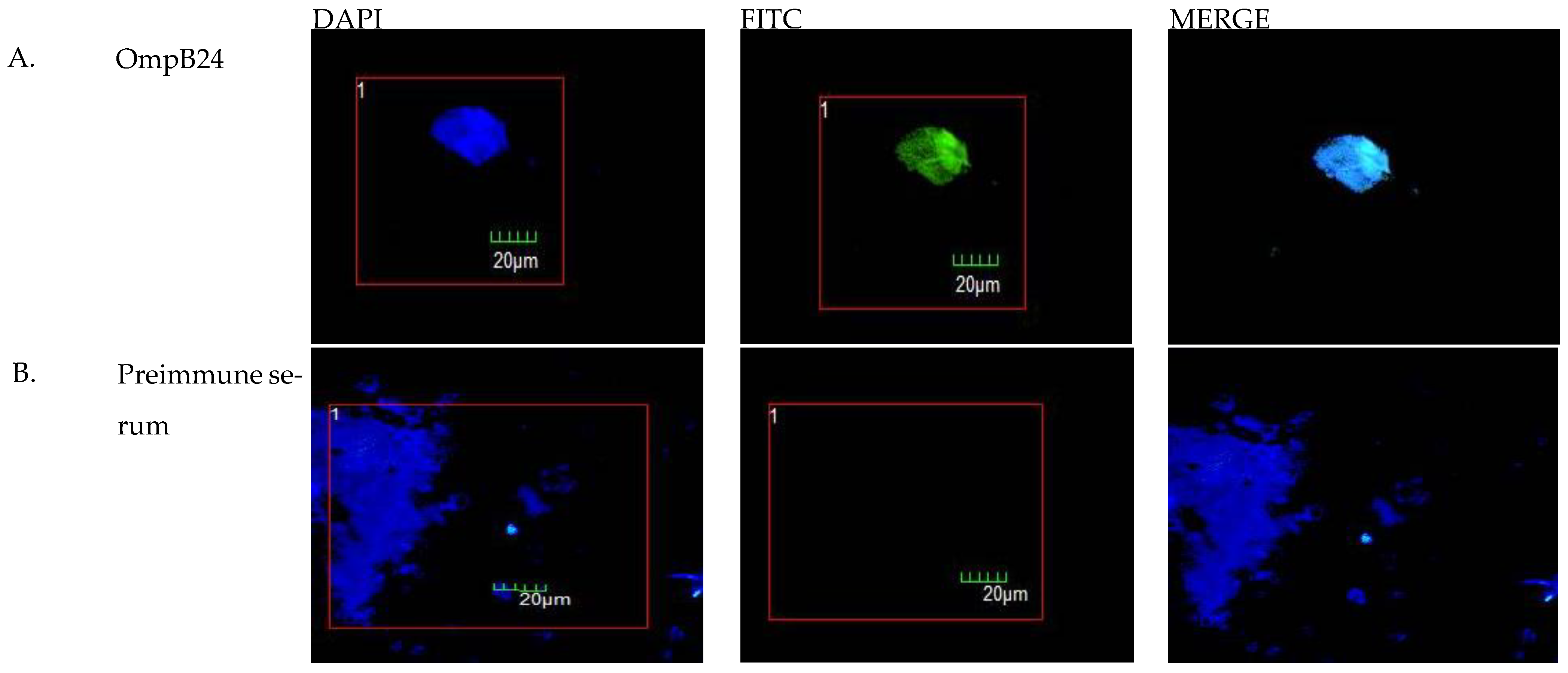
Transfection efficiency was assessed by indirect immunofluorescence (IFI) using a previously generated mouse antiserum against full-length recombinant OmpB-24 and a FITC-labeled anti-mouse secondary antibody. Viable transfected cells were identified by confocal microscopy using DAPI counterstaining and observing at least 5 different fields. The mean number of viable transfected cells was 90.86% vs. 0% for non-transfected cells (Figure 2), which was corroborated by fluorescence-activated cell sorting (FACS) using indirect staining against OmpB-24. (Data not shown).

3.3. Flow Cytometric Analysis

TCD4+INF-γ+ and TCD8+INF-γ+ Lymphocytes Induced by Human Macrophages Transfected with pVAX1-OmpB24

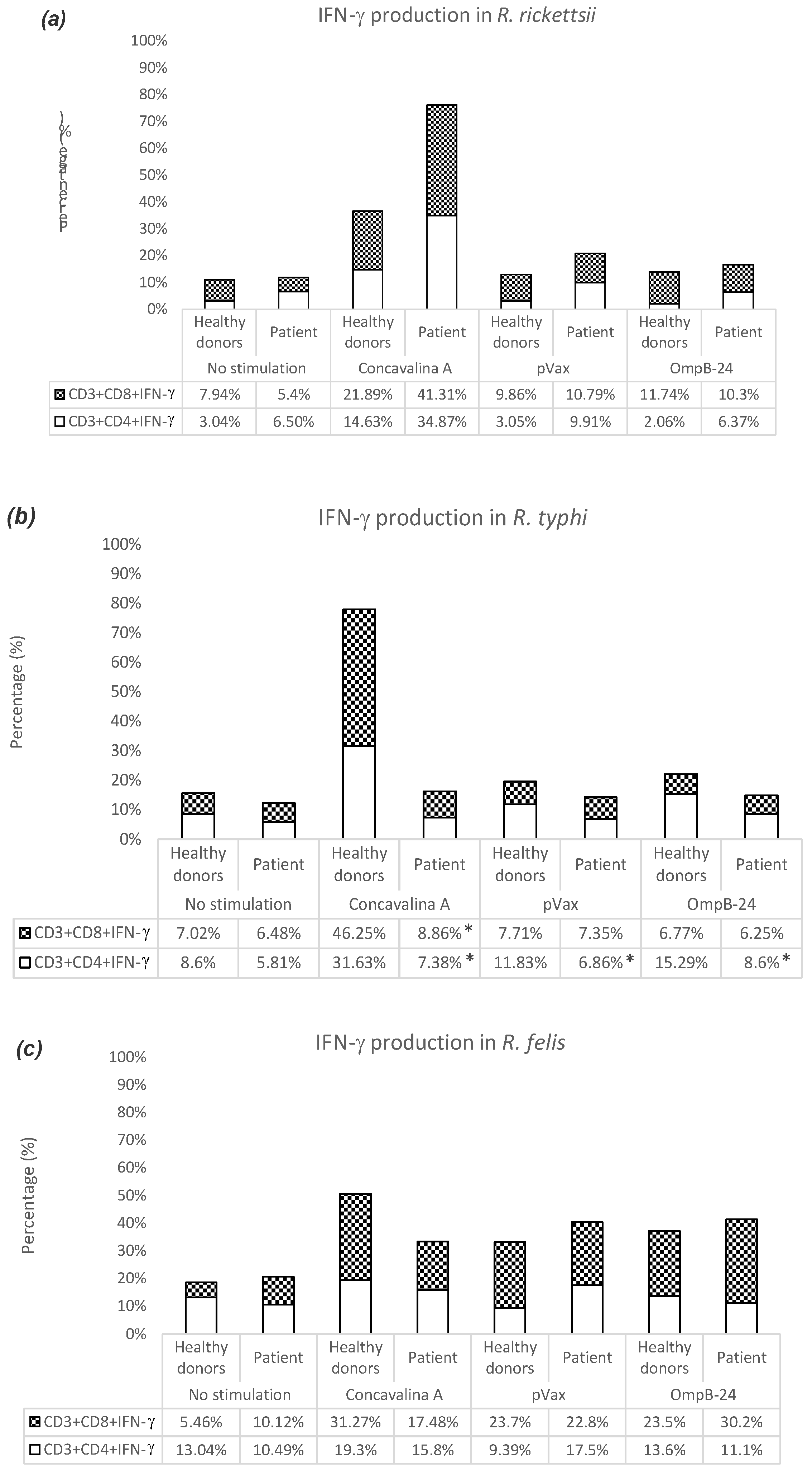
In the co-cultures of cells stimulated with pVAX1-OmpB24, from healthy donors and patients infected with R. rickettsii, higher percentages of TCD8+INF-γ+ were observed: 11.74% and 10.3%, respectively (Figure 3a). Meanwhile, the co-cultures of cells from healthy donors and patients infected with R. typhi, showed increases in TCD4+INF-γ+ of 15.29% and 8.6%, respectively, with significant differences between the two groups (Figure 3b). In the case of co-cultures of cells from healthy donors and patients infected with R. felis, figures of 23.5% and 30.2% respectively were recorded for the population of TCD8+INF-γ+ (Figure 3c).
When determining the antigenic presentation of transfected macrophages, higher percentages of CD40+, CD80+, HLA-I and HLA-II were observed in macrophages from healthy donors transfected with the OmpB-24 plasmid compared to those from patients infected with R. felis, R. typhi, and R. rickettsii. (Table 2). However, when evaluating the MFI (Mediam Fluorescence Intensity) in the group of patients with R. felis, a higher expression of CD40+, CD80+, HLA-I and HLA-II was observed compared with the controls (Table 3). Macrophages from patients stimulated with OmpB-24 showed a low percentage compared to healthy subjects, but had a higher IMF.
The results showed a higher percentage of CD14+CD80+ cells in the co-cultures of healthy donors and patients infected with R. rickettsii, with OmpB-24 stimulation and an increase in the CD14+HLA-I population. In contrast, in the co-cultures of healthy donors and patients infected with R. typhi, an increase in the percentage of CD14+CD40+ was observed, while the CD14+HLA-I population was increased with the stimulation of OmpB-24. In the co-cultures of healthy donors and patients infected with R. felis, an increase occured in the CD14+HLA-I population, compared to the CD14+HLA-II population. However, the difference between the two populations was minimal (Table 2).
The FMI results showed higher CD80 expression in CD14+ macrophages of healthy donors and patients infected with R. rickettsii compared to OmpB-24 stimulation, and higher expression of the HLA-I. On the other hand, in macrophages from healthy donors and patients infected with R. typhi, greater expression of CD40 was observed, while the expression of HLA-I was increased with OmpB-24 stimulation. In macrophages from healthy donors and patients infected with R. felis, an increased expression of CD80 and HLA-I was observed, and the expression of HLA-II was similar to HLA-I in the patient group (Table 3).

3.4. PD1 Expression in TCD4+ and TCD8+ Lymphocytes

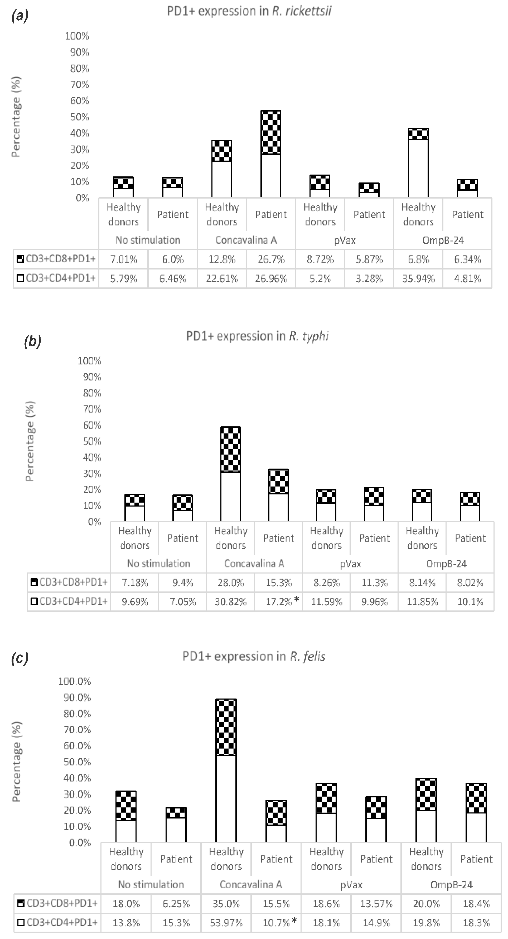
In co-cultures of cells stimulated with OmpB-24 from healthy donors, a greater population of TCD4+PD1+ was observed, with 35.94%, while in patients with R. rickettsii the highest percentage was found in TCD8+PD-1+, with 6.34% (Figure 4a). Cells from healthy donors and patients with R. typhi under the stimulation of OmpB-24 showed increases in TCD4+PD1+ of 11.85% and 10.1%, respectively (Figure 4b). In the case of healthy donors and patients with R. felis, there was an increase in the response of TCD8+PD1+. However, the difference in percentages was minimal compared to TCD4+PD1+ (Figure 4c). Interestingly, TCD4+PD1+ and TCD8+PD1+ cells from the group of patients with R. rickettsii and R. typhi in co-cultures with ConA showed an increase in the production of INF-γ.

4. Discussion

The social impact of the development of a vaccine against Rickettsia is important since there are currently no commercial methods for the diagnosis of rickettsiosis in the acute phase, which is found mainly in vulnerable communities away from specialized health services where patients are in critical conditions that eventually become fatal cases [20]. Furthermore, the initial clinical considerations are like other pathologies such as Dengue or chikungunya which delays the diagnosis and treatment [21].
The nature of rickettsial antigens that elicit humoral and cellular responses has been the subject of thorough investigations, primarily directed toward vaccine development. However, the immunodominant rickettsial antigens responsible for stimulating CD4+ and CD8+ cells have not yet been clearly identified and existing studies have been based on animal models. In recent years, ex vivo assays have been done using presenting cells (macrophages or DC) maintained in cultures with sensitized lymphocytes, and the determination of the cytokine response generated under this strategy has created high expectations, with high predictive value and a human profile, resulting in potential antigens to be used in the development of human vaccines [22,23]. The use of autologous systems has the advantage of matching the main HLA loci for cases in which antigenic recognition is required. In addition, it allows for the anticipated selection of cell clones. In the present study, an autologous system was used with macrophages transfected with the vaccine candidate Rickettsia pVAX1-OmpB24 [24,25,26].
Macrophage transfection by cationic liposomes is a highly effective technique which guarantees efficient delivery of DNA plasmids into cells. Transfection efficiency has been evaluated by methods such as fluorescent microscopy, gene reporter activity, PCR, flow cytometry or viability assays [27,28,29]. PCR, confocal microscopy, and flow cytometry were used to evaluate the transfection efficiency of this plasmid. Confocal microscopy and flow cytometry reported 90.8% of viable cells, underscoring a low percentage of cytotoxic damage to transfected cells. It is known that DNA liposome transfection enhances the generation of specific TCD8 lymphocytes through MHC-I presentation; however, there are several reports of TCD4 lymphocyte-oriented immune responses with these delivery systems [30,31]. It has been hypothesized that a common epitope for MHC I or II molecules could elicit a TCD4 or CD8 response, and that liposome transfection could trigger a cross presentation through both MHC molecules [32].
After antigenic contact, T lymphocytes can have different effector responses. IFN-γ is a cytokine produced mainly by CD4+ lymphocytes, NK cells and macrophages, and it is important for the control or elimination of intracellular pathogens [33]. In heterologous models, levels of IL-2 and IFN-γ related to CD4+ lymphocytes have been detected [17]. The present study shows an approach to the immune response generated by the plasmid pVAX1-OmpB24, using an autologous system of human cells. It was observed that CD4+IFN-γ+ lymphocytes are found in a higher percentage than CD8+IFN-γ+ lymphocytes, which suggests that the plasmid could be inducing a cellular immune response, similar to that obtained in murine models [14]. The presence of CD4+IFN-γ+ and CD8+IFN-γ+ lymphocytes are related to the cellular response and the elimination of intracellular pathogens. However, patients presented a different CD4+IFN-γ+ expression from that of healthy subjects, and a higher rate of CD4+ lymphocyte proliferation. Variables such as the age of the subjects can influence the immune response. Increased age is linked to immunosenescence, which translates to a decrease in virgin cells compared to existing memory cells [34]. This data shows that during a first infection the immune response is oriented towards cellular infection, for the elimination of the pathogen through mechanisms dependent on IFN-γ [1].
The immune system responds to rickettsial infection. However, there is a decrease in the percentage of CD4+ and CD45+ lymphocytes, as well as an increase in monocytes, which maintain high levels of TNF-α [35]. IFN-γ and TNF-α are cytokines that activate phagocytic cells such as macrophages, and the release of IL-12 influences the presentation of antigens to CD8+lymphocytes, inducing the destruction of cells infected by bacteria [36]. Meanwhile, the presence of IL-4 and IFN-γ favosr antigen presentation to CD4+ lymphocytes. It has been reported that infected endothelial cells can carry out autophagy and promote antigen presentation to B or CD4+ lymphocytes mediated by MHC-II [37,38].
After contact with antigens, T lymphocytes can have activating or inhibitory responses. PD1 (Programmed dead-1) is a molecule that inhibits some lymphocyte functions such as cytokine production, cell proliferation and cytotoxicity. The interaction of PD1 with its ligands PD-L1 and PD-L2 is important in the control of T lymphocyte activation in infectious diseases, since it limits the response of subpopulations of effector cells to avoid tissue damage [39]. The expression level of PD1 in T lymphocytes is a useful marker to establish the degree of cellular exhaustion, which seems to correlate with chronic infections [40]. It has been reported in Mycobacterium tuberculosis infections that PD1 has an important role in inhibiting the cellular response [41,42]. Cellular exhaustion could explain the results found in autologous co-cultures of healthy subjects and patients (post-R. rickettsii infection). In homologous co-cultures of healthy subjects and patients (post-R. felis infection) a negative regulation by PD1 was not seen, possibly explaining the low degree of pathogenicity that is observed in the clinical evolution of these patients. The result observed in co-cultures of healthy subjects and patients (post-R. typhi infection), could be associated with other molecules such as CTLA-4, as it occurs in infections with Leishmania donovani and Mycobacteria However, studies are required to prove this [43,44]. This preliminary data will allow further studies to determine the role of PD1 in Rickettsia infections.
As for the molecules related to antigenic presentation in macrophages, low expression of HLA-I in patients was observed. This could be due to the degradation of the plasmid containing pVAX1-OmpB24, which prevents its being processed by the cytosolic pathway, coupled with the time at which co-cultures were analyzed. It is important to consider these aspects and perform the expression kinetics of this molecule in subsequent studies. The high percentage of HLA-I expression in healthy subjects seems to indicate processing of the rickettsial protein by the cytosolic pathway in the first interaction with Rickettsia. No statistical differences were found in antigen presentation by HLA-II expression, probably due to the limited number of patients in the study [17,45,46,47]. The high expression levels of CD80 in macrophages of healthy subjects seems to correlate with the activation of lymphocytes, due to the interaction of CD40L/CD40, and an increase in cytokines such as IL-12 and IFN-γ that promote their differentiation [48]. The high expression of CD80 becomes detectable 24 h after activation, reaching maximum levels between 48 and 72 h. However, it will be necessary to perform an expression kinetics on this molecule in further studies [45,46,47,49]. In macrophages of healthy subjects, there seems to be a greater antigenic presentation mediated by HLA I molecules that allow lymphocytes to interact and express CD80 as a second activation signal, since in the absence of stimulation its expression is low or null [50]. Interaction between MHC-peptide-TLRs, co-stimulatory molecules (CD80 with its ligand CD28) or cytokines provides intracellular signals that allow the stimulation and differentiation of effector cells [50,51].
The differences found in the autologous co-cultures of patients who became ill with R. rickettsii, R. typhi and R. felis could be due to the pathogenicity of the species and the genetic diversity of the individuals. Knowledge regarding these immune responses is accumulating, and efforts have been undertaken to identify antigenic components of Rickettsiae that may be useful as a vaccine [1,3,52,53,54,55].
There are limitations in this study due to the sample size. Although the immune response shows an important behavior, the sample size prevents extrapolation to a larger population.

5. Conclusions

The results indicate that for the first time an autologous evaluation model from PBMC can be proposed as an alternative to the use of murine models. This data also suggests cross-reaction of T cells to conserved epitopes of OmpB from SFG and TG Rickettsiae, which makes this protein a promising vaccine candidate for vaccination against a broad range of Rickettsiae. Activation of TCD4+IFN-γ+ and TCD8+IFN-γ+ lymphocytes has been demonstrated in co-cultures of cells from patients infected with R. rickettsii, R. typhi and R. felis species, and in co-cell cultures from healthy donors, stimulated with the Rickettsia vaccine candidate pVAX1-OmpB24, respectively, together with the involvement of PD1 expression in the inhibition of TCD4+ and TCD8+ lymphocytes after Rickettsia infections

Author Contributions

Conceptualization: K.D.-R., L.D.-M., J.A.-L., C.P.-B. and J.B.-R.; data curation K.D.-R., L.D.-M., C.P.-B., J.B.-R. and J.M.; formal analysis: K.D.-R., L.D.-M., C.P.-B. and J.B.-R.; funding acquisition: K.D.-R.; investigation: K.D.-R., L.D.-M., C.P.-B., J.B.-R., J.M.-C., G.V.-P., J.M. and A.K.-G.; methodology: K.D.-R., L.D.-M., J.B.-R., G.V.-P., J.A.-L., A.K.-G., G.P.-L. and J.M.-C.; project administration: K.D.-R. and F.P.-M.; visualization: K.D.-R., L.D.-M., C.P.-B., C.L.-C. and J.M.; writing—original draft: K.D.-R., C.P.-B., L.D.-M. and J.M.; writing—review and editing: K.D.-R., C.P.-B., L.D.-M., J.M., J.M.-C. and O.E.Z. All authors have read and agreed to the published version of the manuscript.

Funding

This work was supported by CONACyT grant CONACYT FOSISS 261885.

Institutional Review Board Statement

The work was approved by the Research Ethics Committee of the Dr. Hideyo Noguchi Regional Research Center (Merida, Yucatan, Mexico) (CIE-05-2017).

Informed Consent Statement

Informed consent was obtained from all subjects involved in the study.

Data Availability Statement

The datasets generated and analysed during the present study are available from the corresponding author upon reasonable request.

Acknowledgments

The authors are grateful to Kitzia Valdez, Zulma Fonseca, Karina López, Raul Tello, Yuri Cupul, Harold Miranda, Karla Arroyo, Henrry Noh., Miguel Rosado Vallado and Liliana Villanueva Lizama for their technical support.

Conflicts of Interest

The authors declare no conflict of interest.

References

  1. Osterloh, A. The Neglected Challenge: Vaccination against Rickettsiae. PLoS Negl. Trop. Dis. 2020, 14, e0008704. [Google Scholar] [CrossRef]
  2. Moreira, J.; Bressan, C.S.; Brasil, P.; Siqueira, A.M. Epidemiology of Acute Febrile Illness in Latin America. Clin. Microbiol. Infect. 2018, 24, 827–835. [Google Scholar] [CrossRef] [Green Version]
  3. Blanton, L.S.; Wilson, N.M.; Quade, B.R.; Walker, D.H. Susceptibility of Rickettsia Rickettsii to Tigecycline in a Cell Culture Assay and Animal Model for Rocky Mountain Spotted Fever. Am. J. Trop. Med. Hyg. 2019, 101, 1091–1095. [Google Scholar] [CrossRef]
  4. Helgren, T.R.; Chen, C.; Wangtrakuldee, P.; Edwards, T.E.; Staker, B.L.; Abendroth, J.; Sankaran, B.; Housley, N.A.; Myler, P.J.; Audia, J.P.; et al. Rickettsia Prowazekii Methionine Aminopeptidase as a Promising Target for the Development of Antibacterial Agents. Bioorg. Med. Chem. 2017, 25, 813–824. [Google Scholar] [CrossRef] [PubMed] [Green Version]
  5. Gong, W.; Qi, Y.; Xiong, X.; Jiao, J.; Duan, C.; Wen, B. Rickettsia Rickettsii Outer Membrane Protein YbgF Induces Protective Immunity in C3H/HeN Mice. Hum. Vaccines Immunother. 2015, 11, 642–649. [Google Scholar] [CrossRef] [PubMed] [Green Version]
  6. Gong, W.; Xiong, X.; Qi, Y.; Jiao, J.; Duan, C.; Wen, B. Surface Protein Adr2 of Rickettsia Rickettsii Induced Protective Immunity against Rocky Mountain Spotted Fever in C3H/HeN Mice. Vaccine 2014, 32, 2027–2033. [Google Scholar] [CrossRef]
  7. Riley, S.P.; Cardwell, M.M.; Chan, Y.G.Y.; Pruneau, L.; Del Piero, F.; Martinez, J.J. Failure of a Heterologous Recombinant Sca5/OmpB Protein-Based Vaccine to Elicit Effective Protective Immunity against Rickettsia Rickettsii Infections in C3H/HeN Mice. Pathog. Dis. 2015, 73, ftv101. [Google Scholar] [CrossRef] [Green Version]
  8. Giordano, M. Patologia, parassitologia ed igiene del paesi caldi. J. Am. Med. Assoc. 1951, 145, 358–359. [Google Scholar] [CrossRef]
  9. Cox, H.R. Cultivation of Rickettsiae of the Rocky Mountain Spotted Fever, Typhus and Q Fever Groups in the Embryonic Tissues of Developing Chicks. Science 1941, 94, 399–403. [Google Scholar] [CrossRef]
  10. Kenyon, R.H.; Pedersen, C.E. Preparation of Rocky Mountain Spotted Fever Vaccine Suitable for Human Immunization. J. Clin. Microbiol. 1975, 1, 500–503. [Google Scholar] [CrossRef]
  11. Clements, M.L.; Wisseman, C.L.; Woodward, T.E.; Fiset, P.; Dumler, J.S.; McNamee, W.; Black, R.E.; Rooney, J.; Hughes, T.P.; Levine, M.M. Reactogenicity, Immunogenicity, and Efficacy of a Chick Embryo Cell-Derived Vaccine for Rocky Mountain Spotted Fever. J. Infect. Dis. 1983, 148, 922–930. [Google Scholar] [CrossRef] [PubMed]
  12. DuPont, H.L.; Hornick, R.B.; Dawkins, A.T.; Heiner, G.G.; Fabrikant, I.B.; Wisseman, C.L.; Woodward, T.E. Rocky Mountain Spotted Fever: A Comparative Study of the Active Immunity Induced by Inactivated and Viable Pathogenic Rickettsia Rickettsii. J. Infect. Dis. 1973, 128, 340–344. [Google Scholar] [CrossRef] [PubMed]
  13. Crocquet-Valdes, P.A.; Díaz-Montero, C.M.; Feng, H.M.; Li, H.; Barrett, A.D.; Walker, D.H. Immunization with a Portion of Rickettsial Outer Membrane Protein A Stimulates Protective Immunity against Spotted Fever Rickettsiosis. Vaccine 2001, 20, 979–988. [Google Scholar] [CrossRef] [PubMed]
  14. Wang, P.; Xiong, X.; Jiao, J.; Yang, X.; Jiang, Y.; Wen, B.; Gong, W. Th1 Epitope Peptides Induce Protective Immunity against Rickettsia Rickettsii Infection in C3H/HeN Mice. Vaccine 2017, 35, 7204–7212. [Google Scholar] [CrossRef] [PubMed]
  15. Moderzynski, K.; Heine, L.; Rauch, J.; Papp, S.; Kuehl, S.; Richardt, U.; Fleischer, B.; Osterloh, A. Cytotoxic Effector Functions of T Cells Are Not Required for Protective Immunity against Fatal Rickettsia Typhi Infection in a Murine Model of Infection: Role of TH1 and TH17 Cytokines in Protection and Pathology. PLoS Negl. Trop. Dis. 2017, 11, e0005404. [Google Scholar] [CrossRef] [Green Version]
  16. Meng, Y.; Xiong, X.; Qi, Y.; Duan, C.; Gong, W.; Jiao, J.; Wen, B. Protective Immunity against Rickettsia Heilongjiangensis in a C3H/HeN Mouse Model Mediated by Outer Membrane Protein B-Pulsed Dendritic Cells. Sci. China Life Sci. 2015, 58, 287–296. [Google Scholar] [CrossRef] [PubMed] [Green Version]
  17. Dzul-Rosado, K.; Balam-Romero, J.; Valencia-Pacheco, G.; Lugo-Caballero, C.; Arias-León, J.; Peniche-Lara, G.; Zavala-Castro, J. Immunogenicity of OmpA and OmpB Antigens from Rickettsia Rickettsii on Mononuclear Cells from Rickettsia Positive Mexican Patients. J. Vector Borne Dis. 2017, 54, 317. [Google Scholar] [CrossRef]
  18. Blair, P.J.; Jiang, J.; Schoeler, G.B.; Moron, C.; Anaya, E.; Cespedes, M.; Cruz, C.; Felices, V.; Guevara, C.; Mendoza, L.; et al. Characterization of Spotted Fever Group Rickettsiae in Flea and Tick Specimens from Northern Peru. J. Clin. Microbiol. 2004, 42, 4961–4967. [Google Scholar] [CrossRef] [Green Version]
  19. Choi, Y.-J.; Jang, W.-J.; Kim, J.-H.; Ryu, J.-S.; Lee, S.-H.; Park, K.-H.; Paik, H.-S.; Koh, Y.-S.; Choi, M.-S.; Kim, I.-S. Spotted Fever Group and Typhus Group Rickettsioses in Humans, South Korea. Emerg. Infect. Dis. 2005, 11, 237–244. [Google Scholar] [CrossRef]
  20. Zavala-Castro, J.E.; Zavala-Velázquez, J.E.; Walker, D.H.; Arcila, E.E.R.; Laviada-Molina, H.; Olano, J.P.; Ruiz-Sosa, J.A.; Small, M.A.; Dzul-Rosado, K.R. Fatal Human Infection with Rickettsia Rickettsii, Yucatán, Mexico. Emerg. Infect. Dis. 2006, 12, 672–674. [Google Scholar] [CrossRef]
  21. Zavala-Velazquez, J.E.; Ruiz-Sosa, J.; Vado-Solis, I.; Billings, A.N.; Walker, D.H. Serologic Study of the Prevalence of Rickettsiosis in Yucatán: Evidence for a Prevalent Spotted Fever Group Rickettsiosis. Am. J. Trop. Med. Hyg. 1999, 61, 405–408. [Google Scholar] [CrossRef]
  22. Álvarez de Haro, N. Cultivos Celulares Para el Desarrollo de Vacunas DNA Frente a Virus de Peces Utilizando el Modelo de Trucha Arco Iris/Rhabdovirus de la Septicemia Hemorrágica Vírica (VHSV). Ph.D. Thesis, Universidad de León, León, Spain, 2015. [Google Scholar] [CrossRef]
  23. Li, Z.; Díaz-Montero, C.M.; Valbuena, G.; Yu, X.-J.; Olano, J.P.; Feng, H.-M.; Walker, D.H. Identification of CD8 T-Lymphocyte Epitopes in OmpB of Rickettsia Conorii. Infect. Immun. 2003, 71, 3920–3926. [Google Scholar] [CrossRef] [PubMed] [Green Version]
  24. Dolskiy, A.A.; Grishchenko, I.V.; Yudkin, D.V. Cell Cultures for Virology: Usability, Advantages, and Prospects. Int. J. Mol. Sci. 2020, 21, 7978. [Google Scholar] [CrossRef] [PubMed]
  25. Okita, K.; Matsumura, Y.; Sato, Y.; Okada, A.; Morizane, A.; Okamoto, S.; Hong, H.; Nakagawa, M.; Tanabe, K.; Tezuka, K.; et al. A More Efficient Method to Generate Integration-Free Human IPS Cells. Nat. Methods 2011, 8, 409–412. [Google Scholar] [CrossRef] [PubMed] [Green Version]
  26. Ventura, C.; Bisceglia, H.; Girerd-Chambaz, Y.; Burdin, N.; Chaux, P. HLA-DR and HLA-DP Restricted Epitopes from Human Cytomegalovirus Glycoprotein B Recognized by CD4+ T-Cell Clones from Chronically Infected Individuals. J. Clin. Immunol. 2012, 32, 1305. [Google Scholar] [CrossRef] [Green Version]
  27. Dokka, S.; Toledo, D.; Shi, X.; Ye, J.; Rojanasakul, Y. High-Efficiency Gene Transfection of Macrophages by Lipoplexes. Int. J. Pharm. 2000, 206, 97–104. [Google Scholar] [CrossRef]
  28. Osorio, J.S.; Bionaz, M. Plasmid Transfection in Bovine Cells: Optimization Using a Realtime Monitoring of Green Fluorescent Protein and Effect on Gene Reporter Assay. Gene 2017, 626, 200–208. [Google Scholar] [CrossRef]
  29. Duarte, F.B.; Brígido, M.d.M.; Melo, E.d.O.; Báo, S.N.; Martins, C.F. Strategies for Transfection of Bovine Mesenchymal Stem Cells with PBC1-Anti-CD3 Vector. Anim. Biotechnol. 2022, 33, 1014–1024. [Google Scholar] [CrossRef]
  30. Mbawuike, I.N.; Zhang, Y.; Wang, Y.; Song, L. Cationic Liposome-Mediated Enhanced Generation of Human HLA-Restricted RSV-Specific CD8+ CTL+. J. Clin. Immunol. 2002, 22, 164–175. [Google Scholar] [CrossRef]
  31. Schwendener, R.A. Liposomes as Vaccine Delivery Systems: A Review of the Recent Advances. Ther. Adv. Vaccines 2014, 2, 159–182. [Google Scholar] [CrossRef]
  32. U’Ren, L.; Kedl, R.; Dow, S. Vaccination with Liposome--DNA Complexes Elicits Enhanced Antitumor Immunity. Cancer Gene Ther. 2006, 13, 1033–1044. [Google Scholar] [CrossRef] [PubMed] [Green Version]
  33. Walker, D.H.; Popov, V.L.; Feng, H.M. Establishment of a Novel Endothelial Target Mouse Model of a Typhus Group Rickettsiosis: Evidence for Critical Roles for Gamma Interferon and CD8 T Lymphocytes. Lab. Investig. J. Tech. Methods Pathol. 2000, 80, 1361–1372. [Google Scholar] [CrossRef] [Green Version]
  34. Gruver, A.; Hudson, L.; Sempowski, G. Immunosenescence of Ageing. J. Pathol. 2007, 211, 144–156. [Google Scholar] [CrossRef]
  35. Zavala, J.; Ruiz, A.; Zavala, J. Las Rickettsias Del Grupo de Las Fiebres Manchadas: Respuesta Inmune y Sus Proteínas Inmunodominantes. Rev. Médica Chile 2004, 132, 381–387. [Google Scholar] [CrossRef] [Green Version]
  36. Mansueto, P.; Vitale, G.; Cascio, A.; Seidita, A.; Pepe, I.; Carroccio, A.; di Rosa, S.; Rini, G.B.; Cillari, E.; Walker, D.H. New Insight into Immunity and Immunopathology of Rickettsial Diseases. Clin. Dev. Immunol. 2011, 2012, e967852. [Google Scholar] [CrossRef] [PubMed]
  37. Secretaría de Salud Guía de Práctica Clínica SS-595-13 “Prevención, Diagnóstico y Tratamiento de la Fiebre Manchada por Rickettsia Rickettsii en Población Pediátrica y Adulta en el Primer y Segundo Nivel de Atención”. Centro Nacional de Excelencia Tecnologica en Salud. Mexico: Secretaria de Salud. 2013. Available online: https://www.actuamed.com.mx/informacion-medica/prevencion-diagnostico-y-tratamiento-de-fiebre-manchada-por-rickettsia-rickettsii (accessed on 14 December 2022).
  38. Ávila-Aguirre, L.M.; Martínez-Díaz, H.C.; Betancourt-Ruiz, P.; Hidalgo, M. 12. Historia de la rickettsiosis en Colombia. Fondo Editor. Biogénesis 2020, 1, 218–239. [Google Scholar]
  39. Sampedro-Núñez, M.; Serrano-Somavilla, A.; Adrados, M.; Cameselle-Teijeiro, J.M.; Blanco-Carrera, C.; Cabezas-Agricola, J.M.; Martínez-Hernández, R.; Martín-Pérez, E.; Muñoz de Nova, J.L.; Díaz, J.Á.; et al. Analysis of Expression of the PD-1/PD-L1 Immune Checkpoint System and Its Prognostic Impact in Gastroenteropancreatic Neuroendocrine Tumors. Sci. Rep. 2018, 8, 17812. [Google Scholar] [CrossRef] [PubMed] [Green Version]
  40. Wykes, M.N.; Lewin, S.R. Immune Checkpoint Blockade in Infectious Diseases. Nat. Rev. Immunol. 2018, 18, 91–104. [Google Scholar] [CrossRef]
  41. Jurado, J.O.; Alvarez, I.B.; Pasquinelli, V.; Martínez, G.J.; Quiroga, M.F.; Abbate, E.; Musella, R.M.; Chuluyan, H.E.; García, V.E. Programmed Death (PD)-1:PD-Ligand 1/PD-Ligand 2 Pathway Inhibits T Cell Effector Functions during Human Tuberculosis. J. Immunol. 2008, 181, 116–125. [Google Scholar] [CrossRef] [Green Version]
  42. Shen, L.; Gao, Y.; Liu, Y.; Zhang, B.; Liu, Q.; Wu, J.; Fan, L.; Ou, Q.; Zhang, W.; Shao, L. PD-1/PD-L Pathway Inhibits M.Tb-Specific CD4+ T-Cell Functions and Phagocytosis of Macrophages in Active Tuberculosis. Sci. Rep. 2016, 6, 38362. [Google Scholar] [CrossRef]
  43. Chang, T.T.; Kuchroo, V.K.; Sharpe, A.H. Role of the B7-CD28/CTLA-4 Pathway in Autoimmune Disease. Curr. Dir. Autoimmun. 2002, 5, 113–130. [Google Scholar] [CrossRef] [PubMed]
  44. Murphy, M.L.; Cotterell, S.E.; Gorak, P.M.; Engwerda, C.R.; Kaye, P.M. Blockade of CTLA-4 Enhances Host Resistance to the Intracellular Pathogen, Leishmania Donovani. J. Immunol. Baltim. 1998, 161, 4153–4160. [Google Scholar] [CrossRef]
  45. Gazi, M.; Caro-Gomez, E.; Goez, Y.; Cespedes, M.A.; Hidalgo, M.; Correa, P.; Valbuena, G. Discovery of a Protective Rickettsia Prowazekii Antigen Recognized by CD8+ T Cells, RP884, Using an In Vivo Screening Platform. PLoS ONE 2013, 8, e76253. [Google Scholar] [CrossRef] [Green Version]
  46. Nair, S.; Archer, G.E.; Tedder, T.F. Isolation and Generation of Human Dendritic Cells. Curr. Protoc. Immunol. 2012, 99, 7–32. [Google Scholar] [CrossRef] [PubMed] [Green Version]
  47. Vázquez, M.B.; Sureda, M.; Rebollo, J. Células dendríticas I: Aspectos básicos de su biología y funciones. Inmunología 2012, 31, 21–30. [Google Scholar] [CrossRef]
  48. Elgueta, R.; Benson, M.J.; de Vries, V.C.; Wasiuk, A.; Guo, Y.; Noelle, R.J. Molecular Mechanism and Function of CD40/CD40L Engagement in the Immune System. Immunol. Rev. 2009, 229, 152–172. [Google Scholar] [CrossRef] [PubMed] [Green Version]
  49. la Mora, J.D.-D.; Licona-Enríquez, J.D.; Leyva-Gastélum, M.; Mora, D.D.-D.L.; Rascón-Alcantar, A.; Álvarez-Hernández, G. A Fatal Case Series of Rocky Mountain Spotted Fever in Sonora, México. Biomédica 2018, 38, 69–76. [Google Scholar] [CrossRef] [PubMed] [Green Version]
  50. Abbas, A.K.; Lichtman, A.H.; Pillai, S. Inmunología Celular y Molecular; Octava; Elsevier: Barcelona, Spain, 2015; ISBN 978-84-9022-909-5. [Google Scholar]
  51. Vasilevko, V.; Ghochikyan, A.; Holterman, M.J.; Agadjanyan, M.G. CD80 (B7-1) and CD86 (B7-2) Are Functionally Equivalent in the Initiation and Maintenance of CD4+ T-Cell Proliferation after Activation with Suboptimal Doses of PHA. DNA Cell Biol. 2002, 21, 137–149. [Google Scholar] [CrossRef]
  52. Galletti, M.F.B.M.; Fujita, A.; Rosa, R.D.; Martins, L.A.; Soares, H.S.; Labruna, M.B.; Daffre, S.; Fogaça, A.C. Virulence Genes of Rickettsia Rickettsii Are Differentially Modulated by Either Temperature Upshift or Blood-Feeding in Tick Midgut and Salivary Glands. Parasit. Vectors 2016, 9, 331. [Google Scholar] [CrossRef] [Green Version]
  53. Hoan, N.X.; Khuyen, N.; Giang, D.P.; Binh, M.T.; Toan, N.L.; Anh, D.T.; Trung, N.T.; Bang, M.H.; Meyer, C.G.; Velavan, T.P.; et al. Vitamin D Receptor ApaI Polymorphism Associated with Progression of Liver Disease in Vietnamese Patients Chronically Infected with Hepatitis B Virus. BMC Med. Genet. 2019, 20, 201. [Google Scholar] [CrossRef]
  54. Niebylski, M.L.; Peacock, M.G.; Schwan, T.G. Lethal Effect of Rickettsia Rickettsii on Its Tick Vector (Dermacentor andersoni). Appl. Environ. Microbiol. 1999, 65, 773–778. [Google Scholar] [CrossRef] [PubMed] [Green Version]
  55. Li, J.; Li, H.; Li, Z.; Liu, Y.; Gao, J.; Zeng, X.; Gou, C.; Zhu, X.; Guo, X.; Pan, L.; et al. Association of Taq I T/C and Fok I C/T polymorphisms of vitamin D receptor gene with outcome of hepatitis B virus infection. Zhonghua Yi Xue Za Zhi 2006, 86, 1952–1956. [Google Scholar] [PubMed]
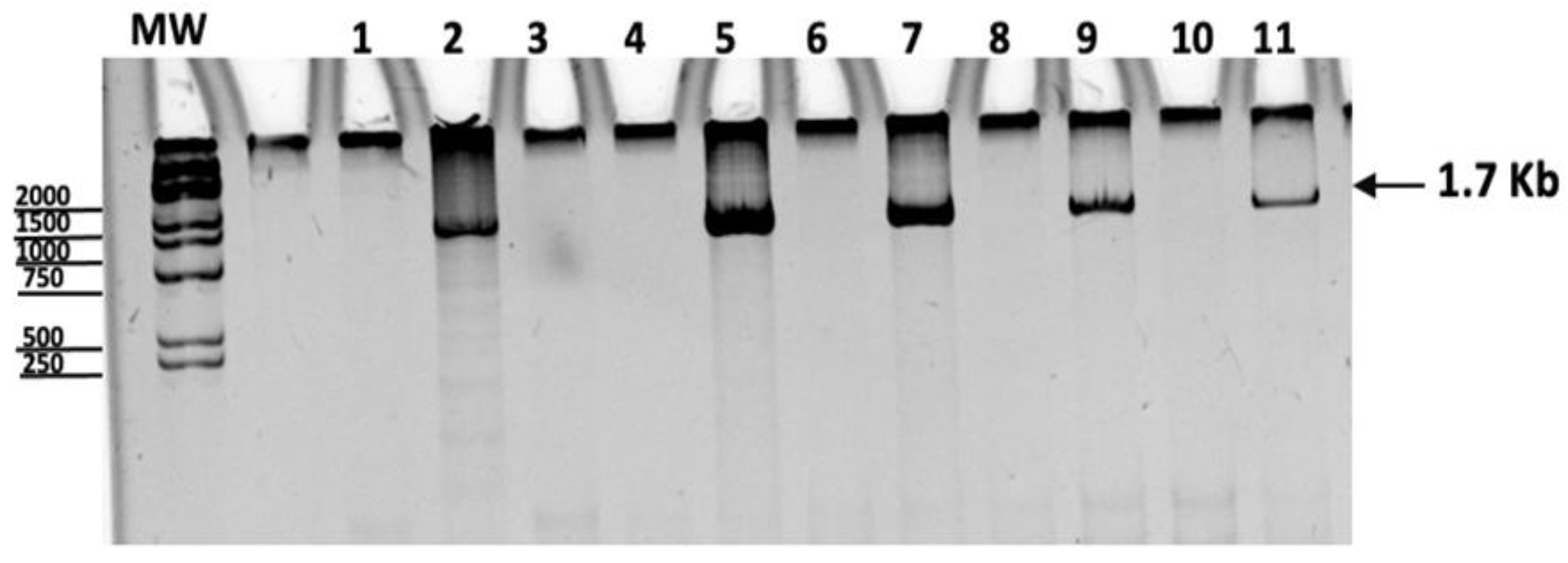
Figure 1. Amplification products by quantitative PCR of macrophages transfected with the plasmid at 24 h. Lane 1: negative control, Lane 2: positive control (pVAX-rOmpB-24 plasmid), Lane 3: non-transfected macrophages. Lanes 4 and 5: Macrophages 12 h after transfection with pVax (4) or OmpB-24 (5), Lanes 6 and 7: Macrophages 24 h after transfection with pVax (6) or OmpB-24 (7), Lanes 8 and 9: Macrophages 48 h after transfection with pVax (8) or OmpB-24 (9), Lanes 10 and 11: Macrophages 72 h after transfection with pVax (10) or OmpB-24 (11).
Vaccines 11 00173 g001
Figure 2. Expression of OmpB-24 in macrophages transfected with pVaX-rOmpB-24. Representative images obtained by confocal microscopy of a single macrophage (20 µm) 24 h after transfection are shown, including (A) OmpB-24 immune serum and (B) Preimmune serum. Staining for viability (DAPI) and OmpB-24 expression (FITC) are shown in the merge row. image was observed at 40× (LSMC FV-1000, Olympus, Tokyo, Japan).
Vaccines 11 00173 g002
Figure 3. Population of T lymphocytes producing INF-γ. Percentage of TCD4+INF-γ+ and TCD8+INF-γ+ in healthy donors and patients post-infection with R. rickettsii (a), R. typhi (b) and R. felis (c), stimulated or not with ConA, pVax and OmpB -24 for 24 h. Values show the average of 5 samples. (*) p-value ≤ 0.05 calculated with the Student’s t-test.
Vaccines 11 00173 g003
Figure 4. PD1 expression in T lymphocyte subpopulations. Percentage of TCD4+PD1+ and TCD8+PD1+ cells in healthy donors and patients post-infection with R. rickettsii (a), R. typhi (b) and R. felis (c), stimulated or not with ConA, pVax and OmpB- 24 for 24 h. Values show the average of 5 samples. (*) p-value ≤ 0.05 calculated with the Student’s t-test.
Vaccines 11 00173 g004
Table 1. Clinical characteristics of the patients.
AgeGenderStateSymptom Onset DateClinical Signs
SpeciesFeverExanthemaHeadacheArthralgias/MyalgiasExposure
26FBaja California24/11/16R. rickettsii+-++Tick
29MBaja California31/08/16R. rickettsii+-++-
54MBaja California18/08/16R. rickettsii+-++Tick
25MBaja California-R. rickettsii++++Tick
51FBaja California21/03/17R. rickettsii++++-
33FYucatan03/10/15R. typhi+-++Fleas
31FYucatan09/11/16R. typhi+++-
39MYucatan14/10/16R. typhi+-++Fleas and Ticks
28MYucatan-R. typhi+---Tick
32MYucatan-R. typhi+----
72MYucatan01/08/13R. felis++-+Tick
43MYucatan01/04/13R. felis+-++Tick
18FYucatan01/05/06R. felis+---Tick
16FYucatan01/11/13R. felis++--Tick
Table 2. Percentage of macrophages (CD14+) expressing CD40+, CD80+, HLA-I and HLA-II. x - refers to the statistical mean.
Macrophage Markers (%)No StimulationStimulation
Concavalina ApVaxOmpB-24
x -
Healthy DonorsPatientHealthy DonorsPatientHealthy DonorsPatientHealthy DonorsPatient
R. rickettsiiCD14+CD40+25.3032.00 a52.8011.9215.2017.70 a9.9712.50 a
CD14+CD80+32.0023.9039.4018.4027.8048.10 a37.3022.40
CD14+HLA-I97.8097.0095.4097.00 a99.2094.1098.1085.50
CD14+HLA-II46.1034.3079.2021.9037.9028.1037.3026.60
R. typhiCD14+CD40+32.7022.1015.4032.20 a32.9020.5039.0018.30
CD14+CD80+16.7016.3011.5021.00 a23.0019.1022.809.91
CD14+HLA-I99.2091.8093.1096.40 a93.7092.2097.0097.30 a
CD14+HLA-II48.0025.2027.7036.10 a40.1028.3053.2033.50
R. felisCD14+CD40+10.406.7023.704.7018.905.2018.507.30
CD14+CD80+16.603.2027.709.2030.409.2025.909.40
CD14+HLA-I98.9096.3097.0096.8097.4097.70 a97.2097.70 a
CD14+HLA-II28.9066.80 a33.0062.10 a44.6092.50 a49.2094.60 a
(a) Higher value observed in patients when compared to their respective control.
Table 3. Mean Fluorescence Intensity (MFI) of CD40, CD80, MHC-I, and MHC-II in macrophages (CD14+). x - refers to the statistical mean.
(MFI)No StimulationStimulation
Concanavalin ApVaxOmpB-24
x -
Healthy DonorsPatientHealthy DonorsPatientHealthy DonorsPatientHealthy DonorsPatient
R. rickettsiiCD14+CD40+46622514479129754008352545372871
CD14+CD80+20,569247629,801261941,331227529,8013332
CD14+HLA-I51,05453,824 a62,13365,078 a58,74855,55238,63042,342 a
CD14+HLA-II10,110811750392000600012,000 a10,0008000
R. typhiCD14+CD40+12705136 a21153991 a16334924 a51915439 a
CD14+CD80+3132277118394316 a24004352 a19464325 a
CD14+HLA-I80,09255,41734,83065,897 a77,77676,40182,27673,816
CD14+HLA-II76979795 a332510,248 a697211,197 a801014,788 a
R. felisCD14+CD40+5125189,210 a2799465,000 a6531340,706 a147,873321,110 a
CD14+CD80+222347,918 a227049,923 a233295,271 a2917125,643 a
CD14+HLA-I47,42975,097 a51,13265,897 a62,588165,240 a58,60674,065 a
CD14+HLA-II915672,806 a764678,536 a12,69876,929 a15,17178,807 a
(a) Higher value observed in patients when compared to their respective control.
Disclaimer/Publisher’s Note: The statements, opinions and data contained in all publications are solely those of the individual author(s) and contributor(s) and not of MDPI and/or the editor(s). MDPI and/or the editor(s) disclaim responsibility for any injury to people or property resulting from any ideas, methods, instructions or products referred to in the content.