
Suppressing the Substance P-NK1R Signalling Protects Mice against Sepsis-Associated Acute Inflammatory Injury and Ferroptosis in the Liver and Lungs


Abstract
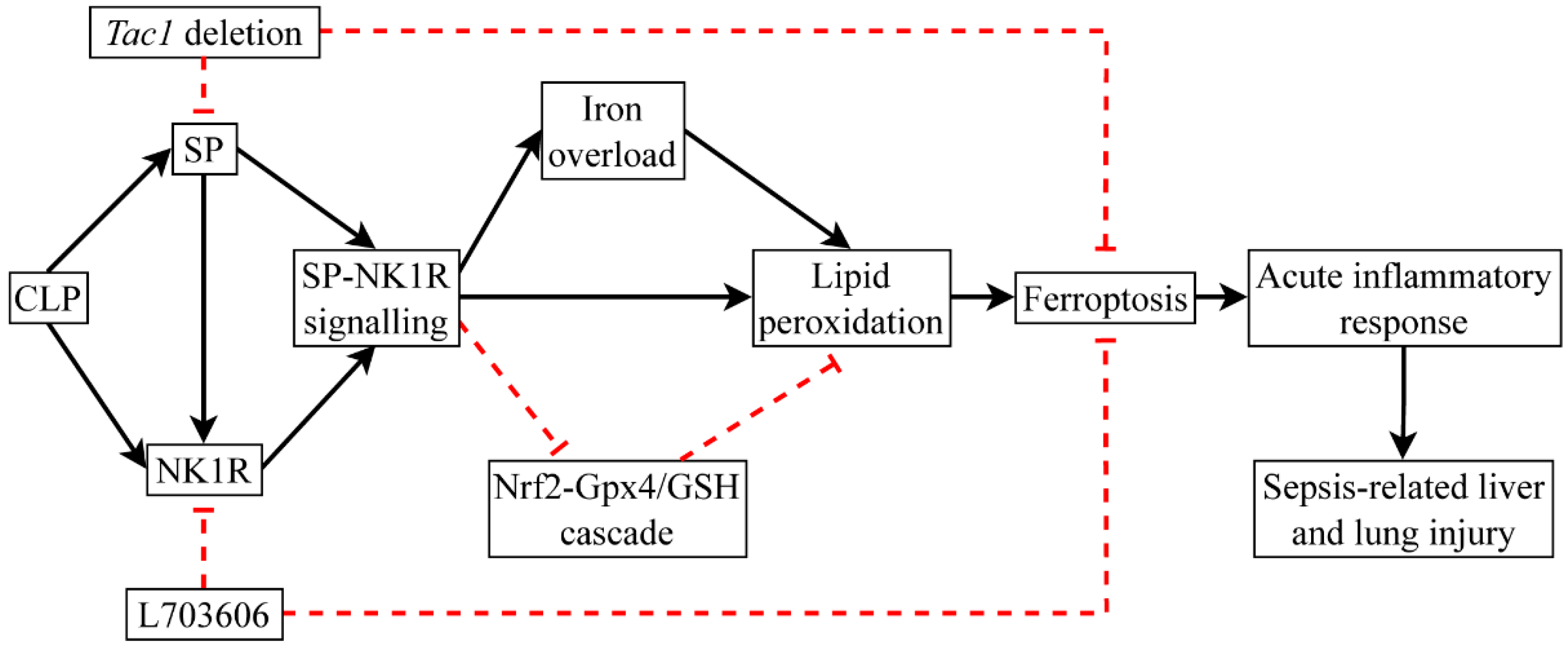
1. Introduction
2. Materials and Methods
2.1. Group Setting
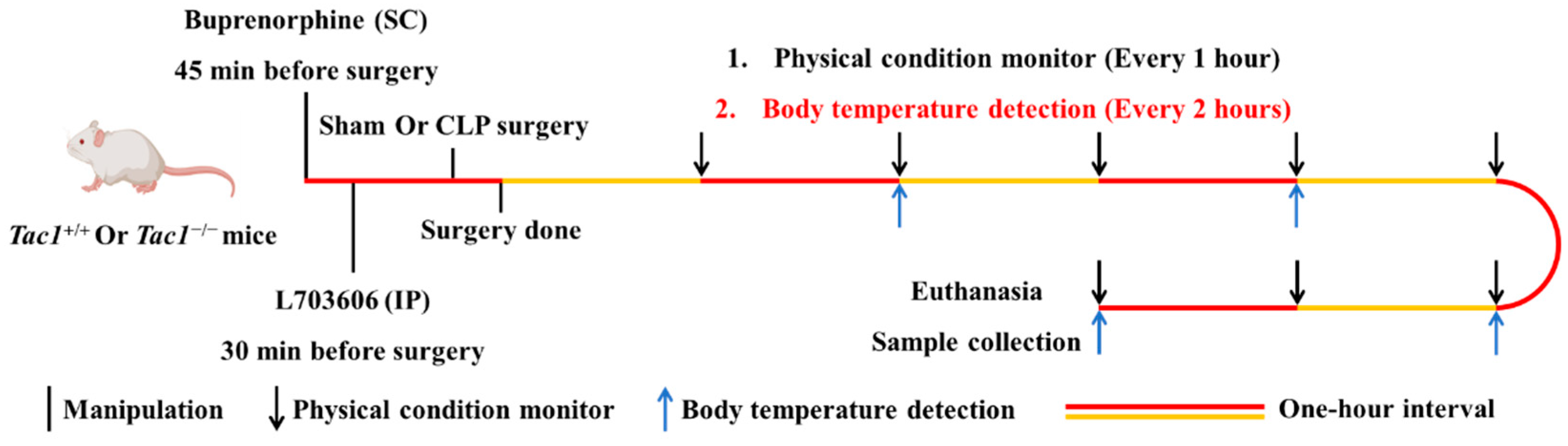
2.2. Sepsis Establishment, Manipulations, and Sample Collection in Mice
2.3. Evaluation of Sepsis Severity in Mice
2.4. Measurement of Lung W/D Ratio
2.5. Determination of SP in Tissues
2.6. Measurement of Pro-Inflammatory Cytokines and Chemokines in Tissues
2.7. Western Blotting
2.8. Immunohistochemistry Staining
2.9. Measurement of Iron, Malondialdehyde (MDA), and GSH in Tissues
2.10. Histological Analysis
2.11. Statistical Analysis
3. Results
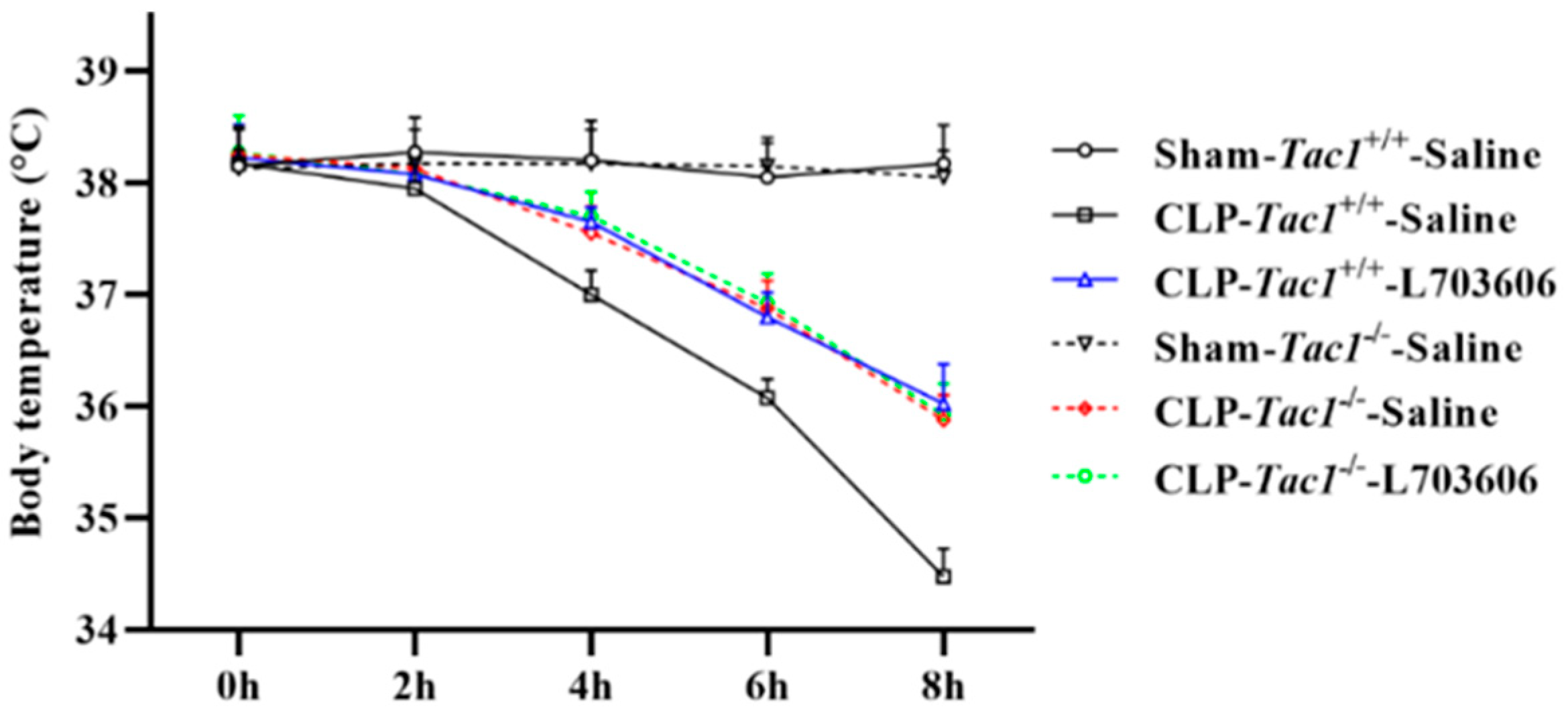
3.1. Suppressing SP-NK1R Signalling Improved the Physical Condition of Mice following CLP Surgery-Induced Sepsis
3.2. Concentration of SP in the Liver and Lungs in Mice
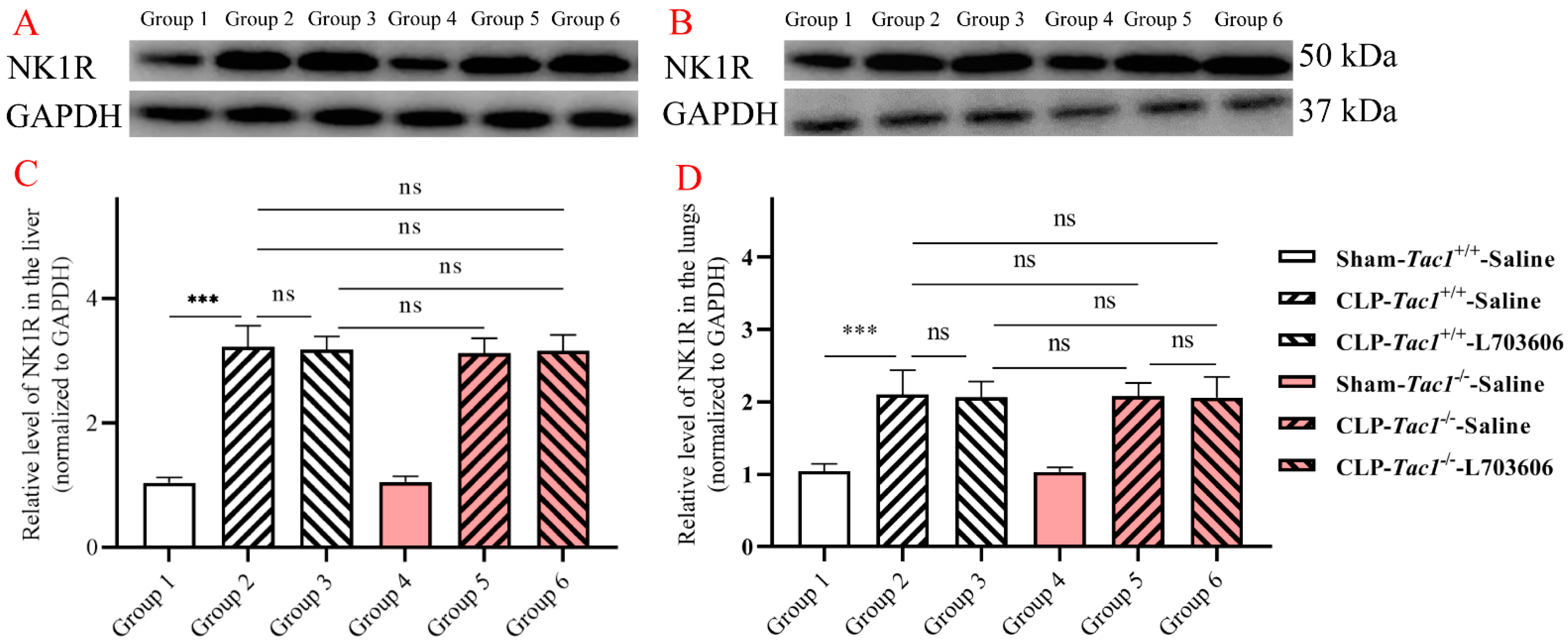
3.3. Expression of NK1R in the Liver and Lungs in Mice
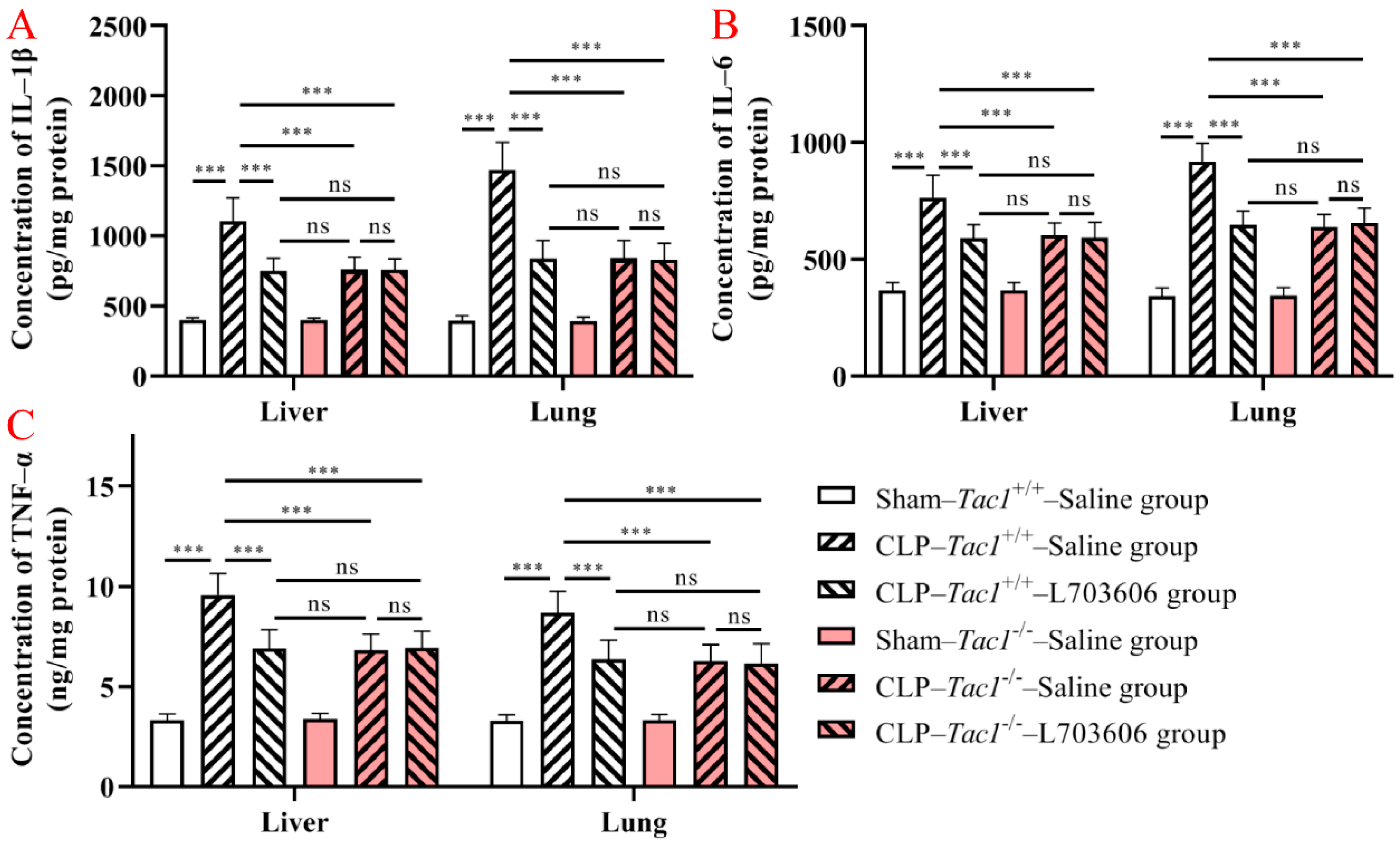
3.4. Suppressing SP-NK1R Signalling Attenuated the Increase in the Concentrations of IL-1β, IL-6, and TNF-α in the Liver and Lungs in Mice following CLP-Surgery-Induced Sepsis
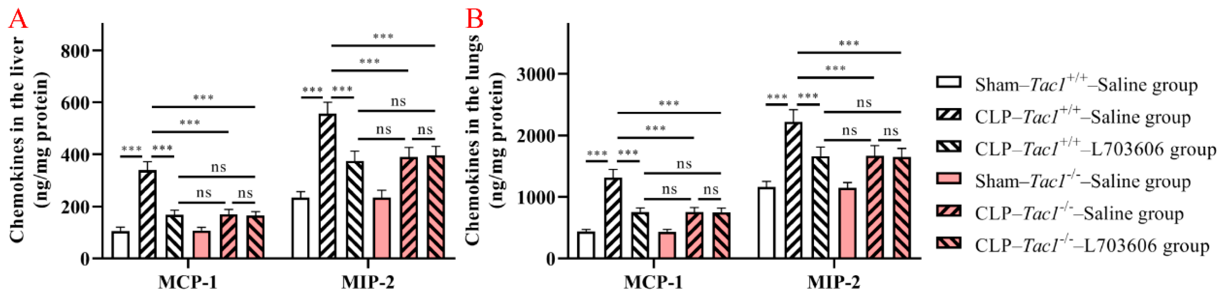
3.5. Suppressing SP-NK1R Signalling Attenuated the Increase in the Concentrations of MCP-1 and MIP-2 in the Liver and Lungs in Mice following CLP-Surgery-Induced Sepsis
3.6. Suppressing SP-NK1R Signalling Attenuated the Increase in the Concentrations of Iron in the Liver and Lungs in Mice following CLP-Surgery-Induced Sepsis
3.7. Suppressing SP-NK1R Signalling Attenuated the Increase in the Concentration of MDA in the Liver and Lungs in Mice following CLP-Surgery-Induced Sepsis
3.8. Suppressing SP-NK1R Signalling Attenuated the Decrease in the Concentration of GSH in the Liver and Lungs in Mice following CLP-Surgery-Induced Sepsis
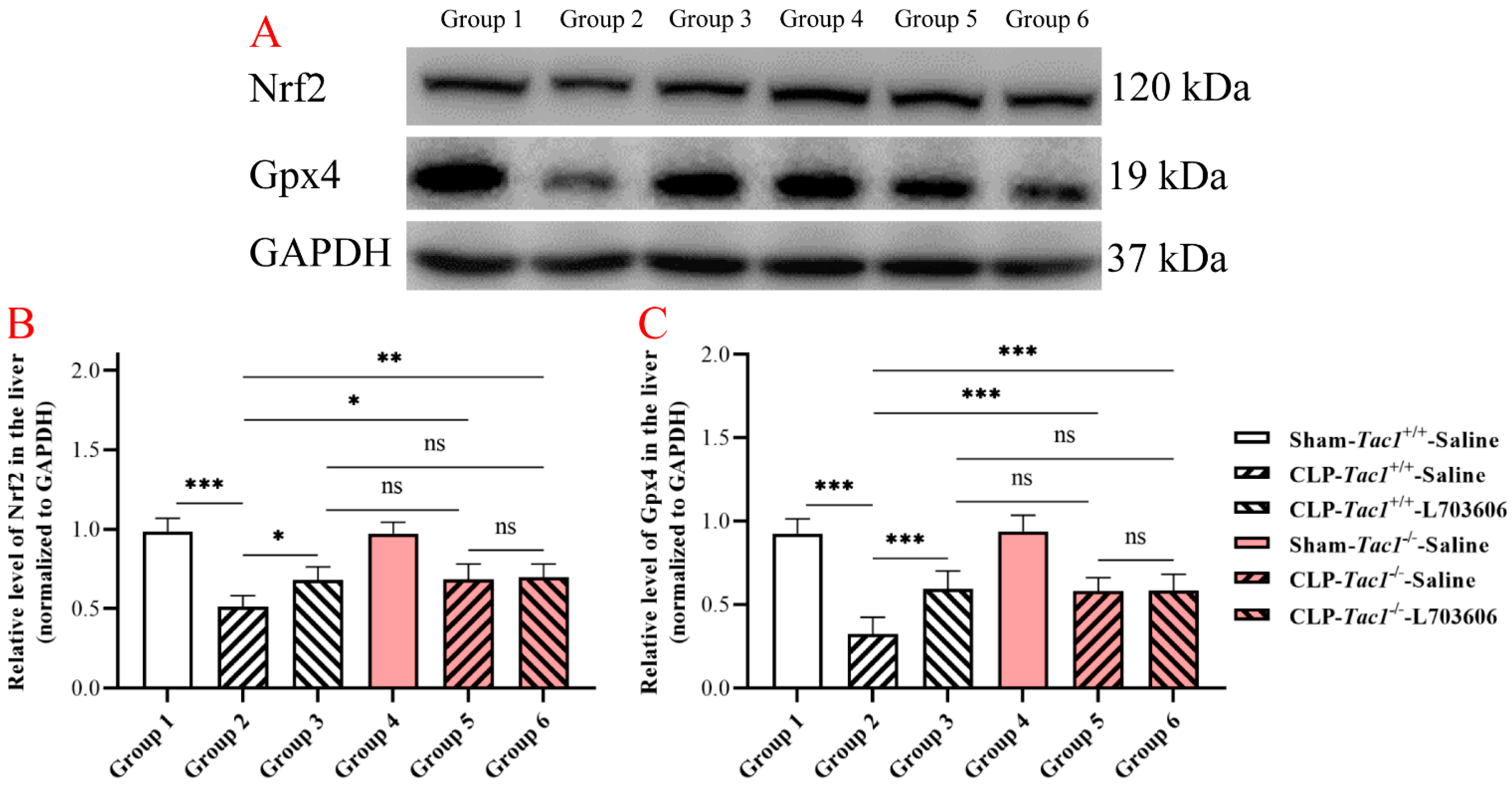
3.9. Suppressing SP-NK1R Signalling Attenuated the Decrease in the Expressions of Nrf2 and Gpx4 in the Liver in Mice following CLP-Surgery-Induced Sepsis
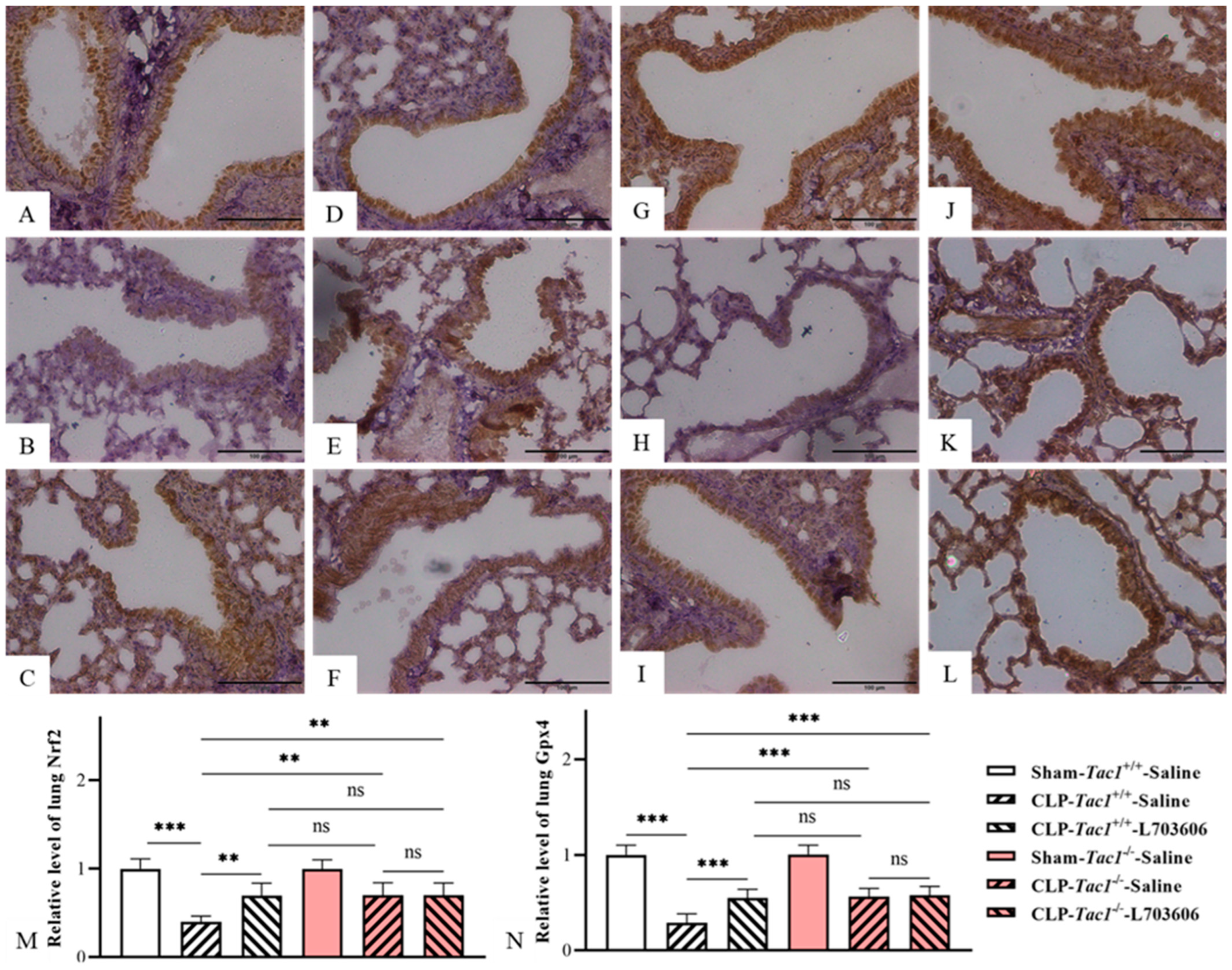
3.10. Suppressing SP-NK1R Signalling Attenuated the Decreases in the Expressions of Nrf2 and Gpx4 in the Lungs in Mice following CLP-Surgery-Induced Sepsis
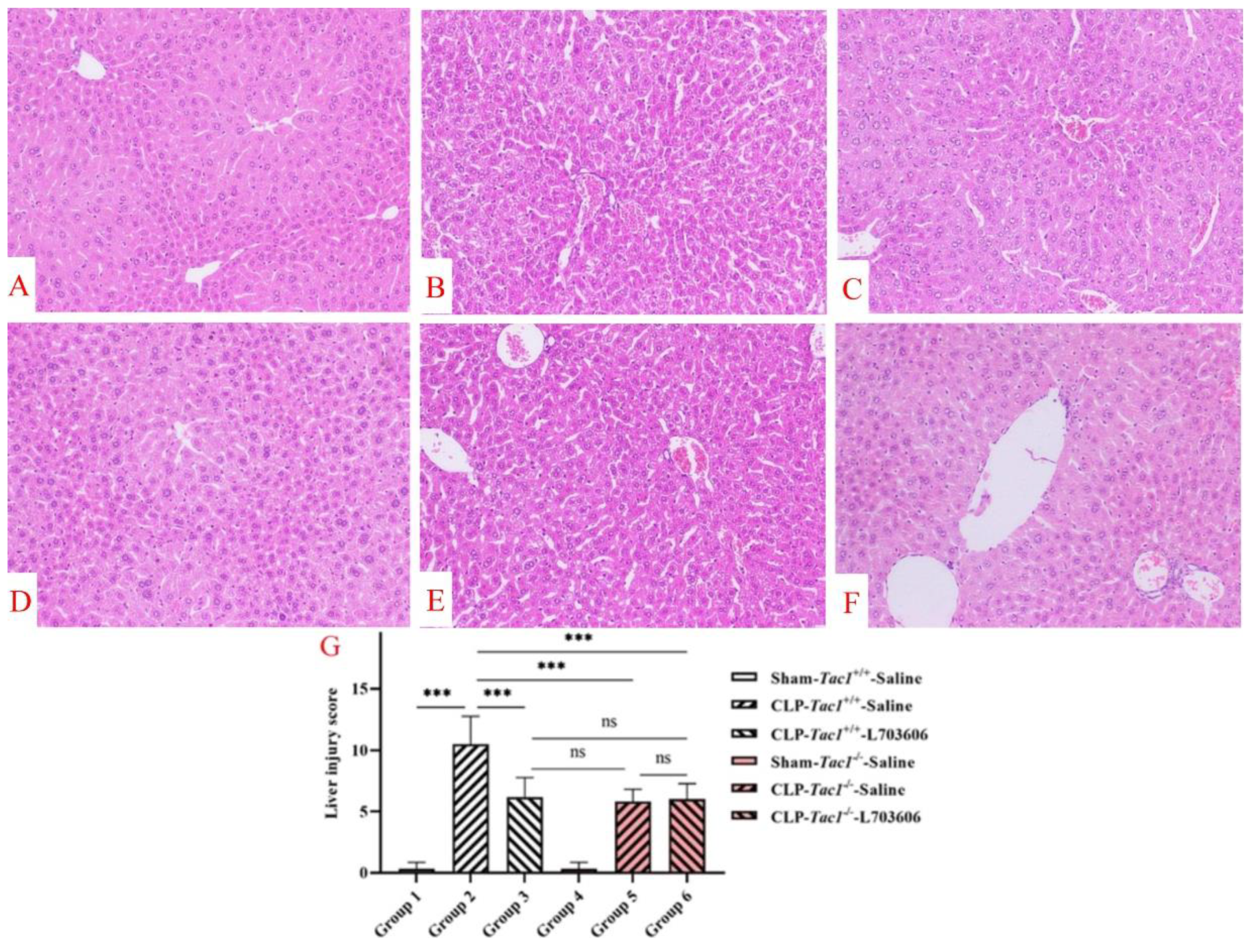
3.11. Suppressing SP-NK1R Signalling Mitigated the Severity of Liver Inflammatory Injury in Mice following CLP-Surgery-Induced Sepsis
3.12. Suppressing SP-NK1R Signalling Mitigated the Severity of Lung Inflammatory Injury in Mice following CLP-Surgery-Induced Sepsis
4. Discussion
5. Conclusions
Author Contributions
Funding
Institutional Review Board Statement
Informed Consent Statement
Data Availability Statement
Acknowledgments
Conflicts of Interest
References
- Cecconi, M.; Evans, L.; Levy, M.; Rhodes, A. Sepsis and septic shock. Lancet 2018, 392, 75–87. [Google Scholar] [CrossRef]
- Singer, M.; Deutschman, C.S.; Seymour, C.W.; Shankar-Hari, M.; Annane, D.; Bauer, M.; Bellomo, R.; Bernard, G.R.; Chiche, J.D.; Coopersmith, C.M.; et al. The Third International Consensus Definitions for Sepsis and Septic Shock (Sepsis-3). JAMA 2016, 315, 801–810. [Google Scholar] [CrossRef] [PubMed]
- Zhang, W.; Jiang, H.; Wu, G.; Huang, P.; Wang, H.; An, H. The pathogenesis and potential therapeutic targets in sepsis. MedComm 2023, 4, e418. [Google Scholar] [CrossRef]
- Steinhoff, M.S.; von Mentzer, B.; Geppetti, P.; Pothoulakis, C.; Bunnett, N.W. Tachykinins and their receptors: Contributions to physiological control and the mechanisms of disease. Physiol. Rev. 2014, 94, 265–301. [Google Scholar] [CrossRef] [PubMed]
- Zhu, Z.; Bhatia, M. Inflammation and Organ Injury the Role of Substance P and Its Receptors. Int. J. Mol. Sci. 2023, 24, 6140. [Google Scholar] [CrossRef]
- Zhu, Z.; Chambers, S.; Zeng, Y.; Bhatia, M. Gases in Sepsis: Novel Mediators and Therapeutic Targets. Int. J. Mol. Sci. 2022, 23, 3669. [Google Scholar] [CrossRef]
- Zhu, Z.; Lian, X.; Bhatia, M. Hydrogen Sulfide: A Gaseous Mediator and Its Key Role in Programmed Cell Death, Oxidative Stress, Inflammation and Pulmonary Disease. Antioxidants 2022, 11, 2162. [Google Scholar] [CrossRef]
- Qu, M.; Wang, Y.; Qiu, Z.; Zhu, S.; Guo, K.; Chen, W.; Miao, C.; Zhang, H. Necroptosis, Pyroptosis, Ferroptosis in Sepsis and Treatment. Shock 2022, 57, 161–171. [Google Scholar] [CrossRef] [PubMed]
- Jiang, X.; Stockwell, B.R.; Conrad, M. Ferroptosis: Mechanisms, biology and role in disease. Nat. Rev. Mol. Cell Biol. 2021, 22, 266–282. [Google Scholar] [CrossRef]
- Liang, D.; Minikes, A.M.; Jiang, X. Ferroptosis at the intersection of lipid metabolism and cellular signaling. Mol. Cell 2022, 82, 2215–2227. [Google Scholar] [CrossRef]
- Tang, D.; Chen, X.; Kang, R.; Kroemer, G. Ferroptosis: Molecular mechanisms and health implications. Cell Res. 2021, 31, 107–125. [Google Scholar] [CrossRef] [PubMed]
- Sun, Y.; Chen, P.; Zhai, B.; Zhang, M.; Xiang, Y.; Fang, J.; Xu, S.; Gao, Y.; Chen, X.; Sui, X.; et al. The emerging role of ferroptosis in inflammation. Biomed. Pharmacother. 2020, 127, 110108. [Google Scholar] [CrossRef] [PubMed]
- Chen, X.; Kang, R.; Kroemer, G.; Tang, D. Ferroptosis in infection, inflammation, and immunity. J. Exp. Med. 2021, 218, e20210518. [Google Scholar] [CrossRef] [PubMed]
- Liu, Y.; Tan, S.; Wu, Y.; Tan, S. The Emerging Role of Ferroptosis in Sepsis. DNA Cell Biol. 2022, 41, 368–380. [Google Scholar] [CrossRef]
- Lei, X.L.; Zhao, G.Y.; Guo, R.; Cui, N. Ferroptosis in sepsis: The mechanism, the role and the therapeutic potential. Front. Immunol. 2022, 13, 956361. [Google Scholar] [CrossRef]
- Huo, L.; Liu, C.; Yuan, Y.; Liu, X.; Cao, Q. Pharmacological inhibition of ferroptosis as a therapeutic target for sepsis-associated organ damage. Eur. J. Med. Chem. 2023, 257, 115438. [Google Scholar] [CrossRef] [PubMed]
- Seibt, T.M.; Proneth, B.; Conrad, M. Role of GPX4 in ferroptosis and its pharmacological implication. Free Radic. Biol. Med. 2019, 133, 144–152. [Google Scholar] [CrossRef] [PubMed]
- Ursini, F.; Maiorino, M. Lipid peroxidation and ferroptosis: The role of GSH and GPx4. Free Radic. Biol. Med. 2020, 152, 175–185. [Google Scholar] [CrossRef]
- Dodson, M.; Castro-Portuguez, R.; Zhang, D.D. NRF2 plays a critical role in mitigating lipid peroxidation and ferroptosis. Redox Biol. 2019, 23, 101107. [Google Scholar] [CrossRef]
- Dai, C.; Chen, X.; Li, J.; Comish, P.; Kang, R.; Tang, D. Transcription factors in ferroptotic cell death. Cancer Gene Ther. 2020, 27, 645–656. [Google Scholar] [CrossRef]
- Zhao, T.; Yu, Z.; Zhou, L.; Wang, X.; Hui, Y.; Mao, L.; Fan, X.; Wang, B.; Zhao, X.; Sun, C. Regulating Nrf2-GPx4 axis by bicyclol can prevent ferroptosis in carbon tetrachloride-induced acute liver injury in mice. Cell Death Discov. 2022, 8, 380. [Google Scholar] [CrossRef] [PubMed]
- He, R.; Liu, B.; Xiong, R.; Geng, B.; Meng, H.; Lin, W.; Hao, B.; Zhang, L.; Wang, W.; Jiang, W.; et al. Itaconate inhibits ferroptosis of macrophage via Nrf2 pathways against sepsis-induced acute lung injury. Cell Death Discov. 2022, 8, 43. [Google Scholar] [CrossRef] [PubMed]
- Wang, Y.; Yan, S.; Liu, X.; Deng, F.; Wang, P.; Yang, L.; Hu, L.; Huang, K.; He, J. PRMT4 promotes ferroptosis to aggravate doxorubicin-induced cardiomyopathy via inhibition of the Nrf2/GPX4 pathway. Cell Death Differ. 2022, 29, 1982–1995. [Google Scholar] [CrossRef] [PubMed]
- Chen, Y.; Fang, Z.M.; Yi, X.; Wei, X.; Jiang, D.S. The interaction between ferroptosis and inflammatory signaling pathways. Cell Death Dis. 2023, 14, 205. [Google Scholar] [CrossRef]
- Wang, F.; He, J.; Xing, R.; Sha, T.; Sun, B. Molecular mechanisms of ferroptosis and their role in inflammation. Int. Rev. Immunol. 2023, 42, 71–81. [Google Scholar] [CrossRef] [PubMed]
- Ang, S.F.; Moochhala, S.M.; MacAry, P.A.; Bhatia, M. Hydrogen sulfide and neurogenic inflammation in polymicrobial sepsis: Involvement of substance P and ERK-NF-κB signaling. PLoS ONE 2011, 6, e24535. [Google Scholar] [CrossRef] [PubMed]
- Sio, S.W.; Ang, S.F.; Lu, J.; Moochhala, S.; Bhatia, M. Substance P upregulates cyclooxygenase-2 and prostaglandin E metabolite by activating ERK1/2 and NF-kappaB in a mouse model of burn-induced remote acute lung injury. J. Immunol. 2010, 185, 6265–6276. [Google Scholar] [CrossRef]
- Koon, H.W.; Zhao, D.; Zhan, Y.; Rhee, S.H.; Moyer, M.P.; Pothoulakis, C. Substance P stimulates cyclooxygenase-2 and prostaglandin E2 expression through JAK-STAT activation in human colonic epithelial cells. J. Immunol. 2006, 176, 5050–5059. [Google Scholar] [CrossRef]
- Gallicchio, M.; Rosa, A.C.; Benetti, E.; Collino, M.; Dianzani, C.; Fantozzi, R. Substance P-induced cyclooxygenase-2 expression in human umbilical vein endothelial cells. Br. J. Pharmacol. 2006, 147, 681–689. [Google Scholar] [CrossRef]
- Cao, Y.Q.; Mantyh, P.W.; Carlson, E.J.; Gillespie, A.M.; Epstein, C.J.; Basbaum, A.I. Primary afferent tachykinins are required to experience moderate to intense pain. Nature 1998, 392, 390–394. [Google Scholar] [CrossRef]
- Bhatia, M.; Slavin, J.; Cao, Y.; Basbaum, A.I.; Neoptolemos, J.P. Preprotachykinin-A gene deletion protects mice against acute pancreatitis and associated lung injury. Am. J. Physiol. Gastrointest. Liver Physiol. 2003, 284, G830–G836. [Google Scholar] [CrossRef]
- Toscano, M.G.; Ganea, D.; Gamero, A.M. Cecal ligation puncture procedure. J. Vis. Exp. 2011, 51, e2860. [Google Scholar] [CrossRef]
- Shrum, B.; Anantha, R.V.; Xu, S.X.; Donnelly, M.; Haeryfar, S.M.; McCormick, J.K.; Mele, T. A robust scoring system to evaluate sepsis severity in an animal model. BMC Res. Notes 2014, 7, 233. [Google Scholar] [CrossRef]
- Muftuoglu, M.A.; Aktekin, A.; Ozdemir, N.C.; Saglam, A. Liver injury in sepsis and abdominal compartment syndrome in rats. Surg. Today 2006, 36, 519–524. [Google Scholar] [CrossRef]
- Jiang, J.; Huang, K.; Xu, S.; Garcia, J.G.N.; Wang, C.; Cai, H. Targeting NOX4 alleviates sepsis-induced acute lung injury via attenuation of redox-sensitive activation of CaMKII/ERK1/2/MLCK and endothelial cell barrier dysfunction. Redox Biol. 2020, 36, 101638. [Google Scholar] [CrossRef]
- Lelubre, C.; Vincent, J.L. Mechanisms and treatment of organ failure in sepsis. Nat. Rev. Nephrol. 2018, 14, 417–427. [Google Scholar] [CrossRef]
- Rochette, L.; Dogon, G.; Rigal, E.; Zeller, M.; Cottin, Y.; Vergely, C. Lipid Peroxidation and Iron Metabolism: Two Corner Stones in the Homeostasis Control of Ferroptosis. Int. J. Mol. Sci. 2022, 24, 449. [Google Scholar] [CrossRef]
- Wang, Y.M.; Gong, F.C.; Qi, X.; Zheng, Y.J.; Zheng, X.T.; Chen, Y.; Yang, Z.T.; Qing, Y.; Mao, E.Q.; Chen, E.Z. Mucin 1 Inhibits Ferroptosis and Sensitizes Vitamin E to Alleviate Sepsis-Induced Acute Lung Injury through GSK3β/Keap1-Nrf2-GPX4 Pathway. Oxid. Med. Cell Longev. 2022, 2022, 2405943. [Google Scholar] [CrossRef]
- Wei, S.; Bi, J.; Yang, L.; Zhang, J.; Wan, Y.; Chen, X.; Wang, Y.; Wu, Z.; Lv, Y.; Wu, R. Serum irisin levels are decreased in patients with sepsis, and exogenous irisin suppresses ferroptosis in the liver of septic mice. Clin. Transl. Med. 2020, 10, e173. [Google Scholar] [CrossRef]
- Michel, F.; Bonnefont-Rousselot, D.; Mas, E.; Drai, J.; Thérond, P. Biomarkers of lipid peroxidation: Analytical aspects. Ann. Biol. Clin. 2008, 66, 605–620. [Google Scholar] [CrossRef]
- Gaddam, R.R.; Chambers, S.; Fraser, R.; Cogger, V.C.; Le Couteur, D.G.; Ishii, I.; Bhatia, M. Cystathionine-Gamma-Lyase-Derived Hydrogen Sulfide-Regulated Substance P Modulates Liver Sieve Fenestrations in Caecal Ligation and Puncture-Induced Sepsis. Int. J. Mol. Sci. 2019, 20, 3191. [Google Scholar] [CrossRef] [PubMed]
- Li, B.; Han, X.; Ye, X.; Ni, J.; Wu, J.; Dai, J.; Wu, Z.; Chen, C.; Wan, R.; Wang, X.; et al. Substance P-regulated leukotriene B4 production promotes acute pancreatitis-associated lung injury through neutrophil reverse migration. Int. Immunopharmacol. 2018, 57, 147–156. [Google Scholar] [CrossRef] [PubMed]
- Chen, X.; Li, J.; Kang, R.; Klionsky, D.J.; Tang, D. Ferroptosis: Machinery and regulation. Autophagy 2021, 17, 2054–2081. [Google Scholar] [CrossRef] [PubMed]


| Antibody | Dilution | Source Catalogue No. |
|---|---|---|
| NK1R | 1:1000 | Thermo Fisher Scientific, Waltham, MA, USA |
| Nrf2 | 1:1000 | Thermo Fisher Scientific, Waltham, MA, USA |
| Gpx4 | 1:1000 | Thermo Fisher Scientific, Waltham, MA, USA |
| GAPDH | 1:1000 | Santa Cruz Biotechnology, Dallas, TX, USA |
| HRP-conjugated secondary antibody | 1:5000 | Santa Cruz Biotechnology, Dallas, TX, USA |
| Group | Sepsis Severity Score |
|---|---|
| Sham-Tac1+/+-Saline group | 0.63 ± 0.74 |
| CLP-Tac1+/+-Saline group | 9.13 ± 2.17 * |
| CLP-Tac1+/+-L703606 group | 4.63 ± 1.41 # |
| Sham-Tac1−/−-Saline group | 0.75 ± 0.71 |
| CLP-Tac1−/−-Saline group | 4.50 ± 1.41 # |
| CLP-Tac1−/−-L703606 group | 4.88 ± 1.64 # |
| Group | Tissue SP Concentration (ng/g Protein) | |
|---|---|---|
| Liver | Lung | |
| Sham-Tac1+/+-Saline group | 26.95 ± 2.95 | 25.13 ± 2.09 |
| CLP-Tac1+/+-Saline group | 390.82 ± 32.40 * | 151.46 ± 6.34 * |
| CLP-Tac1+/+-L703606 group | 237.36 ± 19.83 # | 104.17 ± 8.12 # |
| Sham-Tac1−/−-Saline group | <the LLOD | <the LLOD |
| CLP-Tac1−/−-Saline group | <the LLOD | <the LLOD |
| CLP-Tac1−/−-L703606 group | <the LLOD | <the LLOD |
| Group | Tissue Iron Concentration (μg/g Protein) | |
|---|---|---|
| Liver | Lung | |
| Sham-Tac1+/+-Saline group | 2.75 ± 0.19 | 1.03 ± 0.06 |
| CLP-Tac1+/+-Saline group | 4.33 ± 0.70 * | 2.14 ± 0.24 * |
| CLP-Tac1+/+-L703606 group | 3.51 ± 0.32 # | 1.53 ± 0.15 & |
| Sham-Tac1−/−-Saline group | 2.77 ± 0.17 | 1.02 ± 0.07 |
| CLP-Tac1−/−-Saline group | 3.48 ± 0.31 & | 1.58 ± 0.15 & |
| CLP-Tac1−/−-L703606 group | 3.40 ± 0.33 & | 1.57 ± 0.14 & |
| Group | Tissue MDA Concentration (μg/g Protein) | |
|---|---|---|
| Liver | Lung | |
| Sham-Tac1+/+-Saline group | 2.467 ± 0.15 | 5.070 ± 0.29 |
| CLP-Tac1+/+-Saline group | 6.297 ± 1.01 * | 15.994 ± 1.82 * |
| CLP-Tac1+/+-L703606 group | 4.947 ± 0.68 & | 10.714 ± 1.14 & |
| Sham-Tac1−/−-Saline group | 2.446 ± 0.17 | 5.117 ± 0.32 |
| CLP-Tac1−/−-Saline group | 5.065 ± 0.53 # | 10.888 ± 0.86 & |
| CLP-Tac1−/−-L703606 group | 4.985 ± 0.52 & | 11.0492 ± 1.08 & |
| Group | Tissue GSH Concentration (μg/mg Protein) | |
|---|---|---|
| Liver | Lung | |
| Sham-Tac1+/+-Saline group | 29.60 ± 1.52 | 2.47 ± 0.10 |
| CLP-Tac1+/+-Saline group | 17.63 ± 2.84 * | 1.08 ± 0.20 * |
| CLP-Tac1+/+-L703606 group | 24.20 ± 2.08 & | 2.10 ± 0.17 & |
| Sham-Tac1−/−-Saline group | 29.72 ± 1.53 | 2.47 ± 0.11 |
| CLP-Tac1−/−-Saline group | 24.33 ± 1.70 & | 2.06 ± 0.14 & |
| CLP-Tac1−/−-L703606 group | 24.67 ± 1.63 & | 2.03 ± 0.14 & |
Disclaimer/Publisher’s Note: The statements, opinions and data contained in all publications are solely those of the individual author(s) and contributor(s) and not of MDPI and/or the editor(s). MDPI and/or the editor(s) disclaim responsibility for any injury to people or property resulting from any ideas, methods, instructions or products referred to in the content. |
© 2024 by the authors. Licensee MDPI, Basel, Switzerland. This article is an open access article distributed under the terms and conditions of the Creative Commons Attribution (CC BY) license (https://creativecommons.org/licenses/by/4.0/).
Share and Cite
Zhu, Z.; Chambers, S.; Bhatia, M. Suppressing the Substance P-NK1R Signalling Protects Mice against Sepsis-Associated Acute Inflammatory Injury and Ferroptosis in the Liver and Lungs. Antioxidants 2024, 13, 300. https://doi.org/10.3390/antiox13030300
Zhu Z, Chambers S, Bhatia M. Suppressing the Substance P-NK1R Signalling Protects Mice against Sepsis-Associated Acute Inflammatory Injury and Ferroptosis in the Liver and Lungs. Antioxidants. 2024; 13(3):300. https://doi.org/10.3390/antiox13030300
Chicago/Turabian StyleZhu, Zhixing, Stephen Chambers, and Madhav Bhatia. 2024. "Suppressing the Substance P-NK1R Signalling Protects Mice against Sepsis-Associated Acute Inflammatory Injury and Ferroptosis in the Liver and Lungs" Antioxidants 13, no. 3: 300. https://doi.org/10.3390/antiox13030300
APA StyleZhu, Z., Chambers, S., & Bhatia, M. (2024). Suppressing the Substance P-NK1R Signalling Protects Mice against Sepsis-Associated Acute Inflammatory Injury and Ferroptosis in the Liver and Lungs. Antioxidants, 13(3), 300. https://doi.org/10.3390/antiox13030300