
Chemically Modified Nanoparticles for Enhanced Antioxidant and Antimicrobial Properties with Cinnamon Essential Oil

 ,
,  ,
,  ,
,  , , ,
, , ,  and
and
Abstract
:1. Introduction
2. Materials and Methods
2.1. Fabrication
2.1.1. Materials
2.1.2. Nanoparticle Modification
2.1.3. Film Fabrication
2.2. Characterization of Nanoparticles
2.2.1. Fourier-Transform Infrared Spectroscopy (FTIR)
2.2.2. X-ray Diffraction (XRD)
2.2.3. Thermogravimetric Analysis (TGA)
2.2.4. X-ray Photoelectron Spectroscopy (XPS)
2.2.5. Antioxidant Activity
2.2.6. Antimicrobial Activity
2.3. Characterization of Films
2.3.1. Fourier-Transform Infrared Spectroscopy (FTIR)
2.3.2. X-ray Diffraction (XRD)
2.3.3. Scanning Electron Microscopy (SEM)
2.3.4. Differential Scanning Calorimetry (DSC)
2.3.5. Water Vapor Permeability (WVP)
2.4. Mechanical Properties
3. Results and Discussion
3.1. Characterization of Nanoparticles
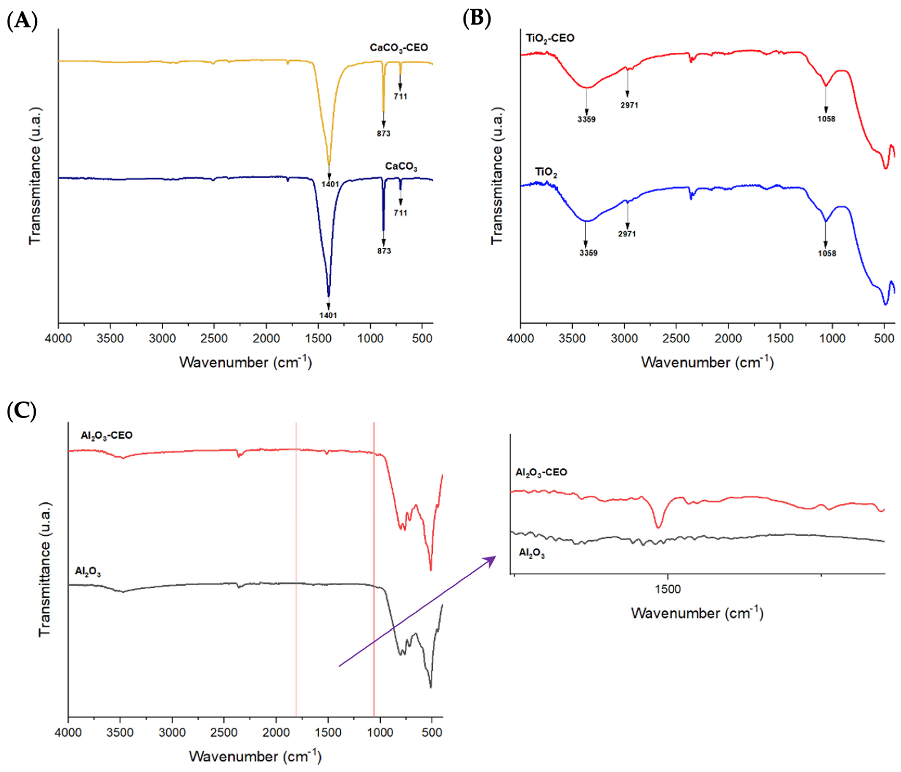
3.1.1. FTIR
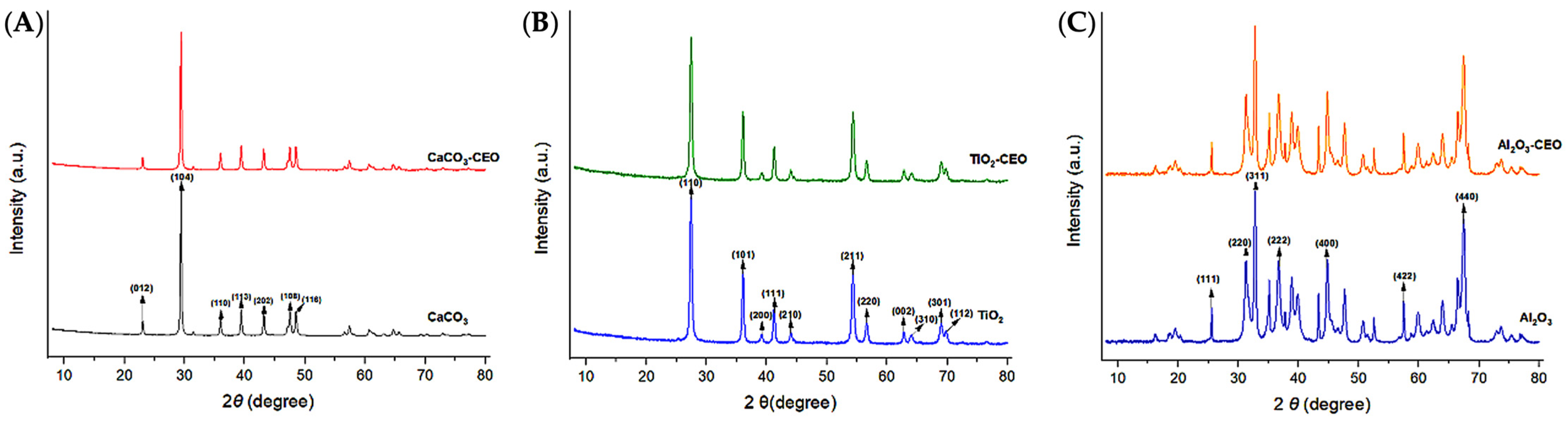
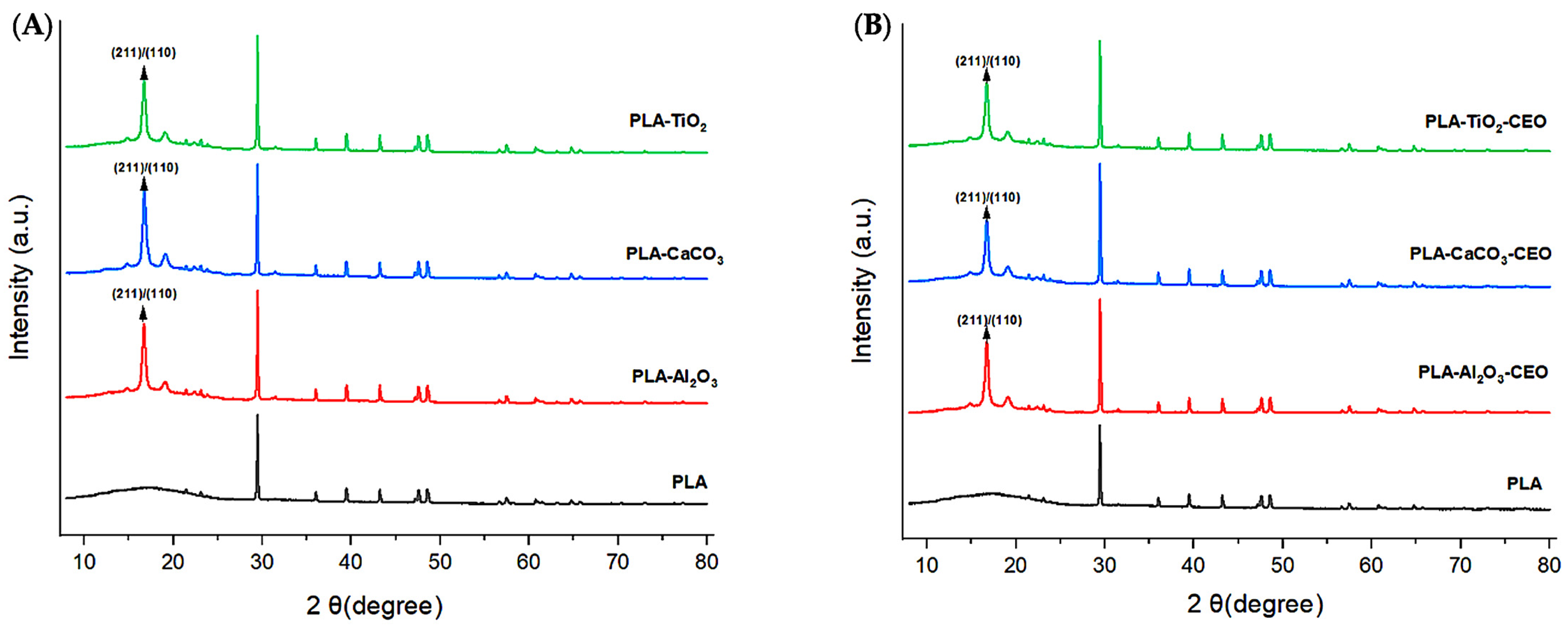
3.1.2. XRD (X-ray Diffraction)
3.1.3. Thermogravimetric Analysis (TGA)
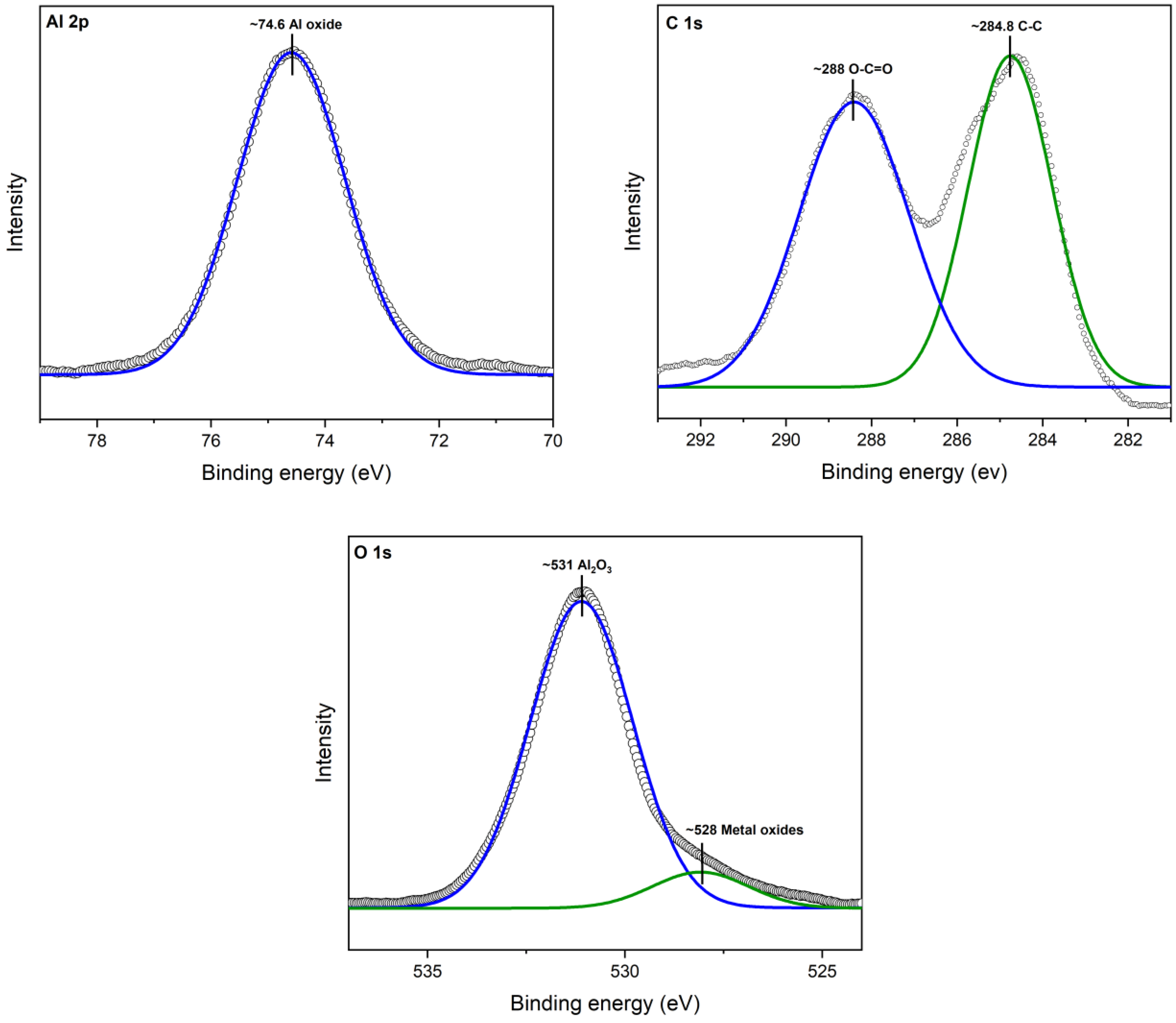
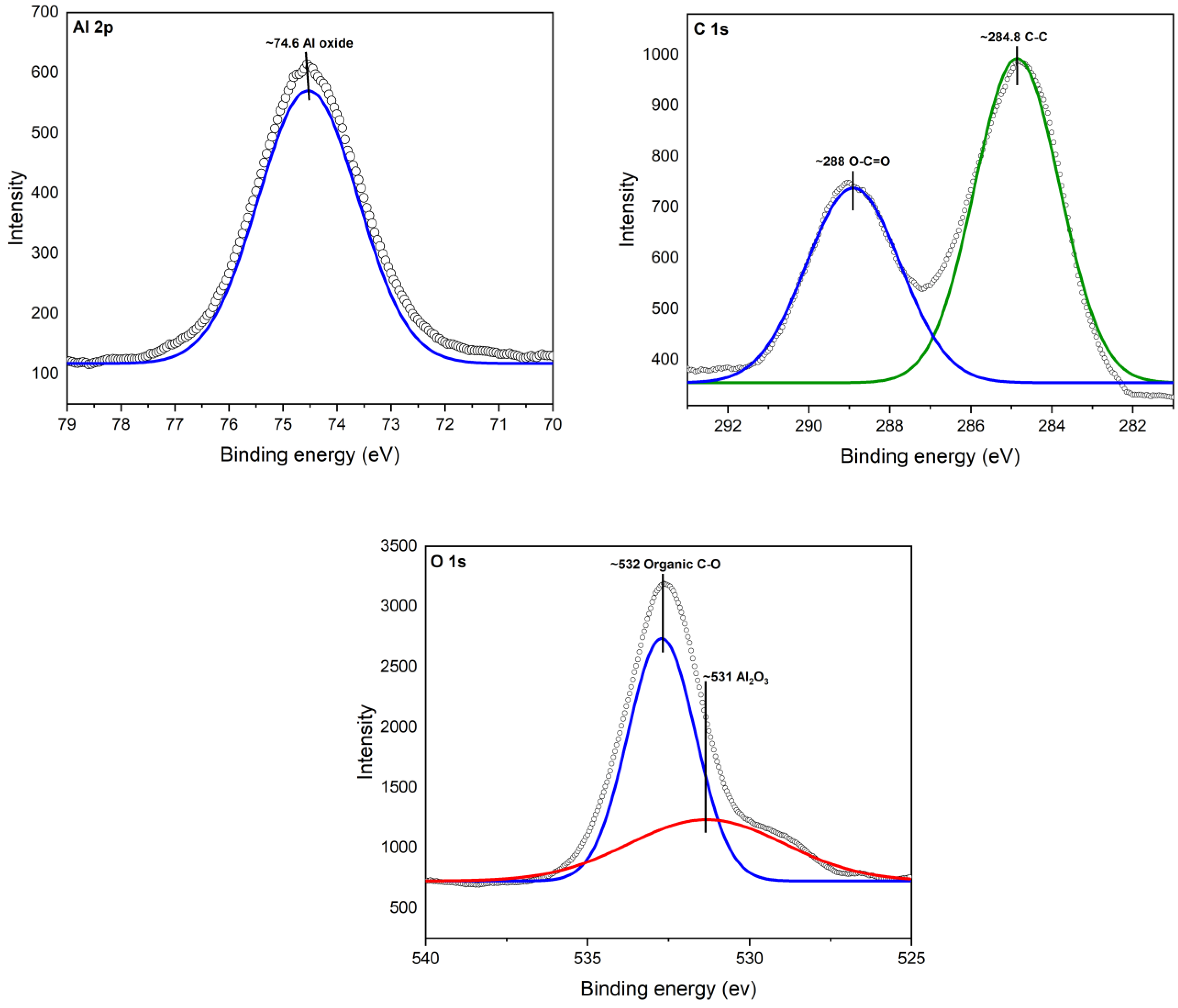
3.1.4. X-ray Photoelectron Spectroscopy (XPS)
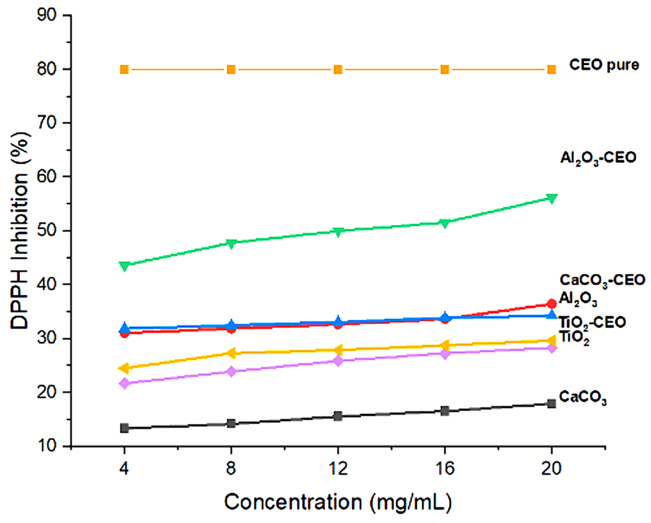
3.1.5. Antioxidant Activity
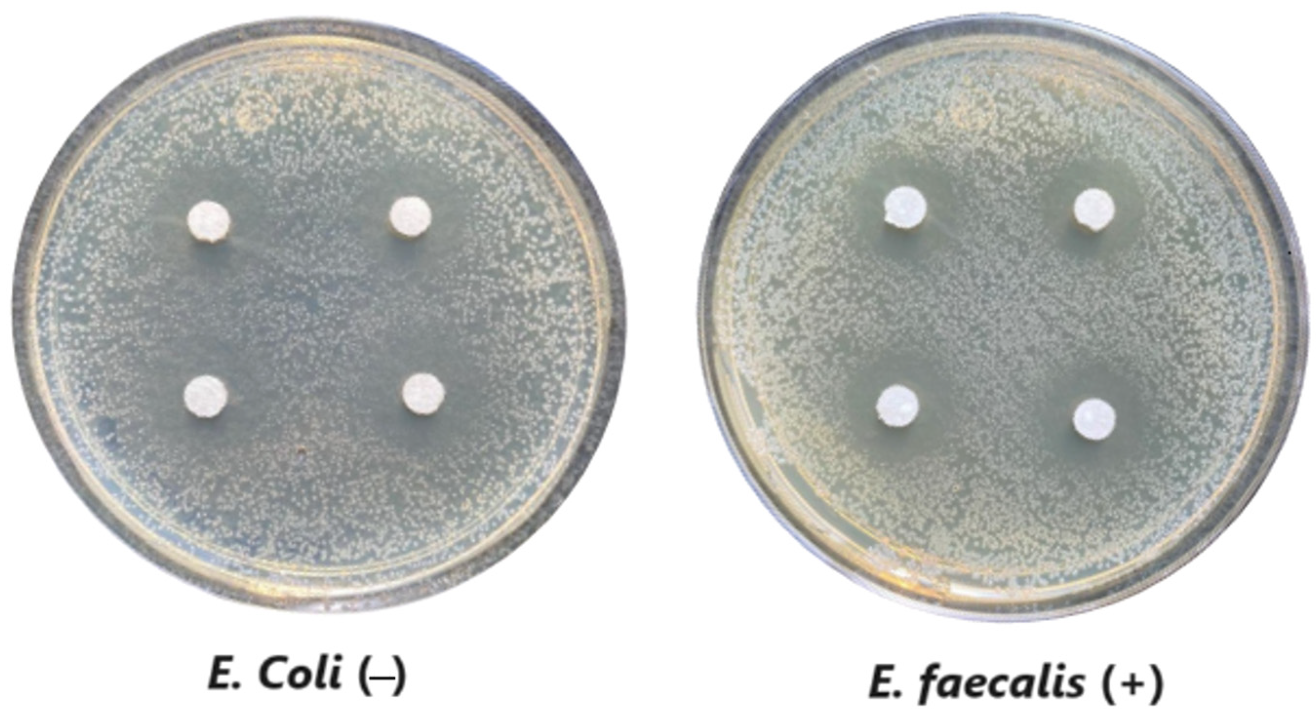
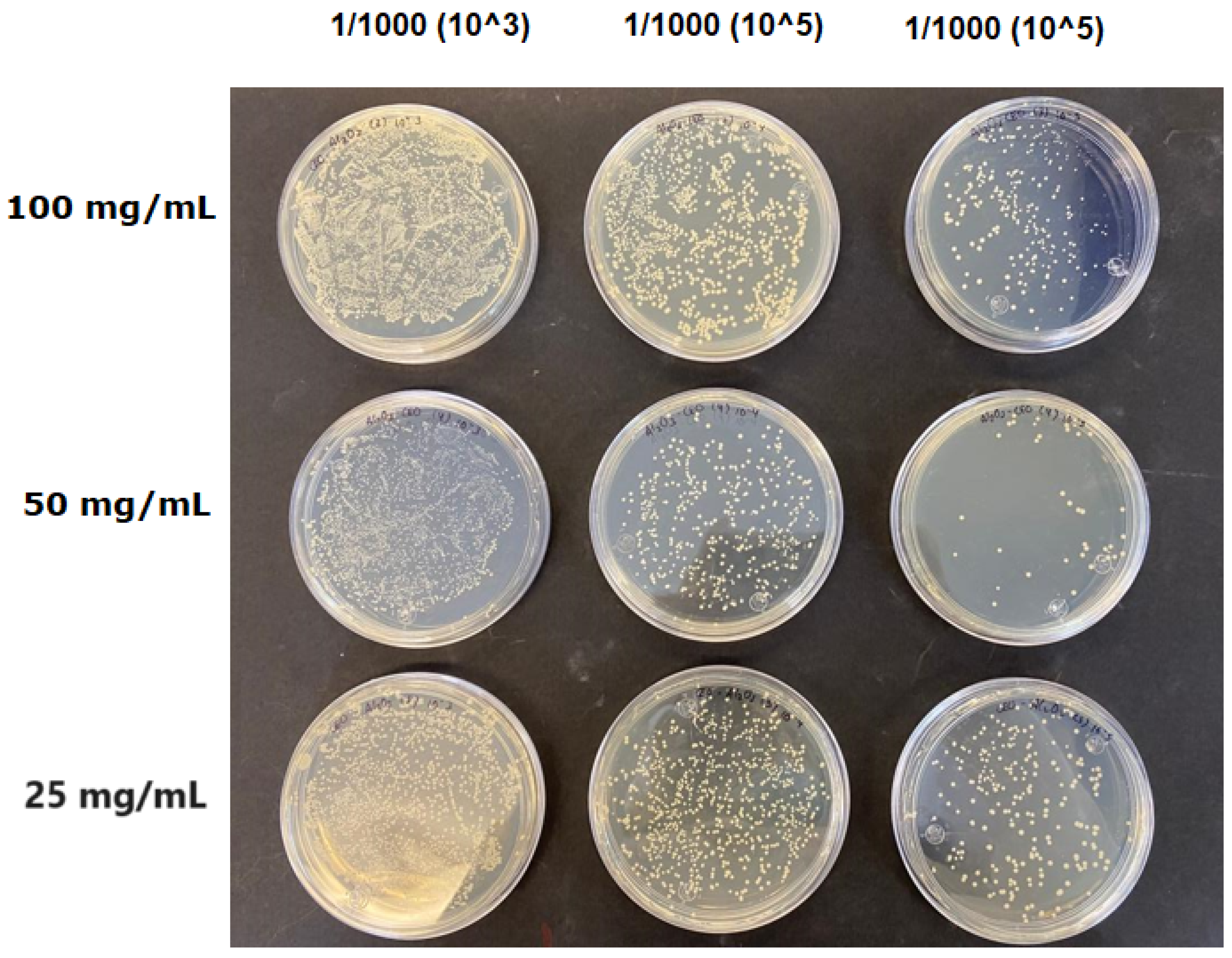
3.1.6. Antimicrobial Evaluation
3.2. Characterization of Films
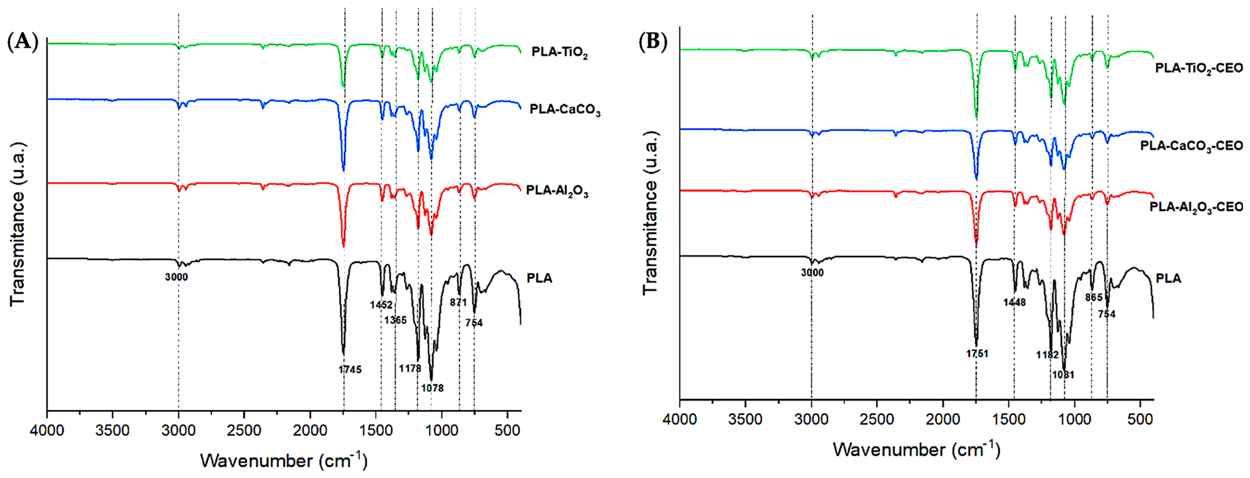
3.2.1. FTIR
3.2.2. XRD
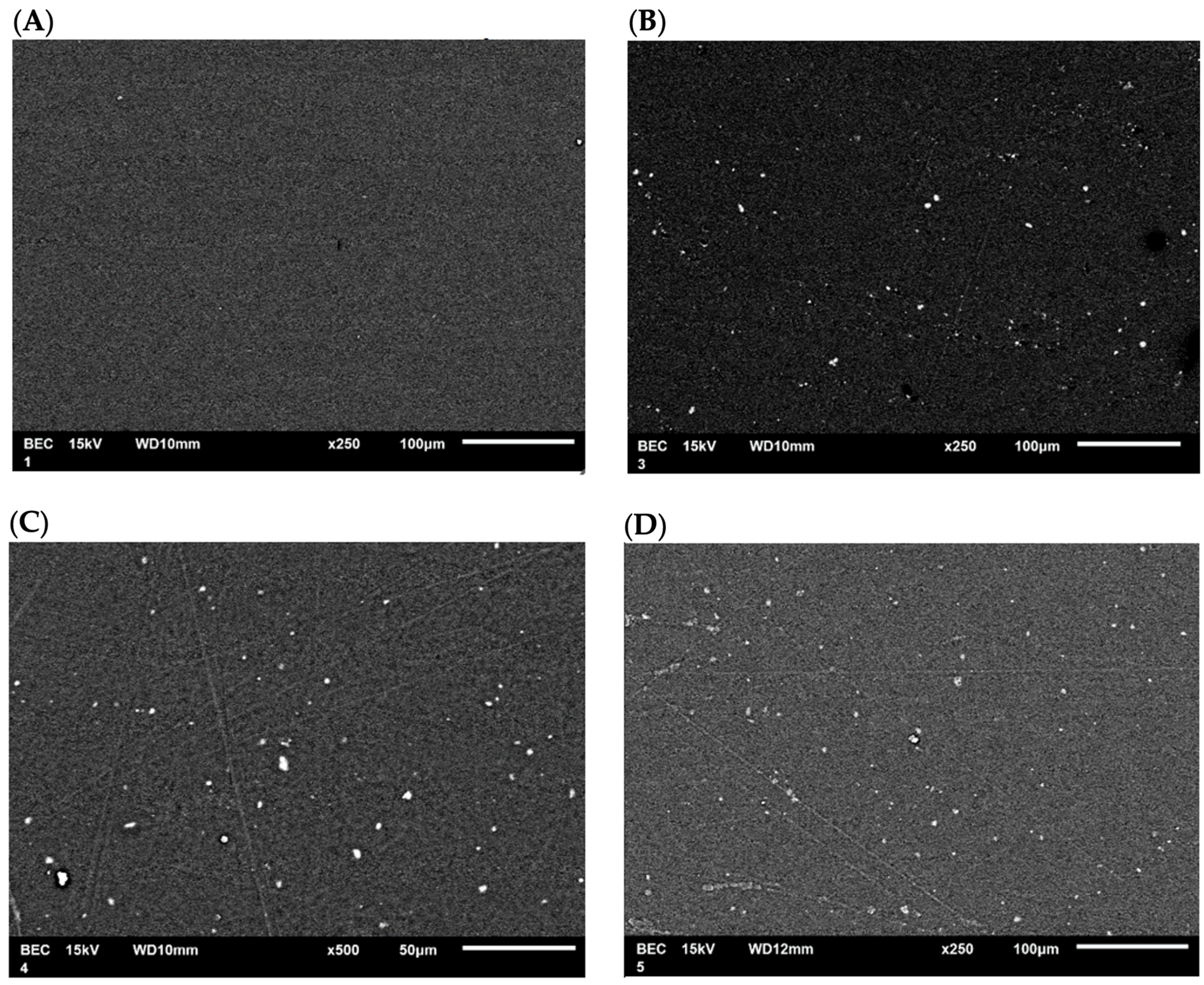
3.2.3. SEM Analysis
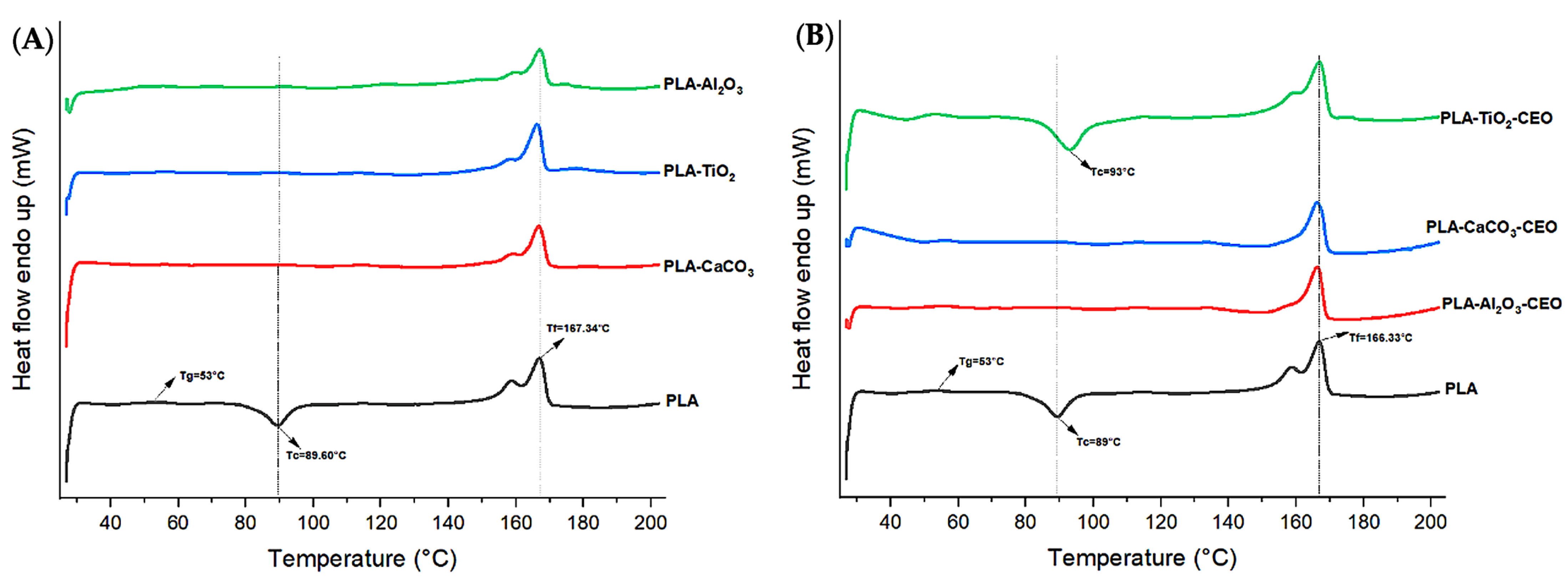
3.2.4. DSC Analysis
3.2.5. Water Vapor Permeability
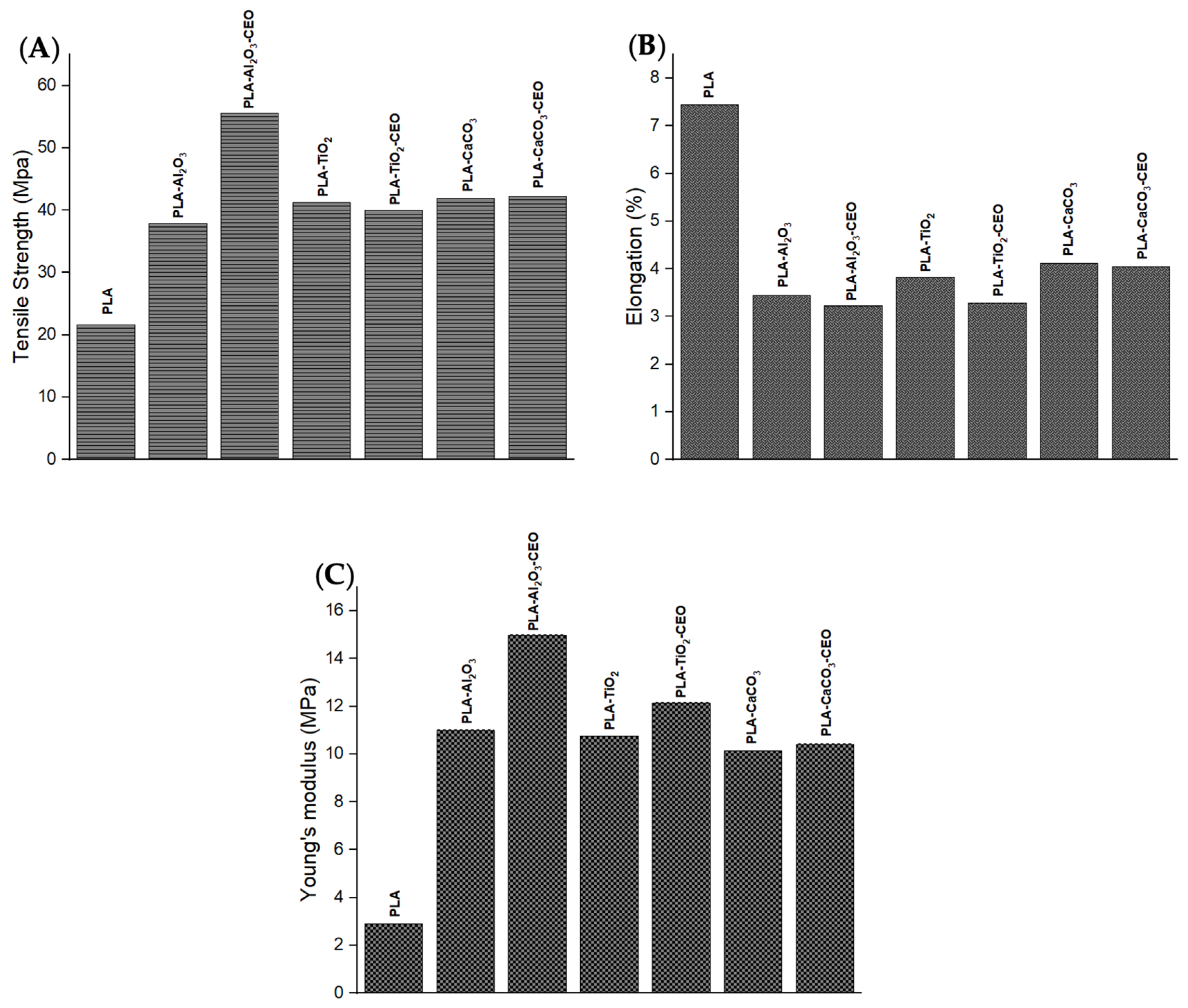
3.3. Mechanical Properties
4. Conclusions
Author Contributions
Funding
Institutional Review Board Statement
Informed Consent Statement
Data Availability Statement
Acknowledgments
Conflicts of Interest
References
- Sana, S.S.; Li, H.; Zhang, Z.; Sharma, M.; Usmani, Z.; Hou, T.; Netala, V.R.; Wang, X.; Gupta, V.K. Recent advances in essential oils-based metal nanoparticles: A review on recent developments and biopharmaceutical applications. J. Mol. Liq. 2021, 333, 115951. [Google Scholar] [CrossRef]
- Sheny, D.S.; Mathew, J.; Philip, D. Synthesis characterization and catalytic action of hexagonal gold nanoparticles using essential oils extracted from Anacardium occidentale. Spectrochim. Acta Part A Mol. Biomol. Spectrosc. 2012, 97, 306–310. [Google Scholar] [CrossRef] [PubMed]
- Karunakaran, G.; Suriyaprabha, R.; Manivasakan, P.; Yuvakkumar, R.; Rajendra, V.; Kannan, N. Screening of in vitro cytotoxicity, antioxidant potential and bioactivity of nano- and micro-ZrO2 and-TiO2 particles. Ecotoxicol. Environ. Saf. 2013, 93, 191–197. [Google Scholar] [CrossRef]
- Brand-Williams, W.; Cuvelier, M.E.; Berset, C. Use of a free radical method to evaluate antioxidant activity. LWT-Food Sci. Technol. 1995, 28, 25–30. [Google Scholar] [CrossRef]
- ASTM International. Standard test method for water vapor transmission of materials. In Annual Book of ASTM Standards; ASTM International: Conshohocken, PA, USA, 1995; pp. 95–96. [Google Scholar]
- ASTM D882-02; Standard Test Method for Tensile Properties of Thin Plastic Sheeting. ASTM: West Conshohocken, PA, USA, 2010. [CrossRef]
- Noah, A.Z.; El Semary, M.A.; Youssef, A.M.; El-Safty, M.A. Enhancement of yield point at high pressure high temperature wells by using polymer nanocomposites based on ZnO & CaCO3 nanoparticles. Egypt. J. Pet. 2017, 26, 33–40. [Google Scholar] [CrossRef]
- Zapata, P.A.; Palza, H.; Díaz, B.; Armijo, A.; Sepúlveda, F.; Ortiz, J.A.; Ramírez, M.P.; Oyarzún, C. Effect of CaCO3 Nanoparticles on the Mechanical and Photo-Degradation Properties of LDPE. Molecules 2019, 24, 126. [Google Scholar] [CrossRef]
- Tizo, M.S.; Blanco, L.A.V.; Cagas, A.C.Q.; Dela Cruz, B.R.B.; Encoy, J.C.; Gunting, J.V.; Mabayo, V.I.F. Efficiency of calcium carbonate from eggshells as an adsorbent for Cadmium removal in aqueous solution. Sustain. Environ. Res. 2018, 28, 326–332. [Google Scholar] [CrossRef]
- Kerru, N.; Gummidi, L.; Bhaskaruni, S.V.H.S.; Maddila, S.N.; Jonnalagadda, S.B. One-pot green synthesis of novel 5,10-dihydro-1H-pyrazolo[1,2-b]phthalazine derivatives with eco-friendly biodegradable eggshell powder as efficacious catalyst. Res. Chem. Intermed. 2020, 46, 3067–3083. [Google Scholar] [CrossRef]
- Dehghani, F.; Kalantariasl, A.; Saboori, R.; Sabbaghi, S.; Peyvandi, K. Performance of carbonate calcium nanoparticles as filtration loss control agent of water-based drilling fluid. SN Appl. Sci. 2019, 1, 1466. [Google Scholar] [CrossRef]
- Sharma, S.; Byrne, M.; Perera, K.Y.; Duffy, B.; Jaiswal, A.K.; Jaiswal, S. Active film packaging based on bio-nanocomposite TiO2 and cinnamon essential oil for enhanced preservation of cheese quality. Food Chem. 2023, 405, 134798. [Google Scholar] [CrossRef]
- Boughendjioua, H.; Amoura, N.; Boughendjioua, Z. Purity Specifications of Constituents of Cinnamon Essential Oil by Fourier Transformed Infrared Spectroscopy Analysis. Indian J. Pharm. Biol. Res. 2017, 5, 36–40. [Google Scholar] [CrossRef]
- Zhou, Y.; Wu, X.; Chen, J.; He, J. Effects of cinnamon essential oil on the physical, mechanical, structural and thermal properties of cassava starch-based edible films. Int. J. Biol. Macromol. 2021, 184, 574–583. [Google Scholar] [CrossRef] [PubMed]
- Han, Y.; Yu, M.; Wang, L. Physical and antimicrobial properties of sodium alginate/carboxymethyl cellulose films incorporated with cinnamon essential oil. Food Packag. Shelf Life 2018, 15, 35–42. [Google Scholar] [CrossRef]
- Shao, P.; Yu, J.; Chen, H.; Gao, H. Development of microcapsule bioactive paper loaded with cinnamon essential oil to improve the quality of edible fungi. Food Packag. Shelf Life 2021, 27, 100617. [Google Scholar] [CrossRef]
- Ahmed, J.; Mulla, M.; Arfat, Y.A.; Bher, A.; Jacob, H.; Auras, R. Compression molded LLDPE films loaded with bimetallic (Ag-Cu) nanoparticles and cinnamon essential oil for chicken meat packaging applications. LWT 2018, 93, 329–338. [Google Scholar] [CrossRef]
- Rodriguez-Navarro, C.; Jimenez-Lopez, C.; Rodriguez-Navarro, A.; Gonzalez-Muñoz, M.T.; Rodriguez-Gallego, M. Bacterially mediated mineralization of vaterite. Geochim. Cosmochim. Acta 2007, 71, 1197–1213. [Google Scholar] [CrossRef]
- Ghadam, A.G.J.; Idrees, M. Characterization of CaCO3 Nanoparticles Synthesized by Reverse Microemulsion Technique in Different Concentrations of Surfactants. Iran. J. Chem. Chem. Eng. 2013, 32, 27–35. [Google Scholar] [CrossRef]
- Render, D.; Samuel, T.; King, H.; Vig, M.; Jeelani, S.; Babu, R.J.; Rangari, V. Biomaterial-Derived Calcium Carbonate Nanoparticles for Enteric Drug Delivery. J. Nanomater. 2016, 2026, 3170248. [Google Scholar] [CrossRef]
- Boudaira, B.; Harabi, A.; Bouzerara, F.; Zenikheri, F.; Foughali, L.; Guechi, A. Preparation and characterization of membrane supports for microfiltration and ultrafiltration using kaolin (DD2) and CaCO3. Desalination Water Treat. 2015, 57, 5258–5265. [Google Scholar] [CrossRef]
- Kirboga, S.; Oner, M.; Akyol, E. The effect of ultrasonication on calcium carbonate crystallization in the presence of biopolymer. J. Cryst. Growth 2014, 401, 266–270. [Google Scholar] [CrossRef]
- Ferreira, A.M.; Vikulina, A.; Volodkin, D. CaCO3 crystals as versatile carriers for controlled delivery of antimicrobials. J. Control Release 2020, 328, 470–489. [Google Scholar] [CrossRef] [PubMed]
- Zhang, W.; Wu, Y.; Wang, J.; Liu, H.; Lu, H.; Zhai, S.; Zhong, Q.; Liu, S.; Zhong, W.; Huang, C.; et al. Adsorption of thallium(I) on rutile nano-titanium dioxide and environmental implications. PeerJ 2019, 7, e6820. [Google Scholar] [CrossRef] [PubMed]
- Pushpamalini, T.; Keerthana, M.; Sangavi, R.; Nagaraj, A.; Kamaraj, P. Comparative analysis of green synthesis of TiO2 nanoparticles using four different leaf extract. Mater. Today Proc. 2021, 40, S180–S184. [Google Scholar] [CrossRef]
- Joni, I.M.; Nulhakim, L.; Panatarani, C. Characteristics of TiO2 particles prepared by simple solution method using TiCl3 precursor. J. Phys. Conf. Ser. 2018, 1080, 012042. [Google Scholar] [CrossRef]
- Jalali, E.; Maghsoudi, S.; Noroozian, E. A novel method for biosynthesis of different polymorphs of TiO2 nanoparticles as a protector for Bacillus thuringiensis from Ultra Violet. Sci. Rep. 2020, 10, 426. [Google Scholar] [CrossRef] [PubMed]
- Saravanan, S.; Balamurugan, M.; Soga, T. Synthesis of Titanium Dioxide Nanoparticles with Desired Ratio of Anatase and Rutile Phases and the Effect of High Temperature Annealing. Trans. Mater. Res. Soc. Jpn. 2018, 43, 255–261. [Google Scholar] [CrossRef]
- Ansari, S.A.; Husain, Q. Immobilization of Kluyveromyces lactis β galactosidase on concanavalin A layered aluminium oxide nanoparticles—Its future aspects in biosensor applications. J. Mol. Catal. B Enzym. 2011, 70, 119–126. [Google Scholar] [CrossRef]
- Romero Toledo, R.; Ruíz Santoyo, V.; Moncada Sánchez, D.; Martínez Rosales, M. Effect of aluminum precursor on physicochemical properties of Al2O3 by hydrolysis/precipitation method. Nova Sci. 2018, 10, 83–99. [Google Scholar] [CrossRef]
- Kim, H.S.; Park, N.-K.; Lee, T.J.; Um, M.-H.; Kang, M. Preparation of Nanosizedα-Al2O3 Particles Using a Microwave Pretreatment at Mild Temperature. Adv. Mater. Sci. Eng. 2012, 2012, 920105. [Google Scholar] [CrossRef]
- Mohammed, A.A.; Khodair, Z.T.; Khadom, A.A. Preparation and investigation of the structural properties of α-Al2O3 nanoparticles using the Sol-Gel method. Chem. Data Collect. 2020, 29, 100531. [Google Scholar] [CrossRef]
- Du, R.; He, L.; Li, P.; Zhao, G. Polydopamine-Modified Al2O3/Polyurethane Composites with Largely Improved Thermal and Mechanical Properties. Materials 2020, 13, 1772. [Google Scholar] [CrossRef] [PubMed]
- Bristy, S.S.; Ahmad, H. Studies on Adsorption Performance of Reactive Dye Remazol Navy RGB (RN-RGB) via Epoxide Functional Magnetic γ-Al2O3 Nanocomposite Particles. J. Sci. Res. 2017, 9, 421–429. [Google Scholar] [CrossRef]
- Siva, T.; Muralidharan, S.; Sathiyanarayanan, S.; Manikandan, E.; Jayachandran, M. Enhanced Polymer Induced Precipitation of Polymorphous in Calcium Carbonate: Calcite Aragonite Vaterite Phases. J. Inorg. Organomet. Polym. Mater. 2017, 27, 770–778. [Google Scholar] [CrossRef]
- Wang, W.; Wang, S.; Liu, Q.; Wang, X.; Zhu, J.; Luo, H.; Ji, S. Study on the efficacy of amorphous calcium carbonate as a consolidant for calcareous matrix. Herit. Sci. 2022, 10, 165. [Google Scholar] [CrossRef]
- Asif, M.; Zafar, M.; Akhter, P.; Hussain, M.; Umer, A.; Razzaq, A.; Kim, W.-Y. Effect of Urea Addition on Anatase Phase Enrichment and Nitrogen Doping of TiO2 for Photocatalytic Abatement of Methylene Blue. Appl. Sci. 2021, 11, 8264. [Google Scholar] [CrossRef]
- Tsega, M.; Dejene, F.B. Morphological, thermal and optical properties of TiO2 nanoparticles: The effect of titania precursor. Mater. Res. Express 2019, 6, 065041. [Google Scholar] [CrossRef]
- Available online: https://www.thermofisher.com/mx/es/home/materials-science/learning-center/periodic-table.html (accessed on 12 November 2023).
- Martínez-Aguilar, V.; Coral Carrillo-Sanchez, P.; Del Angel-Monroy, M.; Sánchez Balderas, G.; Flores-Martínez, N.L.; Pérez, E.; González-Calderón, J.A. Chemical modification of TiO2 with essential oils for its application in active packaging. Polym. Bull. 2023, 80, 2753–2778. [Google Scholar] [CrossRef]
- Liu, S.; Zhao, C.; Cao, Y.; Li, Y.; Zhang, Z.; Nie, D.; Tang, W.; Li, Y. Comparison of Chemical Compositions and Antioxidant Activity of Essential Oils from Litsea Cubeba, Cinnamon, Anise, and Eucalyptus. Molecules 2023, 28, 5051. [Google Scholar] [CrossRef]
- Chen, X.; Shang, S.; Yan, F.; Jiang, H.; Zhao, G.; Tian, S.; Chen, R.; Chen, D.; Dang, Y. Antioxidant Activities of Essential Oils and Their Major Components in Scavenging Free Radicals, Inhibiting Lipid Oxidation and Reducing Cellular Oxidative Stress. Molecules 2023, 28, 4559. [Google Scholar] [CrossRef]
- Kaskatepe, B.; Kiymaci, M.E.; Şimşek, D.; Erol, H.B.; Erdem, S.A. Comparison of the Contents and Antimicrobial Activities of Commercial and Natural Cinnamon Oils. Indian J. Pharm. Sci. 2016, 78, 541–548. [Google Scholar] [CrossRef]
- El Atki, Y.; Aouam, I.; El Kamari, F.; Taroq, A.; Nayme, K.; Timinouni, M.; Badiaa, L.; Abdellaoui, A. Antibacterial activity of cinnamon essential oils and their synergistic potential with antibiotics. J. Adv. Pharm. Technol. Res. 2019, 10, 63–67. [Google Scholar] [CrossRef]
- Parisa, N.; Islami, R.; Amalia, E.; Mariana, M.; Rasyid, R.S.P. Antibacterial Activity of Cinnamon Extract (Cinnamomum burmannii) against Staphylococcus aureus and Escherichia coli In Vitro. BioScientia Med. 2019, 3, 19–28. [Google Scholar] [CrossRef]
- Sunaryono, S.; Halizah, S.N.; Zulaikah, S.; Susanto, H.; Mufti, N.; Taufiq, A. Contribution of ZnO/TiO2 nanocomposite particles towards bacterial growth inhibition. AIP Conf. Proc. 2021, 2353, 030013. [Google Scholar] [CrossRef]
- Al-Azzawi, M.H.; Al-kalifawi, E.J. Antibacterial and Antibiofilm Activity of Calcite (CaCO3) Nanoparticles Synthesized from Chicken Eggshell. Egypt. J. Hosp. Med. 2023, 90, 2275–2282. [Google Scholar] [CrossRef]
- Vergis, J.; Gokulakrishnan, P.; Agarwal, R.K.; Kumar, A. Essential Oils as Natural Food Antimicrobial Agents: A Review. Crit. Rev. Food Sci. Nutr. 2013, 55, 1320–1323. [Google Scholar] [CrossRef] [PubMed]
- Carrol, X.A.; Agnes, J.; Ajith, P.; Muthu, S.; Rejith, S.G.; Anand, D.P. Synthesis, characterization and antibacterial activity of aluminium oxide nanoparticles. J. Xi’an Shiyou Univ. 2022, 18, 263–267. Available online: https://www.xisdxjxsu.asia/viewarticle.php?aid=849 (accessed on 2 September 2023).
- Aumeeruddy-Elalfi, Z.; Gurib-Fakim, A.; Mahomoodally, F. Antimicrobial, antibiotic potentiating activity and phytochemical profile of essential oils from exotic and endemic medicinal plants of Mauritius. Ind. Crops Prod. 2015, 71, 197–204. [Google Scholar] [CrossRef]
- Nanasombat, S.; Wimuttigosol, P. Antimicrobial and antioxidant activity of spice essential oils. Food Sci. Biotechnol. 2011, 20, 45–53. [Google Scholar] [CrossRef]
- Fang, H.; Zhang, L.; Chen, A.; Wu, F. Improvement of Mechanical Property for PLA/TPU Blend by Adding PLA-TPU Copolymers Prepared via In Situ Ring-Opening Polymerization. Polymers 2022, 14, 1530. [Google Scholar] [CrossRef]
- Siriprom, W.; Sangwaranatee, N.; Chantarasunthon, K.; Teanchai, K.; Chamchoi, N. Characterization and analyzation of the poly (L-lactic acid) (PLA) films. Mater. Today Proc. 2018, 5, 14803–14806. [Google Scholar] [CrossRef]
- Xu, T.; Gao, C.; Feng, X.; Huang, M.; Yang, Y.; Shen, X.; Tang, X. Cinnamon and clove essential oils to improve physical, thermal and antimicrobial properties of chitosan-gum arabic polyelectrolyte complexed films. Carbohydr. Polym. 2019, 217, 116–125. [Google Scholar] [CrossRef] [PubMed]
- Zhang, Y.; Zhou, L.; Zhang, C.; Show, P.L.; Du, A.; Fu, J.; Ashokkumar, V. Preparation and characterization of curdlan/polyvinyl alcohol/thyme essential oil blending film and its application to chilled meat preservation. Carbohydr. Polym. 2020, 247, 116670. [Google Scholar] [CrossRef] [PubMed]
- Famil Zirak, M.; Tabari, M. PLA-SiO2 nanocomposite films: Morphological and mechanical properties and specific end-use characteristics. Nanomed. Res. J. 2018, 3, 140–145. [Google Scholar] [CrossRef]
- Heydari-Majd, M.; Ghanbarzadeh, B.; Shahidi-Noghabi, M.; Abdolshahi, A.; Dahmardeh, S.; Malek Mohammadi, M. Poly(lactic acid)-based bionanocomposites: Effects of ZnO nanoparticles and essential oils on physicochemical properties. Polym. Bull. 2020, 79, 97–119. [Google Scholar] [CrossRef]
- Qin, Y.; Li, W.; Liu, D.; Yuan, M.; Li, L. Development of active packaging film made from poly (lactic acid) incorporated essential oil. Prog. Org. Coat. 2017, 103, 76–82. [Google Scholar] [CrossRef]
- Sasaki, S.; Asakura, T. Helix Distortion and Crystal Structure of the α-Form of Poly(l-lactide). Macromolecules 2023, 36, 8385–8390. [Google Scholar] [CrossRef]
- Mathew, A.P.; Oksman, K.; Sain, M. Mechanical properties of biodegradable composites from poly lactic acid (PLA) and microcrystalline cellulose (MCC). J. Appl. Polym. Sci. 2005, 97, 2014–2025. [Google Scholar] [CrossRef]
- Yu, F.; Fei, X.; He, Y.; Li, H. Poly(lactic acid)-based composite film reinforced with acetylated cellulose nanocrystals and ZnO nanoparticles for active food packaging. Int. J. Biol. Macromol. 2021, 186, 770–779. [Google Scholar] [CrossRef]
- Pan, H.; Wang, X.; Jia, S.; Lu, Z.; Bian, J.; Yang, H.; Han, L.; Zhang, H. Fiber-induced crystallization in polymer composites: A comparative study on poly(lactic acid) composites filled with basalt fiber and fiber powder. Int. J. Biol. Macromol. 2021, 183, 45–54. [Google Scholar] [CrossRef]
- Farid, T.; Herrera, V.N.; Kristiina, O. Investigation of crystalline structure of plasticized poly (lactic acid)/Banana nanofibers composites. IOP Conf. Ser. Mater. Sci. Eng. 2018, 369, 012031. [Google Scholar] [CrossRef]
- Yang, C.; Zhu, B.; Wang, J.; Qin, Y. Structural changes and nano-TiO2 migration of poly(lactic acid)-based food packaging film contacting with ethanol as food simulant. Int. J. Biol. Macromol. 2019, 139, 85–93. [Google Scholar] [CrossRef] [PubMed]
- Chu, Z.; Zhao, T.; Li, L.; Fan, J.; Qin, Y. Characterization of Antimicrobial Poly (Lactic Acid)/Nano-Composite Films with Silver and Zinc Oxide Nanoparticles. Materials 2017, 10, 659. [Google Scholar] [CrossRef] [PubMed]
- Mallick, S.; Ahmad, Z.; Touati, F.; Bhadra, J.; Shakoor, R.A.; Al-Thani, N.J. PLA-TiO2 nanocomposites: Thermal, morphological, structural, and humidity sensing properties. Ceram. Int. 2018, 44, 16507–16513. [Google Scholar] [CrossRef]
- Shankar, S.; Wang, L.-F.; Rhim, J.-W. Incorporation of zinc oxide nanoparticles improved the mechanical, water vapor barrier, UV-light barrier, and antibacterial properties of PLA-based nanocomposite films. Mater. Sci. Eng. C 2018, 93, 289–298. [Google Scholar] [CrossRef] [PubMed]
- Noori, N.; Khanjari, A.; Rezaeigolestani, M.; Karabagias, I.K.; Mokhtari, S. Development of Antibacterial Biocomposites Based on Poly(lactic acid) with Spice Essential Oil (Pimpinella anisum) for Food Applications. Polymers 2021, 13, 3791. [Google Scholar] [CrossRef] [PubMed]
- Rezaeigolestani, M.; Misaghi, A.; Khanjari, A.; Basti, A.A.; Abdulkhani, A.; Fayazfar, S. Antimicrobial evaluation of novel poly-lactic acid based nanocomposites incorporated with bioactive compounds in-vitro and in refrigerated vacuum-packed cooked sausages. Int. J. Food Microbiol. 2017, 260, 1–10. [Google Scholar] [CrossRef]
- Liu, D.; Li, H.; Jiang, L.; Chuan, Y.; Yuan, M.; Chen, H. Characterization of Active Packaging Films Made from Poly(Lactic Acid)/Poly(Trimethylene Carbonate) Incorporated with Oregano Essential Oil. Molecules 2016, 21, 695. [Google Scholar] [CrossRef]
- Arrieta, M.P.; López, J.; Ferrándiz, S.; Peltzer, M.A. Characterization of PLA-limonene blends for food packaging applications. Polym. Test. 2013, 32, 760–768. [Google Scholar] [CrossRef]
- Yakdoumi, F.Z.; Hadj-Hamou, A.S. Effectiveness assessment of TiO2-Al2O3 nano-mixture as a filler material for improvement of packaging performance of PLA nanocomposite films. J. Polym. Eng. 2020, 40, 848–858. [Google Scholar] [CrossRef]
- Javidi, Z.; Hosseini, S.F.; Rezaei, M. Development of flexible bactericidal films based on poly(lactic acid) and essential oil and its effectiveness to reduce microbial growth of refrigerated rainbow trout. LWT-Food Sci. Technol. 2016, 72, 251–260. [Google Scholar] [CrossRef]
- Singh, T.; Gangil, B.; Ranakoti, L.; Joshi, A. Effect of silica nanoparticles on physical, mechanical, and wear properties of natural fiber reinforced polymer composites. Polym. Compos. 2021, 42, 2396–2407. [Google Scholar] [CrossRef]


| Sample | Bacteria | Concentrations | |
|---|---|---|---|
| E. coli | E. faecalis 29 | ||
| CaCO3 |  | 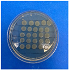 | + 200 mg/mL 100 mg/mL 50 mg/mL 25 mg/mL − |
| CaCO3-CEO |  | 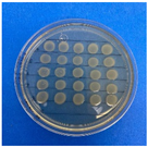 | + 200 mg/mL 100 mg/mL 50 mg/mL 25 mg/mL − |
| TiO2 | 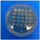 | 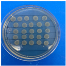 | + 200 mg/mL 100 mg/mL 50 mg/mL 25 mg/mL − |
| TiO2-CEO | 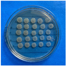 | 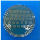 | + 200 mg/mL 100 mg/mL 50 mg/mL 25 mg/mL − |
| Al2O3 | 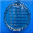 |  | + 200 mg/mL 100 mg/mL 50 mg/mL 25 mg/mL − |
| Al2O3-CEO |  |  | + 200 mg/mL 100 mg/mL 50 mg/mL 25 mg/mL − |
| Sample | Cold Crystallization | Melting | Crystallinity | |||
|---|---|---|---|---|---|---|
| Tg (°C) | Tc (°C) | (J/g) | Tm (°C) | (J/g) | Xc (%) | |
| PLA | 53 | 89.51 | 22.78 | 166.79 | 40.89 | 18.61 |
| PLA-TiO2-CEO | 56 | 93 | 39.23 | 166.09 | 46.82 | 7.80 |
| PLA-CaCO3-CEO | 57 | - | - | 166.56 | 18.93 | 19.45 |
| PLA-Al2O3-CEO | 56 | - | - | 166.32 | 41.72 | 42.87 |
| PLA-TiO2 | - | - | 166.61 | 37.21 | 38.24 | |
| PLA-CaCO3 | - | - | 166.90 | 26.53 | 27.27 | |
| PLA-Al2O3 | - | - | 167.47 | 39.24 | 40.33 |
Disclaimer/Publisher’s Note: The statements, opinions and data contained in all publications are solely those of the individual author(s) and contributor(s) and not of MDPI and/or the editor(s). MDPI and/or the editor(s) disclaim responsibility for any injury to people or property resulting from any ideas, methods, instructions or products referred to in the content. |
© 2023 by the authors. Licensee MDPI, Basel, Switzerland. This article is an open access article distributed under the terms and conditions of the Creative Commons Attribution (CC BY) license (https://creativecommons.org/licenses/by/4.0/).
Share and Cite
López-Cano, A.A.; Martínez-Aguilar, V.; Peña-Juárez, M.G.; López-Esparza, R.; Delgado-Alvarado, E.; Gutiérrez-Castañeda, E.J.; Del Angel-Monroy, M.; Pérez, E.; Herrera-May, A.L.; Gonzalez-Calderon, J.A. Chemically Modified Nanoparticles for Enhanced Antioxidant and Antimicrobial Properties with Cinnamon Essential Oil. Antioxidants 2023, 12, 2057. https://doi.org/10.3390/antiox12122057
López-Cano AA, Martínez-Aguilar V, Peña-Juárez MG, López-Esparza R, Delgado-Alvarado E, Gutiérrez-Castañeda EJ, Del Angel-Monroy M, Pérez E, Herrera-May AL, Gonzalez-Calderon JA. Chemically Modified Nanoparticles for Enhanced Antioxidant and Antimicrobial Properties with Cinnamon Essential Oil. Antioxidants. 2023; 12(12):2057. https://doi.org/10.3390/antiox12122057
Chicago/Turabian StyleLópez-Cano, Aaron A., Verónica Martínez-Aguilar, Mariana G. Peña-Juárez, Ricardo López-Esparza, Enrique Delgado-Alvarado, Emmanuel J. Gutiérrez-Castañeda, Mayra Del Angel-Monroy, Elías Pérez, Agustín L. Herrera-May, and J. Amir Gonzalez-Calderon. 2023. "Chemically Modified Nanoparticles for Enhanced Antioxidant and Antimicrobial Properties with Cinnamon Essential Oil" Antioxidants 12, no. 12: 2057. https://doi.org/10.3390/antiox12122057
APA StyleLópez-Cano, A. A., Martínez-Aguilar, V., Peña-Juárez, M. G., López-Esparza, R., Delgado-Alvarado, E., Gutiérrez-Castañeda, E. J., Del Angel-Monroy, M., Pérez, E., Herrera-May, A. L., & Gonzalez-Calderon, J. A. (2023). Chemically Modified Nanoparticles for Enhanced Antioxidant and Antimicrobial Properties with Cinnamon Essential Oil. Antioxidants, 12(12), 2057. https://doi.org/10.3390/antiox12122057