

Mental Imagery in Dreams of Congenitally Blind People


 , , and
Abstract
:1. Introduction
2. Methods and Materials
2.1. Dream Selection
2.2. Dream Analysis
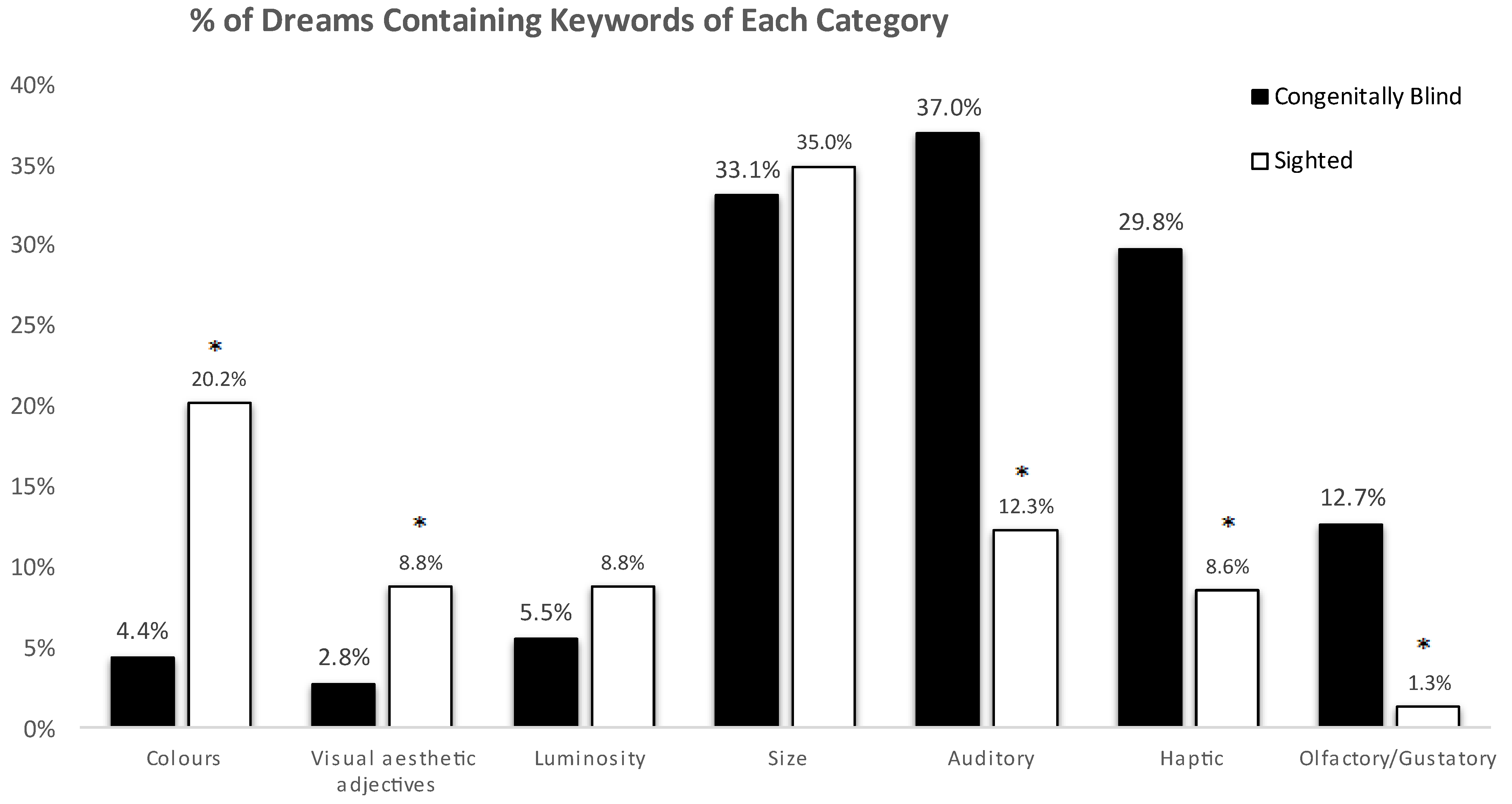
3. Results
4. Discussion
Supplementary Materials
Author Contributions
Funding
Data Availability Statement
Acknowledgments
Conflicts of Interest
References
- Dance, C.J.; Ward, J.; Simner, J. What is the Link Between Mental Imagery and Sensory Sensitivity? Insights from Aphantasia. Perception 2021, 50, 757–782. [Google Scholar] [CrossRef] [PubMed]
- Pearson, J.; Naselaris, T.; Holmes, E.A.; Kosslyn, S.M. Mental Imagery: Functional Mechanisms and Clinical Applications. Trends Cogn. Sci. 2015, 19, 590–602. [Google Scholar] [CrossRef] [PubMed]
- Wasserman, D.; Gullone, S.; Duncan, I.; Veronese, M.; Gnoni, V.; Higgins, S.; Birdseye, A.; Gelegen, E.C.; Goadsby, P.J.; Ashkan, K.; et al. Restricted truncal sagittal movements of rapid eye movement behaviour disorder. NPJ Park. Dis. 2022, 8, 26. [Google Scholar] [CrossRef] [PubMed]
- Hobson, J.A. REM sleep and dreaming: Towards a theory of protoconsciousness. Nat. Rev. Neurosci. 2009, 10, 803–813. [Google Scholar] [CrossRef] [PubMed]
- Ilic, K.; Bertani, R.; Lapteva, N.; Drakatos, P.; Delogu, A.; Raheel, K.; Soteriou, M.; Mutti, C.; Steier, J.; Carmichael, D.W.; et al. Visuo-spatial imagery in dreams of congenitally and early blind: A systematic review. Front. Integr. Neurosci. 2023, 17, 1204129. [Google Scholar] [CrossRef] [PubMed]
- Amadeo, M.; Gomez, E. Eye Movements, Attention and Dreaming in Subjects with Lifelong Blindness. Can. Psychiatr. Assoc. J. 1966, 11, 501–507. [Google Scholar] [CrossRef]
- Berger, R.J.; Olley, P.; Oswald, I. The EEG, eye-movements and dreams of the blind. Q. J. Exp. Psychol. 1962, 14, 183–186. [Google Scholar] [CrossRef]
- Christensen, J.A.E.; Aubin, S.; Nielsen, T.; Ptito, M.; Kupers, R.; Jennum, P. Rapid eye movements are reduced in blind individuals. J. Sleep Res. 2019, 28, e12866. [Google Scholar] [CrossRef]
- Holzinger, B. The Dreams of the Blind: In Consideration of the Congenital and Adventitiously Blind. J. Sleep Res. 2000, 9, 83. [Google Scholar]
- Hurovitz, C.S.; Dunn, S.; Domhoff, G.W.; Fiss, H. The dreams of blind men and women: A replication and extension of previous findings. Dreaming 1999, 9, 183–193. [Google Scholar] [CrossRef]
- Kerr, N.H.; Foulkes, D.; Schmidt, M. The structure of laboratory dream reports in blind and sighted subjects. J. Nerv. Ment. Dis. 1982, 170, 286–294. [Google Scholar] [CrossRef] [PubMed]
- Kirtley, D.D. The Psychology of Blindness; Nelson-Hall: Oxford, UK, 1975; p. xv. 312p. [Google Scholar]
- Meaidi, A.; Jennum, P.; Ptito, M.; Kupers, R. The sensory construction of dreams and nightmare frequency in congenitally blind and late blind individuals. Sleep Med. 2014, 15, 586–595. [Google Scholar] [CrossRef] [PubMed]
- Staunton, H.; O’Rourke, K. The creation of a topographical world and its contents in the dreams of the congenitally blind. Dreaming 2012, 22, 53–57. [Google Scholar] [CrossRef]
- Andrade, M.J.O. Do congenitally blind people have visual dreams? Sleep Sci. 2021, 14, 190–192. [Google Scholar] [CrossRef] [PubMed]
- Lopes Da Silva, F.H. Visual dreams in the congenitally blind? Trends Cogn. Sci. 2003, 7, 328–330. [Google Scholar] [CrossRef] [PubMed]
- Aleman, A.; van Lee, L.; Mantione, M.H.M.; Verkoijen, I.G.; de Haan, E.H.F. Visual imagery without visual experience: Evidence from congenitally totally blind people. NeuroReport 2001, 12, 2601–2604. [Google Scholar] [CrossRef] [PubMed]
- Sadato, N.; Pascual-Leone, A.; Grafman, J.; Ibañez, V.; Deiber, M.P.; Dold, G.; Hallett, M. Activation of the primary visual cortex by Braille reading in blind subjects. Nature 1996, 380, 526–528. [Google Scholar] [CrossRef] [PubMed]
- Striem-Amit, E.; Amedi, A. Visual cortex extrastriate body-selective area activation in congenitally blind people “Seeing” by using sounds. Curr. Biol. 2014, 24, 687–692. [Google Scholar] [CrossRef]
- Röder, B.; Rösler, F.; Hennighausen, E. Different cortical activation patterns in blind and sighted humans during encoding and transformation of haptic images. Psychophysiology 1997, 34, 292–307. [Google Scholar] [CrossRef]
- Kujala, T.; Huotilainen, M.; Sinkkonen, J.; Ahonen, A.I.; Alho, K.; Hämälä:inen, M.S.; Ilmoniemi, R.J.; Kajola, M.; Knuutila, J.E.T.; Lavikainen, J.; et al. Visual cortex activation in blind humans during sound discrimination. Neurosci. Lett. 1995, 183, 143–146. [Google Scholar] [CrossRef]
- Kujala, T.; Palva, M.J.; Salonen, O.; Alku, P.; Huotilainen, M.; Järvinen, A.; Näätänen, R. The role of blind humans’ visual cortex in auditory change detection. Neurosci. Lett. 2005, 379, 127–131. [Google Scholar] [CrossRef] [PubMed]
- Kupers, R.; Fumal, A.; de Noordhout, A.M.; Gjedde, A.; Schoenen, J.; Ptito, M. Transcranial magnetic stimulation of the visual cortex induces somatotopically organized qualia in blind subjects. Proc. Natl. Acad. Sci. USA 2006, 103, 13256. [Google Scholar] [CrossRef] [PubMed]
- Liotti, M.; Ryder, K.; Woldorff, M.G. Auditory attention in the congenitally blind: Where, when and what gets reorganized? Neurorep. An. Int. J. Rapid Commun. Res. Neurosci. 1998, 9, 1007–1012. [Google Scholar] [CrossRef] [PubMed]
- Müller, F.; Niso, G.; Samiee, S.; Ptito, M.; Baillet, S.; Kupers, R. A thalamocortical pathway for fast rerouting of tactile information to occipital cortex in congenital blindness. Nat. Commun. 2019, 10, 5154. [Google Scholar] [CrossRef] [PubMed]
- De Volder, A.G.; Toyama, H.; Kimura, Y.; Kiyosawa, M.; Nakano, H.; Vanlierde, A.; Wanet-Defalque, M.C.; Mishina, M.; Oda, K.; Ishiwata, K.; et al. Auditory triggered mental imagery of shape involves visual association areas in early blind humans. Neuroimage 2001, 14, 129–139. [Google Scholar] [CrossRef] [PubMed]
- Vetter, P.; Bola, L.; Reich, L.; Bennett, M.; Muckli, L.; Amedi, A. Decoding Natural Sounds in Early “Visual” Cortex of Congenitally Blind Individuals. Curr. Biol. 2020, 30, 3039–3044.e2. [Google Scholar] [CrossRef] [PubMed]
- Raz, N.; Amedi, A.; Zohary, E. V1 activation in congenitally blind humans is associated with episodic retrieval. Cereb. Cortex 2005, 15, 1459–1468. [Google Scholar] [CrossRef]
- Weeks, R.; Horwitz, B.; Aziz-Sultan, A.; Tian, B.; Wessinger, C.M.; Cohen, L.G.; Hallett, M.; Rauschecker, J.P. A positron emission tomographic study of auditory localization in the congenitally blind. J. Neurosci. 2000, 20, 2664–2672. [Google Scholar] [CrossRef]
- Amedi, A.; Raz, N.; Azulay, H.; Malach, R.; Zohary, E. Cortical activity during tactile exploration of objects in blind and sighted humans. Restor. Neurol. Neurosci. 2010, 28, 143–156. [Google Scholar] [CrossRef]
- Marks, G.A.; Shaffery, J.P.; Oksenberg, A.; Speciale, S.G.; Roffwarg, H.P. A functional role for REM sleep in brain maturation. Behav. Brain Res. 1995, 69, 1–11. [Google Scholar] [CrossRef]
- Frank, M.G.; Issa, N.P.; Stryker, M.P. Sleep Enhances Plasticity in the Developing Visual Cortex. Neuron 2001, 30, 275–287. [Google Scholar] [CrossRef] [PubMed]
- Foulkes, D. Children’s Dreams: Longitudinal Studies; Wiley: New York, NY, USA, 1982. [Google Scholar]
- Foulkes, D. Children’s Dreaming and the Development of Consciousness; Harvard University Press: Cambridge MA, USA, 1999. [Google Scholar]
- Siclari, F.; Valli, K.; Arnulf, I. Dreams and nightmares in healthy adults and in patients with sleep and neurological disorders. Lancet Neurol. 2020, 19, 849–859. [Google Scholar] [CrossRef] [PubMed]
- Eagleman, D.M.; Vaughn, D.A. The Defensive Activation Theory: REM Sleep as a Mechanism to Prevent Takeover of the Visual Cortex. Front. Neurosci. 2021, 15, 632853. [Google Scholar] [CrossRef] [PubMed]
- Simor, P.; van der Wijk, G.; Nobili, L.; Peigneux, P. The microstructure of REM sleep: Why phasic and tonic? Sleep Med. Rev. 2020, 52, 101305. [Google Scholar] [CrossRef] [PubMed]
- Braun, A.R.; Balkin, T.J.; Wesenten, N.J.; Carson, R.E.; Varga, M.; Baldwin, P.; Selbie, S.; Belenky, G.; Herscovitch, P. Regional cerebral blood flow throughout the sleep-wake cycle. An H2(15)O PET study. Brain 1997, 120 Pt 7, 1173–1197. [Google Scholar] [CrossRef] [PubMed]
- Tononi, G. An information integration theory of consciousness. BMC Neurosci. 2004, 5, 42. [Google Scholar] [CrossRef] [PubMed]
- Andrillon, T.; Nir, Y.; Cirelli, C.; Tononi, G.; Fried, I. Single-neuron activity and eye movements during human REM sleep and awake vision. Nat. Commun. 2015, 6, 7884. [Google Scholar] [CrossRef] [PubMed]
- Bértolo, H.; Mestre, T.; Barrio, A.; Antona, B. Rapid Eye Movements (REMs) and Visual Dream Recall in Both Congenitally Blind and Sighted Subjects; SPIE: Cergy Pontoise, France, 2017; Volume 10453. [Google Scholar]
- Bértolo, H.; Paiva, T.; Pessoa, L.; Mestre, T.; Marques, R.; Santos, R. Visual dream content, graphical representation and EEG alpha activity in congenitally blind subjects. Brain Res. Cogn. Brain Res. 2003, 15, 277–284. [Google Scholar] [CrossRef]
- Bértolo, H. Visual Imagery without Visual Perception. Psicologica 2005, 26, 173–188. [Google Scholar]
- Barrett, J.; Ehrlichman, H. Bilateral hemispheric alpha activity during visual imagery. Neuropsychologia 1982, 20, 703–708. [Google Scholar] [CrossRef]
- Cantero, J.L.; Atienza, M.; Salas, R.M.; Gómez, C. Alpha power modulation during periods with rapid oculomotor activity in human REM sleep. NeuroReport For. Rapid Commun. Neurosci. Res. 1999, 10, 1817–1820. [Google Scholar] [CrossRef] [PubMed]
- Williamson, S.J.; Kaufman, L.; Lu, Z.L.; Wang, J.Z.; Karron, D. Study of human occipital alpha rhythm: The alphon hypothesis and alpha suppression. Int. J. Psychophysiol. 1997, 26, 63–76. [Google Scholar] [CrossRef] [PubMed]
- Kerr, N.H.; Domhoff, G.W. Do the Blind Literally “See” in Their Dreams? A Critique of a Recent Claim That They Do. Dreaming 2004, 14, 230–233. [Google Scholar] [CrossRef]
- Schneider, A.; Domhoff, G.W. The Quantitative Study of Dreams. Available online: http://dreamresearch.net/ (accessed on 1 May 2021).
- Moverley, M.; Schredl, M.; Göritz, A.S. Media dreaming and media consumption—An online study. Int. J. Dream. Res. 2018, 11, 127–134. [Google Scholar] [CrossRef]
- Wilmer, H.H.; Sherman, L.E.; Chein, J.M. Smartphones and Cognition: A Review of Research Exploring the Links between Mobile Technology Habits and Cognitive Functioning. Front. Psychol. 2017, 8, 605. [Google Scholar] [CrossRef] [PubMed]
- Hall, C.S.; Van De Castle, R.L. The Content Analysis of Dreams; Appleton-Century-Crofts: East Norwalk, CT, USA, 1966; p. xiv. 320p. [Google Scholar]
- Carbon, C.-C.; Jakesch, M. A Model for Haptic Aesthetic Processing and Its Implications for Design. Proc. IEEE 2013, 101, 2123–2133. [Google Scholar] [CrossRef]
- Voss, U.; Holzmann, R.; Tuin, I.; Hobson, J.A. Lucid dreaming: A state of consciousness with features of both waking and non-lucid dreaming. Sleep 2009, 32, 1191–1200. [Google Scholar] [CrossRef]
- Mota-Rolim, S.A.; Erlacher, D.; Tort, A.B.L.; Araujo, J.F.; Ribeiro, S. Different kinds of subjective experience during lucid dreaming may have different neural substrates. Int. J. Dream. Res. 2010, 3, 33–35. [Google Scholar] [CrossRef]
- Murzyn, E. Do we only dream in colour? A comparison of reported dream colour in younger and older adults with different experiences of black and white media. Conscious. Cogn. 2008, 17, 1228–1237. [Google Scholar] [CrossRef]
- Schwitzgebel, E. Why did we think we dreamed in black and white? Stud. Hist. Philos. Sci. Part. A 2002, 33, 649–660. [Google Scholar] [CrossRef]
- Fazekas, P.; Nemeth, G.; Overgaard, M. White dreams are made of colours: What studying contentless dreams can teach about the neural basis of dreaming and conscious experiences. Sleep Med. Rev. 2019, 43, 84–91. [Google Scholar] [CrossRef] [PubMed]
- Hasson, U.; Levy, I.; Behrmann, M.; Hendler, T.; Malach, R. Eccentricity Bias as an Organizing Principle for Human High-Order Object Areas. Neuron 2002, 34, 479–490. [Google Scholar] [CrossRef] [PubMed]
- Pulvermuller, F. Neural reuse of action perception circuits for language, concepts and communication. Prog. Neurobiol. 2018, 160, 127–134. [Google Scholar] [CrossRef]
- Papadopoulos, R.K. The Handbook of Jungian Psychology: Theory, Practice and Applications; Routledge: London, UK, 2006. [Google Scholar]
- Pascual-Leone, A.; Hamilton, R. The metamodal organization of the brain. Progress. Brain Res. 2001, 134, 427–445. [Google Scholar]
- Vanlierde, A.; De Volder, A.G.; Wanet-Defalque, M.C.; Veraart, C. Occipito-parietal cortex activation during visuo-spatial imagery in early blind humans. Neuroimage 2003, 19, 698–709. [Google Scholar] [CrossRef] [PubMed]
- Bach-y-Rita, P.; Collins, C.C.; Saunders, F.A.; White, B.; Scadden, L. Vision substitution by tactile image projection. Nature 1969, 221, 963–964. [Google Scholar] [CrossRef] [PubMed]
- Abboud, S.; Hanassy, S.; Levy-Tzedek, S.; Maidenbaum, S.; Amedi, A. EyeMusic: Introducing a “visual” colorful experience for the blind using auditory sensory substitution. Restor. Neurol. Neurosci. 2014, 32, 247–257. [Google Scholar] [CrossRef]
- Buchs, G.; Heimler, B.; Amedi, A. The Effect of Irrelevant Environmental Noise on the Performance of Visual-to-Auditory Sensory Substitution Devices Used by Blind Adults. Multisens. Res. 2019, 32, 87–109. [Google Scholar] [CrossRef]
- Bavelier, D.; Neville, H.J. Cross-modal plasticity: Where and how? Nat. Rev. Neurosci. 2002, 3, 443–452. [Google Scholar] [CrossRef]
- Burton, H. Visual Cortex Activity in Early and Late Blind People. J. Neurosci. 2003, 23, 4005. [Google Scholar] [CrossRef]
- Amedi, A.; Stern, W.M.; Camprodon, J.A.; Bermpohl, F.; Merabet, L.; Rotman, S.; Hemond, C.; Meijer, P.; Pascual-Leone, A. Shape conveyed by visual-to-auditory sensory substitution activates the lateral occipital complex. Nat. Neurosci. 2007, 10, 687–689. [Google Scholar] [CrossRef]
- Likova, L.T. Drawing enhances cross-modal memory plasticity in the human brain: A case study in a totally blind adult. Front. Hum. Neurosci. 2012, 6, 44. [Google Scholar] [CrossRef]

| Dreambank CODE | Sex | Age | Years of Education | Occupation | Nature/Degree of Blindness | # of Dream Reports |
|---|---|---|---|---|---|---|
| 1 | F | 32 | 18 | Unemployed | C/T | 10 |
| 2 | F | 52 | 12 | Envelope stuffer | C/T | 37 |
| 3 | F | 44 | 18 | Factory worker (retired) | C/T | 32 |
| 4 | F | 44 | 13 | Medical transcriptionist | C/T | 9 |
| 5 | M | 45 | 16 | Human resources management | C/T | 61 |
| 6 | M | 46 | 12 | Small engine repairs | C/T | 12 |
| 7 | F | 18 | 13 | College student | C/T | 19 |
| Colours | ^white^ or ^black^ or ^gold^ or ^silver^ or ^copper^ or ^bronze^ or ^red^ or ^green^ or ^orange^ or ^violet^ or ^purple^ or ^blue^ or ^yellow^ or ^gr[ae]y^ |
| Aesthetic adjectives | pretty or beaut- or gorgeous or handsome or ugly or disgust or attractive |
| Luminosity | dark or bright or ^light^ or ^lit^ or ^shin(ing|e|ed)^ or illumi or ^sun(^|ny)^ |
| Size | ^big(|ger)^ or enormous or huge or ^long^ or ^larg(e|er)^ or ^gi(ant|gantic)^ or ^ta(ll|ller)^ or ^smal(l|ler|lest)^ or t[i|ee]ny or little or ^thi(n|nner|nnest)^ |
| Auditory | ^hea(r|rd|ring)^ or sound or ^lou(d|dly|der)^ or ^quiet^ or nois |
| Haptic/Touch | ^touc(h|hing|ed)^ or ^fe(el|lt)^ or ^smooth^ or ^soft^ or ^co(ol|ld) or ^h(eat|ot)^ or ^pai(n|ful)^ or ^hurt^ or ^warm |
| Olfactory/Gustatory | smel(l|t) or scent or tast(y|e) |
| Dream | “[…] We went over to a table that was up against the wall, at one end of the studio. The top was covered by a white chiffon tablecloth, very voluminous. It was a gorgeous thing, very soft and full and beautiful. And on the table were two big silver candelabras with candles in them, and I think they were lit. Neither R. nor I were content with the way the tablecloth was arranged, so while we waited for the music to come on, we went over to the tablecloth to rearrange it in nicer folds […]” |
| Q: | Do you think R. told you that the tablecloth was white? |
| E: | No. I just knew it and I had a visual impression of white which I can’t describe except that it was just devoid of any darkness, no color. |
| Q: | Do you often have this sensation? |
| E: | No, it’s just as unusual as having a color impression, for me, that is. Actually, of course, I never have any idea of dark and light, neither in the day nor in the night. It’s just nothing at all, but this was a real visual impression of white, at least it was to me. It may just be my conception of white, but there it was. |
| Q: | What about the candelabra, did you have an impression of the color silver or do you mean you knew it was of the metal silver? |
| E: | Well, I knew it was silver metal because it was very smooth to touch, but I also had the impression of silver and the way I know silver is that it’s like white only shiny. |
Disclaimer/Publisher’s Note: The statements, opinions and data contained in all publications are solely those of the individual author(s) and contributor(s) and not of MDPI and/or the editor(s). MDPI and/or the editor(s) disclaim responsibility for any injury to people or property resulting from any ideas, methods, instructions or products referred to in the content. |
© 2023 by the authors. Licensee MDPI, Basel, Switzerland. This article is an open access article distributed under the terms and conditions of the Creative Commons Attribution (CC BY) license (https://creativecommons.org/licenses/by/4.0/).
Share and Cite
Kang, J.; Bertani, R.; Raheel, K.; Soteriou, M.; Rosenzweig, J.; Valentin, A.; Goadsby, P.J.; Tahmasian, M.; Moran, R.; Ilic, K.; et al. Mental Imagery in Dreams of Congenitally Blind People. Brain Sci. 2023, 13, 1394. https://doi.org/10.3390/brainsci13101394
Kang J, Bertani R, Raheel K, Soteriou M, Rosenzweig J, Valentin A, Goadsby PJ, Tahmasian M, Moran R, Ilic K, et al. Mental Imagery in Dreams of Congenitally Blind People. Brain Sciences. 2023; 13(10):1394. https://doi.org/10.3390/brainsci13101394
Chicago/Turabian StyleKang, Jungwoo, Rita Bertani, Kausar Raheel, Matthew Soteriou, Jan Rosenzweig, Antonio Valentin, Peter J. Goadsby, Masoud Tahmasian, Rosalyn Moran, Katarina Ilic, and et al. 2023. "Mental Imagery in Dreams of Congenitally Blind People" Brain Sciences 13, no. 10: 1394. https://doi.org/10.3390/brainsci13101394
APA StyleKang, J., Bertani, R., Raheel, K., Soteriou, M., Rosenzweig, J., Valentin, A., Goadsby, P. J., Tahmasian, M., Moran, R., Ilic, K., Ockelford, A., & Rosenzweig, I. (2023). Mental Imagery in Dreams of Congenitally Blind People. Brain Sciences, 13(10), 1394. https://doi.org/10.3390/brainsci13101394