Abstract
A review that summarizes the most recent technological developments in the field of ultrafast structural dynamics with focus on the use of ultrashort X-ray and electron pulses follows. Atomistic views of chemical processes and phase transformations have long been the exclusive domain of computer simulators. The advent of femtosecond (fs) hard X-ray and fs-electron diffraction techniques made it possible to bring such a level of scrutiny to the experimental area. The following review article provides a summary of the main ultrafast techniques that enabled the generation of atomically resolved movies utilizing ultrashort X-ray and electron pulses. Recent advances are discussed with emphasis on synchrotron-based methods, tabletop fs-X-ray plasma sources, ultrabright fs-electron diffractometers, and timing techniques developed to further improve the temporal resolution and fully exploit the use of intense and ultrashort X-ray free electron laser (XFEL) pulses.
1. Introduction
The present review article summarizes some of the key advances in the development of ultrafast structural techniques over the last decade. Pioneering experiments including a historical compilation can be found elsewhere [1,2,3,4,5,6,7]. After more than 30 years of continuous improvements in femtosecond (fs, 1 fs = 10−15 s) hard X-ray and fs-electron sources, ultrafast diffractometers confer the prerequisite spatial and temporal resolutions to capture atoms in motion. The characteristic length-scale that such ultrafast cameras had to resolve is evident—this is the size of an atom or a bond length (dc)~1 Å = 10−10 m = 0.1 nm. In order to determine their necessary shutter speed or temporal resolution, however, one must assume a characteristic speed (vc) for atomic motion. There are faster and slower processes in nature but as a general rule of thumb, vc~103 m/s−1 = 1 nm ps−1 (1 ps = 10−12 s), which is roughly the speed of sound in most solid-state organic materials and liquids results a good estimate [8]. Note that structural chemists, biologists, and condensed matter physicists are usually interested in rather low-frequency, high-amplitude atomic displacements. Soft vibrational modes are those that govern chemical reactivity through the anharmonicities encountered along the potential energy landscape as we approach the bond dissociation threshold or the transition state [9]. Protein function, on the other hand, seems to rely on collective low-frequency vibrational modes that couple to large amplitude protein motions for which thermal bath fluctuations and entropic factors play key roles (vide infra examples in Section 2.4.2 and Section 2.4.3). In addition, very interesting condensed matter physics arises from low-frequency collective excitations (vide infra examples in Section 2.1, Section 3.5 and Section 3.6). Two examples are the famous amplitude phonon mode (known as amplitudon) that modulates the charge density wave (CWD) gap in Peierls distorted systems, [10] and the lattice vibrations responsible for the formation of Cooper pairs that lead to the foundation of the BCS theory for superconductivity [11]. Thus, a characteristic time (Δtc) or temporal resolution of about Δtc = dc vc−1 = 0.1 nm(1 nm ps−1)−1 = 0.1 ps = 100 fs suffices to monitor a vast variety of structurally relevant phenomena.
Ultrafast diffraction belongs to a family of techniques known as pump-probe in which two well-synchronized pulses, a pump and a probe, are implemented. The pump is a fs-optical pulse coming from a master fs-laser and is responsible for initiating structural changes by bringing the system out of equilibrium. The probe is an ultrashort X-ray or electron pulse, which scatters off the irradiated sample volume to generate a diffraction pattern downstream. A series of diffraction patterns obtained for different time delays (t) between these two pulses is then recorded in a stroboscopic fashion. Structural refinement finally brings time-dependent changes in diffracted intensities to real-space information to render a “molecular movie” that unveils details about electron density and nuclear reorganization. The success of ultrafast structural dynamics, therefore, relies on the ability of producing ultrashort hard X-ray pulses or electron bursts as well as maintaining an outstanding synchronization between probe and pump pulses. Recall these are repetitive stroboscopic experiments so both the durations of the pump and probe pulses and their relative timing are vital factors in addition to source brightness. The synchronization noise is referred to as timing jitter and has been one of the key issues when radio frequency (RF) fields are utilized for accelerating or compressing electron bunches.
It should be noted that a good source of intense electron bursts is not only the heart of any time-resolved electron diffractometer, but also the first component required to generate hard X-ray pulses at synchrotron and X-ray free electron laser (XFEL) facilities. Furthermore, RF technology is fundamental to operate electron injectors, linacs (linear accelerators), boosters, and storage rings. Thus, circumventing the problem of the timing jitter between the driving fs-laser and RF fields has been essential to the success of ultrafast structural experiments performed with both RF-based fs-electron diffraction (FED) setups (vide infra Section 3.6 and Section 3.7) and XFELs (vide infra Section 2.5). A brief description of the main techniques that enabled monitoring atomic motion with the aforementioned time resolution of ~100 fs is highlighted in the application-based examples which follow.
2. Structural Techniques Involving Ultrashort X-ray Pulses
2.1. Laser Slicing at Third-Generation Synchrotron Facilities
Synchrotron radiation continues to be one of the most powerful sources of polychromatic light capable of covering a wide spectral range that extends from THz to hard X-rays [12]. Depending on the facility, type of insertion device, beam optics, and required degree of monochromaticity; X-ray fluxes at synchrotrons typically range from about 104 to 108 photons/pulse (at the sample position). Moreover, photon energies >6 keV (photon wavelengths <2.1 Å) are often required in order to attain atomic spatial resolution. The temporal resolution of synchrotron sources is dictated by the length (duration) of the stored electron bunch, which commonly ranges from 50 ps to 200 ps (fwhm, full width at half maximum) [13,14].
Laser slicing techniques [15,16,17,18,19,20,21,22] enabled the production of ~100 fs (fwhm) hard X-ray pulses at third-generation synchrotron facilities such as the Advanced Light Source (ALS) and the Swiss Light Source (SLS). Figure 1 illustrates the laser slicing principle originally proposed by Zholents and Zolotorev [16] and demonstrated by Schoenlein et al. [15] at ALS. Here, an intense fs-laser pulse with a field strength of ~109 V m–1 co-propagates with the stored electron bunch through a wiggler. Under optimal resonant conditions, the high laser peak intensity introduces an energy modulation in the underlying electrons of several MeV. This energy modulation is much larger than the energy spread of the unperturbed electrons in the bunch and, therefore, energy-modulated electrons can be transversely separated from the main beam through the implementation of a bending magnet, and subsequently used to produce fs-X-ray pulses in another bend. Finally, an aperture is applied to select one of the spatially separated fs-X-ray pulses, which then can be used for probing the ultrafast structural changes. Note that timing jitter here is negligible since the electron beam modulation (or fs-X-ray pulse) and the photoexcitation pulse originate from the same initial laser pulse via beam splitters. All pulse durations below are given in fwhm if not stated otherwise.

Figure 1.
Schematic of the fs-laser slicing method for generating fs-hard X-ray pulses at ALS. Laser interaction with the electron bunch in a resonantly tuned wiggler introduces an energy modulation. The modulated electrons are spatially separated by a dispersive bend. Thus, subsequently produced fs-X-ray pulses will follow a slightly different path and can be selected for probing. Reproduced from Reference [15] with permission from Science AAAS.
Figure 2 shows recent results obtained by Huber et al. [20] at the FEMTO slicing facility of SLS for the prototypical Peierls 1D charge-density wave (CDW) system, K0.3MoO3. The amplitudon in this material has a frequency (period) of 1.7 THz (590 fs) [23] at T ~ 0 K. Fs-X-ray diffraction experiments were performed in a grazing incidence geometry while the K0.3MoO3 single crystalline sample was maintained well below Tc. The crystal was excited by 100-fs laser pulses centered at λ = 800 nm and probed by 80-fs X-ray pulses with λ = 1.8 Å. The relative intensity changes of the -reflection of the superlattice lattice (SL) were measured as a function of t (delay) at various photoexcitation fluences (Figure 2a). Results were modeled (Figure 2d) by implementing an equation of motion derived from a double-well potential (Figure 2b). Huber et al. [20] were able to observe the coherent lattice dynamics associated with the CDW formation. The authors interpreted the results as a rearrangement (atomic motion) of the SL distortion evolving along a generalized excitation-dependent potential energy surface. Their observed recovery of the SL distortion remains unclear. The results presented in Figure 2 reflect typical measurements that should be performed in order to extract valuable physical information from time-dependent diffraction data; i.e., many time points as well as different photoexcitation levels are usually required. Thus, depending on the probed volume, diffraction efficiency, and X-ray beam brightness, several weeks of continuous data collection may be necessary. In addition, sample degradation may introduce additional challenges during extensive data acquisition. Laser slicing enabled the generation of fs-X-ray pulses in synchrotrons at the expense of lowering the X-ray beam flux to about 102–103 photons/pulse, which is comparable to the effective brightness of a fs-hard X-ray plasma source (see below).
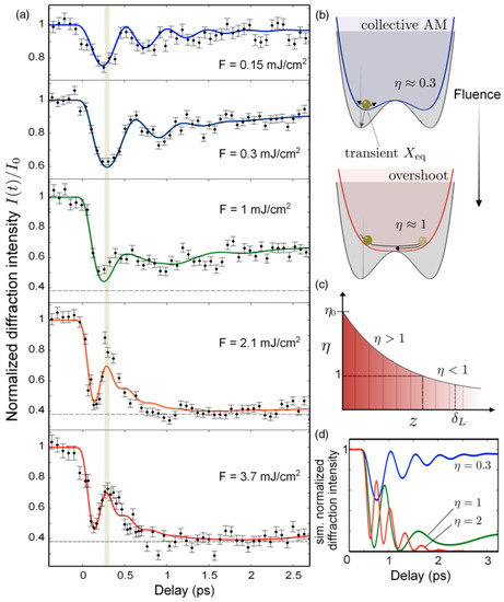
Figure 2.
Results from FEMTO line (laser slicing) at the Swiss Light Source (SLS). (a) Time evolution of the normalized SL diffraction peak intensity at different excitation fluences (F). The horizontal dashed line in the higher fluence scan represents saturation background. Solid lines in red are fits obtained by implementing an equation of motion derived from a double-well potential that includes a phenomenological damping term. They grey vertical line denotes the time at which the SL diffraction intensity recovers and peaks, t = 0.35 ps. (b) Double-well potential; the grey potential corresponds to the unperturbed charge-density wave (CDW) state (η = 0). Note that η is a scaled excitation parameter proportional to F so that η = 1 corresponds to the threshold value for overshooting, i.e., where the double-well shape of the potential vanishes (red curve). The green circle represents the collective atomic displacement of the SL distortion or dimmers along the structural coordinate of the Peierls distortion. The transient quasi-equilibrium position after excitation is labeled Xeq. (c) Schematic of the inhomogeneously excited crystal in the case of a high fluence excitation that leads to η0 > 1, the z axis corresponds to the axis perpendicular to the surface plane; δL corresponds to the laser penetration depth. (d) Simulated diffraction signals of a thin layer for certain values of η. Reproduced from Reference [20] with permission from the American Physical Society (APS).
In addition to laser slicing, the operation of synchrotrons in the low alpha mode has also allowed the production of low-intensity X-ray pulses as short as 0.6 ps (rms, root mean square) [24]. There is a legitimate need for attaining ultrashort as well as ultrabright hard X-ray and electron pulses. Generally speaking, excitation fluences must be sufficiently high to transform at least 1% of the molecules (or unit cells) in order to discern structural changes from background signals and a very limited number of materials are able to survive multiple excitation cycles; metals such (Al, Au, etc.), semiconductors (Si, GaAs, InSb), ceramics and ceramic-like materials (Al2O3, ZnO, YBCO, etc.) are some typical examples. However, radiation labile specimens such as organic compounds and biomolecular systems only allow for a very limited number of pump shots before showing clear signs of degradation. At such excitation intensities, thermal effects, long-lived photochemical intermediates and photoproducts alter the specimen structure.
2.2. Table-Top fs Hard X-ray Plasma Diffractometers
Table-top fs-X-ray plasma diffractometers have been successfully implemented to unveil ultrafast structural dynamics in different materials [25,26,27,28,29,30,31,32,33,34,35,36,37,38,39]. These time-resolved instruments are based on the use of chirped-pulse amplification [40] with pulse energies in the range of 1–100 mJ depending on the repetition rate of the laser and the average power. Fs-hard X-ray pulses are produced when a solid target is irradiated by fs-laser pulses carrying a peak intensity Ipeak > 1016 W cm–2. The generation of fs-X-ray pulses from a plasma source involves the extraction of electrons from the metal target by strong field ionization with a small laser spot. This step is followed by electron acceleration in vacuum and electron re-entrance into the target material owing to the strong electric field. Then, X-ray photons are generated via collisional inner-shell ionization followed by radiative transitions of outer-shell electrons into inner-core holes. Such processes occur every cycle of the electric field of the fs-laser driving pulse.
The source is the main challenge in the design of table-top fs-X-ray plasma diffractometers. The surface of the target material is ablated after each laser shot and, therefore, it must be translated to preserve surface uniformity. Cu tape of about 20 µm in thickness is often utilized as target material producing the characteristic Cu Kα and Kβ X-ray emission lines. The emission of X-ray photons is highly isotropic (all directions). Hence, the implementation of X-ray optics becomes crucial to obtain a decent photon flux at the sample position. The schematic of a table-top fs-X-ray plasma diffractometer is shown in Figure 3. The X-ray source is driven by 50-fs laser pulses with a central wavelength, λ = 800 nm. After losses associated with X-ray collection, this instrument delivers at the sample position an effective X-ray photon flux of 5000 Cu Kα photons/pulse with a repetition rate of 1 kHz [38].
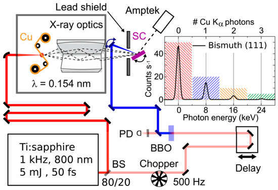
Figure 3.
Layout of the table-top fs-X-ray plasma diffractometer at the Max Born Institute. In this example, a (1 1 1)-oriented single crystalline (SC) Bi sample is photoexcited by 400-nm laser pulses generated by frequency doubling in a β-barium borate (BBO) crystal. The pump arm is mechanically chopped. A photodiode (PD) is used to determine the state of the sample (i.e., excited or unexcited) on shot to shot basis. Diffracted X-ray photons are measured by an energy-resolving CdTe diode (Amptek). The inset shows a spectrum with a very low X-ray flux on the detector. Figure adapted from Reference [38] with permission from the authors.
Figure 4 illustrates some representative measurements obtained by Freyer et al. [39] with the table-top fs-X-ray plasma diffractometer shown in Figure 3 but in a different configuration to perform experiments in transmission geometry (i.e., these measurements required the implementation of a large area high-resolution detector). Freyer et al. [39] studied a polycrystalline sample of [Fe(bpy)3]2+(PF6−)2. The authors were able to determine the electron density changes that follow a charge transfer reaction (Figure 4g–h). Recall that X-ray photons scatter off the electron density, i.e., they interact with the electron cloud and, therefore, variations in diffracted intensities (Figure 4b–f) correspond to changes in electron density arising from charge redistribution and/or nuclear rearrangements. Freyer et al. [39] carried out a careful analysis of the electron density maps that applies to centrosymmetric crystals under relatively low excitation fluence. The authors found that each photoexcited complex largely affects its surroundings, highlighting the many-body character of this charge transfer. They concluded that the PF6− counterions play a key role in charge relocation with a behaviour in the solid state that is markedly different from that in diluted liquid solutions where the spatial separation among solvated anions and cations is much larger. Similar anionic effects were found in (EDO-TTF)2PF6 by Gao et al. [41] through the implementation of FED.
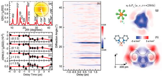
Figure 4.
Results from the above table-top fs-X-ray plasma source. (a) Radially integrated and normalized diffracted intensity I(2θ)/I0(0 2 2) from [Fe(bpy)3]2+(PF6−)2 in powder form. The inset shows a diffraction pattern recorded after an integration time of 8 h. The data have been normalized to the intensity of the (0 2 2)-Bragg reflection without photoexcitation and the asterisks mark the peaks originating from non-equivalent sets of lattice planes. (b–f) Temporal evolution of normalized changes of Bragg diffraction intensities as a function of scattering angle (b) and selected Bragg reflections (c–f). The red lines are continuous B-splines of the data. (g–h) Contour plots of 2D projections () obtained by integrating the changes in electron density (Δρ(r, t = +250 fs)) along the w-direction. The dashed lines indicate the contours of stationary electron density corresponding to (g) the six fluorine atoms and the phosphorus atom in the middle, and (h) the Fe atom, and one bipyridine unit. Please note that η denotes the fraction of unit cells undergoing photoinduced changes. Figure adapted from Reference [39] with permission for AIP Publishing LLC.
More recent efforts in the development of fs-hard X-ray plasma sources driven by mid-infrared (mid-IR) lasers demonstrated an increase in brightness of about 25 times [42]. The emission efficiency of X-ray photons increases with the kinetic energy of the re-entering electron, which is proportional to Ipeak λ2. Thus, the application of intense fs-mid-IR lasers may bring the effective brightness of table-top fs-hard X-ray diffractometers to the level of ~105–106 photons/pulse (at sample position). This is about one order of magnitude below the flux achieved at the Sub-Picosecond Photon Source (SPPS) [43]. SPPS was a test facility that operated at the Stanford Linear Accelerator Center (SLAC) before the Linac Coherent Light Source (LCLS) became available in 2009 (see section below). In addition to the above, fs-mid-IR drivers have been recently applied for the generation of coherent ultrahigh harmonics in gases reaching keV-photon energies [44,45]. Although high harmonic generation (HHG) is not yet an alternative for the production of fs-hard X-ray pulses for coherent diffraction, HHG sources with photon frequencies between the vacuum UV and soft X-ray regime have found applications in fs-spectroscopies and attosecond (att, 1 att = 10–18 s) metrologies [44,45,46,47,48,49,50,51,52,53,54,55,56,57,58,59,60,61,62,63,64,65].
2.3. Introduction to Fourth-Generation Fs-Hard X-ray Sources: X-ray Free Electron Lasers
Since the first ultrabright shot of fs-hard X-rays at LCLS in 2009, four additional XFEL user facilities [66,67,68,69,70,71] became operational: SACLA (Japan, 2011), PAL-XFEL (Korea, 2016), SwissFEL (Switzerland, 2016), and EuXFEL (Germany, 2017). Figure 5 shows the schematic of SACLA. In an XFEL, ultrabright electron bunches are created in an RF-photoinjector or a DC electron gun and accelerated by a linac to kinetic energies that normally range from 5 GeV to 15 GeV (up to 17.5 GeV at EuXFEL). As the case of synchrotrons, the duration of the X-ray pulses corresponds to the length of the electron bunches in the undulator. Magnetic chicanes are commonly used to compress the electron bursts before they arrive to the undulator. Depending on the electron bunch charge, X-ray pulses with typical lengths ranging from 5 fs to 400 fs can be readily obtained via self-amplified spontaneous emission (SASE), and even Fourier limited X-ray pulses through the implementation of self-seeding [72]. In addition, sub-fs X-ray pulses are also attainable by selectively spoiling the transverse emittance of the electron beam [72,73]. With up to ~1013 X-ray photons/pulse XFELs produce peak fluxes which are about 104–105 times brighter than synchrotrons. Perhaps the only disadvantage of XFELs over synchrotrons is the fact that each electron bunch in an XFEL experiences a single pass before hitting a block at the end of the beam line. Such high X-ray flux, relatively small bandwidth ~10–3–10–2 (SASE), and short pulse duration ~100 fs provide XFELs outstanding single-shot diffraction quality for ultrafast structural dynamics. In the following sections, the main structural techniques currently implemented at XFEL facilities will be introduced.

Figure 5.
Schematic of SACLA: a compact XFEL. EG, 500 keV electron gun; DF, deflector with collimator; SHB, 238 MHz subharmonic buncher; BS, 476 MHz booster; L-CC, L-band correction cavity; L-APS, L-band alternating periodic structure (APS) typed standing-wave cavity; C-CAT, C-band correction acceleration tube; S(C)-TWA, S(C)-band travelling-wave acceleration tube; BCn (n ≈ 1–3), nth bunch compressor; EDS, electron beam diagnostic section; SM, switching magnet; UND, undulator line; BLn (n = 1, 3), nth beam line; XDS, X-ray diagnostic system; EHA, experimental hall; OH, optical hutch; EH, experimental hutch. Only two beam lines beamlines (BL1 and BL3) were available in 2012. SACLA has the capability to install five independent FEL beamlines, and there are three currently in operation. Please refer to Reference [66] for details about each insertion device. Image adapted with permission from Springer Nature.
2.4. Main Applications at X-ray Free Electron Lasers (XFELs) for the Study of Structure and Dynamics of Matter
2.4.1. Serial Fs-X-ray Crystallography
Serial fs-nanocrystallography (SFX) [74,75,76,77,78,79,80,81,82,83,84,85,86,87,88,89,90,91,92] has been one of the first methods implemented at XFELs. The approach is illustrated in Figure 6a. Here, a µm-sized crystal is intercepted by the intense X-ray pulse to generate a diffraction pattern which encodes the relative orientation between the crystal and the X-ray beam. The process is repeated a number of times until sufficient amount of data is accumulated for structural refinement. The success of this technique relies on an approach called self-terminating diffraction gates, i.e., obtaining sufficient diffraction information when a sample crystal falls apart while an ultra-intense X-ray pulse propagates through it. Experiments at LCLS and simulations involving intense X-ray pulses with energies of up to 3 mJ [1 × 10¹³ photons/pulse @ 2 keV (λ = 6 Å)] and durations between 30 fs and 400 fs showed that a unique mechanism to ultrashort pulses allows structural information to be collected from crystalline samples using high radiation doses without the requirement for the pulse to terminate before the onset of sample damage. During exposure to a single X-ray pulse focused into a 10 µm2 spot with Ipeak~1017 W cm–2 a large fraction of the atoms in a protein crystal absorb a photon. This leads to an ultrafast cascade of secondary energy relaxation processes, lattice temperatures of ~500,000 K, high pressure, and the ultimate explosion of the sample. The onset of disorder is illustrated in Figure 6b for a small model molecule. As can be observed, the long-range order is completely gone in about 100 fs, and therefore the sample may lose its crystallinity during the transit time of the X-ray pulse.
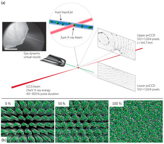
Figure 6.
Serial fs-nanocrystallography (SFX) approach. (a) A suspension of µm-crystals flows in a liquid jet across the X-ray beam, and diffraction is recorded using a pair of 512 × 1024-pixel pnCCD detectors. The lower half detector is placed further from the beam center than the upper half detector to increase the accessible range of scattering angles. Thousands of individual diffraction patterns are recorded from single µm-crystals with pulse lengths up to 300 fs to a resolution of 0.76 nm. (b) Onset of disorder in a crystalline lattice during an ultra-intense 100-fs X-ray pulse. Sample: lysergic acid diethylamide. Atomic displacements were calculated using Cretin [93]. For this small molecule, crystalline order is largely destroyed by 100 fs. Adapted from Reference [92] with permission from Springer Nature.
Figure 7 shows the effect of disorder on the accumulation of Bragg peak intensities for an intense X-ray pulse of duration Δt = 150 fs. Since disorder affects higher-resolution peaks in a stronger manner, the counting turns off sooner for higher-resolution Bragg reflections. The aforementioned experiments and modelling demonstrate that SFX does not require X-ray pulses to be shorter than the onset of long-range structural disorder. Instead, the impulsive heating leading to large-amplitude uncorrelated displacement of atoms destroys the crystalline order and shuts down Bragg diffraction. Therefore, SFX measurements in combination with simulations offer a way to characterize the order-to-disorder dynamics, correct for Bragg termination, find optimal X-ray pulse parameters, and perform structural refinement. The success of SFX is highlighted by the determination of a multitude of protein structures from (typically) sub-10 µm crystals [74,75,76,77,78,79,80,81,82,83,84,85,86,87,88,89,90,91,92].
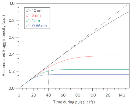
Figure 7.
Plot of relative accumulation of Bragg signal for Ipeak = 1 × 10¹⁷ W cm–2 and a pulse duration Δt = 150 fs. Bragg intensities initially accumulate at the same rate proportional to Ipeak. As expected, disorder affects higher-resolution peaks sooner (alike to the Debye–Waller effect) and termination of Bragg intensity accumulation may occur during the transit of the X-ray pulse through the crystal, leading to apparent pulse lengths (Δtapp) shorter than the duration of the incident pulse. In this case, Δtapp = 40 fs (black vertical dashed line), which is close to the turn-off time for the highest-resolution peaks at 0.76 nm. Figure adapted from Reference [92] with permission from Springer Nature.
It should be mentioned that the necessity of fs-X-ray pulses for attaining high-resolution structures of radiation-sensitive specimens is questionable. Serial millisecond crystallography at a synchrotron beamline, for instance, equipped with the same high-viscosity injector and a high frame-rate detector allowed for typical protein crystallographic experiments to be performed at room temperature [94]. In addition, fs-electron diffraction has also enabled the structural determination of radiation labile organic crystals at room temperature [41]. Low-dose micro-electron diffraction [95,96] (microED) is, in fact, likely to become the most convenient and economical approach for determining the structure of sub-µm crystals. Samples must, however, withstand the high vacuum conditions in the electron column or otherwise be kept at cryogenic temperatures. The main advantage of fs-X-ray probes relies on their ability to elucidate the dynamics of large protein systems. These large molecules are difficult to resolve with FED owing to the relatively low transverse coherence length (high transverse emittance) of fs-electron sources [4,5] (vide infra Section 3.2 and Section 3.3).
It has been argued that the combined use of a large crystal and XFEL pulses should provide inherently better structures than those obtained by SFX. In this technique the crystal must be very large and the distance between successive X-ray shots greater than the diameter of the damaged irradiated spot. This approach has been successfully applied at SACLA to obtain the structure of bovine heart cytochrome c oxidase (CcO) at 1.9 Å [97] and photosystem II (PS II) at 1.95 Å [98]. However, it has been shown that SFX can indeed deliver sub-1.5 Å from crystals of photoactive yellow protein (PYP) at room temperature [81].
Another possible method to bring µm-crystals to the X-ray beam path is the implementation of fixed-target crystallography chips [99,100,101,102]. Very recently, Schulz et al. [99] demonstrated a hit-and-return (HARE) approach based on the use of fixed-target crystallography chips mounted on a translation stage system. The HARE method enables time-resolved serial synchrotron crystallography with time resolution from milliseconds to seconds or longer. Figure 8 shows one of employed crystallography chips and some characteristic time-resolved structural results obtained from µm-crystals of Rhodopseudomonas palustris fluoroacetate dehalogenase (FAcD) soaked with a photocaged substrate as a model system. For more information vide infra reference [99].
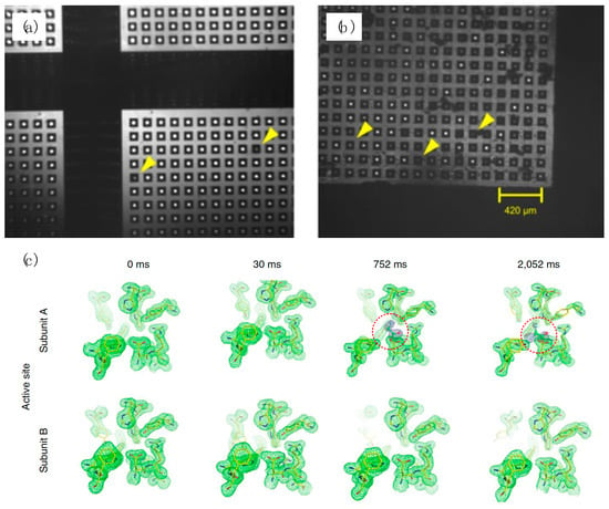
Figure 8.
Hit-and-return (HARE) method. (a) An empty chip is shown as a control. The yellow arrows point out underdeveloped features, which appear semi-transparent under infrared (IR) light illumination. (b) Loaded chip with yellow arrows representing crystals sitting in the features of the chip. Crystals sitting inside of the features can be clearly distinguished from crystals on its surface and from empty features. (c) Changes in fluoroacetate dehalogenase (FAcD) electron density as a function of t for the active site subunits A and B. Electron density maps are at a cutoff of 2σ (green, protein; blue, ligand (highlighted with red circle)). Figure adapted from Reference [99] with permission from Springer Nature.
2.4.2. Time-Resolved Fs-X-ray Crystallography and Scattering
Since the very first ultrafast structural experiments at SPPS [43,103,104], the brightness of XFELs has increased by about one million fold, allowing for the study of more complex systems and photoinduced phenomena such as protein dynamics in crystalline [105,106,107,108,109,110,111,112] and liquid phases, [113] bond formation in the [Au(CN)2–]3 trimer in solution, [114,115] lattice dynamics in individual gold nanoparticles, [116] ultrafast melting of charge and orbital order in PCMO, [117] non-linear lattice dynamics [118] and CWD order in YBCO, [119] visualization of breathing modes in nanocrystals at extreme photoexcitation conditions, [120] photoinduced insulator-to-metal (IM) transitions, [121] warm dense matter physics, [122] and ultrafast crystallization [123] and melting [124] in shock-compressed crystals.
Figure 9a–d shows time-resolved X-ray diffraction results obtained by Barends et al. [107] at LCLS in the investigation of the ultrafast structural dynamics that follows CO photodissociation in Myoglobin-CO (MbCO). Early time-dependent structural changes are clearly visible. In this work, horse heart MbCO µm-crystals were injected in random orientations within a thin liquid microjet to be intercepted by the XFEL beam, i.e., the same sample delivery method commonly implemented in the aforementioned SFX experiments. Photolyzing 150-fs optical laser pulses (centered at λ = 532 nm) were used to trigger the structural changes, while time-delayed 80-fs XFEL pulses (λ = 1.8 Å) probed the structural rearrangements. A time stamping tool was implemented in order to bring the timing error from ~300 fs (intrinsic uncorrected shot-to-shot timing noise) to ~50 fs (see Section 2.5 below). Differential electron density maps were calculated from the differential structure factors (light minus dark). Barends et al. [107] found that their results are consistent with the conceptual view of Mb dynamics, in which ultrafast modes of the heme couple with collective lower-frequency protein modes [125,126] and subsequent large-scale protein motions.
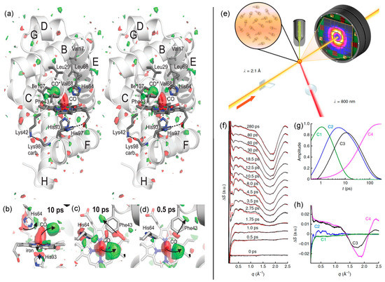
Figure 9.
Time-resolved fs-X-ray diffraction and scattering. Left panel: (a) stereo image showing the structure of Mb, as well as the electron density difference following photoexcitation for t = +0.5 ps (contoured at +3σ (green) and –3σ (red)). There are clear structural changes in the vicinity of the heme. (b–d) Difference electron density maps showing bound (–3σ contour level, red) and photodissociated (+3σ, contour level green) CO, doming of the heme, out-of-plane movement of the iron, and concomitant movement of His93 away from the heme, as well as rotation of His64 and movement of Phe43, which is displaced in different directions at t = +0.5 ps and t = +10 ps. The orientation in (b) is rotated by 90° with respect to the orientation in (c) and (d). Figure adapted from reference [107] with permission from Science AAAS. Right panel: Time-dependent changes in wide-angle X-ray scattering (WAXS) data recorded from detergent-solubilized samples of RCvir. (e) Experimental setup illustrating the microjet of solubilized RCvir (green), X-ray detector, XFEL beam (orange) and 800-nm pump laser (red). (f) Time-resolved WAXS structure factor difference, ΔS(q, t) = Slight(q, t) – Sdark(q), recorded as a function of t. Linear sums of the four basis spectra (red) shown in (h) are superimposed upon the experimental difference data (black). (g) Time-dependent amplitudes of the components C1–C4 used to extract the basis spectra. (h) Basis spectra extracted from the experimental data by spectral decomposition. Figure adapted from Reference [113] with permission from Springer Nature.
Figure 9e–h illustrates a very similar experimental layout implemented by Arnlund et al. [113] to perform time-resolved (TR) wide-angle X-ray scattering (WAXS) measurements. Here, Arnlund et al. [113] used multiphoton excitation of the photosynthetic reaction center of B. viridis (RCvir) to demonstrate that TR-WAXS data recorded using XFEL radiation can capture ultrafast protein conformational changes in solution. The probe beam consisted of 40-fs X-ray pulses containing 2.6 × 10¹² photons/pulse (λ = 2.1 Å) focused into a 10-µm2 spot. Detergent-solubilized RCvir samples were photoactivated using 500-fs optical pulses (λ = 800-nm) with a fluence of 530 mJ cm−2; this level of excitation leads to considerable multiphoton absorption since it is very close to the universal threshold for plasma formation (~1 J cm−2) [127]. Figure 9g depicts the separation of TR-WAXS data (Figure 9f) into their major components by spectral decomposition. The extracted spectral components are presented in Figure 9h. C1 is an ultrafast component, C2 is a protein component that displays the expected oscillatory features, and C3 and C4 are components that relate to heating effects. Arnlund et al. [113] concluded that XFELs facilitate the direct visualization of ultrafast protein motions. However, it should be noted that the interpretation of TR-WAXS data from solution phase requires the assistance of calculated structure factors from molecular dynamics (MD) simulations [128,129]. Moreover, depending on the solute under investigation the input of quantum mechanically optimized geometries [129,130] may be necessary. Time-resolved X-ray absorption spectroscopy (TR-XAS) is another approach for the study of molecular dynamics in solution with great sensitivity to structural changes.
2.4.3. Fs-X-ray Absorption Spectroscopy
Fs-X-ray absorption spectroscopy (fs-XAS), often in combination with fs-X-ray emission and other relevant spectroscopies, [131] has emerged as a powerful technique to monitor structural dynamics in solution, [132,133,134,135,136,137,138,139,140] crystalline phases, [141,142,143] and even at surfaces. [144] XAS is element-specific and depending on the element and core-shell involved in the process, X-ray photon energies usually range from 100 eV to 100 keV. There are two main regions in XAS: X-ray photoexcitation below the ionization threshold (IE) gives rise to pre-edge bands that arise from transitions to unoccupied or partially filled orbitals. For photon energies slightly above IE, there is a jump in the absorption cross-section and the quasi-bound photoelectron with low kinetic energy, and therefore high cross-section, undergoes multiple-scattering events with the surrounding atoms. This leads to a strong oscillatory behavior in the X-ray absorption spectrum. This region is denoted as XANES (X-ray absorption near-edge structure) or NEXAFS (near-edge X-ray absorption fine structure). As the kinetic energy of the photoelectron increases, the cross-section decreases and weaker modulations, in the so-called EXAFS (extended X-ray absorption fine structure) region appear as a consequence of single scattering contributions. The surrounding molecular arrangement is encoded in the observed absorption features, and therefore the structural information can be recovered through the application of well-established data analysis protocols. [145,146]
Initial pioneering fs-XAS experiments were performed at the femtosecond lines of SLS [133,134,143] and ALS [135,136] via laser slicing (see Section 2.1 above). With the advent of XFELs, the enhancement in signal to noise ratio (SNR) in fs-XAS data is remarkable. The main challenge, however, in the use of XFEL radiation for fs-XAS experiments is the intrinsic spectral instability arising from the stochastic nature of SASE. The rather broad SASE bandwidth (for spectroscopy) requires the use of a monochromator while large variations in the central wavelength results in large shot-to-shot intensity fluctuations of the order of 100%. There are normalization protocols that implement non-invasive shot intensity monitors to account for such large fluctuations. The application of dispersive spectrometers seems to be another suitable detection approach. [147,148] Figure 10a (red symbols) illustrate results obtained by Gawelda et al. [140] at the micro-XAS synchrotron beam line of SLS. Here, intense 100-fs pump pulses (λ = 400 nm) excite an aqueous solution of [Fe(bpy)3]2+ while 100-ps tunable monochromatic hard X-ray pulses probe the photoinduced changes by XANES at t = +50 ps. The differential XANES spectrum is shown in Figure 10b together with fs-XANES measurements (blue stars) performed by Bressler et al. [133] at the FEMTO line of SLS (laser slicing, see Section 2.1) for the same system at t = + 300 fs utilizing 100-fs hard X-ray pulses. The lack of brightness caused by laser slicing is evident. Recent results obtained by Mara et al. [138] at LCLS (see Figure 10, bottom panel) illustrate the near 1010 increase in ray flux. They implemented Fe K-edge fs-XANES and fs-XES to directly probe the ligand environment of the iron and its spin state, respectively. Mara et al. [138] were able to unambiguously revealed the geometric and electronic structural dynamics of photoexcited ferrous cytochrome c (cyt c), providing a direct correlation between axial ligand rebinding to the Fe and the temperature decay. The latter enabled experimental quantification of a protein entatic contribution towards the stabilization and control of an active site metal-ligand bond.
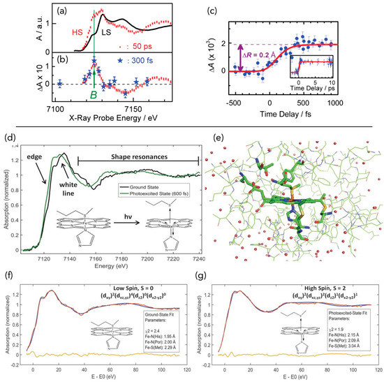
Figure 10.
Time-resolved X-ray absorption spectroscopy (XAS). Top panels (a–c): time-resolved X-ray absorption near-edge structure (XANES) for aqueous [Fe(bpy)3]2+. (a) Fe K-edge XANES spectrum of the low spin (LS) ground state (black trace) and of the high spin (HS) quintet state (red dots). The latter is determined from the LS spectrum and the transient spectrum measured at t = +50 ps after fs-laser excitation (Gawelda et al. [140] synchrotron microXAS line at SLS). (b) Transient XANES spectrum (difference between red symbols and black trace shown in panel (a)). The blue stars represent the transient spectrum recorded at t = +300 fs via laser slicing at SLS. (c) Time scan of the signal (blue points) at the B-feature (arrow in panel (b)) as a function of t. The inset shows a long time scan up to a 10-ps time delay. The red trace is the simulated signal assuming a simple kinetic model to describe the spin conversion process. The vertical arrow displays the expected effect of an elongation of 0.2 Å for the Fe-N bond distance difference between the LS and HS states. Error bars are ±1 standard deviation. Figure adapted from reference [133] with permission from Science AAAS. Bottom panels (d–g): Ultrafast x-ray absorption spectra of ferrous cyt c. (d) Superimposed spectra of ground-state (GS, black) and excited-state (ES, green) cyt c, showing the edge, white line, and shape resonance regions. (e) Cyt c heme structural model used in simulations. Fits (blue) to the GS (f) and ES (g) spectra (red) and associated structural parameters, derived from simulations. The fit residuals are in yellow at the bottom with the obtained structures above. Figure adapted from Reference [138] with permission from Science AAAS.
2.5. Timing Techniques at XFEL Facilities
The intrinsic time resolution of XFELs is limited by synchronization noise (or timing jitter in rms) to about 100–200 fs [72,149,150] with 100 fs reflecting short-term pulse-to-pulse variations. In order to fully exploit the potential of ultrashort hard X-ray pulses for monitoring faster dynamic phenomena different schemes have been proposed and implemented for the characterization of X-ray pulses. [149,150,151,152,153,154,155,156,157,158] Note that any useful characterization tool should provide single-shot sensitivity since a large component of the XFEL timing noise comes from shot-to-shot fluctuations. Ideally, one would like to access the temporal and spectral profiles of each X-ray pulse as well as its relative timing with respect to the fs-optical driving pulse; being the latter the bare-minimum. Among all current options, single-shot, single-cycle terahertz (THz) pulse streaking presents the advantage of characterizing both the temporal pulse shape of the free electron laser (FEL) pulse and its relative arrival time. The experimental arrangement implemented by Grguraš et al. [149] at FLASH (DESY, Hamburg) is shown in Figure 11a. A~3 mJ, 50-fs laser pulse (λ = 800 nm) is split into two parts; most of the pulse energy is used for THz generation via pulse front tilting for phase matched optical rectification in a LiNbO3 non-linear crystal [159,160]. The remainder (tens of µJ) is used for in situ THz-field characterization by electro-optic sampling (EOS) in a ZnTe crystal. Optical rectification produced about 4 µJ, 2-ps, single-cycle THz pulses centered at 0.6 THz. Figure 11b–d illustrates the THz streaking principle. In order to determine both the temporal shape and the relative arrival time, the FEL pulse should be within the streaking window of the THz vector potential denoted in Figure 11d by Δt, which in this case Δt~600 fs. Note that the streaking window should be larger than the intrinsic timing jitter to guarantee that most (if not all) FEL pulses become potentially useful (i.e., they are timed). Grguraš et al. [149] demonstrated the use of single-cycle THz pulses for the characterization of extreme ultraviolet (XUV) FEL pulses. Appropriate target atoms can be chosen to extend THz streaking into the hard X-ray regime where the main disadvantage is the large reduction in the photoionization cross-section when compared to soft X-rays [72,161].
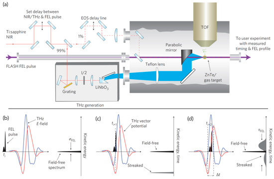
Figure 11.
Single-shot, single-cycle THz streaking. (a) Experimental setup: A fs-laser pulse from a Ti:Sapphire, appropriately delayed with respect to the free electron laser (FEL) pulse, is split into two parts. Most of the pulse energy is used for tilted-wavefront THz generation in LiNbO3. The remaining part can be used for characterizing the THz pulse via electro-optic sampling (EOS). This could be done in situ (prior streaking measurements) in a zinc telluride (ZnTe) nonlinear crystal positioned at the gas target location. In the streaking measurement, the collinear FEL photon pulse ejects a burst of photoelectrons from the gas, with a temporal profile identical to the incident soft XUV/X-ray FEL pulse. The THz pulse is used to streak the photoelectron burst and consequently characterize the FEL pulse. (b–d) Schematic of the operational principle: Blue and red curves represent the electric field and corresponding vector potential of the single-cycle THz pulse. The vertical axis corresponds to the kinetic energy of the photoelectron emission, which is determined by a time of flight (TOF) spectrometer. (a) The FEL pulse does not overlap with the THz pulse, and therefore the kinetic energy distribution of photoelectrons ejected by the FEL pulse is unaffected. (c) The FEL pulse overlaps with an extreme of the THz vector potential, leading to a maximally downshifted photoelectron spectrum with minimized spectral broadening. (c) Temporal overlap occurs near the zero crossing within the effective streaking window (Δt) of the vector potential where the time of arrival as well as the temporal profile can be determined. Figure adapted from Reference [149] with permission from Springer Nature.
The aforementioned THz streaking method is probably one of the most elegant characterization tools, which is sensitive to fluctuations in SASE amplification and in combination with a dispersive X-ray spectrometer [147,148] could provide a quasi-complete description of XFEL pulses. However, the most popular timing methods at XFELs, owing to simplicity, are spatial [150,154,155,156,157,162,163] and spectral [150,152] encoding. These are cross-correlation techniques based on X-ray induced material changes and confer a means to determine the relative timing between the fs-optical pump pulse and the X-ray probe pulse. Figure 12 illustrates a spectrogram approach recently introduced by Hartmann et al. [158] that combines the best features (or avoids the disadvantages) of spatial and spectral encoding to provide an outstanding timestamping error of sub-1 fs (rms). With such a timing method in hand, and routine 5-fs optical and 5-fs SASE X-ray pulses, the effective time-resolution of XFELs is now down to the 5-fs level providing the possibility to monitor high frequency vibrational modes and electronic coherences.
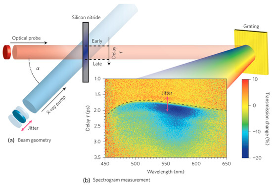
Figure 12.
Schematic of the single-shot geometry for measurement of the spectrogram. (a) The X-ray and optical beams are crossed in a silicon nitride membrane and their relative delay is encoded in the spatial beam profile of the optical probe. The crossing angle α and beam diameters define the time window in which the X-ray-induced absorption is probed. (b) Measured spectrogram using an unpumped normalization spectrogram to calculate the change in transmission. The result of the two-step edge-finding algorithm to determine the X-ray arrival time is overlaid as a black dashed line. Figure adapted from Reference [158] with permission from Springer Nature.
3. Structural Techniques Involving Ultrashort Electron Pulses
3.1. Introduction to Femtosecond Electron Diffraction (FED)
Since the very first stroboscopic experiments utilizing electron pulses as structural probes in time-resolved diffraction experiments in the 1980s, [164,165], there has been a continuous progress in the development of brighter and faster electron diffractometers. This advancement has occurred alongside several breakthrough ultrafast electron diffraction (UED) discoveries in the gas phase, [166,167,168,169,170,171,172,173] and in solid crystalline materials measured in reflection [174,175,176,177,178] and transmission [41,179,180,181,182,183,184,185,186,187,188,189,190,191,192,193,194,195,196] geometries. Great progress has been achieved in terms of FED (or UED) technology, with three main types of approaches being widely implemented in our research community (ordered in increasing design complexity and cost): (i) Compact (direct current, DC) FED designs, (ii) Hybrid (DC-RF) FED setups (i.e., with electron pulse rebunching) and (iii) Relativistic (RF) FED instruments. Figure 13 presents the layout of a compact FED setup with a conventional DC fs-electron source. The main difference among FED instruments is configuration/design of the electron gun, which is highlighted in Figure 13 by a dashed box. In a DC fs-electron source, electrons are generated via photoemission and accelerated by a DC electric field. A magnetic lens (ML) is used to focus the diffraction pattern on the detector screen. Note that the ML could be placed after the sample (akin to an objective lens in a transmission electron microscope (TEM)). This configuration greatly reduces the electron pulse propagation distance to the sample and, therefore, improves the temporal resolution (vide infra).
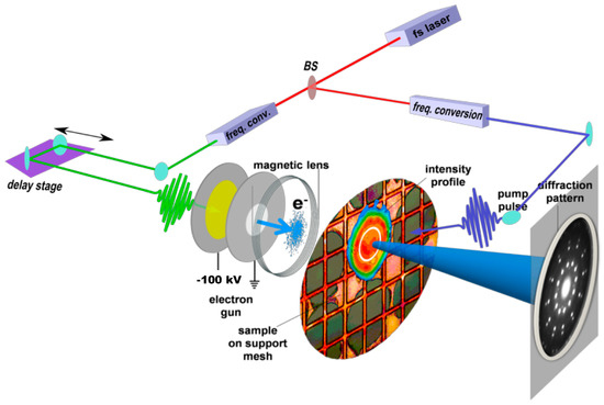
Figure 13.
Femtosecond electron diffraction (FED) layout for a compact electron gun design. In the present compact FED setup, a fs-optical pulse (green) generates, via the photoelectric effect, a fs-electron bunch by back-illuminating an ultrathin ~20-nm thick gold film. Electrons are accelerated by a 100 kV DC potential drop. A magnetic lens can be implemented before or after the sample to focus the diffraction pattern on the detector. Typical electron beam diameters at the sample position range from 50 to 500 µm. Figure adapted from Reference [181] with permission from Science AAAS.
A 300 kV DC-FED setup currently under development at the Ultrafast electron Imaging Lab (UeIL) of the University of Waterloo is shown in Figure 14. As can be observed, FED systems are laboratory scale tools, which depending on their design may even fit on top of optical tables. Care should be taken when operating FED instruments at acceleration voltages >30 kV; energetic electrons produce via corona discharge may lead to the generation of X-rays when impacting the walls of the vacuum chamber. Therefore, proper radiation shielding in addition to dosage monitors are a must in FED laboratories.

Figure 14.
300-kV DC FED apparatus at the Ultrafast electron Imaging Lab (UeIL). Left panel displays the 300-kV power supply, and the optical table with beam transport optics. Right panel shows the 300-kV FED instrument surrounded by a radiation enclosure.
3.2. Electrons as Structural Probes: Fermionic Nature and Source Brightness
The electron is an interesting elementary particle to play with. Its spin of one-half makes the electron a fermion and as such “no two electrons can share the same state” (Pauli exclusion principle). Therefore, an equivalent process to that of light amplification by stimulated emission of radiation (laser), which arises from the bosonic behaviour of photons, is not possible for electrons. One of the challenges is thus to develop an electron source with sufficient coherence and brightness to resolve the ensuing atomic motions. There is in fact a fundamental “quantum limit” to electron beam brightness (BQ) enforced by the Heisenberg uncertainty principle. Note that the minimum angular divergence a laser beam can achieve is also given by the uncertainty principle. However, there is no fundamental boundary to laser brightness besides practical and physical constraints usually dictated by the damaging threshold of amplifying laser medium. [40] The definition of electron beam brightness (in German, “Richstrahlwert”) was initially introduced by von Borries and Ruska [197] in 1939, (Equation (1)):
where I is the current; the areal spot size at focal point; and the beam divergence in sr.
Equation (1) has now evolved into different quasi-equivalent forms depending on the background of the scientific community. Beam accelerator scientists prefer working in position-momentum phase space owing to the invariance of the six-dimensional (6D) phase-space density () or volume () imposed by the Liouville’s theorem (as long as beam dynamics can be described by Hamilton’s equations).
V6D is the volume occupied by the electron bunch in this 6D phase space (x, , y, , z, ), which is proportional to the product of the normalized rms beam emittances , (Equation (2)):
where and p = x, y, or z. Hence, (Equation (3)):
where are variances or rms spreads in positions x, y, z; and momenta , , .
The result from Equation (3) is a true figure of merit that can be compared against the quantum degenerate limit, (Equation (4)):
where is known as Compton wavelength.
Note that for a paraxial electron beam propagating in the z direction, the brightness can also be written as, (Equation (5)):
where and are the energy and time variances, respectively.
The latter expression (Obtained after considering a given longitudinal momentum (or electron kinetic energy) such thus and ) in Equation (5) is referred to as “brilliance” and is a common figure of merit to characterize photon beams. Note that by assuming a value for and removing such dependence from Equation (5) we recover Equation (1). For very instructive derivations related to electron beam/source brightness please vide infra references [198,199,200,201,202,203,204,205,206,207].
3.3. Transverse Electron Beam Properties: Transverse Emittance and Coherence Length
Regardless of the chosen definition of brightness, a reality check shows that even the most advanced electron sources present a that is several orders of magnitude below the quantum limit, . There is still a lot of room for improvement and in most practical applications the transverse and longitudinal phase spaces are weakly coupled; therefore it is more convenient to treat them separately. The transverse electron beam coherence length () is defined as follows, (Equation (6)):
where is the de Broglie wavelength and the rms angular spread or variance.
(or ) is an essential electron beam parameter. For instance, it is known that in XFELs the value of at the entrance of the undulator corresponds to the wavelength of the X-rays produced () according to, (Equation (7)):
where v is the electron speed, and is the Lorentz factor.
Equation (7) links electron beam and X-ray beam properties and highlights the importance of attaining more intense and coherent electron sources. Thus, by reducing (or increasing ) one could in principle achieve shorter photon wavelengths or reduce the length of the linac [208]. The design and implementation of better and more efficient photocathodes is currently an area of active research. Generally speaking, the spatial resolution provided by a given photon or electron source is determined by its transverse () and longitudinal or temporal () coherences. The expression for longitudinal coherence of photons is easily deduced by replacing by in Equation (6).
Thus, for photons, (Equation (8)):
and (Equation (9)):
Then, given , and therefore ; we obtain (Equation (10)):
On the other hand, for electrons becomes, (Equation (11)):
and considering , we obtain and, (Equation (12)):
Note that assuming and combining Equations (6) and (12), we obtained , which seems to indicate that transverse and longitudinal coherences should have a similar effect on the spatial resolution of X-ray and electron diffractometers. However, the small de Broglie wavelength or small scattering angles () of keV-electrons makes the effect of negligible. Equivalently, one could introduce an effective longitudinal coherence length by noting that their effects on the diffraction peak widths are weighted by their relative projections on the screen. Hence, the spatial resolution in FED experiments is limited by , which is about 3 nm for conventional electron sources. In the case of synchrotron and XFEL sources, the transverse coherence, given by Equation (8), is in the order of several µm and thus, governs the spatial resolution. Considering typical XFEL beam parameters and bandwidth 10–3, we obtain . This suffices to spatially resolve systems with relatively large unit cells or large molecules such as proteins. Moreover, if necessary, the bandwidth can be reduced through the use of a monochromator in order to increase . This is perhaps the main advantage of XFELs over FED instruments, which have not yet succeeded in monitoring protein dynamics. On the other hand, the scattering cross section for electrons is approximately 105–106 higher than X-rays for the same energy and are about 1000-fold less damaging [209,210,211]. These facts make electrons a better probe of matter at the nanoscale.
3.4. Temporal Resolution in the Absence of Repulsive Interactions
For decades the main challenge has been to break the ps-temporal resolution barrier in FED experiments. Electrons are not only fermions but also charged particles, and therefore it is extremely challenging to keep them together and preserve the temporal resolution. Coulomb repulsion or space-charge effects lengthen the pulse during its propagation towards the sample. In fact, the instrument response function of FED setups is given by the electron pulse duration at the sample position (the pump pulse is usually much shorter). The use of single-electron or low-density electron sources resulted in the first and simplest alternative to reduce space-charge effects and preserve the temporal resolution [176,177,212,213]. However, there are only few samples capable of withstanding the repetitive photoexcitation conditions necessary to build a statistically meaningful (high SNR) diffraction pattern. Time-resolved electron diffraction experiments employing single-electron bursts rely on the accumulation of multiple events averaged over ∼108 pump-probe cycles to obtain a diffraction pattern with sufficient quality. To collect the data in a reasonable amount of time, single-electron sources often require the use of high repetition rate lasers >100 kHz. This procedure leads to accumulated heating effects that can significantly deteriorate the sample and even introduce artifacts, see supplementary information in Reference [41]. Therefore, it is essential to reach both ultrashort and ultrabright electron pulses to enable the study of structural dynamics in susceptible solid-state molecular materials. High-repetition electron sources are then best suited for gas phase experiments (molecular beams) in which the sample is refreshed between successive excitation pulses.
Nevertheless, it is interesting to consider the limiting case in which the interactions among electrons within the bunch are switched off. This limit offers an idea of the “optimal” temporal resolution achievable by a DC FED instrument, such as the one shown above in Figure 13. Neglecting relativistic effects, the longitudinal broadening () of the electron pulse can be approximated by , where is the initial velocity spread of the photoemitted electrons and is the electron pulse transit time from the cathode to the sample plane , where and are the cathode-anode and anode-sample distances, respectively, and is the electron speed in the propagation direction after exiting the anode aperture. Considering that the temporal pulse broadening () can be calculated as , we arrive to the following expression, [4,5] (Equation (13)):
where V is the applied DC voltage that satisfies , and is the initial momentum spread, .
Although Equation (13) suitably approximates the temporal spread gained in the cathode-anode region through a very simple and straightforward derivation, it was found to over exaggerate the pulse broadening occurring in the drift region. A more detailed derivation that includes relativistic effects shows that, [214] Equation (14):
and Equation (15):
Equation (15) has been derived for an applied constant DC electric field (E), and therefore provides the following compact expression for the temporal broadening in the absence of space-charge effects, Equation (16):
Optimal photoemission conditions in the limit of low electron density bursts with = 10 MV m–1 have shown to deliver electron pulses as short as 80 fs. [215] Ideally one would like to preserve such temporal resolution with pulses containing >103 electrons in a beam with a transverse size of about 100 µm.
3.5. Bright Compact Direct Current (DC) FED Setups
It was not until the work of Siwick et al. [179] demonstrating the first fully resolved structural transition using 600 fs electron pulses that it was realized that space-charge effects could be diminished by reducing the electron propagation distance and maximizing the extraction field, . Detailed N-body simulations and mean-field analytic models provided a greater understanding of pulse propagation dynamics of sub-relativistic electron bunches [216,217,218,219]. Nowadays, the use of charge-tracking programs, [220,221] that are required for the proper design of FED instrumentation, is routine. Historically, charge-tracking algorithms and powerful software packages were implemented by accelerator physicists, yet for a while UED scientists were largely unaware of this development due to the lack of interaction between communities.
Despite having a much simpler design than that of MeV relativistic machines, compact FEDs have provided unprecedented results in the study of novel crystalline materials. Several representative experiments performed in transmission and reflection geometries with compact FED instruments are shown in Figure 15 and Figure 16, respectively. FED has been found to be particularly advantageous for the investigation of 2D materials [183,185,186,222]. Figure 15a,b presents results obtained by Eichberger et al. [183] for the study of 1T-TaS2, a prototypical CDW system. FED has enabled the observation of atomic motions on timescales short enough to follow even the effect of non-equilibrium electronic distribution in this strongly correlated crystal. Since FED provides diffraction information over the entire 2D Brillouin zone at once, it is capable of unveiling different processes that give rise to changes in diffracted intensities. Figure 15c,d illustrates FED measurements gathered by Waldecker et al. [190] in the investigation of Ge2Sb2Te5 (GST), a phase-change compound commonly used for optical data storage. In this study, the combination of transient absorption/reflectivity and FED data allowed the authors obtain a microscopic picture of the initial steps in the photoinduced amorphization process in GST. Figure 15e–m presents FED results in Me4P[Pt(dmit)2]2 (Me4P = tetramethylphosphonium, dmit = 1,3-dithiol-2-thione-4,5-dithiolate), a crystalline system with a relatively large unit cell (a = 2.89 Å, b = 12.6 Å, c = 37.4 Å). Ishikawa et al. [223] applied transient reflectivity and FED to elucidate the key molecular motions that couple to an orbital-driven charge transfer reaction, i.e., a dimer expansion and a librational mode. These results illustrate the potential of advanced FED instruments to address fundamental structural phenomena occurring in increasingly complex molecular systems. On the other hand, FED experiments in reflection geometry (reflection high-energy electron diffraction, RHEED) with 30 keV electrons enabled the investigation of an indium (In) monolayer adsorbed on a (1 1 1)-Si surface (see Figure 16). At room temperature, the In atoms form regular arrays of quasi-one-dimensional (4 × 1) zigzag wires, separated by rows of silicon atoms as shown in Figure 16c. Around 125 K, a first-order Peierls-like transition to the (8 × 2) CDW phase occurs that is accompanied by a metal-to-insulator transition, see Figure 16a. With a temporal resolution of about 330 fs achieved via the implementation of pump pulse front tilting, [176,177]. Frigge et al. [178] enabled the observation of In–In bond breaking and formation that drive the structural transition through the non-thermal excitation of critically damped soft phonon modes.
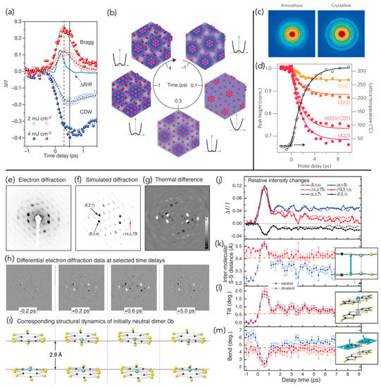
Figure 15.
Compact FED setups, measurements in transmission geometry. (a–b) Time evolution of the CDW state in 1T-TaS2 upon photoexcitation. (a) The circles correspond to FED and the solid traces to transient reflectivity data, respectively. A detailed analysis of the evolution of Bragg and CDW peaks, and optical measurements provides an atomically resolved movie highlighting the interplay between electronic and nuclear degrees of freedom. (b) Intense perturbation of the electronic system gives rise to smearing of the electron density modulation (t < 0.1 ps), driving the lattice towards the undistorted state (t < 0.3 ps). At the same time, the energy is transferred from the electronic subsystem to phonons, resulting in the recovery of the electron density modulation and thermal disordering of the lattice (t < 1 ps). The CDW order is nearly recovered at t < 4ps, after which time the sample is thermalized at a somewhat higher temperature. Figure adapted from Reference 183 with permission from Springer Nature. (c–d) Transient optical properties and structural transitions in phase-change material Ge2Sb2Te5 (GST). (c) Diffraction patterns of crystalline and amorphous states recorded with 92-keV fs-electron pulses. (d) Below-threshold dynamics in crystalline GST showing the time evolution of diffraction peaks and the extracted temperature rise. Figure adapted from Reference 190 with permission from Springer Nature. (e–m) Collective modes coupled to charge transfer in Me4P[Pt(dmit)2]2. (e) Experimental diffraction pattern of Me4P[Pt(dmit)2]2 at 90 K. (f) Simulated diffraction showing the contributions of individual reflections and selected peaks. (g) Simulated difference pattern for the thermal phase transition: charge-separated phase → high-temperature (metallic) phase. (h) Differential FED data [Ion(t) − Ioff] at different t following photoexcitation. (i) Snapshots of the structural dynamics constructed from the FED data. (j) Temporal profiles of the relative intensity changes of selected diffraction peaks. (k–m) Dynamics of key structural modes involved in the photoinduced phase transition of dimers initially neutral (blue) and divalent (red), compared with the thermal phase transition (dashed orange lines). Figure adapted from Reference [223] with permission from Science AAAS.
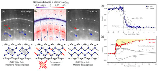
Figure 16.
Structural transitions at surfaces, measurements in reflection geometry. (a–c) Fs-reflection high-energy electron diffraction (RHEED) patterns and surface structures. Diffraction patterns at T = 30 K before (a) and 6 ps after (c) laser excitation. Surface structures are sketched below, with In and Si atoms depicted as filled blue circles and open circles, respectively; In–In bonds are shaded grey. (a) In the insulating phase, the In atoms form distorted hexagons in an (8 × 2) reconstructed unit cell. (c) In the metallic phase, the In atoms rearrange to form zigzag chains with (4 × 1) structure. (b) Normalized difference between RHEED patterns (c) and (a). All of the (8 × 2) ground-state features disappear (blue), while most (4 × 1) spots gain intensity (red). (d–e) Time evolution of the diffraction intensities following photoexcitation with F = 6.7 mJ cm−2. (d) Transient intensity of the (00) spot and an (8 × 2) spot (rescaled) as a function of t. The solid line is an exponential fit to the data for the (00) spot, from which a time constant of τtrans = 350 fs, reflecting the structural change, is extracted. The (8 × 2) spot vanishes completely to the background level (dashed grey line). (e) Intensity of a fourth-order spot and the thermal diffuse background. The transient dip in the intensity of the fourth-order spot (yellow shaded area) at t = +6 ps indicates surface heating by ΔT = 80 K (Debye–Waller effect), which coincides with the increase in background intensity. The fourth-order spot intensity is described (solid red curve) by the superposition of the two dashed lines, representing incoherent thermal motion (heating and subsequent cooling with time constants of 2.2 ps and 30 ps, respectively) and the structural transition with τtrans = 350 fs. Figure adapted from Reference [178] with permission from Springer Nature.
In all aforementioned FED experiments, the temporal resolution was in the range of 100 – 350-fs. In pursuit of a different DC fs-electron gun concept capable of producing sub-100-fs, and ideally sub-30-fs multi-electron bunches, Petruk et al. [224] found that is governed the strength of the electric field at the cathode’s surface since most of the temporal broadening occurs during the initial stage of electron propagation [225]. Hence, a more general expression for Equation (16) follows, Equation (17):
where is the magnitude of the geometrical electric field at the cathode’s surface, i.e., in the area where electrons are born.]
Equation (17) stresses the need to achieve both high electric field strengths and low initial momentum (or energy) spread at the cathode’s surface in order to minimize the temporal broadening caused by as well as additional broadening arising from instabilities in the high-voltage power supply [224]. Note that affects both the temporal and spatial resolutions (see definition of in Equation (6)).
Figure 17 illustrates a new DC fs-electron source concept capable of generating multi-electron bursts as short as 30 fs (12 fs rms). The idea here is to shape the cathode in order to increase the geometrical electric field at the surface, i.e., in the area from which electrons are photoemitted. The main challenge is then to avoid vacuum breakdown (i.e., electrical discharges).
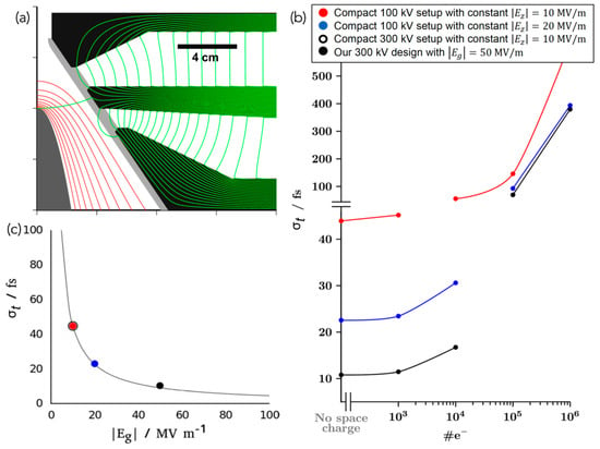
Figure 17.
Ultrafast electron source concept for the generation of <15-fs (rms) multi-electron bursts. (a) Schematic of the electron source concept. Equipotential and magnetic field lines calculated using Poisson Superfish, [226] are shown in red and green, respectively with a value of = 50 MV m–1. (b,c) Results from ASTRA simulations. (b) Electron pulse duration (, in fs) as a function of the number of electrons per bunch (#e–). Black trace corresponds to our 300-kV DC FED design with da = 5 cm and a total cathode-sample distance dT = 10 cm. Blue and red traces correspond to compact 100 kV DC FED setups (with flat electrodes) with dT = 2 cm and constant electrostatic fields of = 20 MV m–1 and 10 MV m–1, respectively. (c) Dependence of electron pulse duration (without space-charge effects) as a function of . Electrons in simulations were generated at the cathode surface considering a temporal (longitudinal) Gaussian profile of 6 fs (rms) [14 fs (fwhm)], an initial energy spread of 0.2 eV, and a lateral (transverse) Gaussian spot size of 50 µm (rms). The solid trace in panel (c) was obtained from a fit implementing Equation (20). Figure adapted from Reference [224].
The critical surface vacuum breakdown is given by, Equation (18):
where is the magnitude of the critical surface vacuum breakdown field, and are microscopic and geometrical field enhancement factors, respectively, and Vc is the critical applied potential drop over the cathode-anode separation distance, da.]
Note that , where is the magnitude of the average critical applied field and equals what we refer to as the geometrical critical surface electric field, . Thus, Equation (19):
Values of have been found to be 6.5–10 GV m–1 for refractory metals [227,228,229] while is governed by the surface roughness with 100 for mirror-like finished and for a roughness free surface. Note that field emitters approach the limit , and thus . For a bulky surface with = 100, we obtain 60–100 MV m–1, and therefore <60 MV m–1 should in principle be achievable.
A more detailed analysis of these and additional simulations show that the temporal broadening in the absence of space-charge effects follows, Equation (20)
where is the initial energy spread and k is a constant that depends on the type of initial electron distribution (k = 991 fs MV m–1 eV–1/2 for isotropic).]
It should be noted that Equation (20) and Equation (16) are equivalent since , and therefore . Initial preliminary tests at UeIL indicate, however, that achieving > 30 MV m–1 seems to require ultrahigh vacuum conditions, which are currently out of reach in our FED vacuum chamber designed to obtain a background pressure of 10–7 torr. A next-generation FED setup will be developed at UeIL to attain 30-fs multielectron bursts.
3.6. Ultrabright Hybrid DC-Radio Frequency (RF) FED Setups: Compression of Electron Pulses
Electron pulse compression methods are based on either the static [230,231,232,233,234] or active [235,236,237,238,239] application of electric or magnetic fields that can reverse the longitudinal momentum-position distribution of the electrons in the bunch. These electron beam components effectively act as a temporal lens and the sample is to be positioned at the temporal focal point. Among all proposed and/or demonstrated approaches for electron pulse rebunching, the implementation of a cylindrical TM010-RF cavity [235,236,237,238,239] became the most widely accepted and successful method. Figure 18 illustrates the electron pulse compression principle in a hybrid 100-kV DC-RF FED instrument. The RF pill lens provides an axial electric field which is uniform along the cavity axis and varies in time according to ; f ≈ 3 GHz is the resonant frequency of the cavity and a phase that assures that the electron pulse does not experiences any net gain or loss of energy. The choice of a frequency of 3 GHz was made to benefit from the existing S-band technology used by the RF accelerator community, in which this frequency is a standard.

Figure 18.
Layout of a hybrid DF-RF FED setup. The electron bunch is represented by a colored gradient where faster electrons are in violet and slower electrons in red. The electron pulse is generated via the photoelectric effect. This part is essentially the same as in a compact DC electron gun. The longitudinal momentum-space distributions within the electron bunch are illustrated below. The effect of the axial RF electric field is to invert the longitudinal momentum–space distribution causing the electron bunch to recompress during its propagation towards the sample, which is located at the temporal focal point. Magnetic lenses (MLs) are used to control the transverse dimension of the electron beam. Figure adapted from Reference 4 with permission from IOP Publishing.
N-particle simulations [236] and an experimental demonstration [237] showed that such an approach can indeed provide ultrashort electron bursts containing #e–106. However, laser-RF synchronization noise has limited (until very recently, see below) the instrument response of hybrid DF-RF FED setups to about 300–400 fs [238,239]. Ultrabright fs-electron bunches provided unique insights into photoinduced phenomena in strongly correlated materials including labile and poorly conductive organic crystals. Figure 19 shows some characteristic results obtained by Gao et al. [41] and Morrison et al. [189] in the FED study of (EDO-TTF)2PF6 and VO2, respectively.
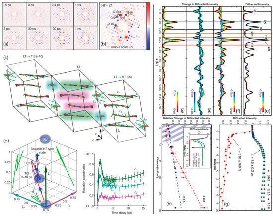
Figure 19.
Movies recorded with 100-kV hybrid DC-RF FED setups. Key molecular motions leading to charge delocalization in (EDO-TTF)2PF6. (a–c) Photoinduced time-dependent structural changes in (EDO-TTF)2PF6. (a) Differences between the diffraction patterns of the photoinduced phase and the initial low temperature (LT) phase as a function of t. (b) Difference between the diffraction patterns of the high-temperature (HT) and LT phases. Formation of a photo-induced transient intermediate structure (TIS). (c) Unit cells illustrating the arrangement of EDO-TTF molecules and PF6 counter-ions in the LT-unit cell. Major structural changes are illustrated using different background colours in the central unit cell: flat EDO-TTF moieties have a green background, bent EDO-TTF moieties have a pink background, and PF6 anions have a cyan background. Green sticks denote the atomic displacements from the initial LT structure to the TIS (left) and the HT-type structure (right); displacements were magnified 10 and 5 times, respectively, for clarity. (d) Evolution of phase-transition parameter in a -Cartesian configuration space (left) and each of its components as a function of t in the formation of TIS (right), F, B and P denote flat, bent EDO-TTF, and PF6 moieties, respectively. Figure adapted from Reference 41 with permission from Springer Nature. Photoinduced metal-like phase of monoclinic VO2. (e–e) Photoinduced time-dependent structural changes: (e) Background-subtracted FED data from t = 0 to 20 ps. Vertical lines indicate different groups of reflections that are relevant to monitor the monoclinic (M1) to rutile (R) photoinduced structural transition. (f) Overall differences in the diffraction intensities from –0.5 to 20 ps. (g) Time-resolved diffraction intensity changes showing fast (~300 fs) and slow (~1.6 ps) dynamics. (h) Fluence dependence of the fast and slow signal amplitudes as measured for two selected Bragg reflections; inset, transient transmittance measurements. (i) Diffraction difference spectrum for the fast dynamics from –0.5 to 1.5 ps. The change in diffracted intensity is plotted with respect to –1 ps. (j) Diffraction intensity difference for the slow dynamics. The change in diffracted intensity from 2 to 10 ps (referenced to 2 ps) is shown. Figure adapted from Reference 189 with permission from Science AAAS.
Gao et al. [41] studied the formation of a light-induced TIS in (EDO-TTF)2PF6 that is distinct from its LT and HT phases but resembles optical properties of the HT metallic phase [240,241]. Morrison et al., [189] on the other hand, discovered a metal-like M1 phase of VO2; see hatched region in Figure 19c, in which electronic and nuclear degrees of freedom are decoupled, suggesting that the insulating properties of VO2 may arise from Mott physics.
Different synchronization methods have been put forward to reduce the timing jitter noise in hybrid ultrabright DC-RF FED setups [242,243,244] in order to bring this technology to its maximum potential. An elegant and convenient approach that solved this long-lasting problem has been recently demonstrated by Otto et al. [244] who brought the noise level (long term >10 h) below 50 fs (rms)—limited by the resolution of their streak camera. Their lockbox is based on the use of an ultrabroadband photodiode (12.5 GHz), a low phase-noise CW amplifier, and a bandpass filter that produces the high harmonic signal that drives the RF cavity and acts as a reference in a phase feedback loop, i.e., a phase comparator that feeds from an antenna integrated into the cavity. With the time arrival jitter under long-term control, more exciting results are expected to come from these ultrabright electron sources as Siwick’s team plans to commercialize their lockbox.
In addition to RF pill lens for electron pulse compression, the use of intense few-cycle THz pulses has recently emerged as a means to control ultrabright electron bunches [245]. Figure 20 illustrates the experimental layout implemented by Kealhofer et al. [245] for THz compression and electron pulse characterization. The authors used butterfly-shaped metal resonators to mediate the interaction between the electrons and the THz fields. The first resonator in the beam path acts as a compressor whereas a second unit serves as a streaking device. It should be noted that the implementation of THz pulses instead of RF cavities nearly eliminates the timing jitter drifts that were found to be as small as 4 fs (rms). Thus, Kealhofer et al. [245] demonstrated a technique that will certainly find applications in the field of ultrafast structural dynamics.
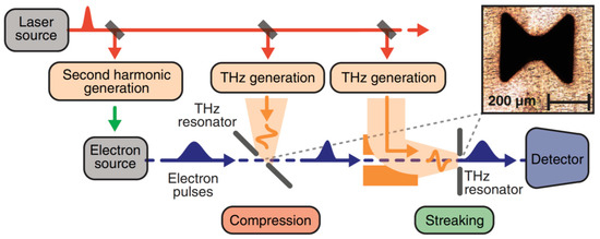
Figure 20.
All-optical control and metrology of electron pulses. Experimental setup: a 1-ps laser pulse from a Yb:YAG regenerative amplifier is frequency doubled and generates an electron pulse from a thin-film gold photocathode. The laser also drives two optical-rectification stages, each generating single-cycle THz pulses with energy of up to 40 nJ. (Inset) THz resonator structures are laser-machined in a 30-mm-thick aluminum foil. The first element, used for compression, is oriented at 45° to the electron beam, providing time-dependent longitudinal forces on the electrons. The second THz resonator, used for streaking, is oriented normal to the beam, resulting in time-dependent transverse deflection. Figure adapted from Reference 245 with permission from Science AAAS.
3.7. Ultrabright Relativistic FED Setups: RF-Photoinjectors
RF-photoinjectors are capable of producing ultrabright and ultrashort electron bursts, which are currently exploited as probes in relativistic FEDs. The use of radiofrequency allows for routine extraction fields 100 MVm−1 to be attained. Additionally, RF-photoinjectors are capable of producing ultrabright and ultrashort pulses of electrons with kinetic energies that are usually within the range of 1–5 MeV for FED measurements.
At these energies, the space-charge effects that leads to pulse broadening are largely suppressed due to relativistic effects that diminish Coulombic repulsion in by a factor of γ3 in both the longitudinal and transverse directions [246]. The progress in the development of relativistic FED setups over the last 10 years has been tremendous. Photoinjectors were originally optimized for higher bunch charges #e– 109 (instead of #e– 106-7) as well as for higher kinetic energies to be used as sources in XFELs. Relativistic FED was found to be of great importance to XFELs since the generation of high-quality low emittance electron beams are beneficial to both technologies. University-based pioneering MeV FED setups have been developed at UCLA [184,207] and Osaka, [187,247] and later launched at BNL, [192] SLAC [171,172,173,193,194,195,196] and DESY [248,249]. The amount of existing knowhow in accelerator centers has been critical to obtain stable RF-photoinjectors with minimal laser-RF timing noise to reach 100 fs (rms) temporal resolution [250].
Recent results obtained at the MeV-UED line of SLAC are shown in Figure 21 for the study of CH3I in the gas phase. Relativistic FED is the ideal tool for monitoring structural dynamics in molecular beams. Electron pulses moving at near the speed of light are necessary to avoid temporal broadening caused by velocity mismatch between the optical pump pulse and the electron probe pulse [4,251]. Yang et al. [173] conducted the first direct observation of a nuclear wave-packet with atomic spatiotemporal resolution during nonadiabatic processes involving conical intersections. More experiments with similar level of details are expected for the investigation of ultrafast structural dynamics in the gas phase.
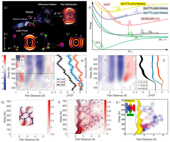
Figure 21.
Relativistic ultrafast gas-phase electron diffraction. (a) Schematic of relativistic FED experiment. (b) A model for a CF3I molecule oriented along the laser polarization axis. (c) Simulated pair distribution function (PDF) for a molecular ensemble with a angular distribution. (d) A model for a CF3I molecule oriented perpendicular to the laser polarization. (e) Simulated PDF for a molecular ensemble with a angular distribution. The laser polarization is indicated by the double-headed arrow. Gray, light green, and purple represent carbon, fluorine, and iodine, respectively. (f) Potential energy surfaces depicting the two-channel excitation in CF3I along the C–I bond length coordinate, with major states labeled and reaction pathways marked by arrows. Color coding indicates different electronic states and highlight the complexity of reaction. (g) Experimental ΔPDF⊥. The dashed line at 200 fs shows a rough separation between contributions from the one-photon and the two-photon channels. (h) Experimental time evolution of ΔPDF⊥ at 2.14, 2.52, and 2.90 Å. A comb illustrates the first two periods of the C–I stretching vibration. (i) Simulated ΔPDF⊥ of the two-photon channel. (j) Simulated time evolution of ΔPDF⊥ at 2.14, 2.52, and 2.90 Å. Figure adapted from Reference [173] with permission from Science AAAS.
3.8. Compact Ultrafast Low-Energy FED: Metallic Tip as Coherent Electron Sources
The use of sharp field emission tips triggered by fs-laser pulses [252,253,254,255,256,257,258,259,260,261,262] provides a unique alternative to reach the required transverse source coherence for the ultrafast dynamical study of large molecular structures. Low-energy fs-electron sources based on field emitters are attracting increasing interest in the community. Emission from an atom wide tip should in principle reach the quantum limit opening opportunities for lensless imaging (or point projection) [259,263,264,265] and coherent diffraction [260,266]. Figure 22 outlines results obtained by Müller et al. [259] for the study of ultrafast photocurrents in nanowires. The authors implemented a point projection that conferred sub-100 fs temporal and 10 nm spatial resolutions. Müller et al. [259] demonstrated an imaging tool capable of monitoring small electric fields around semiconductor nanowires (NWs) and photocurrents by taking advantage of the high sensitivity of sub-keV fs-electron pulses combined with the large magnification of their point projection source.
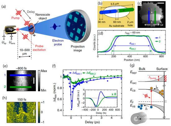
Figure 22.
Setup for time-resolved low-energy electron point projection mode (PPM). (a) PPM operating principle: photoelectrons, generated from a nanotip by an ultrashort laser pulse, are accelerated towards the sample positioned several µm away from the tip. A pump laser pulse, variably delayed from the electron probe, photoexcites the sample. An electrostatic lens is used to switch between divergent and collimated (not shown here) diffraction modes. (b–d) Point projection microscopy of axially doped NWs. InP NWs (radius 15 nm, length 3.5 mm) with p-i-n axial doping profile and 60-nm i-segment in the center are spanned across 2-µm holes in a gold substrate. Instead of being a real shadow image of the objects shape, projection images are strongly influenced by local fields surrounding the NW. In addition, a spatial inhomogeneity (marked by the white arrows in (c) is observed (d). (e–g) Femtosecond imaging of ultrafast photocurrents. (e) Projection image of the same NW as in panel (b) recorded in pulsed fs-PPM mode at negative t. Photoexcitation by an ultrashort laser pulse leads to a transient, spatially inhomogeneous change of the projected NW diameter (h, normalized difference plot). Data recorded at 70 eV electron energy; scale bars: 500 nm. Different dynamical behavior and amplitudes of the transient diameter change ΔdNW are observed for the p- and n-doped segments along the NW (f), where an empirical three-exponential function was fitted to the data. Both segments show a fast initial photoinduced effect with 10–90 rise times in the p- and n-segments of 140 and 230 fs, respectively, followed by multi-exponential decay on the fs-to-few picosecond time scale. As ΔdNW is directly proportional to the transient electric field change, the derivative ΔdNW/dt plotted in the inset in (f) is a direct measure of the instantaneous photocurrent inside the NW. Surface states cause effective radial doping leading to band bending at the NW surface as sketched in (g), where r is the radial coordinate, causing a radial photocurrent of electrons, je, and holes, jh, after photoexcitation. This leads to a pump-induced transient shift Δpump of the energies of the conduction band edge (ECB) and valence band edge (EVB), and hence a shift of the vacuum level (Evac) (red shaded area), compared to the reference level Eref (given by the environment), with the magnitude of the shift depending on the specific band bending and doping level. Figure adapted from Reference [259] with permission from Springer Nature.
The advances in tip-based photoemitters and nanofabrication techniques have made it possible to achieve ps-resolution with ultrafast low-energy electron diffraction (ULEED) in backscattering mode. Storeck, Vogelgesang et al. [261,262] developed a novel µm-sized electron gun, which can be brought close to the investigated surface to diminish temporal broadening. The µm-sized electron source is presented in Figure 23 alongside ULEED results obtained in the study of 1T-TaS2. The significantly improved spatial resolution of this tip source allowed for spot-profile analysis, resolving the phase-ordering kinetics in the nascent incommensurate charge density wave phase.
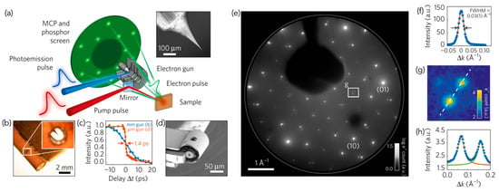
Figure 23.
Ultrafast low-energy electron diffraction (ULEED) setup and high-resolution diffraction pattern from 1T-TaS2. (a) Schematic of the experimental setup for ULEED. Inset: electron micrograph of an electrochemically etched tungsten tip used as a photoemitter. (b) mm-sized laser-driven electron gun. Inset: tungsten tip, visible through the hole for laser illumination. (c) Electron pulse durations of the mm-sized electron gun (16.4 ps at 100 eV) and the µm-sized electron gun (1.4 ps at 50 eV). (d) µm-sized electron gun prepared by nanofabrication. (e) LEED pattern of the near commensurate CDW room-temperature phase, recorded with pulsed 100-eV electrons from the mm-sized electron gun (logarithmic colour scale). A retarding voltage of −20 V is applied at the detector front plate. (f) Line profile of the (1 ) diffraction peak, illustrating high transversal coherence of the source. The fitted spot width of 0.03 Å–1 (fwhm) corresponds to a transfer width of 21 nm. (g) Close-up of region marked in e, showing second-order CDW diffraction spots (h), Line profile of CDW diffraction spots shown in g, fitted with Lorentzian peak profiles. Figure adapted from reference 262 with permission from Springer Nature.
4. Conclusions
The future of fs-X-ray and fs-electron sources is very bright with several XFEL facilities readily available for use and a relatively small but quickly growing community of FED developers. There are other promising techniques for the generation of ultrashort electron pulses, which have not been discussed here. For instance, laser plasma electron accelerators [267,268,269,270,271,272,273] are quickly improving through the reduction of the large energy spread that has limited their application as FED sources. Another interesting candidate is the cold-atom electron source, [202,274,275,276,277,278,279,280,281] which has recently been proposed as an alternative to photoinjectors owing to the large reduction in electron beam emittance that should considerably improve the X-ray beam properties. Moreover, important advances have been made in the development and implementation of dynamic and ultrafast electron microscopes [3,282,283,284,285,286,287,288,289]. We are already transiting the era of atomically resolved movies, with more advanced experiments and exciting discoveries soon to come.
Funding
G.S. would like to acknowledge support from the Canada Research Chair program, the National Science and Engineering Research Council of Canada, Canada Foundation for Innovation, and the Government of Ontario. G.S. is thankful for the support provided by WIN (through the Interdisciplinary Research Program Funds) and Transformative Quantum Technologies (via the Quantum Quest Seed Fund program).
Acknowledgments
GS would like to thank Kostyantyn Pichugin and Tyler Lott (UeIL) for their very useful suggestions.
Conflicts of Interest
The author declares no conflict of interest.
References
- Zewail, A.H. 4D Ultrafast Electron Diffraction, Crystallography, and Microscopy. Annu. Rev. Phys. Chem. 2006, 57, 65–103. [Google Scholar] [CrossRef] [PubMed]
- Chergui, M.; Zewail, A.H. Electron and X-ray Methods of Ultrafast Structural Dynamics: Advances and Applications. ChemPhysChem 2009, 10, 28–43. [Google Scholar] [CrossRef] [PubMed]
- Zewail, A.H. Four-Dimensional Electron Microscopy. Science 2010, 328, 187–193. [Google Scholar] [CrossRef] [PubMed]
- Sciaini, G.; Miller, R.J.D. Femtosecond Electron Diffraction: Heralding the Era of Atomically Resolved Dynamics. Rep. Prog. Phys. 2011, 74, 096101. [Google Scholar] [CrossRef]
- Hada, M.; Pichugin, K.; Sciaini, G. Ultrafast Structural Dynamics with Table Top Femtosecond Hard X-ray and Electron Diffraction Setups. Eur. Phys. J. Spec. Top. 2013, 222, 1093–1123. [Google Scholar] [CrossRef]
- Elsaesser, T.; Woerner, M. Perspective: Structural Dynamics in Condensed Matter Mapped by Femtosecond X-ray Diffraction. J. Chem. Phys. 2014, 140, 020901. [Google Scholar] [CrossRef] [PubMed]
- Miller, R.J.D. Femtosecond Crystallography with Ultrabright Electrons and X-rays: Capturing Chemistry in Action. Science 2014, 343, 1108–1116. [Google Scholar] [CrossRef]
- Rumble, J. CRC Handbook of Chemistry and Physics, 99th ed.; CRC Press: Boca Raton, FL, USA, 2018. [Google Scholar]
- Polanyi, J.C.; Zewail, A.H. Direct Observation of the Transition State. Acc. Chem. Res. 1995, 28, 119–132. [Google Scholar] [CrossRef]
- Grüner, G. The Dynamics of Charge-Density Waves. Rev. Mod. Phys. 1988, 60, 1129–1181. [Google Scholar] [CrossRef]
- Schrieffer, J.R. Theory of Superconductivity; CRC Press: Boca Raton, FL, USA, 2018. [Google Scholar]
- Allen, P.G.; Mini, S.M.; Perry, D.L.; Stock, S.R. Applications of Synchrotron Radiation Techniques to Materials Science IV: Volume 678, 1st ed.; Cambridge University Press: Cambridge, UK, 2001. [Google Scholar]
- Schotte, F.; Lim, M.; Jackson, T.A.; Smirnov, A.V.; Soman, J.; Olson, J.S.; Phillips, G.N.; Wulff, M.; Anfinrud, P.A. Watching a Protein as It Functions with 150-Ps Time-Resolved X-ray Crystallography. Science 2003, 300, 1944–1947. [Google Scholar] [CrossRef] [PubMed]
- Collet, E.; Lemée-Cailleau, M.-H.; Cointe, M.B.-L.; Cailleau, H.; Wulff, M.; Luty, T.; Koshihara, S.-Y.; Meyer, M.; Toupet, L.; Rabiller, P.; et al. Laser-Induced Ferroelectric Structural Order in an Organic Charge-Transfer Crystal. Science 2003, 300, 612–615. [Google Scholar] [CrossRef] [PubMed]
- Schoenlein, R.W.; Chattopadhyay, S.; Chong, H.H.W.; Glover, T.E.; Heimann, P.A.; Shank, C.V.; Zholents, A.A.; Zolotorev, M.S. Generation of Femtosecond Pulses of Synchrotron Radiation. Science 2000, 287, 2237–2240. [Google Scholar] [CrossRef] [PubMed]
- Zholents, A.A.; Zolotorev, M.S. Femtosecond X-ray Pulses of Synchrotron Radiation. Phys. Rev. Lett. 1996, 76, 912–915. [Google Scholar] [CrossRef] [PubMed]
- Cavalleri, A.; Wall, S.; Simpson, C.; Statz, E.; Ward, D.W.; Nelson, K.A.; Rini, M.; Schoenlein, R.W. Tracking the Motion of Charges in a Terahertz Light Field by Femtosecond X-ray Diffraction. Nature 2006, 442, 664–666. [Google Scholar] [CrossRef] [PubMed]
- Beaud, P.; Johnson, S.L.; Streun, A.; Abela, R.; Abramsohn, D.; Grolimund, D.; Krasniqi, F.; Schmidt, T.; Schlott, V.; Ingold, G. Spatiotemporal Stability of a Femtosecond Hard—X-ray Undulator Source Studied by Control of Coherent Optical Phonons. Phys. Rev. Lett. 2007, 99, 174801. [Google Scholar] [CrossRef] [PubMed]
- Johnson, S.L.; Beaud, P.; Milne, C.J.; Krasniqi, F.S.; Zijlstra, E.S.; Garcia, M.E.; Kaiser, M.; Grolimund, D.; Abela, R.; Ingold, G. Nanoscale Depth-Resolved Coherent Femtosecond Motion in Laser-Excited Bismuth. Phys. Rev. Lett. 2008, 100, 155501. [Google Scholar] [CrossRef]
- Huber, T.; Mariager, S.O.; Ferrer, A.; Schäfer, H.; Johnson, J.A.; Grübel, S.; Lübcke, A.; Huber, L.; Kubacka, T.; Dornes, C.; et al. Coherent Structural Dynamics of a Prototypical Charge-Density-Wave-to-Metal Transition. Phys. Rev. Lett. 2014, 113, 026401. [Google Scholar] [CrossRef] [PubMed]
- Lantz, G.; Neugebauer, M.J.; Kubli, M.; Savoini, M.; Abreu, E.; Tasca, K.; Dornes, C.; Esposito, V.; Rittmann, J.; Windsor, Y.W.; et al. Coupling between a Charge Density Wave and Magnetism in an Heusler Material. Phys. Rev. Lett. 2017, 119, 227207. [Google Scholar] [CrossRef]
- Porer, M.; Fechner, M.; Bothschafter, E.M.; Rettig, L.; Savoini, M.; Esposito, V.; Rittmann, J.; Kubli, M.; Neugebauer, M.J.; Abreu, E.; et al. Ultrafast Relaxation Dynamics of the Antiferrodistortive Phase in Ca Doped SrTiO3. Phys. Rev. Lett. 2018, 121, 055701. [Google Scholar] [CrossRef]
- Travaglini, G.; Wachter, P.; Marcus, J.; Schlenker, C. The Blue Bronze K0.3MoO3: A New One-Dimensional Conductor. Solid State Commun. 1981, 37, 599–603. [Google Scholar] [CrossRef]
- Martin, I.P.S.; Rehm, G.; Thomas, C.; Bartolini, R. Experience with Low-Alpha Lattices at the Diamond Light Source. Phys. Rev. Spec. Top. Accel. Beams 2011, 14, 040705. [Google Scholar] [CrossRef]
- Murnane, M.M.; Kapteyn, H.C.; Rosen, M.D.; Falcone, R.W. Ultrafast X-ray Pulses from Laser-Produced Plasmas. Science 1991, 251, 531–536. [Google Scholar] [CrossRef] [PubMed]
- Rose-Petruck, C.; Jimenez, R.; Guo, T.; Cavalleri, A.; Siders, C.W.; Rksi, F.; Squier, J.A.; Walker, B.C.; Wilson, K.R.; Barty, C.P.J. Picosecond–Milliångström Lattice Dynamics Measured by Ultrafast X-ray Diffraction. Nature 1999, 398, 310–312. [Google Scholar] [CrossRef]
- Siders, C.W.; Cavalleri, A.; Sokolowski-Tinten, K.; Tóth, C.; Guo, T.; Kammler, M.; von Hoegen, M.H.; Wilson, K.R.; von der Linde, D.; Barty, C.P.J. Detection of Nonthermal Melting by Ultrafast X-ray Diffraction. Science 1999, 286, 1340–1342. [Google Scholar] [CrossRef] [PubMed]
- Cavalleri, A.; Tóth, C.; Siders, C.W.; Squier, J.A.; Ráksi, F.; Forget, P.; Kieffer, J.C. Femtosecond Structural Dynamics in VO2 during an Ultrafast Solid-Solid Phase Transition. Phys. Rev. Lett. 2001, 87, 237401. [Google Scholar] [CrossRef] [PubMed]
- Rousse, A.; Rischel, C.; Fourmaux, S.; Uschmann, I.; Sebban, S.; Grillon, G.; Balcou, P.; Förster, E.; Geindre, J.P.; Audebert, P.; et al. Non-Thermal Melting in Semiconductors Measured at Femtosecond Resolution. Nature 2001, 410, 65–68. [Google Scholar] [CrossRef]
- Sokolowski-Tinten, K.; Blome, C.; Blums, J.; Cavalleri, A.; Dietrich, C.; Tarasevitch, A.; Uschmann, I.; Förster, E.; Kammler, M.; Horn-von-Hoegen, M.; et al. Femtosecond X-ray Measurement of Coherent Lattice Vibrations near the Lindemann Stability Limit. Nature 2003, 422, 287–289. [Google Scholar] [CrossRef] [PubMed]
- Hagedorn, M.; Kutzner, J.; Tsilimis, G.; Zacharias, H. High-Repetition-Rate Hard X-ray Generation with Sub-Millijoule Femtosecond Laser Pulses. Appl. Phys. B 2003, 77, 49–57. [Google Scholar] [CrossRef]
- Bargheer, M.; Zhavoronkov, N.; Gritsai, Y.; Woo, J.C.; Kim, D.S.; Woerner, M.; Elsaesser, T. Coherent Atomic Motions in a Nanostructure Studied by Femtosecond X-ray Diffraction. Science 2004, 306, 1771–1773. [Google Scholar] [CrossRef]
- Serbanescu, C.G.; Chakera, J.A.; Fedosejevs, R. Efficient Kα X-ray Source from Submillijoule Femtosecond Laser Pulses Operated at Kilohertz Repetition Rate. Rev. Sci. Instrum. 2007, 78, 103502. [Google Scholar] [CrossRef]
- Hada, M.; Okimura, K.; Matsuo, J. Characterization of Structural Dynamics of VO2 Thin Film on c-Al2O3 Using in-Air Time-Resolved X-ray Diffraction. Phys. Rev. B 2010, 82, 153401. [Google Scholar] [CrossRef]
- Hada, M.; Okimura, K.; Matsuo, J. Photo-Induced Lattice Softening of Excited-State VO2. Appl. Phys. Lett. 2011, 99, 051903. [Google Scholar] [CrossRef]
- Zamponi, F.; Rothhardt, P.; Stingl, J.; Woerner, M.; Elsaesser, T. Ultrafast Large-Amplitude Relocation of Electronic Charge in Ionic Crystals. Proc. Natl. Acad. Sci. USA 2012, 109, 5207–5212. [Google Scholar] [CrossRef] [PubMed]
- Giles, C.; Celestre, R.; Tasca, K.R.; Dias, C.S.B.; Vescovi, R.; Faria, G.; Ferbonink, G.F.; Nome, R.A. Compact Arrangement for Femtosecond Laser-Induced Generation of Broadband Hard X-ray Pulses. In High-Brightness Sources and Light-Driven Interactions (2018), Paper ET1B.3, Proceedings of the Compact EUV and X-ray Light Sources, Strasbourg, France, 26–28 March 2018; OSA: Washington, DC, USA. [CrossRef]
- Holtz, M.; Hauf, C.; Weisshaupt, J.; Salvador, A.-A.H.; Woerner, M.; Elsaesser, T. Towards Shot-Noise Limited Diffraction Experiments with Table-Top Femtosecond Hard X-ray Sources. Struct. Dyn. 2017, 4, 054304. [Google Scholar] [CrossRef] [PubMed]
- Freyer, B.; Zamponi, F.; Juvé, V.; Stingl, J.; Woerner, M.; Elsaesser, T.; Chergui, M. Ultrafast Inter-Ionic Charge Transfer of Transition-Metal Complexes Mapped by Femtosecond X-ray Powder Diffraction. J. Chem. Phys. 2013, 138, 144504. [Google Scholar] [CrossRef] [PubMed]
- Strickland, D.; Mourou, G. Compression of Amplified Chirped Optical Pulses. Opt. Commun. 1985, 55, 219–221. [Google Scholar] [CrossRef]
- Gao, M.; Lu, C.; Jean-Ruel, H.; Liu, L.C.; Marx, A.; Onda, K.; Koshihara, S.; Nakano, Y.; Shao, X.; Hiramatsu, T.; et al. Mapping Molecular Motions Leading to Charge Delocalization with Ultrabright Electrons. Nature 2013, 496, 343–346. [Google Scholar] [CrossRef]
- Weisshaupt, J.; Juvé, V.; Holtz, M.; Ku, S.; Woerner, M.; Elsaesser, T.; Ališauskas, S.; Pugžlys, A.; Baltuška, A. High-Brightness Table-Top Hard X-ray Source Driven by Sub-100-Femtosecond Mid-Infrared Pulses. Nat. Photonics 2014, 8, 927–930. [Google Scholar] [CrossRef]
- Bentson, L.; Bolton, P.; Bong, E.; Emma, P.; Galayda, J.; Hastings, J.; Krejcik, P.; Rago, C.; Rifkin, J.; Spencer, C.M. FEL Research and Development at the SLAC Sub-Picosecond Photon Source, SPPS. Nucl. Instrum. Methods Phys. Res. A 2003, 507, 205–209. [Google Scholar] [CrossRef]
- Shiner, A.D.; Trallero-Herrero, C.; Kajumba, N.; Bandulet, H.-C.; Comtois, D.; Légaré, F.; Giguère, M.; Kieffer, J.-C.; Corkum, P.B.; Villeneuve, D.M. Wavelength Scaling of High Harmonic Generation Efficiency. Phys. Rev. Lett. 2009, 103, 073902. [Google Scholar] [CrossRef] [PubMed]
- Popmintchev, T.; Chen, M.-C.; Popmintchev, D.; Arpin, P.; Brown, S.; Alisauskas, S.; Andriukaitis, G.; Balciunas, T.; Mucke, O.D.; Pugzlys, A.; et al. Bright Coherent Ultrahigh Harmonics in the KeV X-ray Regime from Mid-Infrared Femtosecond Lasers. Science 2012, 336, 1287–1291. [Google Scholar] [CrossRef] [PubMed]
- Corkum, P.B. Plasma Perspective on Strong Field Multiphoton Ionization. Phys. Rev. Lett. 1993, 71, 1994–1997. [Google Scholar] [CrossRef] [PubMed]
- Bauer, M.; Lei, C.; Read, K.; Tobey, R.; Gland, J.; Murnane, M.M.; Kapteyn, H.C. Direct Observation of Surface Chemistry Using Ultrafast Soft-X-ray Pulses. Phys. Rev. Lett. 2001, 87, 025501. [Google Scholar] [CrossRef]
- Stolow, A.; Bragg, A.E.; Neumark, D.M. Femtosecond Time-Resolved Photoelectron Spectroscopy. Chem. Rev. 2004, 104, 1719–1758. [Google Scholar] [CrossRef] [PubMed]
- Itatani, J.; Levesque, J.; Zeidler, D.; Niikura, H.; Pépin, H.; Kieffer, J.C.; Corkum, P.B.; Villeneuve, D.M. Tomographic Imaging of Molecular Orbitals. Nature 2004, 432, 867–871. [Google Scholar] [CrossRef] [PubMed]
- Corkum, P.B.; Krausz, F. Attosecond Science. Nat. Phys. 2007, 3, 381–387. [Google Scholar] [CrossRef]
- Krausz, F.; Ivanov, M. Attosecond Physics. Rev. Mod. Phys. 2009, 81, 163–234. [Google Scholar] [CrossRef]
- Goulielmakis, E.; Loh, Z.-H.; Wirth, A.; Santra, R.; Rohringer, N.; Yakovlev, V.S.; Zherebtsov, S.; Pfeifer, T.; Azzeer, A.M.; Kling, M.F.; et al. Real-Time Observation of Valence Electron Motion. Nature 2010, 466, 739–743. [Google Scholar] [CrossRef]
- Sansone, G.; Kelkensberg, F.; Pérez-Torres, J.F.; Morales, F.; Kling, M.F.; Siu, W.; Ghafur, O.; Johnsson, P.; Swoboda, M.; Benedetti, E.; et al. Electron Localization Following Attosecond Molecular Photoionization. Nature 2010, 465, 763–766. [Google Scholar] [CrossRef] [PubMed]
- Schultze, M.; Fieß, M.; Karpowicz, N.; Gagnon, J.; Korbman, M.; Hofstetter, M.; Neppl, S.; Cavalieri, A.L.; Komninos, Y.; Mercouris, T.; et al. Delay in Photoemission. Science 2010, 328, 1658–1662. [Google Scholar] [CrossRef]
- Torres, R.; Siegel, T.; Brugnera, L.; Procino, I.; Underwood, J.G.; Altucci, C.; Velotta, R.; Springate, E.; Froud, C.; Turcu, I.C.E.; et al. Revealing Molecular Structure and Dynamics through High-Order Harmonic Generation Driven by Mid-IR Fields. Phys. Rev. A 2010, 81, 051802. [Google Scholar] [CrossRef]
- Petersen, J.C.; Kaiser, S.; Dean, N.; Simoncig, A.; Liu, H.Y.; Cavalieri, A.L.; Cacho, C.; Turcu, I.C.E.; Springate, E.; Frassetto, F.; et al. Clocking the Melting Transition of Charge and Lattice Order in 1T-TaS2 with Ultrafast Extreme-Ultraviolet Angle-Resolved Photoemission Spectroscopy. Phys. Rev. Lett. 2011, 107, 177402. [Google Scholar] [CrossRef] [PubMed]
- Rohwer, T.; Hellmann, S.; Wiesenmayer, M.; Sohrt, C.; Stange, A.; Slomski, B.; Carr, A.; Liu, Y.; Avila, L.M.; Kalläne, M.; et al. Collapse of Long-Range Charge Order Tracked by Time-Resolved Photoemission at High Momenta. Nature 2011, 471, 490–493. [Google Scholar] [CrossRef] [PubMed]
- Eich, S.; Stange, A.; Carr, A.V.; Urbancic, J.; Popmintchev, T.; Wiesenmayer, M.; Jansen, K.; Ruffing, A.; Jakobs, S.; Rohwer, T.; et al. Time- and Angle-Resolved Photoemission Spectroscopy with Optimized High-Harmonic Pulses Using Frequency-Doubled Ti:Sapphire Lasers. J. Electron Spectrosc. 2014, 195, 231–236. [Google Scholar] [CrossRef]
- Gariepy, G.; Leach, J.; Kim, K.T.; Hammond, T.J.; Frumker, E.; Boyd, R.W.; Corkum, P.B. Creating High-Harmonic Beams with Controlled Orbital Angular Momentum. Phys. Rev. Lett. 2014, 113, 153901. [Google Scholar] [CrossRef] [PubMed]
- Gessner, O.; Gühr, M. Monitoring Ultrafast Chemical Dynamics by Time-Domain X-ray Photo- and Auger-Electron Spectroscopy. Acc. Chem. Res. 2016, 49, 138–145. [Google Scholar] [CrossRef] [PubMed]
- Marangos, J.P. Development of High Harmonic Generation Spectroscopy of Organic Molecules and Biomolecules. J. Phys. B Atomic Mol. Opt. Phys. 2016, 49, 132001. [Google Scholar] [CrossRef]
- Forbes, R.; Makhija, V.; Veyrinas, K.; Stolow, A.; Lee, J.W.L.; Burt, M.; Brouard, M.; Vallance, C.; Wilkinson, I.; Lausten, R.; et al. Time-Resolved Multi-Mass Ion Imaging: Femtosecond UV-VUV Pump-Probe Spectroscopy with the PImMS Camera. J. Chem. Phys. 2017, 147, 013911. [Google Scholar] [CrossRef]
- Nicholson, C.W.; Lücke, A.; Schmidt, W.G.; Puppin, M.; Rettig, L.; Ernstorfer, R.; Wolf, M. Beyond the Molecular Movie: Dynamics of Bands and Bonds during a Photoinduced Phase Transition. Science 2018, 362, 821–825. [Google Scholar] [CrossRef]
- Young, L.; Ueda, K.; Gühr, M.; Bucksbaum, P.H.; Simon, M.; Mukamel, S.; Rohringer, N.; Prince, K.C.; Masciovecchio, C.; Meyer, M.; et al. Roadmap of Ultrafast X-ray Atomic and Molecular Physics. J. Phys. B Atomic Mol. Opt. Phys. 2018, 51, 032003. [Google Scholar] [CrossRef]
- Bhattacherjee, A.; Leone, S.R. Ultrafast X-ray Transient Absorption Spectroscopy of Gas-Phase Photochemical Reactions: A New Universal Probe of Photoinduced Molecular Dynamics. Acc. Chem. Res. 2018, 51, 3203–3211. [Google Scholar] [CrossRef]
- Tanaka, H.; Ishikawa, T.; Aoyagi, H.; Asaka, T.; Asano, Y.; Azumi, N.; Bizen, T.; Ego, H.; Fukami, K.; Fukui, T.; et al. A Compact X-ray Free-Electron Laser Emitting in the Sub-Ångström Region. Nat. Photonics 2012, 6, 540–544. [Google Scholar]
- Doerr, A. The New XFELs. Nat. Methods 2018, 15, 33. [Google Scholar] [CrossRef]
- Abela, R.; Beaud, P.; van Bokhoven, J.A.; Chergui, M.; Feurer, T.; Haase, J.; Ingold, G.; Johnson, S.L.; Knopp, G.; Lemke, H.; et al. Perspective: Opportunities for Ultrafast Science at SwissFEL. Struct. Dyn. 2017, 4, 061602. [Google Scholar] [CrossRef] [PubMed]
- Milne, C.J.; Schietinger, T.; Aiba, M.; Alarcon, A.; Alex, J.; Anghel, A.; Arsov, V.; Beard, C.; Beaud, P.; Bettoni, S.; et al. SwissFEL: The Swiss X-ray Free Electron Laser. Appl. Sci. 2017, 7, 720. [Google Scholar] [CrossRef]
- Arthur, J.; Materlik, G.; Tatchyn, R.; Winick, H. The LCLS: A Fourth Generation Light Source Using the SLAC Linac. Rev. Sci. Instrum. 1995, 66, 1987–1989. [Google Scholar] [CrossRef]
- Cartlidge, E. European XFEL to Shine as Brightest, Fastest X-ray Source. Science 2016, 354, 22–23. [Google Scholar] [CrossRef]
- Helml, W.; Grguraš, I.; Juranić, P.; Düsterer, S.; Mazza, T.; Maier, A.; Hartmann, N.; Ilchen, M.; Hartmann, G.; Patthey, L.; et al. Ultrashort Free-Electron Laser X-ray Pulses. Appl. Sci. 2017, 7, 915. [Google Scholar] [CrossRef]
- Emma, P.; Bane, K.; Cornacchia, M.; Huang, Z.; Schlarb, H.; Stupakov, G.; Walz, D. Femtosecond and Subfemtosecond X-ray Pulses from a Self-Amplified Spontaneous-Emission–Based Free-Electron Laser. Phys. Rev. Lett. 2004, 92, 074801. [Google Scholar] [CrossRef]
- Zhang, H.; Qiao, A.; Yang, D.; Yang, L.; Dai, A.; de Graaf, C.; Reedtz-Runge, S.; Dharmarajan, V.; Zhang, H.; Han, G.W.; et al. Structure of the Full-Length Glucagon Class B G-Protein-Coupled Receptor. Nature 2017, 546, 259–264. [Google Scholar] [CrossRef]
- Ishigami, I.; Zatsepin, N.A.; Hikita, M.; Conrad, C.E.; Nelson, G.; Coe, J.D.; Basu, S.; Grant, T.D.; Seaberg, M.H.; Sierra, R.G.; et al. Crystal Structure of CO-Bound Cytochrome c Oxidase Determined by Serial Femtosecond X-ray Crystallography at Room Temperature. Proc. Natl. Acad. Sci. USA 2017, 114, 8011–8016. [Google Scholar] [CrossRef] [PubMed]
- Young, I.D.; Ibrahim, M.; Chatterjee, R.; Gul, S.; Fuller, F.D.; Koroidov, S.; Brewster, A.S.; Tran, R.; Alonso-Mori, R.; Kroll, T.; et al. Structure of Photosystem II and Substrate Binding at Room Temperature. Nature 2016, 540, 453–457. [Google Scholar] [CrossRef] [PubMed]
- Andersson, R.; Safari, C.; Dods, R.; Nango, E.; Tanaka, R.; Yamashita, A.; Nakane, T.; Tono, K.; Joti, Y.; Båth, P.; et al. Serial Femtosecond Crystallography Structure of Cytochrome c Oxidase at Room Temperature. Sci. Rep. 2017, 7, 4518. [Google Scholar] [CrossRef] [PubMed]
- Ayyer, K.; Yefanov, O.M.; Oberthür, D.; Roy-Chowdhury, S.; Galli, L.; Mariani, V.; Basu, S.; Coe, J.; Conrad, C.E.; Fromme, R.; et al. Macromolecular Diffractive Imaging Using Imperfect Crystals. Nature 2016, 530, 202–206. [Google Scholar] [CrossRef] [PubMed]
- Nakane, T.; Hanashima, S.; Suzuki, M.; Saiki, H.; Hayashi, T.; Kakinouchi, K.; Sugiyama, S.; Kawatake, S.; Matsuoka, S.; Matsumori, N.; et al. Membrane Protein Structure Determination by SAD, SIR, or SIRAS Phasing in Serial Femtosecond Crystallography Using an Iododetergent. Proc. Natl. Acad. Sci. USA 2016, 113, 13039–13044. [Google Scholar] [CrossRef] [PubMed]
- Zhang, H.; Unal, H.; Gati, C.; Han, G.W.; Liu, W.; Zatsepin, N.A.; James, D.; Wang, D.; Nelson, G.; Weierstall, U.; et al. Structure of the Angiotensin Receptor Revealed by Serial Femtosecond Crystallography. Cell 2015, 161, 833–844. [Google Scholar] [CrossRef] [PubMed]
- Schmidt, M.; Pande, K.; Basu, S.; Tenboer, J. Room Temperature Structures beyond 1.5 Å by Serial Femtosecond Crystallography. Struct. Dyn. 2015, 2, 041708. [Google Scholar] [CrossRef]
- Rodriguez, J.A.; Ivanova, M.I.; Sawaya, M.R.; Cascio, D.; Reyes, F.E.; Shi, D.; Sangwan, S.; Guenther, E.L.; Johnson, L.M.; Zhang, M.; et al. Structure of the Toxic Core of α-Synuclein from Invisible Crystals. Nature 2015, 525, 486–490. [Google Scholar] [CrossRef]
- Liu, W.; Wacker, D.; Gati, C.; Han, G.W.; James, D.; Wang, D.; Nelson, G.; Weierstall, U.; Katritch, V.; Barty, A.; et al. Serial Femtosecond Crystallography of G Protein–Coupled Receptors. Science 2013, 342, 1521–1524. [Google Scholar] [CrossRef]
- Redecke, L.; Nass, K.; DePonte, D.P.; White, T.A.; Rehders, D.; Barty, A.; Stellato, F.; Liang, M.; Barends, T.R.M.; Boutet, S.; et al. Natively Inhibited Trypanosoma Brucei Cathepsin B Structure Determined by Using an X-ray Laser. Science 2013, 339, 227–230. [Google Scholar] [CrossRef]
- Johansson, L.C.; Arnlund, D.; White, T.A.; Katona, G.; DePonte, D.P.; Weierstall, U.; Doak, R.B.; Shoeman, R.L.; Lomb, L.; Malmerberg, E.; et al. Lipidic Phase Membrane Protein Serial Femtosecond Crystallography. Nat. Methods 2012, 9, 263–265. [Google Scholar] [CrossRef] [PubMed]
- Fromme, P.; Spence, J.C. Femtosecond Nanocrystallography Using X-ray Lasers for Membrane Protein Structure Determination. Curr. Opin. Struct. Biol. 2011, 21, 509–516. [Google Scholar] [CrossRef] [PubMed]
- Chapman, H.N.; Fromme, P.; Barty, A.; White, T.A.; Kirian, R.A.; Aquila, A.; Hunter, M.S.; Schulz, J.; DePonte, D.P.; Weierstall, U.; et al. Femtosecond X-ray Protein Nanocrystallography. Nature 2011, 470, 73–77. [Google Scholar] [CrossRef] [PubMed]
- Boutet, S.; Lomb, L.; Williams, G.J.; Barends, T.R.M.; Aquila, A.; Doak, R.B.; Weierstall, U.; DePonte, D.P.; Steinbrener, J.; Shoeman, R.L.; et al. High-Resolution Protein Structure Determination by Serial Femtosecond Crystallography. Science 2012, 337, 362–364. [Google Scholar] [CrossRef] [PubMed]
- Barends, T.R.M.; Foucar, L.; Botha, S.; Doak, R.B.; Shoeman, R.L.; Nass, K.; Koglin, J.E.; Williams, G.J.; Boutet, S.; Messerschmidt, M.; et al. De Novo Protein Crystal Structure Determination from X-ray Free-Electron Laser Data. Nature 2014, 505, 244–247. [Google Scholar] [CrossRef] [PubMed]
- Kang, Y.; Zhou, X.E.; Gao, X.; He, Y.; Liu, W.; Ishchenko, A.; Barty, A.; White, T.A.; Yefanov, O.; Han, G.W.; et al. Crystal Structure of Rhodopsin Bound to Arrestin by Femtosecond X-ray Laser. Nature 2015, 523, 561–567. [Google Scholar] [CrossRef] [PubMed]
- Sierra, R.G.; Gati, C.; Laksmono, H.; Dao, E.H.; Gul, S.; Fuller, F.; Kern, J.; Chatterjee, R.; Ibrahim, M.; Brewster, A.S.; et al. Concentric-Flow Electrokinetic Injector Enables Serial Crystallography of Ribosome and Photosystem II. Nat. Methods 2016, 13, 59–62. [Google Scholar] [CrossRef] [PubMed]
- Barty, A.; Caleman, C.; Aquila, A.; Timneanu, N.; Lomb, L.; White, T.A.; Andreasson, J.; Arnlund, D.; Bajt, S.; Barends, T.R.M.; et al. Self-Terminating Diffraction Gates Femtosecond X-ray Nanocrystallography Measurements. Nat. Photonics 2012, 6, 35–40. [Google Scholar] [CrossRef]
- Scott, H.A. Cretin—A Radiative Transfer Capability for Laboratory Plasmas. J. Quantum Spectrosc. Radiat. Transf. 2001, 71, 689–701. [Google Scholar] [CrossRef]
- Weinert, T.; Olieric, N.; Cheng, R.; Brünle, S.; James, D.; Ozerov, D.; Gashi, D.; Vera, L.; Marsh, M.; Jaeger, K.; et al. Serial Millisecond Crystallography for Routine Room-Temperature Structure Determination at Synchrotrons. Nat. Commun. 2017, 8, 542. [Google Scholar] [CrossRef]
- Kunde, T.; Schmidt, B.M. Microcrystal Electron Diffraction (MicroED) for Small-Molecule Structure Determination. Angew. Chem. Int. Ed. 2019, 58, 666–668. [Google Scholar] [CrossRef] [PubMed]
- Martynowycz, M.W.; Gonen, T. From Electron Crystallography of 2D Crystals to MicroED of 3D Crystals. Curr. Opin. Colloid Interface Sci. 2018, 34, 9–16. [Google Scholar] [CrossRef] [PubMed]
- Hirata, K.; Shinzawa-Itoh, K.; Yano, N.; Takemura, S.; Kato, K.; Hatanaka, M.; Muramoto, K.; Kawahara, T.; Tsukihara, T.; Yamashita, E.; et al. Determination of Damage-Free Crystal Structure of an X-ray–Sensitive Protein Using an XFEL. Nat. Methods 2014, 11, 734–736. [Google Scholar] [CrossRef] [PubMed]
- Suga, M.; Akita, F.; Hirata, K.; Ueno, G.; Murakami, H.; Nakajima, Y.; Shimizu, T.; Yamashita, K.; Yamamoto, M.; Ago, H.; et al. Native Structure of Photosystem II at 1.95 Å Resolution Viewed by Femtosecond X-ray Pulses. Nature 2015, 517, 99–103. [Google Scholar] [CrossRef] [PubMed]
- Schulz, E.C.; Mehrabi, P.; Müller-Werkmeister, H.M.; Tellkamp, F.; Jha, A.; Stuart, W.; Persch, E.; Gasparo, R.D.; Diederich, F.; Pai, E.F.; et al. The Hit-and-Return System Enables Efficient Time-Resolved Serial Synchrotron Crystallography. Nat. Methods 2018, 15, 901. [Google Scholar] [CrossRef] [PubMed]
- Zarrine-Afsar, A.; Müller, C.; Talbot, F.O.; Miller, R.J.D. Self-Localizing Stabilized Mega-Pixel Picoliter Arrays with Size-Exclusion Sorting Capabilities. Anal. Chem. 2011, 83, 767–773. [Google Scholar] [CrossRef]
- Zarrine-Afsar, A.; Barends, T.R.M.; Mueller, C.; Fuchs, M.R.; Lomb, L.; Schlichting, I.; Miller, R.J.D. Crystallography on a Chip. Acta Crystallogr. D 2012, 68, 321–323. [Google Scholar] [CrossRef] [PubMed]
- Mueller, C.; Marx, A.; Epp, S.W.; Zhong, Y.; Kuo, A.; Balo, A.R.; Soman, J.; Schotte, F.; Lemke, H.T.; Owen, R.L.; et al. Fixed Target Matrix for Femtosecond Time-Resolved and in Situ Serial Micro-Crystallography. Struct. Dyn. 2015, 2, 054302. [Google Scholar] [CrossRef]
- Lindenberg, A.M.; Larsson, J.; Sokolowski-Tinten, K.; Gaffney, K.J.; Blome, C.; Synnergren, O.; Sheppard, J.; Caleman, C.; MacPhee, A.G.; Weinstein, D.; et al. Atomic-Scale Visualization of Inertial Dynamics. Science 2005, 308, 392–395. [Google Scholar] [CrossRef] [PubMed]
- Fritz, D.M.; Reis, D.A.; Adams, B.; Akre, R.A.; Arthur, J.; Blome, C.; Bucksbaum, P.H.; Cavalieri, A.L.; Engemann, S.; Fahy, S.; et al. Ultrafast Bond Softening in Bismuth: Mapping a Solid’s Interatomic Potential with X-rays. Science 2007, 315, 633–636. [Google Scholar] [CrossRef]
- Kupitz, C.; Basu, S.; Grotjohann, I.; Fromme, R.; Zatsepin, N.A.; Rendek, K.N.; Hunter, M.S.; Shoeman, R.L.; White, T.A.; Wang, D.; et al. Serial Time-Resolved Crystallography of Photosystem II Using a Femtosecond X-ray Laser. Nature 2014, 513, 261–265. [Google Scholar] [CrossRef] [PubMed]
- Tenboer, J.; Basu, S.; Zatsepin, N.; Pande, K.; Milathianaki, D.; Frank, M.; Hunter, M.; Boutet, S.; Williams, G.J.; Koglin, J.E.; et al. Time-Resolved Serial Crystallography Captures High-Resolution Intermediates of Photoactive Yellow Protein. Science 2014, 346, 1242–1246. [Google Scholar] [CrossRef] [PubMed]
- Barends, T.R.M.; Foucar, L.; Ardevol, A.; Nass, K.; Aquila, A.; Botha, S.; Doak, R.B.; Falahati, K.; Hartmann, E.; Hilpert, M.; et al. Direct Observation of Ultrafast Collective Motions in CO Myoglobin upon Ligand Dissociation. Science 2015, 350, 445–450. [Google Scholar] [CrossRef] [PubMed]
- Pande, K.; Hutchison, C.D.M.; Groenhof, G.; Aquila, A.; Robinson, J.S.; Tenboer, J.; Basu, S.; Boutet, S.; DePonte, D.P.; Liang, M.; et al. Femtosecond Structural Dynamics Drives the Trans/Cis Isomerization in Photoactive Yellow Protein. Science 2016, 352, 725–729. [Google Scholar] [CrossRef]
- Sauter, N.K.; Echols, N.; Adams, P.D.; Zwart, P.H.; Kern, J.; Brewster, A.S.; Koroidov, S.; Alonso-Mori, R.; Zouni, A.; Messinger, J.; et al. No Observable Conformational Changes in PSII. Nature 2016, 533, E1–E2. [Google Scholar] [CrossRef]
- Nango, E.; Royant, A.; Kubo, M.; Nakane, T.; Wickstrand, C.; Kimura, T.; Tanaka, T.; Tono, K.; Song, C.; Tanaka, R.; et al. A Three-Dimensional Movie of Structural Changes in Bacteriorhodopsin. Science 2016, 354, 1552–1557. [Google Scholar] [CrossRef]
- Shimada, A.; Kubo, M.; Baba, S.; Yamashita, K.; Hirata, K.; Ueno, G.; Nomura, T.; Kimura, T.; Shinzawa-Itoh, K.; Baba, J.; et al. A Nanosecond Time-Resolved XFEL Analysis of Structural Changes Associated with CO Release from Cytochrome c Oxidase. Sci. Adv. 2017, 3, e1603042. [Google Scholar] [CrossRef] [PubMed]
- Coquelle, N.; Sliwa, M.; Woodhouse, J.; Schirò, G.; Adam, V.; Aquila, A.; Barends, T.R.M.; Boutet, S.; Byrdin, M.; Carbajo, S.; et al. Chromophore Twisting in the Excited State of a Photoswitchable Fluorescent Protein Captured by Time-Resolved Serial Femtosecond Crystallography. Nat. Chem. 2018, 10, 31–37. [Google Scholar] [CrossRef] [PubMed]
- Arnlund, D.; Johansson, L.C.; Wickstrand, C.; Barty, A.; Williams, G.J.; Malmerberg, E.; Davidsson, J.; Milathianaki, D.; DePonte, D.P.; Shoeman, R.L.; et al. Visualizing a Protein Quake with Time-Resolved X-ray Scattering at a Free-Electron Laser. Nat. Methods 2014, 11, 923–926. [Google Scholar] [CrossRef]
- Kim, K.H.; Kim, J.G.; Nozawa, S.; Sato, T.; Oang, K.Y.; Kim, T.W.; Ki, H.; Jo, J.; Park, S.; Song, C.; et al. Direct Observation of Bond Formation in Solution with Femtosecond X-ray Scattering. Nature 2015, 518, 385–389. [Google Scholar] [CrossRef] [PubMed]
- Kim, K.H.; Kim, J.G.; Oang, K.Y.; Kim, T.W.; Ki, H.; Jo, J.; Kim, J.; Sato, T.; Nozawa, S.; Adachi, S.; et al. Femtosecond X-ray Solution Scattering Reveals That Bond Formation Mechanism of a Gold Trimer Complex Is Independent of Excitation Wavelength. Struct. Dyn. 2016, 3, 043209. [Google Scholar] [CrossRef] [PubMed]
- Clark, J.N.; Beitra, L.; Xiong, G.; Higginbotham, A.; Fritz, D.M.; Lemke, H.T.; Zhu, D.; Chollet, M.; Williams, G.J.; Messerschmidt, M.; et al. Ultrafast Three-Dimensional Imaging of Lattice Dynamics in Individual Gold Nanocrystals. Science 2013, 341, 56–59. [Google Scholar] [CrossRef] [PubMed]
- Beaud, P.; Caviezel, A.; Mariager, S.O.; Rettig, L.; Ingold, G.; Dornes, C.; Huang, S.-W.; Johnson, J.A.; Radovic, M.; Huber, T.; et al. A Time-Dependent Order Parameter for Ultrafast Photoinduced Phase Transitions. Nat. Mater. 2014, 13, 923–927. [Google Scholar] [CrossRef] [PubMed]
- Mankowsky, R.; Subedi, A.; Först, M.; Mariager, S.O.; Chollet, M.; Lemke, H.T.; Robinson, J.S.; Glownia, J.M.; Minitti, M.P.; Frano, A.; et al. Nonlinear Lattice Dynamics as a Basis for Enhanced Superconductivity in YBa2Cu3O6.5. Nature 2014, 516, 71–73. [Google Scholar] [CrossRef] [PubMed]
- Jang, H.; Lee, W.-S.; Nojiri, H.; Matsuzawa, S.; Yasumura, H.; Nie, L.; Maharaj, A.V.; Gerber, S.; Liu, Y.-J.; Mehta, A.; et al. Ideal Charge-Density-Wave Order in the High-Field State of Superconducting YBCO. Proc. Natl. Acad. Sci. USA 2016, 113, 14645–14650. [Google Scholar] [CrossRef] [PubMed]
- Szilagyi, E.; Wittenberg, J.S.; Miller, T.A.; Lutker, K.; Quirin, F.; Lemke, H.; Zhu, D.; Chollet, M.; Robinson, J.; Wen, H.; et al. Visualization of Nanocrystal Breathing Modes at Extreme Strains. Nat. Commun. 2015, 6, 6577. [Google Scholar] [CrossRef] [PubMed]
- Esposito, V.; Rettig, L.; Bothschafter, E.M.; Deng, Y.; Dornes, C.; Huber, L.; Huber, T.; Ingold, G.; Inubushi, Y.; Katayama, T.; et al. Dynamics of the Photoinduced Insulator-to-Metal Transition in a Nickelate Film. Struct. Dyn. 2018, 5, 064501. [Google Scholar] [CrossRef]
- Fletcher, L.B.; Lee, H.J.; Döppner, T.; Galtier, E.; Nagler, B.; Heimann, P.; Fortmann, C.; LePape, S.; Ma, T.; Millot, M.; et al. Ultrabright X-ray Laser Scattering for Dynamic Warm Dense Matter Physics. Nat. Photonics 2015, 9, 274–279. [Google Scholar] [CrossRef]
- Gleason, A.E.; Bolme, C.A.; Lee, H.J.; Nagler, B.; Galtier, E.; Milathianaki, D.; Hawreliak, J.; Kraus, R.G.; Eggert, J.H.; Fratanduono, D.E.; et al. Ultrafast Visualization of Crystallization and Grain Growth in Shock-Compressed SiO2. Nat. Commun. 2015, 6, 8191. [Google Scholar] [CrossRef] [PubMed]
- Gorman, M.G.; Briggs, R.; McBride, E.E.; Higginbotham, A.; Arnold, B.; Eggert, J.H.; Fratanduono, D.E.; Galtier, E.; Lazicki, A.E.; Lee, H.J.; et al. Direct Observation of Melting in Shock-Compressed Bismuth With Femtosecond X-ray Diffraction. Phys. Rev. Lett. 2015, 115, 095701. [Google Scholar] [CrossRef]
- Goodno, G.D.; Astinov, V.; Miller, R.J.D. Femtosecond Heterodyne-Detected Four-Wave-Mixing Studies of Deterministic Protein Motions. 2. Protein Response. J. Phys. Chem. A 1999, 103, 10630–10643. [Google Scholar] [CrossRef]
- Rosca, F.; Kumar, A.T.N.; Ionascu, D.; Ye, X.; Demidov, A.A.; Sjodin, T.; Wharton, D.; Barrick, D.; Sligar, S.G.; Yonetani, T.; et al. Investigations of Anharmonic Low-Frequency Oscillations in Heme Proteins. J. Phys. Chem. A 2002, 106, 3540–3552. [Google Scholar] [CrossRef][Green Version]
- Hada, M.; Zhang, D.; Pichugin, K.; Hirscht, J.; Kochman, M.A.; Hayes, S.A.; Manz, S.; Gengler, R.Y.N.; Wann, D.A.; Seki, T.; et al. Cold Ablation Driven by Localized Forces in Alkali Halides. Nat. Commun. 2014, 5, 3863. [Google Scholar] [CrossRef]
- Ihee, H.; Lorenc, M.; Kim, T.K.; Kong, Q.Y.; Cammarata, M.; Lee, J.H.; Bratos, S.; Wulff, M. Ultrafast X-ray Diffraction of Transient Molecular Structures in Solution. Science 2005, 309, 1223–1227. [Google Scholar] [CrossRef]
- Ki, H.; Oang, K.Y.; Kim, J.; Ihee, H. Ultrafast X-ray Crystallography and Liquidography. Annu. Rev. Phys. Chem. 2017, 68, 473–497. [Google Scholar] [CrossRef] [PubMed]
- Van Driel, T.B.; Kjær, K.S.; Hartsock, R.W.; Dohn, A.O.; Harlang, T.; Chollet, M.; Christensen, M.; Gawelda, W.; Henriksen, N.E.; Kim, J.G.; et al. Atomistic Characterization of the Active-Site Solvation Dynamics of a Model Photocatalyst. Nat. Commun. 2016, 7, 13678. [Google Scholar] [CrossRef] [PubMed]
- Kraus, P.M.; Zürch, M.; Cushing, S.K.; Neumark, D.M.; Leone, S.R. The Ultrafast X-ray Spectroscopic Revolution in Chemical Dynamics. Nat. Rev. Chem. 2018, 2, 82. [Google Scholar] [CrossRef]
- Bressler, C.; Chergui, M. Ultrafast X-ray Absorption Spectroscopy. Chem. Rev. 2004, 104, 1781–1812. [Google Scholar] [CrossRef] [PubMed]
- Bressler, C.; Milne, C.; Pham, V.-T.; ElNahhas, A.; van der Veen, R.M.; Gawelda, W.; Johnson, S.; Beaud, P.; Grolimund, D.; Kaiser, M.; et al. Femtosecond XANES Study of the Light-Induced Spin Crossover Dynamics in an Iron(II) Complex. Science 2009, 323, 489–492. [Google Scholar] [CrossRef] [PubMed]
- Pham, V.-T.; Penfold, T.J.; van der Veen, R.M.; Lima, F.; El Nahhas, A.; Johnson, S.L.; Beaud, P.; Abela, R.; Bressler, C.; Tavernelli, I.; et al. Probing the Transition from Hydrophilic to Hydrophobic Solvation with Atomic Scale Resolution. J. Am. Chem. Soc. 2011, 133, 12740–12748. [Google Scholar] [CrossRef] [PubMed]
- Huse, N.; Cho, H.; Hong, K.; Jamula, L.; de Groot, F.M.F.; Kim, T.K.; McCusker, J.K.; Schoenlein, R.W. Femtosecond Soft X-ray Spectroscopy of Solvated Transition-Metal Complexes: Deciphering the Interplay of Electronic and Structural Dynamics. J. Phys. Chem. Lett. 2011, 2, 880–884. [Google Scholar] [CrossRef] [PubMed]
- Van Kuiken, B.E.; Cho, H.; Hong, K.; Khalil, M.; Schoenlein, R.W.; Kim, T.K.; Huse, N. Time-Resolved X-ray Spectroscopy in the Water Window: Elucidating Transient Valence Charge Distributions in an Aqueous Fe(II) Complex. J. Phys. Chem. Lett. 2016, 7, 465–470. [Google Scholar] [CrossRef] [PubMed]
- Shelby, M.L.; Lestrange, P.J.; Jackson, N.E.; Haldrup, K.; Mara, M.W.; Stickrath, A.B.; Zhu, D.; Lemke, H.T.; Chollet, M.; Hoffman, B.M.; et al. Ultrafast Excited State Relaxation of a Metalloporphyrin Revealed by Femtosecond X-ray Absorption Spectroscopy. J. Am. Chem. Soc. 2016, 138, 8752–8764. [Google Scholar] [CrossRef] [PubMed]
- Mara, M.W.; Hadt, R.G.; Reinhard, M.E.; Kroll, T.; Lim, H.; Hartsock, R.W.; Alonso-Mori, R.; Chollet, M.; Glownia, J.M.; Nelson, S.; et al. Metalloprotein Entatic Control of Ligand-Metal Bonds Quantified by Ultrafast X-ray Spectroscopy. Science 2017, 356, 1276–1280. [Google Scholar] [CrossRef] [PubMed]
- Miller, N.A.; Deb, A.; Alonso-Mori, R.; Garabato, B.D.; Glownia, J.M.; Kiefer, L.M.; Koralek, J.; Sikorski, M.; Spears, K.G.; Wiley, T.E.; et al. Polarized XANES Monitors Femtosecond Structural Evolution of Photoexcited Vitamin B12. J. Am. Chem. Soc. 2017, 139, 1894–1899. [Google Scholar] [CrossRef] [PubMed]
- Gawelda, W.; Pham, V.-T.; Benfatto, M.; Zaushitsyn, Y.; Kaiser, M.; Grolimund, D.; Johnson, S.L.; Abela, R.; Hauser, A.; Bressler, C.; et al. Structural Determination of a Short-Lived Excited Iron(II) Complex by Picosecond X-ray Absorption Spectroscopy. Phys. Rev. Lett. 2007, 98, 057401. [Google Scholar] [CrossRef] [PubMed]
- Cammarata, M.; Bertoni, R.; Lorenc, M.; Cailleau, H.; Di Matteo, S.; Mauriac, C.; Matar, S.F.; Lemke, H.; Chollet, M.; Ravy, S.; et al. Sequential Activation of Molecular Breathing and Bending during Spin-Crossover Photoswitching Revealed by Femtosecond Optical and X-ray Absorption Spectroscopy. Phys. Rev. Lett. 2014, 113, 227402. [Google Scholar] [CrossRef] [PubMed]
- Bertoni, R.; Cammarata, M.; Lorenc, M.; Matar, S.F.; Létard, J.-F.; Lemke, H.T.; Collet, E. Ultrafast Light-Induced Spin-State Trapping Photophysics Investigated in Fe(Phen)2(NCS)2 Spin-Crossover Crystal. Acc. Chem. Res. 2015, 48, 774–781. [Google Scholar] [CrossRef] [PubMed]
- Santomauro, F.G.; Lübcke, A.; Rittmann, J.; Baldini, E.; Ferrer, A.; Silatani, M.; Zimmermann, P.; Grübel, S.; Johnson, J.A.; Mariager, S.O.; et al. Femtosecond X-ray Absorption Study of Electron Localization in Photoexcited Anatase TiO2. Sci. Rep. 2015, 5, 14834. [Google Scholar] [CrossRef] [PubMed]
- Dell’Angela, M.; Anniyev, T.; Beye, M.; Coffee, R.; Föhlisch, A.; Gladh, J.; Katayama, T.; Kaya, S.; Krupin, O.; LaRue, J.; et al. Real-Time Observation of Surface Bond Breaking with an X-ray Laser. Science 2013, 339, 1302–1305. [Google Scholar] [CrossRef]
- Ressler, T. WinXAS: A Program for X-ray Absorption Spectroscopy Data Analysis under MS-Windows. J. Synchrotron Radiat. 1998, 5, 118–122. [Google Scholar] [CrossRef] [PubMed]
- Ravel, B.; Newville, M. ATHENA, ARTEMIS, HEPHAESTUS: Data Analysis for X-ray Absorption Spectroscopy Using IFEFFIT. J. Synchrotron. Radiat. 2005, 12, 537–541. [Google Scholar] [CrossRef] [PubMed]
- Zhu, D.; Cammarata, M.; Feldkamp, J.M.; Fritz, D.M.; Hastings, J.B.; Lee, S.; Lemke, H.T.; Robert, A.; Turner, J.L.; Feng, Y. A Single-Shot Transmissive Spectrometer for Hard X-ray Free Electron Lasers. Appl. Phys. Lett. 2012, 101, 034103. [Google Scholar] [CrossRef]
- Katayama, T.; Inubushi, Y.; Obara, Y.; Sato, T.; Togashi, T.; Tono, K.; Hatsui, T.; Kameshima, T.; Bhattacharya, A.; Ogi, Y.; et al. Femtosecond X-ray Absorption Spectroscopy with Hard X-ray Free Electron Laser. Appl. Phys. Lett. 2013, 103, 131105. [Google Scholar] [CrossRef]
- Grguraš, I.; Maier, A.R.; Behrens, C.; Mazza, T.; Kelly, T.J.; Radcliffe, P.; Düsterer, S.; Kazansky, A.K.; Kabachnik, N.M.; Tschentscher, T.; et al. Ultrafast X-ray Pulse Characterization at Free-Electron Lasers. Nat. Photonics 2012, 6, 852–857. [Google Scholar] [CrossRef]
- Harmand, M.; Coffee, R.; Bionta, M.R.; Chollet, M.; French, D.; Zhu, D.; Fritz, D.M.; Lemke, H.T.; Medvedev, N.; Ziaja, B.; et al. Achieving Few-Femtosecond Time-Sorting at Hard X-ray Free-Electron Lasers. Nat. Photonics 2013, 7, 215–218. [Google Scholar] [CrossRef]
- Cavalieri, A.L.; Fritz, D.M.; Lee, S.H.; Bucksbaum, P.H.; Reis, D.A.; Rudati, J.; Mills, D.M.; Fuoss, P.H.; Stephenson, G.B.; Kao, C.C.; et al. Clocking Femtosecond X Rays. Phys. Rev. Lett. 2005, 94, 114801. [Google Scholar] [CrossRef] [PubMed]
- Bionta, M.R.; Lemke, H.T.; Cryan, J.P.; Glownia, J.M.; Bostedt, C.; Cammarata, M.; Castagna, J.-C.; Ding, Y.; Fritz, D.M.; Fry, A.R.; et al. Spectral Encoding of X-ray/Optical Relative Delay. Opt. Express 2011, 19, 21855–21865. [Google Scholar] [CrossRef] [PubMed]
- Ding, Y.; Decker, F.-J.; Emma, P.; Feng, C.; Field, C.; Frisch, J.; Huang, Z.; Krzywinski, J.; Loos, H.; Welch, J.; et al. Femtosecond X-ray Pulse Characterization in Free-Electron Lasers Using a Cross-Correlation Technique. Phys. Rev. Lett. 2012, 109, 254802. [Google Scholar] [CrossRef]
- Krupin, O.; Trigo, M.; Schlotter, W.F.; Beye, M.; Sorgenfrei, F.; Turner, J.J.; Reis, D.A.; Gerken, N.; Lee, S.; Lee, W.S.; et al. Temporal Cross-Correlation of X-ray Free Electron and Optical Lasers Using Soft X-ray Pulse Induced Transient Reflectivity. Opt. Express 2012, 20, 11396–11406. [Google Scholar] [CrossRef]
- Schorb, S.; Gorkhover, T.; Cryan, J.P.; Glownia, J.M.; Bionta, M.R.; Coffee, R.N.; Erk, B.; Boll, R.; Schmidt, C.; Rolles, D.; et al. X-ray–Optical Cross-Correlator for Gas-Phase Experiments at the Linac Coherent Light Source Free-Electron Laser. Appl. Phys. Lett. 2012, 100, 121107. [Google Scholar] [CrossRef]
- Riedel, R.; Al-Shemmary, A.; Gensch, M.; Golz, T.; Harmand, M.; Medvedev, N.; Prandolini, M.J.; Sokolowski-Tinten, K.; Toleikis, S.; Wegner, U.; et al. Single-Shot Pulse Duration Monitor for Extreme Ultraviolet and X-ray Free-Electron Lasers. Nat. Commun. 2013, 4, 1731. [Google Scholar] [CrossRef] [PubMed]
- Sato, T.; Togashi, T.; Tono, K.; Inubushi, Y.; Tomizawa, H.; Tanaka, Y.; Adachi, S.; Nakamura, K.; Kodama, R.; Yabashi, M. Development of Ultrafast Pump and Probe Experimental System at SACLA. J. Phys. Conf. Ser. 2013, 425, 092009. [Google Scholar] [CrossRef]
- Hartmann, N.; Helml, W.; Galler, A.; Bionta, M.R.; Grünert, J.; Molodtsov, S.L.; Ferguson, K.R.; Schorb, S.; Swiggers, M.L.; Carron, S.; et al. Sub-Femtosecond Precision Measurement of Relative X-ray Arrival Time for Free-Electron Lasers. Nat. Photonics 2014, 8, 706–709. [Google Scholar] [CrossRef]
- Hebling, J.; Almasi, G.; Kozma, I.; Kuhl, J. Velocity Matching by Pulse Front Tilting for Large Area THz-Pulse Generation. Opt. Express 2002, 10, 1161. [Google Scholar] [CrossRef] [PubMed]
- Hebling, J.; Yeh, K.-L.; Hoffmann, M.C.; Bartal, B.; Nelson, K.A. Generation of High-Power Terahertz Pulses by Tilted-Pulse-Front Excitation and Their Application Possibilities. J. Opt. Soc. Am. B 2008, 25, B6–B19. [Google Scholar] [CrossRef]
- Henke, B.L.; Gullikson, E.M.; Davis, J.C. X-ray Interactions: Photoabsorption, Scattering, Transmission, and Reflection at E = 50–30,000 EV, Z = 1–92. Atomic Data Nucl. Data 1993, 54, 181–342. [Google Scholar] [CrossRef]
- Gahl, C.; Azima, A.; Beye, M.; Deppe, M.; Döbrich, K.; Hasslinger, U.; Hennies, F.; Melnikov, A.; Nagasono, M.; Pietzsch, A.; et al. A Femtosecond X-ray/Optical Cross-Correlator. Nat. Photonics 2008, 2, 165–169. [Google Scholar] [CrossRef]
- Beye, M.; Krupin, O.; Hays, G.; Reid, A.H.; Rupp, D.; de Jong, S.; Lee, S.; Lee, W.-S.; Chuang, Y.-D.; Coffee, R.; et al. X-ray Pulse Preserving Single-Shot Optical Cross-Correlation Method for Improved Experimental Temporal Resolution. Appl. Phys. Lett. 2012, 100, 121108. [Google Scholar] [CrossRef]
- Ischenko, A.A.; Golubkov, V.V.; Spiridonov, V.P.; Zgurskii, A.V.; Akhmanov, A.S.; Vabischevich, M.G.; Bagratashvili, V.N. A Stroboscopical Gas-Electron Diffraction Method for the Investigation of Short-Lived Molecular Species. Appl. Phys. B 1983, 32, 161–163. [Google Scholar] [CrossRef]
- Williamson, S.; Mourou, G.; Li, J.C.M. Time-Resolved Laser-Induced Phase Transformation in Aluminum. Phys. Rev. Lett. 1984, 52, 2364–2367. [Google Scholar] [CrossRef]
- Williamson, J.C.; Dantus, M.; Kim, S.B.; Zewail, A.H. Ultrafast Diffraction and Molecular Structure. Chem. Phys. Lett. 1992, 196, 529–534. [Google Scholar] [CrossRef]
- Dantus, M.; Kim, S.B.; Williamson, J.C.; Zewail, A.H. Ultrafast Electron Diffraction. 5. Experimental Time Resolution and Applications. J. Phys. Chem. 1994, 98, 2782–2796. [Google Scholar] [CrossRef]
- Williamson, J.C.; Cao, J.; Ihee, H.; Frey, H.; Zewail, A.H. Clocking Transient Chemical Changes by Ultrafast Electron Diffraction. Nature 1997, 386, 159–162. [Google Scholar] [CrossRef]
- Ihee, H.; Lobastov, V.A.; Gomez, U.M.; Goodson, B.M.; Srinivasan, R.; Ruan, C.-Y.; Zewail, A.H. Direct Imaging of Transient Molecular Structures with Ultrafast Diffraction. Science 2001, 291, 458–462. [Google Scholar] [CrossRef] [PubMed]
- Dudek, R.C.; Weber, P.M. Ultrafast Diffraction Imaging of the Electrocyclic Ring-Opening Reaction of 1,3-Cyclohexadiene. J. Phys. Chem. A 2001, 105, 4167–4171. [Google Scholar] [CrossRef]
- Yang, J. Diffractive Imaging of Coherent Nuclear Motion in Isolated Molecules. Phys. Rev. Lett. 2016, 117, 153002. [Google Scholar] [CrossRef]
- Yang, J.; Guehr, M.; Vecchione, T.; Robinson, M.S.; Li, R.; Hartmann, N.; Shen, X.; Coffee, R.; Corbett, J.; Fry, A.; et al. Diffractive Imaging of a Rotational Wavepacket in Nitrogen Molecules with Femtosecond Megaelectronvolt Electron Pulses. Nat. Commun. 2016, 7, 11232. [Google Scholar] [CrossRef]
- Yang, J.; Zhu, X.; Wolf, T.J.A.; Li, Z.; Nunes, J.P.F.; Coffee, R.; Cryan, J.P.; Gühr, M.; Hegazy, K.; Heinz, T.F.; et al. Imaging CF3I Conical Intersection and Photodissociation Dynamics with Ultrafast Electron Diffraction. Science 2018, 361, 64–67. [Google Scholar] [CrossRef]
- Ruan, C.-Y.; Lobastov, V.A.; Vigliotti, F.; Chen, S.; Zewail, A.H. Ultrafast Electron Crystallography of Interfacial Water. Science 2004, 304, 80–84. [Google Scholar] [CrossRef]
- Gedik, N.; Yang, D.-S.; Logvenov, G.; Bozovic, I.; Zewail, A.H. Nonequilibrium Phase Transitions in Cuprates Observed by Ultrafast Electron Crystallography. Science 2007, 316, 425–429. [Google Scholar] [CrossRef] [PubMed]
- Baum, P.; Zewail, A.H. Breaking Resolution Limits in Ultrafast Electron Diffraction and Microscopy. Proc. Natl. Acad. Sci. USA 2006, 103, 16105–16110. [Google Scholar] [CrossRef] [PubMed]
- Baum, P.; Yang, D.-S.; Zewail, A.H. 4D Visualization of Transitional Structures in Phase Transformations by Electron Diffraction. Science 2007, 318, 788–792. [Google Scholar] [CrossRef] [PubMed]
- Frigge, T.; Hafke, B.; Witte, T.; Krenzer, B.; Streubühr, C.; Samad Syed, A.; Mikšić Trontl, V.; Avigo, I.; Zhou, P.; Ligges, M.; et al. Optically Excited Structural Transition in Atomic Wires on Surfaces at the Quantum Limit. Nature 2017, 544, 207–211. [Google Scholar] [CrossRef] [PubMed]
- Siwick, B.J.; Dwyer, J.R.; Jordan, R.E.; Miller, R.J.D. An Atomic-Level View of Melting Using Femtosecond Electron Diffraction. Science 2003, 302, 1382–1385. [Google Scholar] [CrossRef] [PubMed]
- Harb, M.; Ernstorfer, R.; Hebeisen, C.T.; Sciaini, G.; Peng, W.; Dartigalongue, T.; Eriksson, M.A.; Lagally, M.G.; Kruglik, S.G.; Miller, R.J.D. Electronically Driven Structure Changes of Si Captured by Femtosecond Electron Diffraction. Phys. Rev. Lett. 2008, 100, 155504. [Google Scholar] [CrossRef] [PubMed]
- Ernstorfer, R.; Harb, M.; Hebeisen, C.T.; Sciaini, G.; Dartigalongue, T.; Miller, R.J.D. The Formation of Warm Dense Matter: Experimental Evidence for Electronic Bond Hardening in Gold. Science 2009, 323, 1033–1037. [Google Scholar] [CrossRef] [PubMed]
- Sciaini, G.; Harb, M.; Kruglik, S.G.; Payer, T.; Hebeisen, C.T.; zu Heringdorf, F.-J.M.; Yamaguchi, M.; Hoegen, M.H.; Ernstorfer, R.; Miller, R.J.D. Electronic Acceleration of Atomic Motions and Disordering in Bismuth. Nature 2009, 458, 56–59. [Google Scholar] [CrossRef]
- Eichberger, M.; Schäfer, H.; Krumova, M.; Beyer, M.; Demsar, J.; Berger, H.; Moriena, G.; Sciaini, G.; Miller, R.J.D. Snapshots of Cooperative Atomic Motions in the Optical Suppression of Charge Density Waves. Nature 2010, 468, 799–802. [Google Scholar] [CrossRef]
- Musumeci, P.; Moody, J.T.; Scoby, C.M.; Gutierrez, M.S.; Westfall, M. Laser-Induced Melting of a Single Crystal Gold Sample by Time-Resolved Ultrafast Relativistic Electron Diffraction. Appl. Phys. Lett. 2010, 97, 063502. [Google Scholar] [CrossRef]
- Erasmus, N.; Eichberger, M.; Haupt, K.; Boshoff, I.; Kassier, G.; Birmurske, R.; Berger, H.; Demsar, J.; Schwoerer, H. Ultrafast Dynamics of Charge Density Waves in 4Hb TaSe2 Probed by Femtosecond Electron Diffraction. Phys. Rev. Lett. 2012, 109, 167402. [Google Scholar] [CrossRef] [PubMed]
- Eichberger, M.; Erasmus, N.; Haupt, K.; Kassier, G.; von Flotow, A.; Demsar, J.; Schwoerer, H. Femtosecond Streaking of Electron Diffraction Patterns to Study Structural Dynamics in Crystalline Matter. Appl. Phys. Lett. 2013, 102, 121106. [Google Scholar] [CrossRef]
- Daraszewicz, S.L.; Giret, Y.; Naruse, N.; Murooka, Y.; Yang, J.; Duffy, D.M.; Shluger, A.L.; Tanimura, K. Structural Dynamics of Laser-Irradiated Gold Nanofilms. Phys. Rev. B 2013, 88, 184101. [Google Scholar] [CrossRef]
- Chatelain, R.P.; Morrison, V.R.; Klarenaar, B.L.M.; Siwick, B.J. Coherent and Incoherent Electron-Phonon Coupling in Graphite Observed with Radio-Frequency Compressed Ultrafast Electron Diffraction. Phys. Rev. Lett. 2014, 113, 235502. [Google Scholar] [CrossRef] [PubMed]
- Morrison, V.R.; Chatelain, R.P.; Tiwari, K.L.; Hendaoui, A.; Bruhács, A.; Chaker, M.; Siwick, B.J. A Photoinduced Metal-like Phase of Monoclinic VO2 Revealed by Ultrafast Electron Diffraction. Science 2014, 346, 445–448. [Google Scholar] [CrossRef] [PubMed]
- Waldecker, L.; Miller, T.A.; Rudé, M.; Bertoni, R.; Osmond, J.; Pruneri, V.; Simpson, R.E.; Ernstorfer, R.; Wall, S. Time-Domain Separation of Optical Properties from Structural Transitions in Resonantly Bonded Materials. Nat. Mater. 2015, 14, 991–995. [Google Scholar] [CrossRef] [PubMed]
- Mancini, G.F.; Latychevskaia, T.; Pennacchio, F.; Reguera, J.; Stellacci, F.; Carbone, F. Order/Disorder Dynamics in a Dodecanethiol-Capped Gold Nanoparticles Supracrystal by Small-Angle Ultrafast Electron Diffraction. Nano Lett. 2016, 16, 2705–2713. [Google Scholar] [CrossRef] [PubMed]
- Li, J.; Yin, W.-G.; Wu, L.; Zhu, P.; Konstantinova, T.; Tao, J.; Yang, J.; Cheong, S.-W.; Carbone, F.; Misewich, J.A.; et al. Dichotomy in Ultrafast Atomic Dynamics as Direct Evidence of Polaron Formation in Manganites. npj Quantum Mater. 2016, 1, 16026. [Google Scholar] [CrossRef]
- Wu, X.; Tan, L.Z.; Shen, X.; Hu, T.; Miyata, K.; Trinh, M.T.; Li, R.; Coffee, R.; Liu, S.; Egger, D.A.; et al. Light-Induced Picosecond Rotational Disordering of the Inorganic Sublattice in Hybrid Perovskites. Sci. Adv. 2017, 3, e1602388. [Google Scholar] [CrossRef]
- Lin, M.-F.; Kochat, V.; Krishnamoorthy, A.; Bassman, L.; Weninger, C.; Zheng, Q.; Zhang, X.; Apte, A.; Tiwary, C.S.; Shen, X.; et al. Ultrafast Non-Radiative Dynamics of Atomically Thin MoSe2. Nat. Commun. 2017, 8, 1745. [Google Scholar] [CrossRef]
- Konstantinova, T.; Rameau, J.D.; Reid, A.H.; Abdurazakov, O.; Wu, L.; Li, R.; Shen, X.; Gu, G.; Huang, Y.; Rettig, L.; et al. Nonequilibrium Electron and Lattice Dynamics of Strongly Correlated Bi2Sr2CaCu2O8+δ Single Crystals. Sci. Adv. 2018, 4, eaap7427. [Google Scholar] [CrossRef] [PubMed]
- Sie, E.J.; Nyby, C.M.; Pemmaraju, C.D.; Park, S.J.; Shen, X.; Yang, J.; Hoffmann, M.C.; Ofori-Okai, B.K.; Li, R.; Reid, A.H.; et al. An Ultrafast Symmetry Switch in a Weyl Semimetal. Nature 2019, 565, 61. [Google Scholar] [CrossRef] [PubMed]
- Von Borries, B.; Ruska, E. Versuche, Rechnungen Und Ergebnisse Zur Frage Des Autflösungsvermögens Beim Übermikroskop. Z. Tech. Phys. 1939, 20, 225–235. [Google Scholar]
- Fink, J.H.; Schumacher, B.W. Characterization of Charged Particle Beam Sources. Nucl. Instrum. Methods 1975, 130, 353–358. [Google Scholar] [CrossRef]
- Callaham, M.B. Quantum-Mechanical Constraints on Electron-Beam Brightness. IEEE J. Quantum Electron. 1988, 24, 1958–1962. [Google Scholar] [CrossRef]
- Spence, J.C.H.; Howells, M.R. Synchrotron Soft X-ray and Field-Emission Electron Sources: A Comparison. Ultramicroscopy 2002, 93, 213–222. [Google Scholar] [CrossRef]
- Floettmann, K. Some Basic Features of the Beam Emittance. Phys. Rev. Spec. Top. Acc. Beams 2003, 6, 034202. [Google Scholar] [CrossRef]
- Luiten, O.J.; Claessens, B.J.; Van Der Geer, S.B.; Reijnders, M.P.; Taban, G.; Vredenbregt, E.J.D. Ultracold Electron Sources. Int. J. Mod. Phys. A 2007, 22, 3882–3897. [Google Scholar] [CrossRef]
- Zolotorev, M.; Commins, E.D.; Sannibale, F. Proposal for a Quantum-Degenerate Electron Source. Phys. Rev. Lett. 2007, 98, 184801. [Google Scholar] [CrossRef]
- Bazarov, I.V.; Dunham, B.M.; Sinclair, C.K. Maximum Achievable Beam Brightness from Photoinjectors. Phys. Rev. Lett. 2009, 102, 104801. [Google Scholar] [CrossRef]
- Reed, B.W.; Armstrong, M.R.; Browning, N.D.; Campbell, G.H.; Evans, J.E.; LaGrange, T.; Masiel, D.J. The Evolution of Ultrafast Electron Microscope Instrumentation. Microsc. Microanal. 2009, 15, 272–281. [Google Scholar] [CrossRef]
- Jarvis, J.D.; Andrews, H.L.; Ivanov, B.; Stewart, C.L.; de Jonge, N.; Heeres, E.C.; Kang, W.-P.; Wong, Y.-M.; Davidson, J.L.; Brau, C.A. Resonant Tunneling and Extreme Brightness from Diamond Field Emitters and Carbon Nanotubes. J. Appl. Phys. 2010, 108, 094322. [Google Scholar] [CrossRef]
- Musumeci, P.; Li, R.K. High Brightness Electron Sources for MeV Ultrafast Diffraction and Microscopy. In Ultrafast Nonlinear Imaging and Spectroscopy II; International Society for Optics and Photonics: Bellingham, WA, USA, 2014; Volume 9198, p. 91980S. [Google Scholar] [CrossRef]
- Leemann, S.C.; Streun, A.; Wrulich, A.F. Beam Characterization for the Field-Emitter-Array Cathode-Based Low-Emittance Gun. Phys. Rev. Spec. Top. Accel. Beams 2007, 10, 071302. [Google Scholar] [CrossRef]
- Henderson, R. The Potential and Limitations of Neutrons, Electrons and X-rays for Atomic Resolution Microscopy of Unstained Biological Molecules. Q. Rev. Biophys. 1995, 28, 171–193. [Google Scholar] [CrossRef] [PubMed]
- Dwyer, J.R.; Hebeisen, C.T.; Ernstorfer, R.; Harb, M.; Deyirmenjian, V.B.; Jordan, R.E.; Miller, R.J.D. Femtosecond Electron Diffraction: ‘Making the Molecular Movie’. Philos. Trans. R. Soc. A 2006, 364, 741–778. [Google Scholar] [CrossRef] [PubMed]
- Miller, R.J.D.; Ernstorfer, R.; Harb, M.; Gao, M.; Hebeisen, C.T.; Jean-Ruel, H.; Lu, C.; Moriena, G.; Sciaini, G. ‘Making the Molecular Movie’: First Frames. Acta Crystallogr. A 2010, 66, 137–156. [Google Scholar] [CrossRef] [PubMed]
- Carbone, F.; Baum, P.; Rudolf, P.; Zewail, A.H. Structural Preablation Dynamics of Graphite Observed by Ultrafast Electron Crystallography. Phys. Rev. Lett. 2008, 100, 035501. [Google Scholar] [CrossRef] [PubMed]
- Lahme, S.; Kealhofer, C.; Krausz, F.; Baum, P. Femtosecond Single-Electron Diffraction. Struct. Dyn. 2014, 1, 034303. [Google Scholar] [CrossRef]
- Gahlmann, A.; Tae Park, S.; Zewail, A.H. Ultrashort Electron Pulses for Diffraction, Crystallography and Microscopy: Theoretical and Experimental Resolutions. Phys. Chem. Chem. Phys. 2008, 10, 2894. [Google Scholar] [CrossRef]
- Aidelsburger, M.; Kirchner, F.O.; Krausz, F.; Baum, P. Single-Electron Pulses for Ultrafast Diffraction. Proc. Natl. Acad. Sci. USA 2010, 107, 19714–19719. [Google Scholar] [CrossRef]
- Siwick, B.J.; Dwyer, J.R.; Jordan, R.E.; Miller, R.J.D. Ultrafast Electron Optics: Propagation Dynamics of Femtosecond Electron Packets. J. Appl. Phys. 2002, 92, 1643–1648. [Google Scholar] [CrossRef]
- Reed, B.W. Femtosecond Electron Pulse Propagation for Ultrafast Electron Diffraction. J. Appl. Phys. 2006, 100, 034916. [Google Scholar] [CrossRef]
- Michalik, A.M.; Sipe, J.E. Analytic Model of Electron Pulse Propagation in Ultrafast Electron Diffraction Experiments. J. Appl. Phys. 2006, 99, 054908. [Google Scholar] [CrossRef]
- Michalik, A.M.; Sipe, J.E. Evolution of Non-Gaussian Electron Bunches in Ultrafast Electron Diffraction Experiments: Comparison to Analytic Model. J. Appl. Phys. 2009, 105, 084913. [Google Scholar] [CrossRef]
- General Particle Tracer (GPT) Package Software. Available online: http://www.pulsar.nl/gpt/ (accessed on 1 February 2019).
- ASTRA. A Space Charge Tracking Algorithm. Available online: http://www.desy.de/~mpyflo/ (accessed on 1 February 2019).
- Han, T.-R.T.; Zhou, F.; Malliakas, C.D.; Duxbury, P.M.; Mahanti, S.D.; Kanatzidis, M.G.; Ruan, C.-Y. Exploration of Metastability and Hidden Phases in Correlated Electron Crystals Visualized by Femtosecond Optical Doping and Electron Crystallography. Sci. Adv. 2015, 1, e1400173. [Google Scholar] [CrossRef] [PubMed]
- Ishikawa, T.; Hayes, S.A.; Keskin, S.; Corthey, G.; Hada, M.; Pichugin, K.; Marx, A.; Hirscht, J.; Shionuma, K.; Onda, K.; et al. Direct Observation of Collective Modes Coupled to Molecular Orbital–Driven Charge Transfer. Science 2015, 350, 1501–1505. [Google Scholar] [CrossRef] [PubMed]
- Petruk, A.; Pichugin, K.; Sciaini, G. Shaped Cathodes for the Production of Ultra-Short Multi-Electron Pulses. Struct. Dyn. 2017, 4, 044005. [Google Scholar] [CrossRef]
- Korobkin, V.V.; Stepanov, B.M.; Fanchenko, S.D.; Schelev, M.Y. Pico-Femtosecond Electron Optical Photography. Opt. Quantum Electron. 1978, 10, 367–381. [Google Scholar] [CrossRef]
- Poisson Superfish. Available online: http://laacg.lanl.gov/laacg/services/serv_codes.phtml (accessed on 1 February 2019).
- Alpert, D. Initiation of Electrical Breakdown in Ultrahigh Vacuum. J. Vac. Sci. Technol. 1964, 1, 35–50. [Google Scholar] [CrossRef]
- Kildemo, M.; Calatroni, S.; Taborelli, M. Breakdown and Field Emission Conditioning of Cu, Mo, and W. Phys. Rev. Spec. Top. Acc. Beams 2004, 7, 092003. [Google Scholar] [CrossRef]
- Slade, P. The Vacuum Interrupter: Theory, Design, and Application, 1st ed.; CRC Press: Boca Raton, FL, USA, 2007. [Google Scholar]
- Weber, P.M.; Carpenter, S.D.; Lucza, T. Reflectron Design for Femtosecond Electron Guns. In Proceedings of the SPIE 2521, Time-Resolved Electron and X-ray Diffraction, San Diego, CA, USA, 1 September 1995. [Google Scholar]
- Kassier, G.H.; Haupt, K.; Erasmus, N.; Rohwer, E.G.; Schwoerer, H. Achromatic Reflectron Compressor Design for Bright Pulses in Femtosecond Electron Diffraction. J. Appl. Phys. 2009, 105, 113111. [Google Scholar] [CrossRef]
- Tokita, S.; Hashida, M.; Inoue, S.; Nishoji, T.; Otani, K.; Sakabe, S. Single-Shot Femtosecond Electron Diffraction with Laser-Accelerated Electrons: Experimental Demonstration of Electron Pulse Compression. Phys. Rev. Lett. 2010, 105, 215004. [Google Scholar] [CrossRef] [PubMed]
- Wang, Y.; Gedik, N. Electron Pulse Compression With a Practical Reflectron Design for Ultrafast Electron Diffraction. IEEE J. Sel. Top. Quantum Electron. 2012, 18, 140–147. [Google Scholar] [CrossRef]
- Mankos, M.; Shadman, K.; Siwick, B.J. A Novel Electron Mirror Pulse Compressor. Ultramicroscopy 2017, 183, 77–83. [Google Scholar] [CrossRef] [PubMed]
- Veisz, L.; Kurkin, G.; Chernov, K.; Tarnetsky, V.; Apolonski, A.; Krausz, F.; Fill, E. Hybrid Dc–Ac Electron Gun for Fs-Electron Pulse Generation. New J. Phys. 2007, 9, 451. [Google Scholar] [CrossRef]
- Van Oudheusden, T.; de Jong, E.F.; van der Geer, S.B.; Op’t Root, W.P.E.M.; Luiten, O.J.; Siwick, B.J. Electron Source Concept for Single-Shot Sub-100 Fs Electron Diffraction in the 100 KeV Range. J. Appl. Phys. 2007, 102, 093501. [Google Scholar] [CrossRef]
- Van Oudheusden, T.; Pasmans, P.L.E.M.; van der Geer, S.B.; de Loos, M.J.; van der Wiel, M.J.; Luiten, O.J. Compression of Subrelativistic Space-Charge-Dominated Electron Bunches for Single-Shot Femtosecond Electron Diffraction. Phys. Rev. Lett. 2010, 105, 264801. [Google Scholar] [CrossRef] [PubMed]
- Chatelain, R.P.; Morrison, V.R.; Godbout, C.; Siwick, B.J. Ultrafast Electron Diffraction with Radio-Frequency Compressed Electron Pulses. Appl. Phys. Lett. 2012, 101, 081901. [Google Scholar] [CrossRef]
- Gao, M.; Jean-Ruel, H.; Cooney, R.R.; Stampe, J.; de Jong, M.; Harb, M.; Sciaini, G.; Moriena, G.; Dwayne Miller, R.J. Full Characterization of RF Compressed Femtosecond Electron Pulses Using Ponderomotive Scattering. Opt. Express 2012, 20, 12048. [Google Scholar] [CrossRef]
- Chollet, M.; Guerin, L.; Uchida, N.; Fukaya, S.; Shimoda, H.; Ishikawa, T.; Matsuda, K.; Hasegawa, T.; Ota, A.; Yamochi, H.; et al. Gigantic Photoresponse in ¼-Filled-Band Organic Salt (EDO-TTF)2PF6. Science 2005, 307, 86–89. [Google Scholar] [CrossRef]
- Onda, K.; Ogihara, S.; Yonemitsu, K.; Maeshima, N.; Ishikawa, T.; Okimoto, Y.; Shao, X.; Nakano, Y.; Yamochi, H.; Saito, G.; et al. Photoinduced Change in the Charge Order Pattern in the Quarter-Filled Organic Conductor (EDO-TTF)2PF6 with a Strong Electron-Phonon Interaction. Phys. Rev. Lett. 2008, 101, 067403. [Google Scholar] [CrossRef] [PubMed]
- Gliserin, A.; Walbran, M.; Baum, P. Passive Optical Enhancement of Laser-Microwave Synchronization. Appl. Phys. Lett. 2013, 103, 031113. [Google Scholar] [CrossRef]
- Gliserin, A.; Walbran, M.; Krausz, F.; Baum, P. Sub-Phonon-Period Compression of Electron Pulses for Atomic Diffraction. Nat. Commun. 2015, 6, 8723. [Google Scholar] [CrossRef] [PubMed]
- Otto, M.R.; René de Cotret, L.P.; Stern, M.J.; Siwick, B.J. Solving the Jitter Problem in Microwave Compressed Ultrafast Electron Diffraction Instruments: Robust Sub-50 Fs Cavity-Laser Phase Stabilization. Struct. Dyn. 2017, 4, 051101. [Google Scholar] [CrossRef] [PubMed]
- Kealhofer, C.; Schneider, W.; Ehberger, D.; Ryabov, A.; Krausz, F.; Baum, P. All-Optical Control and Metrology of Electron Pulses. Science 2016, 352, 429–433. [Google Scholar] [CrossRef] [PubMed]
- Wangler, T.P. RF Linear Accelerators, 2nd ed.; Wiley-VCH: Weinheim, Germany, 2008. [Google Scholar]
- Murooka, Y.; Naruse, N.; Sakakihara, S.; Ishimaru, M.; Yang, J.; Tanimura, K. Transmission-Electron Diffraction by MeV Electron Pulses. Appl. Phys. Lett. 2011, 98, 251903. [Google Scholar] [CrossRef]
- Hachmann, M.; Flöttmann, K. Measurement of Ultra Low Transverse Emittance at REGAE. Nucl. Instrum. Methods Phys. Res. A 2016, 829, 318–320. [Google Scholar] [CrossRef]
- Manz, S.; Casandruc, A.; Zhang, D.; Zhong, Y.; Loch, R.A.; Marx, A.; Hasegawa, T.; Liu, L.C.; Bayesteh, S.; Delsim-Hashemi, H.; et al. Mapping Atomic Motions with Ultrabright Electrons: Towards Fundamental Limits in Space-Time Resolution. Faraday Discuss. 2015, 177, 467–491. [Google Scholar] [CrossRef] [PubMed]
- Weathersby, S.P.; Brown, G.; Centurion, M.; Chase, T.F.; Coffee, R.; Corbett, J.; Eichner, J.P.; Frisch, J.C.; Fry, A.R.; Gühr, M.; et al. Mega-Electron-Volt Ultrafast Electron Diffraction at SLAC National Accelerator Laboratory. Rev. Sci. Instrum. 2015, 86, 073702. [Google Scholar] [CrossRef]
- Williamson, C.J.; Zewail, A.H. Ultrafast Electron Diffraction. Velocity Mismatch and Temporal Resolution in Crossed-Beam Experiments. Chem. Phys. Lett. 1993, 209, 10–16. [Google Scholar] [CrossRef]
- Hommelhoff, P.; Sortais, Y.; Aghajani-Talesh, A.; Kasevich, M.A. Field Emission Tip as a Nanometer Source of Free Electron Femtosecond Pulses. Phys. Rev. Lett. 2006, 96, 077401. [Google Scholar] [CrossRef]
- Hommelhoff, P.; Kealhofer, C.; Kasevich, M.A. Ultrafast Electron Pulses from a Tungsten Tip Triggered by Low-Power Femtosecond Laser Pulses. Phys. Rev. Lett. 2006, 97, 247402. [Google Scholar] [CrossRef] [PubMed]
- Barwick, B.; Corder, C.; Strohaber, J.; Chandler-Smith, N.; Uiterwaal, C.; Batelaan, H. Laser-Induced Ultrafast Electron Emission from a Field Emission Tip. New J. Phys. 2007, 9, 2471. [Google Scholar] [CrossRef]
- Ropers, C.; Solli, D.R.; Schulz, C.P.; Lienau, C.; Elsaesser, T. Localized Multiphoton Emission of Femtosecond Electron Pulses from Metal Nanotips. Phys. Rev. Lett. 2007, 98, 043907. [Google Scholar] [CrossRef] [PubMed]
- Ropers, C.; Elsaesser, T.; Cerullo, G.; Zavelani-Rossi, M.; Lienau, C. Ultrafast Optical Excitations of Metallic Nanostructures: From Light Confinement to a Novel Electron Source. New J. Phys. 2007, 9, 397. [Google Scholar] [CrossRef]
- Paarmann, A.; Gulde, M.; Müller, M.; Schäfer, S.; Schweda, S.; Maiti, M.; Xu, C.; Hohage, T.; Schenk, F.; Ropers, C.; et al. Coherent Femtosecond Low-Energy Single-Electron Pulses for Time-Resolved Diffraction and Imaging: A Numerical Study. J. Appl. Phys. 2012, 112, 113109. [Google Scholar] [CrossRef]
- Hoffrogge, J.; Stein, J.P.; Krüger, M.; Förster, M.; Hammer, J.; Ehberger, D.; Baum, P.; Hommelhoff, P. Tip-Based Source of Femtosecond Electron Pulses at 30 KeV. J. Appl. Phys. 2014, 115, 094506. [Google Scholar] [CrossRef]
- Müller, M.; Paarmann, A.; Ernstorfer, R. Femtosecond Electrons Probing Currents and Atomic Structure in Nanomaterials. Nat. Commun. 2014, 5, 5292. [Google Scholar] [CrossRef]
- Gulde, M.; Schweda, S.; Storeck, G.; Maiti, M.; Yu, H.K.; Wodtke, A.M.; Schäfer, S.; Ropers, C. Ultrafast Low-Energy Electron Diffraction in Transmission Resolves Polymer/Graphene Superstructure Dynamics. Science 2014, 345, 200–204. [Google Scholar] [CrossRef]
- Storeck, G.; Vogelgesang, S.; Sivis, M.; Schäfer, S.; Ropers, C. Nanotip-Based Photoelectron Microgun for Ultrafast LEED. Struct. Dyn. 2017, 4, 044024. [Google Scholar] [CrossRef]
- Vogelgesang, S.; Storeck, G.; Horstmann, J.G.; Diekmann, T.; Sivis, M.; Schramm, S.; Rossnagel, K.; Schäfer, S.; Ropers, C. Phase Ordering of Charge Density Waves Traced by Ultrafast Low-Energy Electron Diffraction. Nat. Phys. 2018, 14, 184–190. [Google Scholar] [CrossRef]
- Scheinfein, M.R.; Qian, W.; Spence, J.C.H. Aberrations of Emission Cathodes: Nanometer Diameter Field-emission Electron Sources. J. Appl. Phys. 1993, 73, 2057–2068. [Google Scholar] [CrossRef]
- Qian, W.; Scheinfein, M.R.; Spence, J.C.H. Brightness Measurements of Nanometer-sized Field-emission-electron Sources. J. Appl. Phys. 1993, 73, 7041–7045. [Google Scholar] [CrossRef]
- Chang, C.-C.; Kuo, H.-S.; Hwang, I.-S.; Tsong, T.T. A Fully Coherent Electron Beam from a Noble-Metal Covered W(111) Single-Atom Emitter. Nanotechnology 2009, 20, 115401. [Google Scholar] [CrossRef] [PubMed]
- Steinwand, E.; Longchamp, J.-N.; Fink, H.-W. Coherent Low-Energy Electron Diffraction on Individual Nanometer Sized Objects. Ultramicroscopy 2011, 111, 282–284. [Google Scholar] [CrossRef] [PubMed]
- Katsouleas, T.; Dawson, J.M. Unlimited Electron Acceleration in Laser-Driven Plasma Waves. Phys. Rev. Lett. 1983, 51, 392–395. [Google Scholar] [CrossRef]
- Faure, J.; Glinec, Y.; Pukhov, A.; Kiselev, S.; Gordienko, S.; Lefebvre, E.; Rousseau, J.-P.; Burgy, F.; Malka, V. A Laser–Plasma Accelerator Producing Monoenergetic Electron Beams. Nature 2004, 431, 541–544. [Google Scholar] [CrossRef] [PubMed]
- Fuchs, J.; Antici, P.; d’Humières, E.; Lefebvre, E.; Borghesi, M.; Brambrink, E.; Cecchetti, C.A.; Kaluza, M.; Malka, V.; Manclossi, M.; et al. Laser-Driven Proton Scaling Laws and New Paths towards Energy Increase. Nat. Phys. 2006, 2, 48–54. [Google Scholar] [CrossRef]
- Esarey, E.; Schroeder, C.B.; Leemans, W.P. Physics of Laser-Driven Plasma-Based Electron Accelerators. Rev. Mod. Phys. 2009, 81, 1229–1285. [Google Scholar] [CrossRef]
- Buck, A.; Nicolai, M.; Schmid, K.; Sears, C.M.S.; Sävert, A.; Mikhailova, J.M.; Krausz, F.; Kaluza, M.C.; Veisz, L. Real-Time Observation of Laser-Driven Electron Acceleration. Nat. Phys. 2011, 7, 543–548. [Google Scholar] [CrossRef]
- Lundh, O.; Lim, J.; Rechatin, C.; Ammoura, L.; Ben-Ismaïl, A.; Davoine, X.; Gallot, G.; Goddet, J.-P.; Lefebvre, E.; Malka, V.; et al. Few Femtosecond, Few Kiloampere Electron Bunch Produced by a Laser-Plasma Accelerator. Nat. Phys. 2011, 7, 219–222. [Google Scholar] [CrossRef]
- Marceau, V.; Varin, C.; Brabec, T.; Piché, M. Femtosecond 240-KeV Electron Pulses from Direct Laser Acceleration in a Low-Density Gas. Phys. Rev. Lett. 2013, 111, 224801. [Google Scholar] [CrossRef] [PubMed]
- Taban, G.; Reijnders, M.P.; Bell, S.C.; van der Geer, S.B.; Luiten, O.J.; Vredenbregt, E.J.D. Design and Validation of an Accelerator for an Ultracold Electron Source. Phys. Rev. Spec. Top. Accel. Beams 2008, 11, 050102. [Google Scholar] [CrossRef]
- McCulloch, A.J.; Sheludko, D.V.; Saliba, S.D.; Bell, S.C.; Junker, M.; Nugent, K.A.; Scholten, R.E. Arbitrarily Shaped High-Coherence Electron Bunches from Cold Atoms. Nat. Phys. 2011, 7, 785–788. [Google Scholar] [CrossRef]
- Saliba, S.D.; Putkunz, C.T.; Sheludko, D.V.; McCulloch, A.J.; Nugent, K.A.; Scholten, R.E. Spatial Coherence of Electron Bunches Extracted from an Arbitrarily Shaped Cold Atom Electron Source. Opt. Express 2012, 20, 3967. [Google Scholar] [CrossRef] [PubMed]
- Engelen, W.J.; van der Heijden, M.A.; Bakker, D.J.; Vredenbregt, E.J.D.; Luiten, O.J. High-Coherence Electron Bunches Produced by Femtosecond Photoionization. Nat. Commun. 2013, 4, 1693. [Google Scholar] [CrossRef] [PubMed]
- McCulloch, A.J.; Sheludko, D.V.; Junker, M.; Scholten, R.E. High-Coherence Picosecond Electron Bunches from Cold Atoms. Nat. Commun. 2013, 4, 1692. [Google Scholar] [CrossRef] [PubMed]
- Van Mourik, M.W.; Engelen, W.J.; Vredenbregt, E.J.D.; Luiten, O.J. Ultrafast Electron Diffraction Using an Ultracold Source. Struct. Dyn. 2014, 1, 034302. [Google Scholar] [CrossRef] [PubMed]
- Thompson, D.J.; Murphy, D.; Speirs, R.W.; van Bijnen, R.M.W.; McCulloch, A.J.; Scholten, R.E.; Sparkes, B.M. Suppression of Emittance Growth Using a Shaped Cold Atom Electron and Ion Source. Phys. Rev. Lett. 2016, 117, 193202. [Google Scholar] [CrossRef]
- Torrance, J.S.; Speirs, R.W.; McCulloch, A.J.; Scholten, R.E. Time-Resolved Brightness Measurements by Streaking. Phys. Rev. Spec. Top. Accel. Beams 2018, 21, 032802. [Google Scholar] [CrossRef]
- Bostanjoglo, O.; Tornow, R.P.; Tornow, W. Nanosecond Transmission Electron Microscopy and Diffraction. J. Phys. E Sci. Instrum. 1987, 20, 556. [Google Scholar] [CrossRef]
- Kleinschmidt, H.; Ziegler, A.; Campbell, G.H.; Colvin, J.D.; Bostanjoglo, O. Phase Transformation Analysis in Titanium at Nanosecond Time Resolution. J. Appl. Phys. 2005, 98, 054313. [Google Scholar] [CrossRef]
- Armstrong, M.R.; Boyden, K.; Browning, N.D.; Campbell, G.H.; Colvin, J.D.; DeHope, W.J.; Frank, A.M.; Gibson, D.J.; Hartemann, F.; Kim, J.S.; et al. Practical Considerations for High Spatial and Temporal Resolution Dynamic Transmission Electron Microscopy. Ultramicroscopy 2007, 107, 356–367. [Google Scholar] [CrossRef] [PubMed]
- Kim, J.S.; LaGrange, T.; Reed, B.W.; Taheri, M.L.; Armstrong, M.R.; King, W.E.; Browning, N.D.; Campbell, G.H. Imaging of Transient Structures Using Nanosecond in Situ TEM. Science 2008, 321, 1472–1475. [Google Scholar] [CrossRef] [PubMed]
- Kwon, O.-H.; Zewail, A.H. 4D Electron Tomography. Science 2010, 328, 1668–1673. [Google Scholar] [CrossRef] [PubMed]
- Barwick, B.; Flannigan, D.J.; Zewail, A.H. Photon-Induced Near-Field Electron Microscopy. Nature 2009, 462, 902–906. [Google Scholar] [CrossRef] [PubMed]
- Carbone, F.; Kwon, O.-H.; Zewail, A.H. Dynamics of Chemical Bonding Mapped by Energy-Resolved 4D Electron Microscopy. Science 2009, 325, 181–184. [Google Scholar] [CrossRef] [PubMed]
- Flannigan, D.J.; Zewail, A.H. 4D Electron Microscopy: Principles and Applications. Acc. Chem. Res. 2012, 45, 1828–1839. [Google Scholar] [CrossRef]
© 2019 by the author. Licensee MDPI, Basel, Switzerland. This article is an open access article distributed under the terms and conditions of the Creative Commons Attribution (CC BY) license (http://creativecommons.org/licenses/by/4.0/).