Abstract
A medical device will emit electromagnetic radiation to its surrounding environment either actively or passively. However, clinicians are unaware as to whether the ambient electromagnetic radiation will exceed the human body’s endurance capacity. In this paper, the mathematical model of electromagnetic parameters devoted to Specific Absorption Rate (SAR) testing of medical devices was established using a Debye Model. Body liquids featuring dielectric properties including the conductivity and permittivity of tissues at various body parts were simulated on the basis of results derived from the model. A simplified anthropomorphic phantom for the SAR test was founded on the basis of geometric parameters by following the principles of resemblance and consistent conductivity. A full-band electromagnetic mathematical model of brain, muscle, heart, lungs, stomach, and kidneys was set up. Electromagnetic radiation levels of a wearable Electrocardiograph monitoring device were measured and found that the maximum electric field intensity was up to 30 V/m, and the electromagnetic radiation SAR value was 0.96 W/kg, which were equivalent to the electromagnetic radiation exposure of the occupational group. The results established that electromagnetic radiation of certain medical devices exceeded the allowed values specified by the World Health Organization (WHO). Therefore, further studies within the field of medicine are required to decide whether additional evaluation measures should be required.
1. Introduction
Electromagnetic exposure brings about extensive effects and hazards to human health, which are mainly in the form of damages and carcinogenic effects to the central nervous system, immunological function, cardiovascular system, reproductive and genetic systems, visual system, etc. [1]. Electromagnetic energy carried by an electromagnetic wave is absorbed by an organism to cause systematic or local body temperature increases, subsequently leading to physiological and behavioral changes. Exposure to high-intensity electromagnetic radiation of different frequencies results in varied impacts on the human body ranging from slight neuropathic pain to excitation of tissue and fever of the skin, which results in a thermal effect and triggers an internal thermoregulatory reaction, increasing body temperature to the extent of endangering human health. The consequence of this can be fatal in some cases. The amount of electromagnetic influence imparted on an organism during the interactions between it and electromagnetic radiation is called the bioelectromagnetic dose. Given that a wide range of factors such as wave (including frequency and polarization), body (including the type of tissue, the posture of the body), and also environment (indoor or outdoor) contribute to the interactions between electromagnetic radiation and the tissues of humans and animals, accurate calculation of the internal electromagnetic field in organisms such as humans and animals can be quite complicated. Presently, the specific absorption rate (SAR) [2] is recognized by the academic circle as a dosimetric measure for electromagnetic exposure evaluation, used for a quantitative description of the energy of an electromagnetic field that is taken in by bio-tissues. It is defined as the power absorbed per mass of tissue and has units of watts per kilogram (W/kg).
Theoretical dosimetry and experimental dosimetry are two approaches to acquire SAR. Theoretical dosimetry refers to theoretical calculation of the internal electromagnetic field distribution or power absorbed by organisms (animal, human, or certain parts of the human body) under the effect of certain electromagnetic fields (far field or near field). It enjoys the advantage of objectively describing the interactions between the electromagnetic field and organism by creating a complicated anthropomorphic voxel phantom based on medical tomography characterized by imaging systems including CT, MRI, and theoretical calculations employing well-proven methods (e.g., Finite Difference Time Domain, FDTD) [3,4]. Academics of the National Radiological Protection Board of the UK, Dimbylow et al. [5], created a male phantom NORMAN in 1997, which is known as the first high-precision anatomical anthropomorphic phantom, with an accuracy up to 2 mm, lattices of whole body up to 9 × 10⁹, and classified into 37 kinds of tissues. Their continuous efforts led to the creation of NAOMI in 2005, the first adult female phantom [6], and proceeded to the establishment of anthropomorphic voxel phantoms of a group of five young children (three boys and two girls) [7] in 2007. Apart from phantom creation, calculations based on anthropomorphic voxel phantoms are drawing ever-increasing attention from scholars in many countries. By calculating electromagnetic radiation SAR under different postures of the human body using an anthropomorphic voxel phantom, Findlay et al. [8] concluded that the majority of radiation sources reached the maximum level within the range of several millimeters from the body’s surface, while the radiation distribution differed greatly with changes of postures. With regard to experimental dosimetry, measurements and calculations based on anthropomorphic phantoms are typically used. The metrics include the electric field intensity, tissue temperature increase, infra-red thermogram, etc. The highly demanding requirements of the experimental measurement method lead to extreme difficulties in practice, but the experimental results are more objective and can better reflect real scenarios compared to theoretical dosimetry. Some current examples of experimental dosimetry include: Instilling fluid that simulates brain water content into a human brain model to study the effect of cell-phone radiation on the brain; instilling fluid that simulates muscle water content into an anthropomorphic phantom for electromagnetic experiment in an effort to determine the relation of radiation frequency and interaction depth of an electromagnetic wave, confirming the distribution characteristics of electromagnetic radiation under different frequencies in tissues of an evenly-distributed anthropomorphic phantom for electromagnetic experiment.
When measuring SAR in an electromagnetic experiment with an anthropomorphic phantom, an essential requirement is to ensure the consistency of the electromagnetic parameters (permittivity and conductivity) of a simulated body liquid and those of the actual tissue being substituted as well as possible (general acceptable levels ≤±5%). However, in real scenarios, the human body consists of nonhomogeneous dispersive media, which means, its electromagnetic parameters are piecewise functions of frequency. Therefore, it is necessary to use simulated body liquid with dielectric properties corresponding to a specific frequency when measuring SAR under different frequencies to align with the real electromagnetic characteristics of bio-tissue. In the telecommunication sector, most communication gadgets have fixed working frequencies (e.g., 900 MHz, 1800 MHz, 1900 MHz etc. are designated for GSM phone) [9]. Therefore, studying electromagnetic parameters of bio-tissue at a fixed frequency point is adequate. Research on the effect of electromagnetic radiation from a cell-phone on the human brain has been on the rise, corresponding with the rapid prevalence of mobile phone use since the end of the last century. Driven by concerns about human health, these studies occasionally managed to attract close attention from academics worldwide, and keep advancing to date. Facts established by the research results, in addition to people’s worries, finally translated to the introduction of standards regarding the regulation of radiation in mobile phone manufacturing within the telecommunication sector.
Research touching on the biological effects on the human body induced by electromagnetic radiation energy from medical devices began to draw recognition from scholars of many countries. However, only preliminary explorations have been carried out [10,11]. Due to the lack of corresponding studies on electromagnetic exposure levels to serve as basic guidance, in addition to the fact that electromagnetic waves are invisible, odorless, and tasteless, people are unaware when ambient electromagnetic radiation exceeds the endurance capacity of the human body until health consequences occur. As a result, the incidence rate of patients and medical workers falling victim to electromagnetic exposure has significantly increased. When experimenting with medical devices in real scenarios, due attention shall be paid to the fact that the research emphasis of the telecommunication sector differs greatly from that of the medical device sector, with the main examples as follows: (a) Considering that the working frequencies of medical devices are discretely distributed due to the fact that the intended purpose and the radiation mechanism differ, it is necessary to study the theoretical and actual distributions of full-band electromagnetic parameters in different human tissues for better measurement of SAR; (b) radiation in the brain is the focus of the majority of research conducted in the telecommunication sector, while the target areas of radiation from medical devices can be multiple parts of the human body. Additionally, in the case of an implanted device, internal exposure is also a possibility. Moreover, in general practice, medical devices must be at a long-time communication status (as is the case with Electrocardiograph (ECG) monitoring); (c) compared to a general population group, the target population subject to electromagnetic radiation of medical devices is usually more sensitive and susceptible to electromagnetic exposure.
Body Area Network (BAN) refers to a system of devices in close proximity to the human body as well as the radiation network formed from the devices, which are located in different body parts for carrying out either active radiofrequency therapy using radio frequency energy or for communication interactions. For instance, the medical sensor applied in wearable BAN is usually stuck on the body surface or implanted to a superficial depth for short-term monitoring or treatment (within 14 days); sensors and actuators used in implanted BAN are installed relatively deeper into the human body, such as the heart, brain, or spinal cord. Enjoying functions of both actual stimulation and physiological status monitoring, implanted BAN is an ideal choice for chronic diseases and monitoring purposes. The medical environment of BAN is an area characterized by high-intensity electromagnetic energy generated by medical devices, where the human body is exposed to electromagnetic waves in close proximity [12]. In contrast to the study in the telecommunication sector, cases are more complicated when the electromagnetic radio frequency energy of medical devices is studied in the context of specific local environments of the human body. Therefore, the study on electromagnetic radiation SAR of BAN medical devices is of representative significance.
In view of the foregoing background, in this paper, electromagnetic radiation SAR of medical devices was measured in the following steps: Firstly, a model was set up to study electromagnetic parameters of different tissues when subjected to electromagnetic exposures within the range of 10 MHz–10 GHz with correction measures employed. Secondly, simulated body liquids were formulated and an anthropomorphic phantom was designed on the basis of electromagnetic parameters acquired through calculations. Finally, regular medical devices and wearable medical devices which could actively emit electromagnetic energy were deployed in the anthropomorphic phantom.
2. Materials and Methods
2.1. Theoretical Calculation of Full-Band Electromagnetic Parameters of Human Tissues
Pethig et al. [13] stated that bio-tissue would exhibit obvious responsive characteristics, including conductivity and dielectric properties, under the influence of an external electric field. Conductivity reflects the responses of free charges (including electrons and various particles) in tissues when an electric field is applied, while dielectric properties feature the responses of trapped charges in bio-tissue molecules under the influence of an external electric field. In real scenarios, it is unpractical to distinguish conductivity from dielectric properties. Thus Gabriel et al. [14,15,16] introduced an equivalent descriptive applied approach. For example, when the dielectric loss is equivalent to the conduction loss, the parameter for the total loss is represented by the equivalent conductivity (); when conduction loss is equivalent to dielectric loss, the parameter for total loss is represented by the equivalent complex permittivity (). In this way, the equivalent complex permittivity is used to represent electrical characteristics of bio-tissues. The relations between the permittivity of bio-tissue and frequency as well as the relaxation time of the external electric field are represented by the Debye Equation [17,18] as follows:
Equation (1) represents the relationship between equivalent complex permittivity and frequency based on a theoretical derivation, in which and should be interpreted as values under a low-frequency limit and high-frequency limit of the permittivity of the dispersion regime in question, and represents the relaxation time. By splitting the real and imaginary parts of , the following equation is derived:
In which,
represents relative permittivity, and represents the dissipation factor, both of which are functions of frequency. Therefore, the equivalent complex permittivity enjoys dispersion characteristics. If a linear response is presented, as for an alternating electric field, the dielectric response of media can be approximated by means of a Taylor-series expansion as follows:
In the equation, represents the limit of the value of conductivity under the condition of . The following formulas are derived from the expansion above:
Equation (7) is expanded, and for the sake of convenience, only the first-order Taylor-series expansion is demonstrated here:
As is shown above, Equation (8) has a logistic dose response with responsive function as follows:
Equations (7) and (8) illustrate the functional relations among permittivity, conductivity, and frequency in a real function domain. Take Equation (7) for instance, it can be deduced from analyzing the formula that the unknown coefficients are , , and . The unknown coefficients are identified by using a least-squares fitting method following the obtainment of electromagnetic parameters of different bio-tissues.
2.2. Formulation and Confirmation of Simulated Body Liquids
From the calculations described in Section 2.1, the electromagnetic parameters of the simulated body liquid at any frequency point were obtained. Then, simulated body liquids were formulated based on the electromagnetic parameters accordingly. A commonly used formula of simulated body liquids: Sodium chloride (edible salt) (purity above 99%), deionized water (minimum impedance at 16 MΩ), hydroxyethyl cellulose (HEC), disinfectant, diethylene glycol butyl ether (DGBE) (purity above 99%), diethylene glycol monoethyl ether, 1,2-propanediol, polyoxyethylene sorbitan monolaurate (Tween 20), emulsifier, and mineral oil. The above agents were added per the designated proportion and order and mixed fully. After measurements confirmed the electromagnetic parameters met the requirements, the experiment was carried out.
In this paper, a vector network analyzer was employed to determine the dielectric parameters of the simulated body liquid using the contact probe method. The contact probe method involves one coaxial transmission line with an open end. Generally, a vessel with a flat bottom as the reflecting field is equipped. The probe was positioned to touch the simulated body liquid before the absorption coefficient or reflection coefficient at the end of open circuit was measured using a vector network analyzer. Then, the sampling wave guided complex reflection coefficient was measured using a vector network analyzer, the permittivity of the sample was calculated based on the change in the short-circuit wave guided complex reflection coefficient before and after loading of the sample.
The test setting consisted of one probe system connected to one port of a vector network analyzer. The vessel for sample measurement was nonmetal as required and large enough to immerse the probe fully. A probe with an external diameter of around 2–4 mm, fit for simulated body liquid measurement with the frequency range of 300 MHz to 3 GHz was adopted. According to the probe dimension, a sample of about 50 mL was needed. Where a probe with an external diameter of 7 mm or above is adopted, the volume of sample shall be increased accordingly. Usually, a larger probe edge facilitates better simulation of the assumed infinite round plane during the absorption calculation. During measurement, the amplitude and phase of the absorption signal of the vector network analyzer were configured. Reference solutions (methanol, ethanol, and deionized water) were used for the calibration. When the probe was dipped into the solution, reflection calibration of a single-port was conducted at the probe plane.
2.3. Study on an Anthropomorphic Phantom for Electromagnetic Experiment
The anthropomorphic phantom plays an important role in the electromagnetic field exposure measurement. An ideal phantom is supposed to accurately simulate the anatomy of the body part it represents in detail, though it is impractical to achieve this goal. Therefore, it is necessary to define and standardize properties, dimension, and material characteristics which can affect SAR measurement. The following two rules for standardization were adopted: (1) Since SAR is mainly associated with permittivity, conductivity, and density of tissue, tissues with consistent or similar electromagnetic parameters are combined; (2) considering the fluidity and heat conductivity of body fluids, tissues in the same circulatory system are combined. In view of the basic rules above, the human tissues were classified into skin (fat), muscle, and organ, among other types. The human body shape, as well as the shape, size, and location of each tissue, were described with voxels of different colors. Take practicability into consideration, an electromagnetic experimental model consisting of an eccentric cylinder of three or four layers and plug-in components was adopted to simulate normal tissues of the human body. The simplified procedure is shown in Figure 1. To be specific, the anthropomorphic phantom had three parts, i.e., head, chest and abdomen, legs, and six kinds of organs were simulated. Main dimensional parameters of the phantom were: Height 170 cm, head diameter 22 cm, shoulder width 48.5 cm, arm length 55 cm, leg length 75 cm. Main organs and tissues simulated were: Brain, muscle, heart, lungs, stomach, kidneys. The internal volume of the brain was 1500 cm3; internal volume of the stomach was 350 cm3; internal volume of the heart was 400 cm3; internal volume of the lungs was 4000 cm3; and the internal volume of the kidney (single) was 150 cm3.
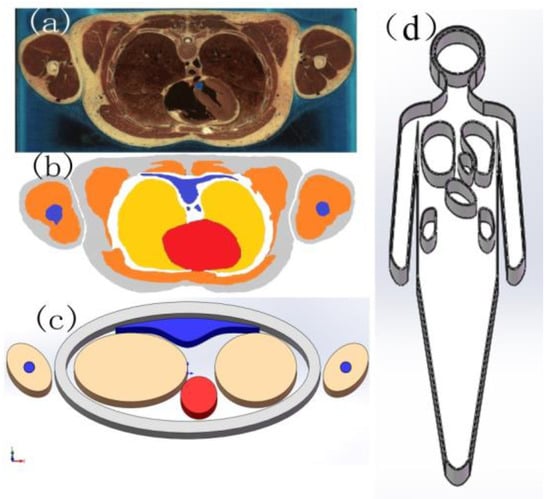
Figure 1.
(a) Human-colored tomographic image data; (b) classified data; (c) phantom designed according to classified data; (d) anthropomorphic phantom used for actual measurement.
2.4. SAR Automatic Test System
Complicated procedures were involved in the evaluation in the distribution of SAR values in the trunk and head concerning medical equipment and wearable medical devices. Apart from the anthropomorphic phantom and simulated body liquids as described in this paper, a professional test system and software were applied.
All experiment results in this paper were obtained through a DASY5 SAR automatic test system manufactured by Switzerland-based SPEAG. As shown in Figure 2, the test system is an international authoritative SAR measurement system. Main performance indicators of the test system were as follows: Field probe should be able to facilitate measurement of three-dimensional electric field intensity of all locations in the phantom exposed to the radiation field; a measurement frequency range of 300 MHz to 3 GHz; and a dynamic range of measurement of at least 10 mW/kg to 100 W/kg. Omni-directional uniformity of the probe within ±1 dB, linearity within ±0.5 d; Field probe diameter of 8 mm; the capability of measuring an unmodulated continuous wave signal as well as a pulse signal with an impulse cycle of 10 Hz to 1 kHz and duty ratio over 4%. Probe positioning accuracy should be finer than ±0.2 mm; the positioning resolution, reflecting the minimum increase in the positional change of the test system when performing the measurement, should not exceed 1 mm.
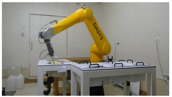
Figure 2.
DASY5 experiment scene for measuring specific absorption rate (SAR).
3. Results
3.1. Calculation of Full-Band Electromagnetic Parameters of Brain, Tissue, Heart, Lungs, Stomach, and Kidneys
Electromagnetic parameters of tissues and organs from the cited reference are as shown in Table 1. Using a least-squares fitting method as per Equation (10), the function expression for full-band electromagnetic parameters of Brain Grey Matter was obtained, as shown in Figure 3:

Table 1.
Electromagnetic parameters of tissues and organs.
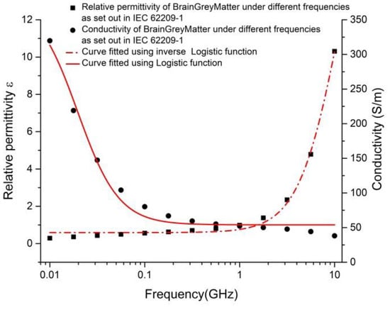
Figure 3.
Values of Brain Grey Matter under different frequencies and results fitted using a logistic function.
In which, the correlation coefficient R2 = 0.95605. By comparing Equations (9) and (11), relevant electromagnetic parameters were identified as = 380.75, = 59.93, and = 0.001587.
Meanwhile, values under a low-frequency limit and high-frequency limit of permittivity of the dispersion regime = 46.38243 and = 39.03617 were highly consistent with corresponding values = 319.7, = 38.11. Thus, verifying that the method was reliable. In the same way, other tissue and an organs’ electromagnetic parameters can be obtained:
By this time, the relation between full-band electromagnetic parameters of human tissues and frequency was derived using a Debye polynomial based on discrete test values of electromagnetic parameters under different frequencies. It should be noted that when conducting a test of tissue parameters at a specific frequency point, considering the uncertainties in the actual test, the system error could be obtained by testing with a reference material (e.g., dimethyl sulfoxide) with identified electromagnetic parameters, and be used for calibration of actual test results.
3.2. Measurement of Electromagnetic Parameters of Simulated Body Liquids
After the function expression of full-band electromagnetic parameters of different tissues was obtained, simulated body liquids matching working frequency points of different devices were formulated according to actual test requirements. Simulated body tissue fluids and trunk tissue fluids at 915 MHz, 1850 MHz, 1880 MHz, and 2450 MHz representing muscular tissue and cardiac muscular tissue were formulated, as shown in Table 2, and a comparison with theoretically calculated results was conducted, with the test errors shown in Table 3.
Table 2.
Formulas and permittivity of simulated trunk tissue fluids satisfying target dielectric parameters.
Table 3.
Comparison for errors between permittivity and conductivity of simulated body liquids under different frequencies and reference values.
It can be inferred from the results that the permittivity of simulated body liquids formulated under different frequencies is not evidently different from the reference value. In common practice, a permittivity difference below 5% is deemed as satisfactory to the requirements. It can also be concluded that the higher the frequency is, the more evident the difference between test value and reference value becomes. Therefore, special attention should be paid to fluid formulation under high frequency.
3.3. Electromagnetic Radiation SAR of a Wearable Medical Device
A wearable ECG sensor detects body information through an electrode, and, after information processing, communicates the information to the other terminal wirelessly. It can be drawn from the working principle of a wearable ECG sensor that apart from the electromagnetic radiation generated from the detection process, additional electromagnetic radiation is produced from the wireless communication process, which is worthy of consideration. The simulated liquids of muscle and heart were tested at 2450 MHz, with the test arrangement adequately reflecting a real working scenario of a wearable ECG sensor; the sensor was directly pasted onto the surface of the test phantom. The device was powered on and started normal operation, refer to Figure 4a. The electromagnetic radiation SAR distribution in key parts of the human body under normal transmission power was tested and charted. To reflect the superimposed effect of electromagnetic radiation SAR at the junction of multiple medical devices, the electromagnetic radiation SAR value of the human body when both cell-phone and sensor were in operation was tested.
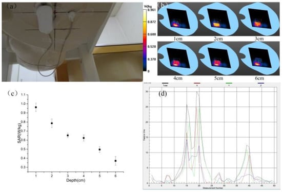
Figure 4.
Test results of a wearable Electrocardiograph (ECG) sensor. (a) Test configuration of ECG sensor, (b) test waterfall chart from SAR test (maximum value of SAR = 0.961 W/kg); (c) test result of SAR at different distance; (d) absolute values of three-dimensional electric field intensity from different locations.
The calculations established the relation between local SAR value of trunk muscular tissue and the distance from the wearable device to the skin surface. The results showed that as the distance from the antenna of the wearable device to skin increased, local SAR value decreased. Moreover, the electric field intensity distribution also confirmed that the SAR value decreased as the distance from the radiation source to head increased, as is shown in Figure 4. It can be seen that the maximum three-dimensional electric field intensity was 30 V/m; electromagnetic radiation SAR value at the surface was 0.961 W/kg, and the value was 0.37 W/kg at a depth of 6 cm from the surface.
4. Discussion
4.1. On the Electromagnetic Characteristics of Different Tissues
As is known, a biological molecule is composed of different types of atoms and has different electric dipole moments. Therefore, different polarization effects are presented under the influence of an external electric field, and different electrical responses are triggered along with a change of frequency. Bio-tissues contain many organic macromolecules and polar groups like –NH2, –OH, –SH, and –COOH with comparatively greater electric dipole moments. When an external electric field is applied, the orientation of the dipoles of polar groups in biological molecules changes, resulting in the high permittivity of bio-tissue. The higher the degree of oriented arrangement, the bigger the permittivity is. Various bio-tissues have a permittivity closely associated with the oriented arrangement of their component biological molecules. Under low frequency, when both macromolecules and small molecules are in an oriented arrangement, the permittivity of a tissue is at its maximum, as the frequency increases, responses are only triggered in polar molecules with relatively smaller molecular weights. The permittivity of the system decreases, but conductivity of bio-tissue increases within the frequency range in question, which indicates when electromagnetic waves are absorbed by a bio-tissue, the higher the frequency, the greater the expected attenuation. Therefore, electromagnetic waves with higher frequencies permeate an organism to shallower depths.
4.2. On the Formulation of Simulated Body Liquids
Considering that bio-tissue is a complex organic system of various constituents (e.g., water, protein, etc.) featuring different relaxation processes, it is extremely hard to formulate a liquid which is capable of simulating the medium characteristics of a real bio-tissue within a wide frequency range; all current tissue models are designed for a certain frequency range. Furthermore, the permittivity of a simulated tissue is subject to the ambient temperature change. In order to simulate the characteristics of a bio-tissue properly, a simulated tissue needs to be studied within a specified temperature range. The basic requirements for the formula of an equivalent simulated tissue are stable and reliable dielectric properties. In this experiment, the Tween simulated body liquid formulation system was adopted. Tween refers to a kind of non-toxic, yellowish thick liquid, miscible with water and salt in any proportion. As it enjoys relatively lower permittivity under a low-frequency range, it gives the phantom lower viscosity and permittivity. Using the Tween system can significantly reduce the amount of materials used. With fewer materials, formulation is less difficult, and therefore, the Tween system offers great advantages for simulated liquid formulation.
4.3. On Radiation Limit and Radiation Exposure Level
According to studies conducted with animals, 4 W/kg is widely recognized as the upper limit for organism exposure to radio frequency. The exposure limit of 2 W/kg for the general public, as stipulated by the ICNIRP (International Commission on Non-Ionizing Radiation Protection), leaves a large safety margin. The ICNIRP standard, formulated on the basis of a thorough analysis of all available scientific materials, can provide a good guarantee of human safety from all identified radio frequency hazards. Thus, it is well supported and received by the WHO (World Health Organization), as well as being a recommended standard of the International Telecommunication Union. To put it simply, the ICNIRP guideline established the recommended limits on the following basis: 4 W/kg is the basic limit of thermal effect when SAR is up to 4 W/kg, the human body would present with significant harmful reactions. According to the basic limit, when a ten-time coefficient of safety is applied for the occupational group, a limit value of 0.4 W/kg is established; when a 50-time coefficient of safety is applied for the general public, a limit value of 0.08 W/kg is established.
It can be drawn from the experiment in this paper that when a wearable device was close to the skin during use, a considerable exposure of electromagnetic radiation to the surface tissue of human body was experienced, which was measured to be up to 0.961 W/kg, and far exceeded the limit value stipulated for the occupational group. Meanwhile, electromagnetic radiation attenuated along with penetration depth, electromagnetic radiation at a depth of 6 cm below the skin was measured to be 0.37 W/kg, which was equivalent to the limit value stipulated for the occupational group. Therefore, it is concluded that the SAR values in tissues and organs change greatly with varied frequencies and penetration depths. As muscle and skin are located in the outer layer of the human body, they usually absorb more radiation energy than internal organs. Moreover, some tissues and organs are more susceptible to electromagnetic radiation, while some are less susceptible. Therefore, different tissues and organs need a comprehensive evaluation based on actual conditions.
5. Conclusions
Due to fact that electromagnetic radiation is purposely used in medical devices for treatment with target users’ who are more susceptible to radiation, electromagnetic radiation hazards related to medical devices deserve more attention than those in the telecommunication sector. Computational modeling of electromagnetic radiation is the basis for the study on SAR using computer simulation, but its results are limited to the correctness of the theoretical model and boundary conditions; the electromagnetic experiment with an anthropomorphic phantom is an essential instrument for the experimental evaluation of SAR, but the electromagnetic experiment phantoms currently in use do not concern specific tissue and the spatial distribution of radiation with results merely reflecting the average electromagnetic exposure level of the human body. Therefore, they cannot satisfy the needs for the evaluation of medical device’s (e.g., cardiac pacemaker, cochlear implant, etc.) electromagnetic field exposure when a specific area is concerned. To this end, an electromagnetic experiment phantom devoted to the evaluation of the electromagnetic exposure of both local organ and tissue, in addition to the whole body was proposed for the first time in this paper. Meanwhile, full-band electromagnetic parameters of different tissues were studied given that medical devices worked under full-band frequency to ensure the accuracy of the SAR measurement. The evaluation and summarization of electromagnetic radiation SAR of BAN medical devices were conducted on the basis of systematic and local electromagnetic experiments on a phantom for the first time. The results established that the electromagnetic radiation of partial equipment exceeded the allowed values specified by the WHO. Therefore, further studies are yet to be carried out in the medical circle to decide whether additional evaluation measures should be set up in this regard.
Author Contributions
S.L. and H.R. conceived and designed the experiments; Z.S. and Q.W. performed the experiments; S.L. and H.W. analyzed the data; S.L. wrote the paper.
Funding
This research was funded by the National Key Research and Development Program of China, grant number 2016YFC0103202.
Conflicts of Interest
The authors declare no conflict of interest.
References
- Soh, P.J.; Vandenbosch, G.A.E.; Wee, F.H.; van den Bosch, A.; Martinez-Vazquez, M.; Schreurs, D. Specific absorption rate (SAR) evaluation of textile antennas. IEEE Antennas Propag. Mag. 2015, 57, 229–240. [Google Scholar] [CrossRef]
- Senic, D.; Sarolic, A.; Holloway, C.L.; Ladbury, J.M. Whole-body specific absorption rate assessment of lossy objects exposed to a diffuse field inside a reverberant environment. IEEE Trans. Electromagn. Compat. 2017, 59, 813–822. [Google Scholar] [CrossRef]
- Wang, J.; Xia, Y.-S.; Yin, W.-Y. Study on SAR distribution of human body on the vehicle platform using a modified fdtd method. IEEE Trans. Electromagn. Compat. 2018, 60, 840–848. [Google Scholar] [CrossRef]
- Guellab, A.; Wu, Q. Modeling human body using four-pole debye model in piecewise linear recursive convolution fdtd method for the sar calculation in the case of vehicular antenna. Int. J. Antennas Propag. 2018, 2018. [Google Scholar] [CrossRef]
- Dimbylow, P.J. Fdtd calculations of the whole-body averaged SAR in an anatomically realistic voxel model of the human body from 1 MHz to 1 GHz. Phys. Med. Biol. 1997, 42, 479–490. [Google Scholar] [CrossRef] [PubMed]
- Dimbylow, P. Resonance behaviour of whole-body averaged specific energy absorption rate (sar) in the female voxel model, naomi. Phys. Med. Biol. 2005, 50, 4053–4063. [Google Scholar] [CrossRef] [PubMed]
- Dimbylow, P.; Bolch, W. Whole-body-averaged SAR from 50 mhz to 4 ghz in the university of florida child voxel phantoms. Phys. Med. Biol. 2007, 52, 6639–6649. [Google Scholar] [CrossRef] [PubMed]
- Findlay, R.P.; Dimbylow, P.J. Effects of posture on fdtd calculations of specific absorption rate in a voxel model of the human body. Phys. Med. Biol. 2005, 50, 3825–3835. [Google Scholar] [CrossRef] [PubMed]
- El Amrani, L.; Mazri, T.; Hmina, N. Specific absorption rate (SAR) in humain body exposed to wirless base station fields. In Proceedings of the 9th International Conference on Electronics, Computers and Artificial Intelligence—ECAI 2017, Targoviste, Romania, 29 June–1 July 2017. [Google Scholar]
- Seo, Y. Evaluation of specific absorption rate and temperature increase induced by artificial medical implants during mri scan. Med. Phys. 2016, 43, 3725. [Google Scholar] [CrossRef]
- Psenakova, Z.; Benova, M. Evaluation of SAR (specific absorption rate) in multilayer structure of biological tissues near ear with cochlear implant. In Proceedings of the 2017 18th International Conference on Computational Problems of Electrical Engineering (CPEE), Kutná Hora, Czech Republic, 11–13 September 2017. [Google Scholar]
- Ahmed, G.; Ul Islam, S.; Shahid, M.; Akhunzada, A.; Jabbar, S.; Khan, M.K.; Riaz, M.; Han, K. Rigorous analysis and evaluation of specific absorption rate (SAR) for mobile multimedia healthcare. IEEE Access 2018, 6, 29602–29610. [Google Scholar] [CrossRef]
- Pethig, R.; Kell, D.B. The passive electrical-properties of biological-systems–Their significance in physiology, biophysics and biotechnology. Phys. Med. Biol. 1987, 32, 933–970. [Google Scholar] [CrossRef] [PubMed]
- Gabriel, S.; Lau, R.W.; Gabriel, C. The dielectric properties of biological tissues: II. Measurements in the frequency range 10 Hz to 20 GHz. Phys. Med. Biol. 1996, 41, 2251–2269. [Google Scholar] [CrossRef] [PubMed]
- Gabriel, S.; Lau, R.W.; Gabriel, C. The dielectric properties of biological tissues: III. Parametric models for the dielectric spectrum of tissues. Phys. Med. Biol. 1996, 41, 2271–2293. [Google Scholar] [CrossRef] [PubMed]
- Gabriel, C.; Gabriel, S.; Corthout, E. The dielectric properties of biological tissues: I. Literature survey. Phys. Med. Biol. 1996, 41, 2231–2249. [Google Scholar] [CrossRef] [PubMed]
- Kanezaki, A.; Hirata, A.; Watanabe, S.; Shirai, H. Effects of dielectric permittivities on skin heating due to millimeter wave exposure. Biomed. Eng. Online 2009, 8. [Google Scholar] [CrossRef] [PubMed]
- Fujiwara, O.; Takai, K. Electrical properties of skin and SAR calculation in a realistic human model for microwave exposure. Trans. Inst. Electr. Eng. Jpn. Part C 1997, 117, 75–80. [Google Scholar] [CrossRef]
© 2018 by the authors. Licensee MDPI, Basel, Switzerland. This article is an open access article distributed under the terms and conditions of the Creative Commons Attribution (CC BY) license (http://creativecommons.org/licenses/by/4.0/).