Article

Dinuclear Copper(II) Complex with Hemiaminal N,O-Donor Ligand

1
Department of Basic Chemical Sciences, Faculty of Pharmacy, Wrocław Medical University, Borowska 211a, 50-556 Wrocław, Poland
2
Department of Biotechnology and Food Microbiology, Faculty of Biotechnology and Food Science, Wrocław University of Environmental and Life Sciences, Chełmońskiego 37, 51-630 Wrocław, Poland
3
Department of Physical and Quantum Chemistry, Faculty of Chemistry, Wroclaw University of Science and Technology, Smoluchowskiego 23, 50-372 Wrocław, Poland
4
Faculty of Chemistry, University of Wrocław, F. Joliot-Curie 14, 50-383 Wrocław, Poland
*
Authors to whom correspondence should be addressed.
Appl. Sci. 2026, 16(1), 136; https://doi.org/10.3390/app16010136
Submission received: 27 November 2025 / Revised: 10 December 2025 / Accepted: 17 December 2025 / Published: 22 December 2025

Abstract

Novel copper(II) coordination compounds with hemiaminal N,O-donor ligands were obtained and synthesized in a one-pot reaction from three appropriate substrates (aldehyde, amine, and copper(II) chloride) in methanol. A dinuclear complex with a [Cu2Cl2(hemiaminal)2(amine)2] coordination mode was obtained. The complex consists of two five-coordinated central Cu(II) cations with square pyramidal geometry and Ci molecular symmetry. The hemiaminal oxygen atom forms a bridge between the two metallic centers, and that coordination bond is a factor stabilizing these hemiaminal moieties, generally regarded as unstable intermediates. We analyzed the energetic and physicochemical properties of the [Cu2Cl2(hemiaminal)2(amine)2] complex using density functional theory (DFT). First of all, we predicted the geometrical parameters, molecular electrostatic potential, HOMO and LUMO energies, and reactivity indices to indicate the free radical scavenging capacity. Based on the topological analysis of charge densities, we also characterized the properties of hydrogen bonds. Moreover, the antimicrobial properties of the complex were investigated, and it exhibited the highest activity against Gram-positive bacteria and Candida albicans.

1. Introduction

Copper(II) complexes with coordinated hemiaminal moieties are interesting functional materials for two main reasons. From the chemical point of view, forming a coordination bond is one of the factors stabilizing the hemiaminal itself; otherwise, a reactive intermediate in a Schiff base formation process is created. Taking into consideration pharmaceutical interests, copper(II) complexes in general and compounds with an imine ligand in particular possess interesting biological activity.
Hemiaminals are tetrahedral intermediates of the multistep addition reaction of a primary or secondary amine to a carbonyl group of an aldehyde or ketone [1]. These products are generally regarded as thermodynamically unstable and subsequently difficult or even impossible to isolate and characterize with direct methods [2]. Under neutral, acidic, or basic pH conditions, these intermediates undergo a further dehydration reaction leading to the formation of an imine (or Schiff base) [3,4,5,6]. The presence of hemiaminals has been observed with DC polarography [7], low-temperature spectroscopy [8], MS spectrometry [9], FTIR liquid-cell spectroscopy [10], in situ cryo-crystallization [2], and solvent-free mechanochemical reactions [11]. Hemiaminals with half-lives ranging from 30 min to over 100 h were observed with spectral methods (NMR) inside a synthetic deep cavitand, whose role was to isolate the reactive tetrahedral center from the environment [12,13,14,15]. Similar shielding was obtained by conducting the addition reaction within the porous crystal network, allowing the direct observation of hemiaminal formation and further dehydration in situ by X-ray crystallography [16] or solid-state 15N-NMR [17]. Hemiaminals have also been trapped within dynamic covalent organogels [18,19]. Several stable hemiaminals have been obtained in the solid state. These compounds were stabilized by either the formation of intramolecular hydrogen bonds [20,21] or by the special electronic effects arising from electron-withdrawing substituents (nitro- or cyano-groups) on a carbonyl component and electron-rich amine (such as 1,2,4-triazole or pyrimidine ring) [22,23,24,25,26,27]. Another important way of stabilizing the hemiaminal functional group involves the formation of metal complexes. An analysis of crystal structures deposited in the CSD database [28] revealed that stabilization was obtained by two coordination modes. The first involved the direct formation of coordinate bonds as N- or O-donors to central metal ions such as manganese [29,30,31,32,33], copper [33,34,35,36,37], cobalt [33,38,39,40,41], zinc [42,43], iron [32], nickel [33,36,44], or vanadium [45,46]. In the second mode, the uncoordinated hemiaminal group was part of a rather rigid metallocyclic ring [47].
Although the hemiaminal functional group occurs in natural compounds [48,49,50] and is a part of the enzymatic Schiff base formation process [3,4,51], few compounds have been tested for pharmaceutical applications, due to their limited half-life. However, stable hemiaminals, such as polycyclic hydrocarbon cages, have been shown to modify the effects of cocaine [52], inhibit nitric oxide synthase [53], and form conjugates with selected non-steroidal anti-inflammatory drugs, thereby improving blood–brain barrier permeability [54]. Hemiaminals containing a nitrobenzyl group and triazole moieties were screened for the treatment of periodontal disease [55].
In the field of pharmaceutical sciences, metallodrugs or drugs containing metal as an essential ingredient are desirable products for therapeutic and diagnostic applications [56]. Following the discovery of the anti-cellular division properties of cisplatin [57], a growing number of metal-containing compounds have been tested for their potential use as anticancer agents [56,58,59,60]. Metal complexes may also possess antiviral, antimicrobial, or antifungal activities and can therefore be used successfully as drugs, especially in combination therapy [56]. Copper is one of the trace elements that are essential for the proper functioning of the human organism, as it acts as a cofactor for many enzymes, including Cu,Zn-superoxide dismutase (which scavenges free radicals), cytochrome c oxidase (which facilitates electron transfer), and ceruloplasmin (which is involved in iron metabolism) [61]. Copper coordination compounds exhibit significant biological activity, making them desirable in pharmaceutical research. In cancer treatment, cuproptosis, a form of cell death caused by copper, has recently gained attention [60]. However, copper complexes may interfere with cancerous pathways by several different mechanisms, including induction of apoptosis, preventing angiogenesis, proteasome inhibition, or regulation of the cellular level of reactive oxygen species [59,62]. Copper(II) complexes are the subject of intensive study for their biological activities such as antimicrobial, antiviral, antifungal, antitumor, anti-inflammatory, and enzyme inhibition properties [63]. Notably, copper complexes can be more effective than common antibiotics as potential antimicrobial agents [63]. Some Cu(II) coordination compounds exhibit stronger antibacterial [64,65,66,67] or antifungal [68,69,70] properties than their initial ligands [71]. A very important subgroup of metal complexes are Schiff base derivatives. These compounds are interesting because of their biological activity and potential medical applications acting as anticancer, antimicrobial, antiviral, antifungal, antioxidant, or antidiabetic agents [72,73,74,75,76,77,78,79,80,81,82]. The hemiaminal intermediate formed during Schiff base synthesis may coordinate with a metal ion to form complexes with interesting biological activity [83]. N,O-donor ligands can form a multidentate coordination motif with a variety of metals [84], which subsequently influences the stability, reactivity or electronic properties of coordination compounds [85]. Copper(II) complexes with N,O-donor ligands have also been studied for their antibacterial [71,86,87,88] or antifungal [88,89,90] activity.
Since copper forms a wide variety of complexes with amino alcoholates [91] and many copper coordination compounds are biologically active, the aim of this study was to synthesize new Cu-hemiaminal (also amino alcohol) derivatives, which may also feature desired pharmacological properties such as antibacterial and antifungal activity. Theoretical calculations were performed to characterize reactivity parameters that may be related to the antimicrobial action of the studied compound. Additionally, the antimicrobial activity of the complex was tested against Gram-positive and Gram-negative bacteria, as well as C. albicans.

2. Materials and Methods

2.1. General Information

The synthesis was performed using commercially available reagents (Sigma-Aldrich, Merck, Darmstadt, Germany) and solvents (Chempur, Piekary Śląskie, Poland) which were not further purified. Elemental analyses were performed using a CHNS Vario EL III analyzer (Elementar Analysensystem GmbH, Hanau, Germany). The uniMELT 2 apparatus (LLG, Meckenheim, Germany) was used for melting point determination. The ATR-IR spectrum was measured using a Nicolet iS50 FT-IR photometer Thermo Fisher Scientific Inc., Waltham, MA, USA).
A single crystal X-ray diffraction measurement was conducted using an XtaLAB Synergy R, DW system (Rigaku Corporation, Tokyo, Japan), four-circle diffractometer with a Cu Kα radiation source and Hybrid Pixel Array Detector at 100 K. Data collection and reduction were performed using the CrysAlisPro 1.171.42.73a program ( (Rigaku Corporation, Tokyo, Japan). The structure was solved by direct methods and further refined using a full-matrix least squares method with anisotropic thermal parameters for non-hydrogen atoms in the SHELXL-2019/2 [92] program. The XP-Interactive Molecular Graphics version 5.1 program (Bruker AXS Inc., Madison, WI, USA) was used for molecular graphics preparation.
The crystallographic data for the complex has been deposited in the Cambridge Crystallographic Data Centre, under the number CCDC2456558. It can be accessed at the CCDC, 12 Union Road, Cambridge, CB2 1EZ, UK [fax: (+44) 1223-336-033; e-mail: deposit@ccdc.cam.ac.uk].

2.2. Synthetic Procedure

A molar ratio of 1:2 of 3-nitrobenzaldehyde (3nb, 86.6 mg, 0.57 mmol) and 4-methylpyridin-2-amine (2a4mp, 123.6 mg, 1.14 mmol) was dissolved in methanol (5 mL) and stirred on a magnetic stirrer at 60 °C for about an hour. Then, an equimolar (to aldehyde) amount of tributylamine (Bu3N, 150 μL) and copper(II) chloride (CuCl2·2H2O, 97.8 mg, 0.57 mmol, in 5 mL of methanol solution) was added (Scheme 1). The reaction mixture was further stirred for an additional 30 minutes. After several days and slow evaporation of the solvent, green crystals of the copper(II) complex (3nb2a4mpCu), which is coordinated with a chloride ion, 4-methylpyridin-2-amine (2a4mp), and a hemiaminal anion [(4-methylpyridin-2-yl)amino](3-nitrophenyl)methanolate (3nb2a4mp), were obtained. The product was filtered off, washed with a small amount of methanol, and air-dried (yield: 200 mg, 75%), mp 147–148 °C d.
Elemental analysis, calculated for formula C38H40Cl2Cu2N10O6 C 49.04, H 4.33, N 15.05, Cl 7.62, found C 48.13, H 4.37, N 14.77, Cl 7.68; the slight difference may arise from the presence of an undefined amount of disordered solvent molecules within the crystal lattice, which was not included in the formula above.
ATR-FTIR 451.74 vs, 595.62 s, 539.49 s, 580.95 m, 621.93 m, 670.62 m, 727.03 s, 764.64 w, 792.60 s, 806.10 s, 858.17 m, 934.34 m, 989.30 m, 1034.14 m, 1069.82 s, 1105.49 m, 1180.22 m, 1189.86 m, 1240.97 w, 1263.15 m, 1282.94 m, 1313.77 m, 1342.21 s, 1465.63 s, 1490.22 s, 1521.08 s, 1557.24 m, 1625.70 s, 3180.04 w, 3299.12 w.
Crystal data 3nb2a4mpCu: 4(C38H40Cl2Cu2N10O6)·CH3OH, M = 7478.26 crystal system: tetragonal, space group: I41/a, a = 22.0776(3) Å, c = 17.1398(3) Å, V = 8354.3(3) Å3, Z = 1, T = 100(2) K, ρc = 1.486 g·cm−3, μ = 2.917 mm−1, θmin = 3.264°, θmax = 72.841°, reflections: 13,846, independent: 4008, Rint = 0.0384, R1 = 0.0438, wR2 = 0.1291, GoF = 1.056.

2.3. Computational Details

The compound structure was optimized using the Gaussian 2016 C.01 software package using the B3LYP method with LanL2DZ and B3LYP ++6-31G** basis sets for copper and the remaining atoms, respectively [93]. Solvent effects were taken into account using the Polarizable Continuum Model (PCM) [94]. The same level of theory was applied to calculate the reactivity parameters. The visualization of estimated parameters was performed in the Avogadro molecular visualization program [95].
Hirshfeld surface analyses were performed using CrystalExplorer 17.5 [96], based on the molecular geometry obtained directly from X-ray diffraction without further optimization. The Hirshfeld surfaces were mapped over the normalized contact distance (dnorm) and visualized to identify regions of significant intermolecular interactions. Two-dimensional fingerprint plots were also generated to quantitatively assess the relative contributions of different contact types to the overall crystal packing.
Quantum Theory of Atoms in Molecules (QTAIM) analyses were carried out using the AIM2000 program package [97]. The required wavefunction (.wfn) files were generated based on the X-ray diffraction molecular geometry using Gaussian 16 [93]. Single-point energy calculations were performed employing the B3LYP density functional with Grimme’s D3 dispersion correction (GD3). The LanL2DZ effective core potential basis set was used for the copper atom, while the 6-31+G(d) basis set was applied to all other elements (C, H, N, O, Cl). No symmetry constraints were applied during the calculations.

2.4. Antimicrobial Activity

The antimicrobial potential of the 3nb2a4mpCu was assessed against a panel of microorganisms comprising (i) Gram-positive bacteria (Bacillus cereus ATCC 10876, Enterococcus hirae ATCC 10541, Staphylococcus aureus ATCC 25923, and Staphylococcus epidermidis KCTC 1917); (ii) Gram-negative bacteria (Escherichia coli ATCC 10536, Klebsiella pneumoniae ATCC 13886, Pseudomonas aeruginosa ATCC 15442, Proteus mirabilis ATCC 21100, and Yersinia enterocolitica DSM 23248); and (iii) the yeast Candida albicans ATCC 10231. All microbial strains were sourced from the culture collection of the Department of Biotechnology and Food Microbiology at Wrocław University of Environmental and Life Sciences. Bacterial strains were maintained in Luria–Bertani (LB) broth (A&A Biotechnology, Gdańsk Poland; consisting of 10 g/L tryptone, 5 g/L yeast extract, and 5 g/L NaCl) at 37 °C, whereas yeast cultures were propagated in yeast peptone dextrose (YPD) medium (A&A Biotechnology, Poland; containing 20 g/L bacteriological peptone, 10 g/L yeast extract, and 20 g/L glucose) at 30 °C. Agar was incorporated into the medium at 2% when required. For preparation of inocula used in the agar diffusion assay, cultures were incubated in 10 mL of the respective medium in 100 mL Erlenmeyer flasks at 180 rpm for 48 h at the appropriate temperature. The cells were subsequently washed with sterile physiological saline and adjusted to an optical density OD600 = 1.
The antimicrobial activity of 3nb2a4mpCu was examined using a standard agar diffusion method. A volume of 100 µL of the standardized inoculum was uniformly spread across the surface of agar plates, which were then allowed to equilibrate for 1 h. Each test disc was loaded with 5 mg of the 3nb2a4mpCu compound (from a 50 mg portion). Plates were incubated for 24 h at either 30 or 37 °C, depending on the microorganism tested. After incubation, the diameters of the inhibition zones surrounding the discs were recorded.

3. Results and Discussion

3.1. Crystal Structure Description

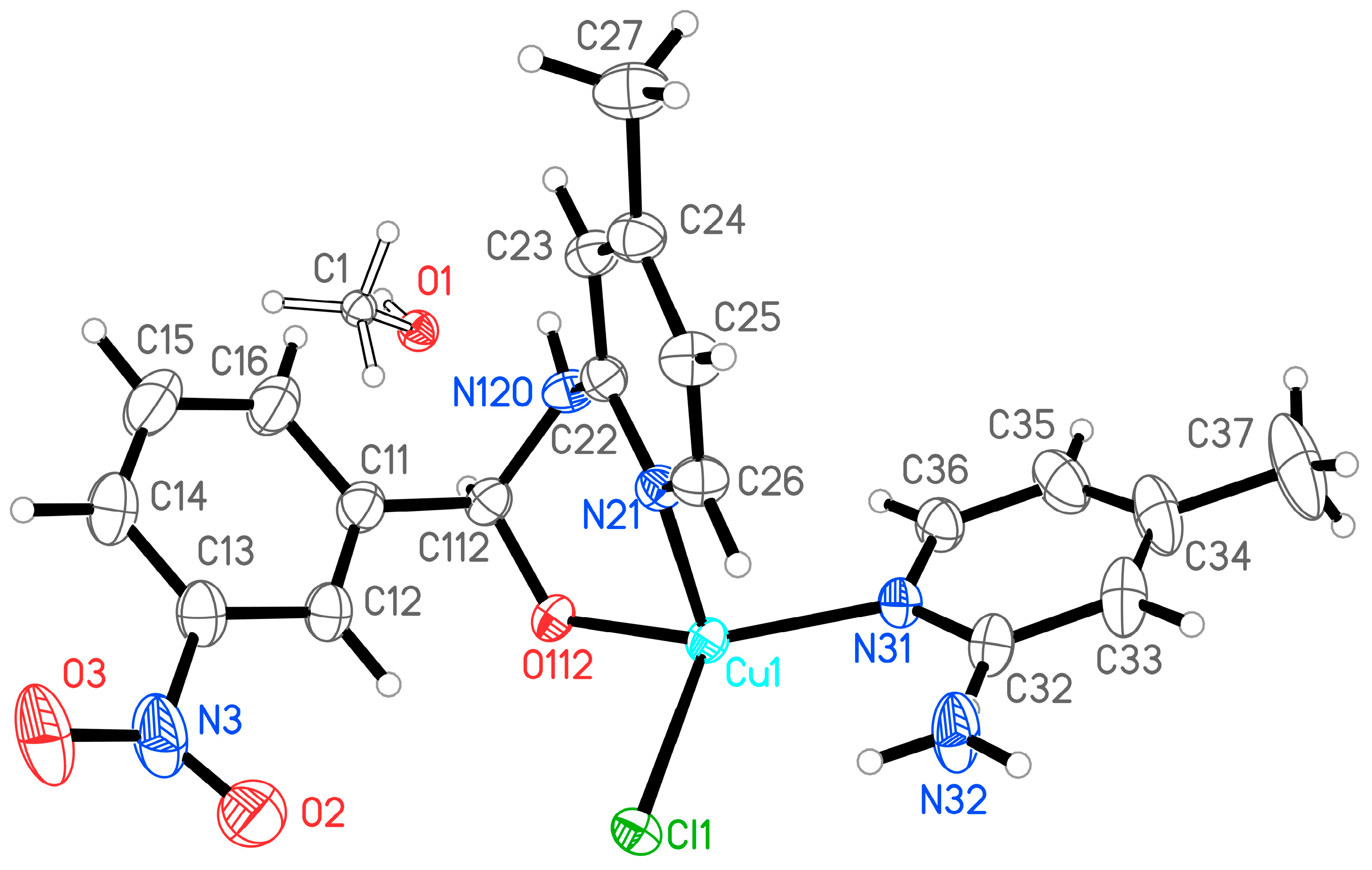
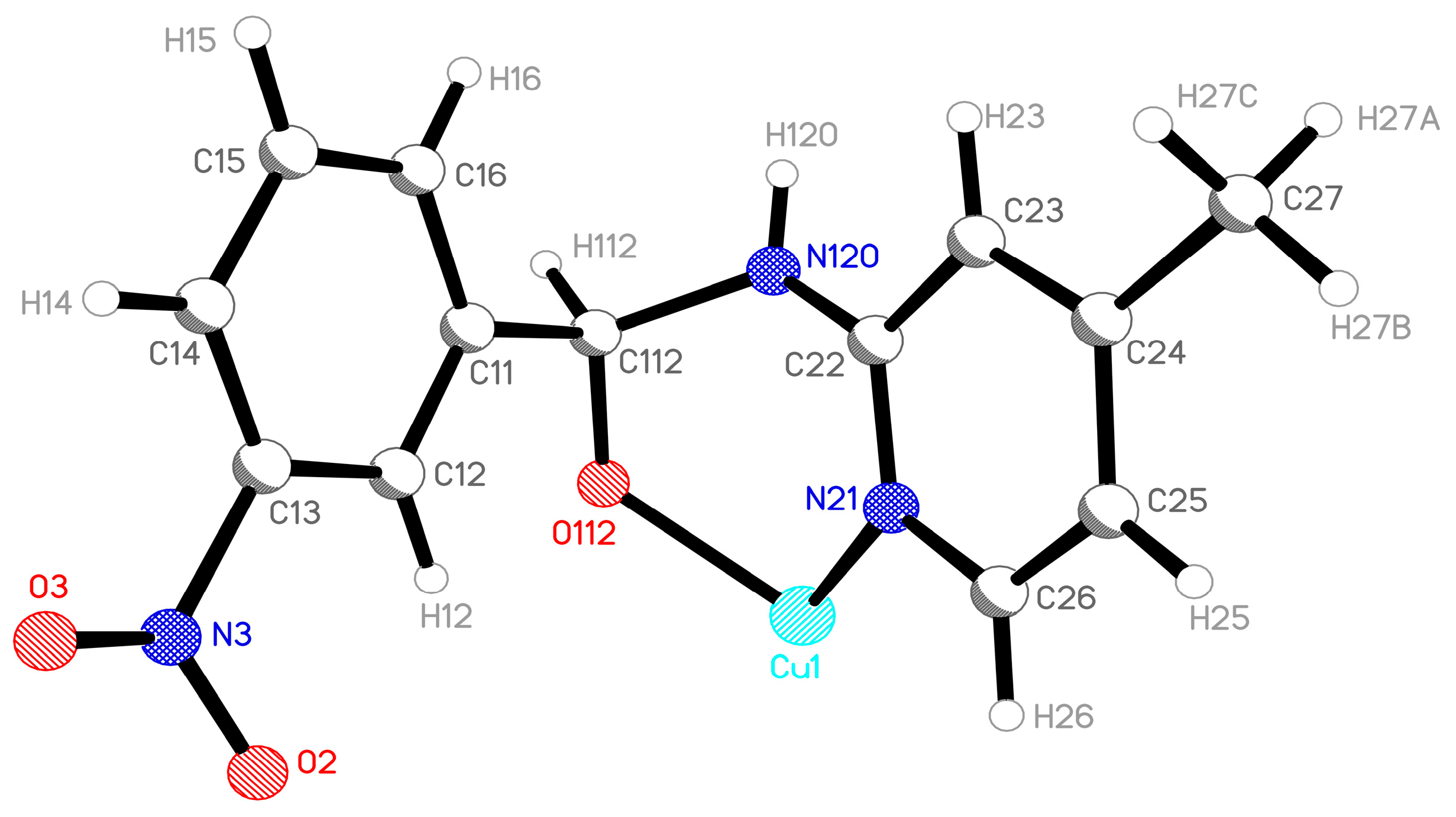
The asymmetric part of the unit cell consists of half a dinuclear complex molecule (3nb2a4mpCu), which comprises a central copper(II) cation that is coordinated by a chloride ion, 4-methylpyridin-2-amine (2a4mp), and a hemiaminal anion, [(4-methylpyridin-2-yl)amino](3-nitrophenyl)methanolate (3nb2a4mp), which acts as a bidentate N,O-donor ligand. Additionally, in the asymmetric part, 1/16 of a disordered solvent molecule methanol solvent molecule occupies the free space (Figure 1). The chloride and the amino group of 2a4mp are disordered in two positions, with an occupancy of 0.75 for the major component and 0.25 for the minor. One oxygen atom from the nitro group of 3nb2a4mp is disordered in two positions that are occupied equally.
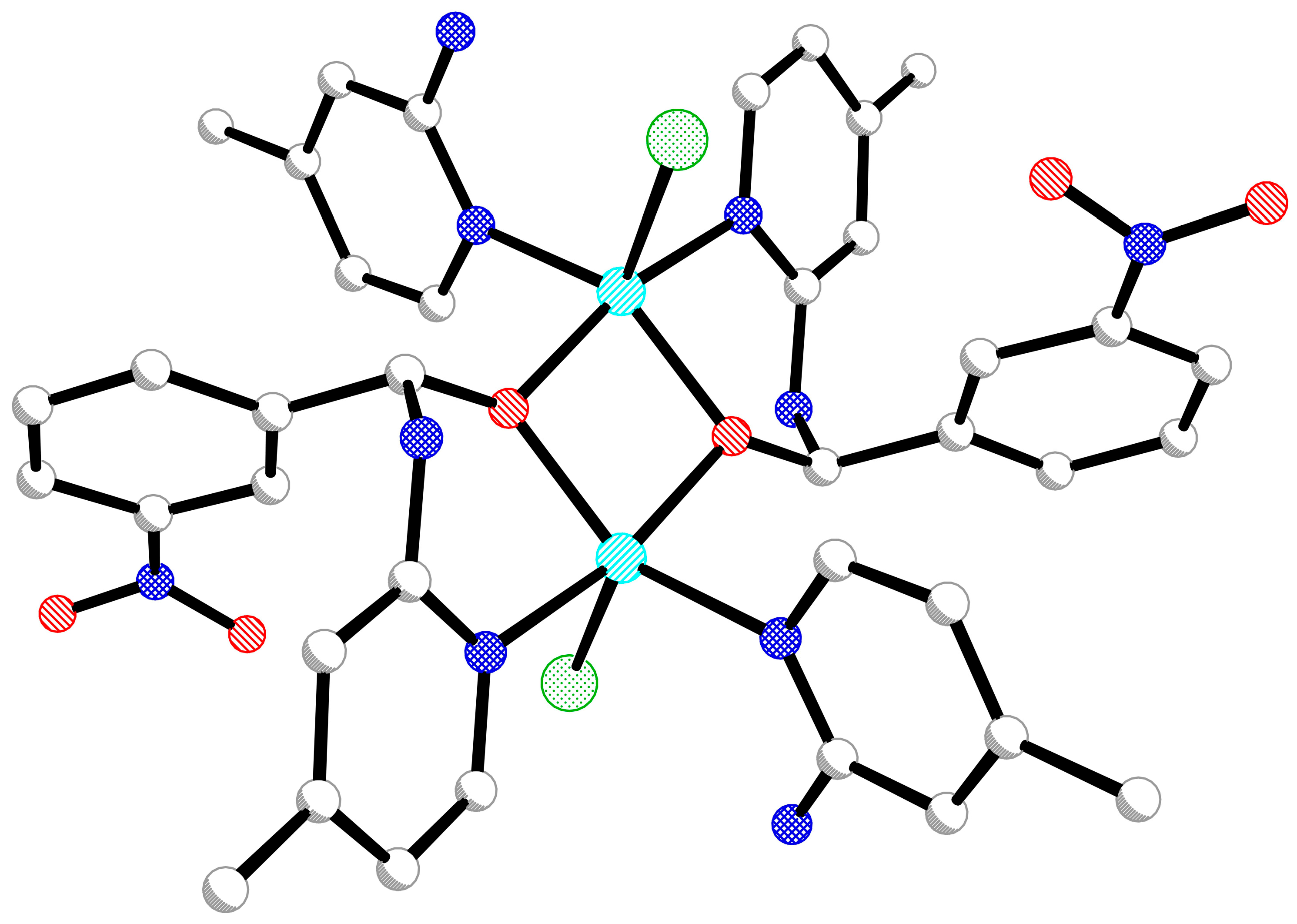
The complex molecule exhibits internal symmetry Ci (Figure 2), and the oxygen atom from the hemiaminalate ion 3nb2a4mp bridges two metallic centers.
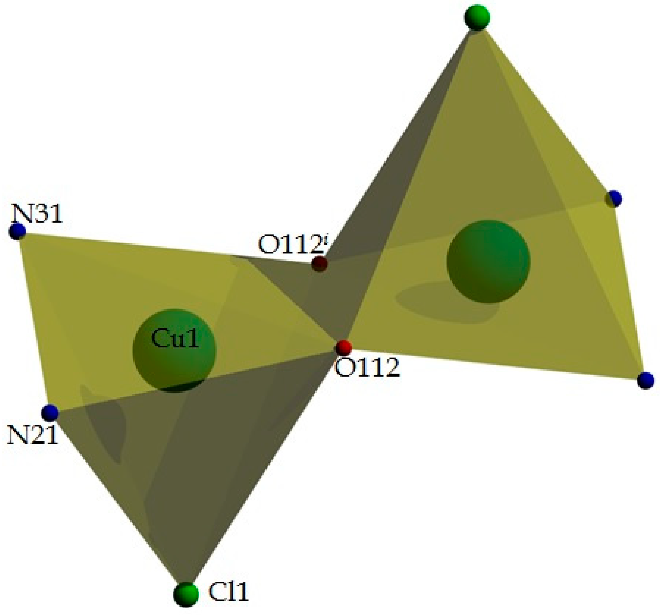
Each Cu(II) ion has a coordination number of 5, and the donor atoms are arranged in a coordination polyhedron that resembles a tetragonal pyramid (Figure 3). The chloride ion Cl1/Cl1A is located at the pyramid’s apex, and two symmetry-related oxygen atoms (O112 and O112i, symmetry code: (i) 1 − x, 1 − y, 1 − z) from 3nb2a4mp ligands bridge the two Cu centers. The O122-O112i edge is shared by two centrosymmetric, tetragonal pyramids. The coordination sphere is filled by two pyridyl nitrogen atoms: N21from 3nb2a4mp and N31 from 2a4mp. Selected geometrical parameters such as bond lengths, valence, and torsion angles are listed in Table 1.
The 3nb2a4mp hemiaminalate ion is coordinated as a bidentate N,O-donor ligand to one metallic center (Figure 4). The C11–C112 and C112–N120 interatomic distances indicate the presence of single bonds between the atoms. Both the C112 and N120 atoms have sp3 hybridization and are chiral centers, both with absolute configuration S. The bidentate mode of 3nb24mp coordination constrains the “+sc” conformation on the C11–C112–N120–C22 atomic linker, which connects the two aromatic rings of the hemiaminal ligand.
Based solely on geometrical criteria, the crystal structure of 3nb2a4mpCu is stabilized by a network of N–H···Cl and C–H···O hydrogen bonds (Table 2). No X–H···π-type interactions are observed in the crystal structure.
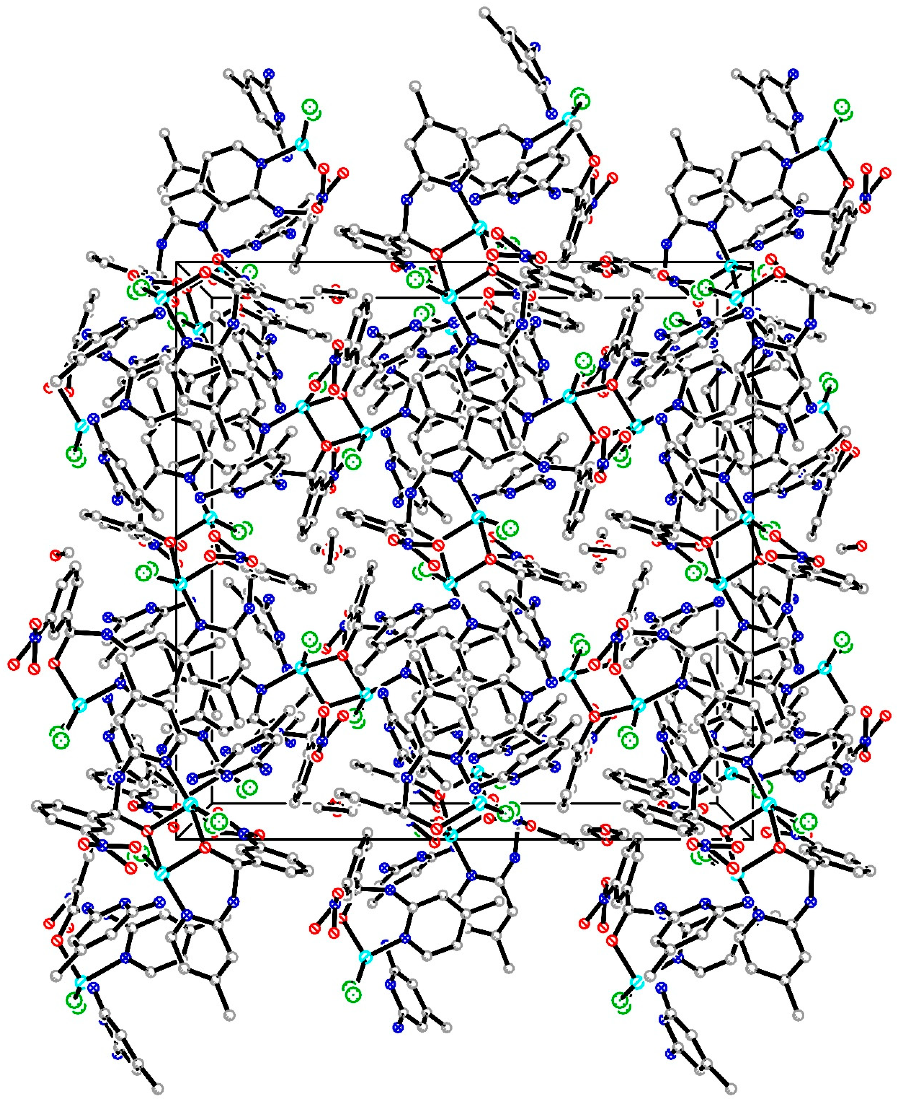
In the crystal structure of 3nb2a4mpCu, channels parallel to the c-axis are formed (Figure 5). Solvent molecules are located within these channels. The methanol molecule (1/16 per the asymmetric unit; 1 per the unit cell) occupies a special position in the lattice on the 4-fold rotoinversion axis 4 ¯ , which entails a disorder model of the solvent in the crystal.
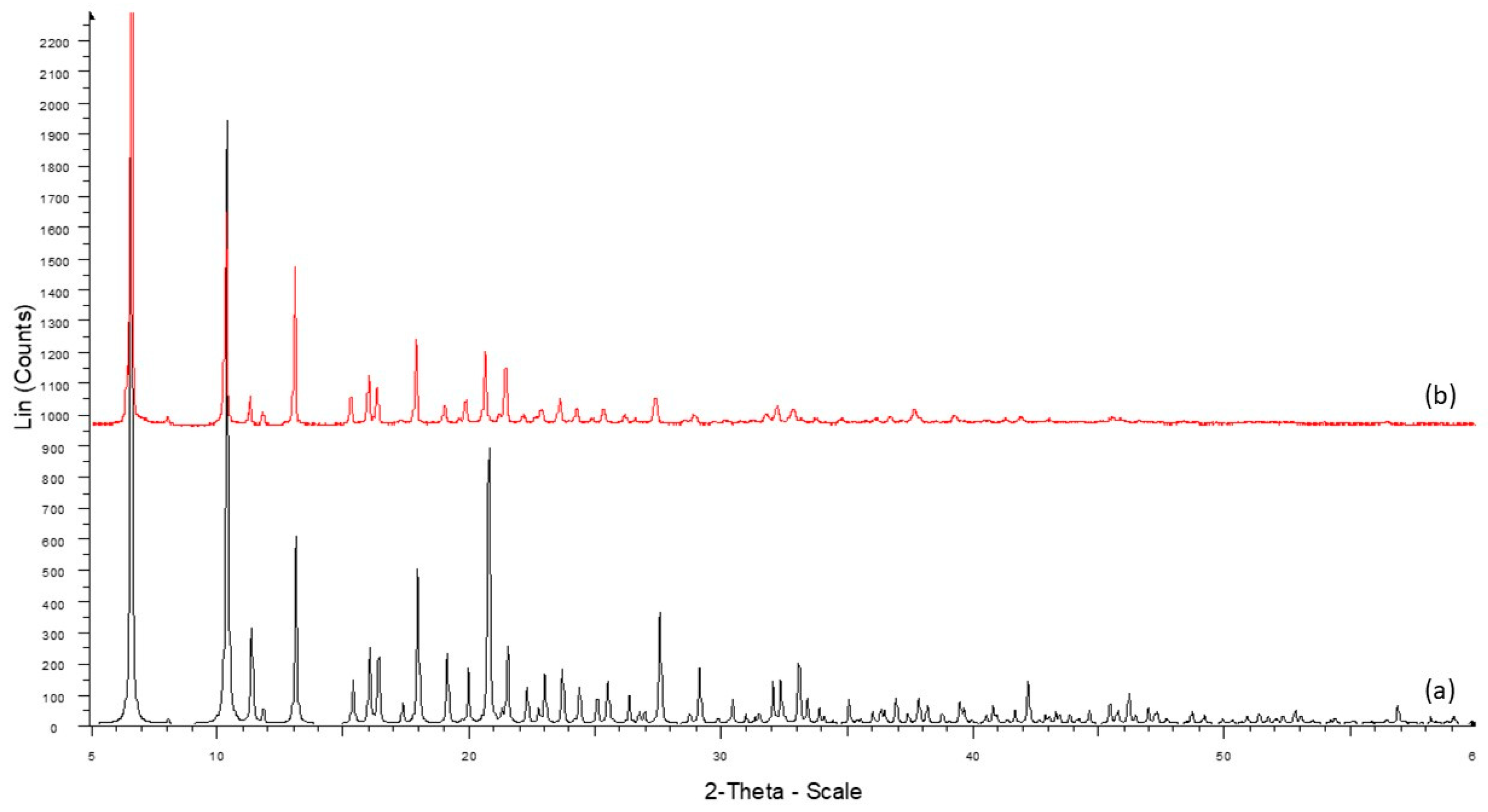
The sample’s homogeneity was confirmed by a powder X-ray diffraction experiment (PXRD). The experimental diffraction pattern obtained for the microcrystalline bulk sample of 3nb2a4mpCu (Figure 6b) is consistent with the pattern calculated for the single crystal structure (Figure 6a). No additional peaks were observed.

3.2. Reactivity Parameters

The reactivity of compounds can be predicted based on computationally obtained chemical descriptors. In the present study, reactivity descriptors were calculated from the highest occupied molecular orbital (HOMO) and lowest unoccupied molecular orbital (LUMO) energies. It is well known that the value of the HOMO energy reflects the electron-donating properties of compounds. On the other hand, the LUMO energy represents its ability to accept electrons. A higher LUMO energy corresponds to a lower tendency to participate in reduction reactions. Table 3 shows the localization of the frontier molecular orbital density of the complex. It was previously proven that the antibacterial activity of compounds can be a function of their LUMO energy. Several studies have demonstrated that certain regions characterized by a high contribution of LUMO play a significant role in biological activities [98,99]. The calculated HOMO/LUMO gap of the compound (0.673 eV) indicates high chemical reactivity. It is generally accepted that a lower energy gap (<2 eV) is associated with lower chemical stability and greater environmental sensitivity. As demonstrated by the data, the ionization potential (I) value is indicative of high compound reactivity and greater susceptibility to oxidation. The electron affinity (A) is the ability of a molecule to accept an extra electron and indicates its potential to participate in nucleophilic reactions. The value of the ionization potential in this case is related to the molecule’s low stability (6.846 eV). Other key parameters that describe a compound’s resistance to electronic deformation and its ability to participate in chemical reactions are chemical hardness and softness. As presented in Table 3, the chemical softness of the analyzed compounds is equal to 2.972 eV−1. As established, soft molecules are characterized by greater stability. The results also revealed that the chemical potential that defines the tendency of the compound to accept electrons is equal to −6.510 eV. The values of the remaining parameters, electronegativity and electrophilicity index, are also presented in Table 3.

3.3. Hirshfeld Surface Analyses

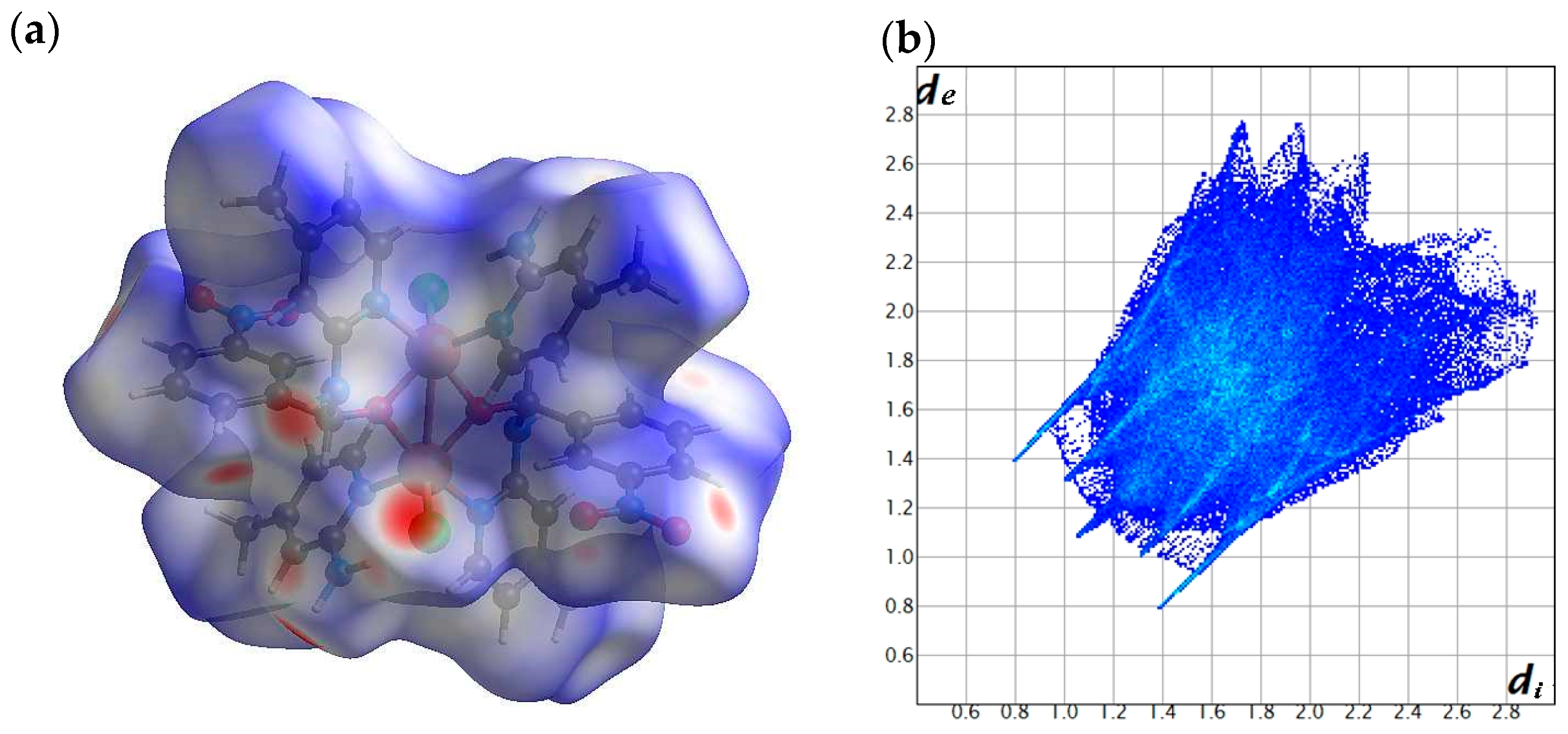
The Hirshfeld surface analysis, combined with 2D fingerprint plots, was utilized to explore the intermolecular interactions in the crystal structures of the synthesized compound (Figure 7). This approach enabled the identification of dominant interactions and their relative contributions to the overall crystal packing. The Hirshfeld surface analysis highlighted the presence of close contacts involving chlorine atoms and hydrogen atoms bonded to nitrogen—specifically those from the N–H group of the imine moiety and the NH2 group of the amine. These interactions correspond to the most pronounced regions of hydrogen bonding within the crystal structure. This is particularly evident from the intense red regions observed on the dnorm surface, which signify contacts shorter than the sum of van der Waals radii. Such pronounced red spots strongly suggest the presence of significant N–H···X (X = electronegative atom) hydrogen bonds, reinforcing the role of these groups in stabilizing the crystal packing.
The analysis further revealed the primary types of intermolecular interactions present in the studied structure. The largest contribution to the total surface arises from H···H contacts (41.2%). These contacts dominate in organic structures due to the high hydrogen content and the small size of hydrogen atoms, enabling close packing through weak dispersive interactions. In contrast, more directional and specific interactions, such as H··O/O··H interactions, were also observed, comprising 23.6% of the surface at a contact distance of approximately de + di ~ 2.2 Å. Although less abundant, Cl··H/H··Cl contacts still play a significant role in stabilizing the crystal packing, contributing 7.5%, and occurring at a similar distance (de + di ~ 2.2 Å).
The Hirshfeld fingerprint plot of the Cu(II) complex showed sharp spikes assigned to H··Cl and Cl···H interactions, and a broad central region corresponding to O··H/H··O hydrogen bonds, highlighting key contributions to the crystal packing.

3.4. QTAIM

Following the Hirshfeld surface analysis, which identified key intermolecular contacts involving N–H···Cl and C–H···O interactions, a topological study using the QTAIM was carried out to provide deeper insight into the nature of these interactions. By analyzing the electron density topology, QTAIM enables a more precise evaluation of bond characteristics beyond geometric proximity. The topological parameters extracted at bond critical points (BCPs) are summarized in Table 4. These parameters include electron density ρ(r), the Laplacian of electron density ∇2(r), kinetic energy density G(r), potential energy density V(r), and total energy density H(r).
The results showed that, for the intramolecular N–H···Cl interaction, the electron density at the BCP is relatively low (ρ(r) = 0.0186 a.u.), with a positive Laplacian (∇2(r) = 0.0595 a.u.), and a slightly negative potential energy density (V(r) = −0.0016 a.u.). Similarly, for the intermolecular N–H···Cl interaction, the values ρ(r) = 0.0194 a.u., ∇2(r) = 0.0652 a.u., and V(r) = −0.0166 a.u. were obtained. The weaker intermolecular C–H···O interaction presented an even lower electron density (ρ(r) = 0.0054 a.u.), a positive Laplacian (∇2(r) = 0.0242 a.u.), and a slightly negative V(r) (−0.0038 a.u.). According to the classification proposed by Koch and Popelier [100], hydrogen bonds with low ρ(r), positive ∇2(r), and negative V(r) values are indicative of weak electrostatic interactions. Furthermore, the |V(r)|/G(r) ratios, ranging from 0.20 to 0.28, confirmed the primarily electrostatic character of these bonds, which is in agreement with Hirshfeld surface findings highlighting the predominance of H···Cl and H···O interactions in crystal packing.

3.5. Antimicrobial Activity

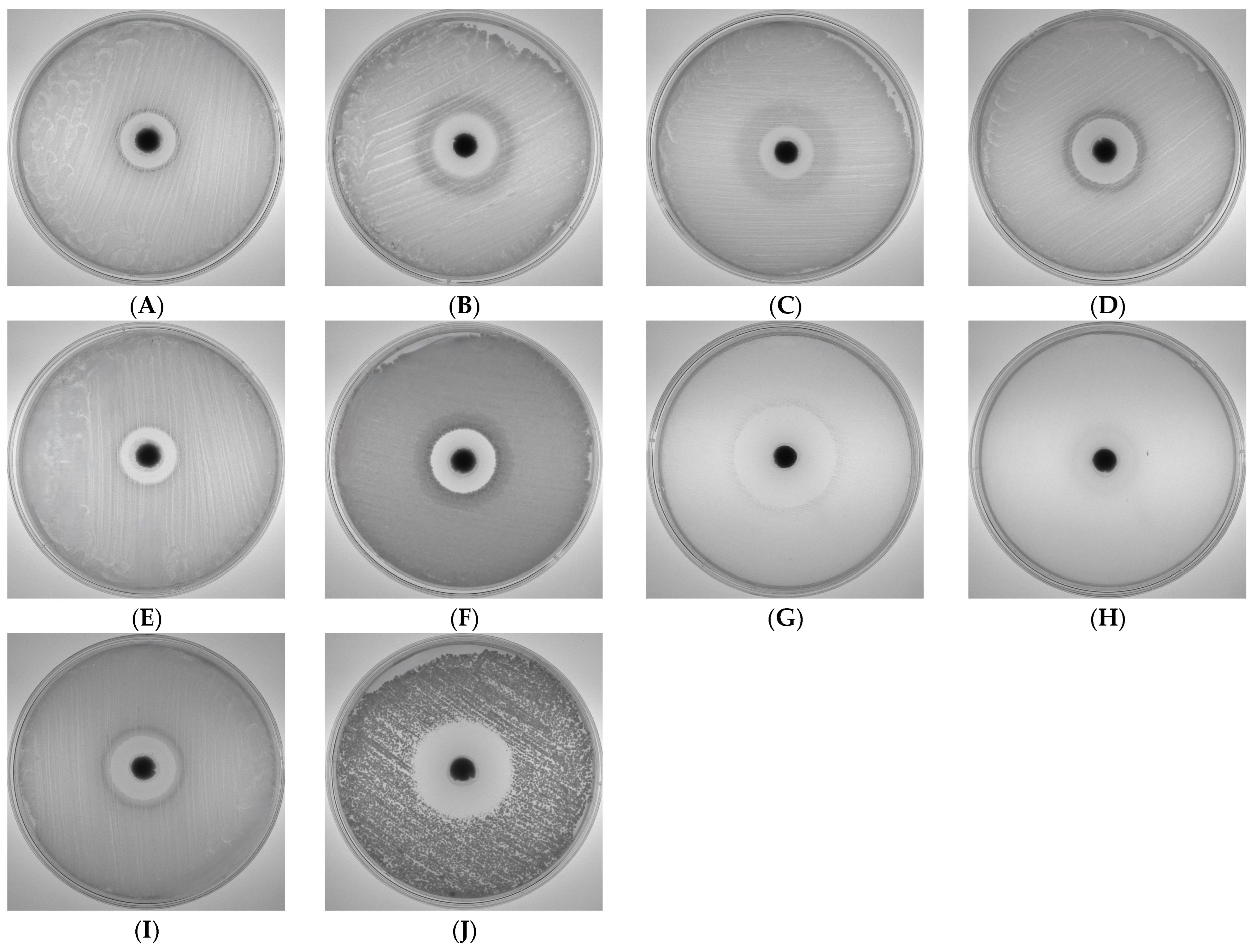
The antimicrobial activity of 3nb2a4mpCu was evaluated against a range of Gram-positive and Gram-negative bacterial strains, as well as the yeast C. albicans, using an agar diffusion method. The results demonstrated a broad-spectrum antimicrobial effect, with varying degrees of inhibition depending on the microorganism tested (Table 5, Figure 8).
Among Gram-negative bacteria, K. pneumoniae exhibited the largest inhibition zone (22.6 mm), followed closely by Y. enterocolitica (22.2 mm). Other Gram-negative species such as E. coli, P. aeruginosa, and P. mirabilis displayed inhibition zones ranging from 18.0 to 19.5 mm. The results indicate that 3nb2a4mpCu exhibits considerable efficacy against Gram-negative bacteria, including K. pneumoniae and P. aeruginosa, which are known for their intrinsic resistance to many antimicrobial agents [101,102].
For Gram-positive bacteria, S. epidermidis displayed the highest susceptibility, with an inhibition zone of 35.2 mm, followed by S. aureus (23.1 mm), B. cereus (21.2 mm), and E. hirae (18.1 mm). The exceptionally large inhibition zone observed for S. epidermidis (35.2 mm) can be attributed to the pronounced susceptibility of this species to copper-based compounds. Numerous studies have shown that S. epidermidis exhibits rapid loss of viability upon exposure to both ionic and complexed forms of copper, a phenomenon linked to copper-induced oxidative stress, membrane damage, and disruption of essential metabolic pathways [103]. This species-specific sensitivity is therefore consistent with the extensive growth inhibition recorded in our assay. Under the applied experimental conditions, the dose of 5 mg per disc appears sufficient to achieve the maximal antibacterial effect of the complex. The significant inhibition of S. aureus, a major pathogen responsible for hospital-acquired infections, highlights the potential clinical relevance of 3nb2a4mpCu. Previous studies have reported the efficacy of copper-based compounds against S. aureus due to their ability to generate reactive oxygen species and disrupt bacterial membranes [104].
Interestingly, C. albicans exhibited a high degree of susceptibility, with an inhibition zone of 32.3 mm, suggesting that 3nb2a4mpCu possesses potent antifungal properties. This observation is consistent with prior research demonstrating the antifungal efficacy of metal-based antimicrobial agents against C. albicans, which is a common opportunistic pathogen responsible for infections in immunocompromised individuals [105].
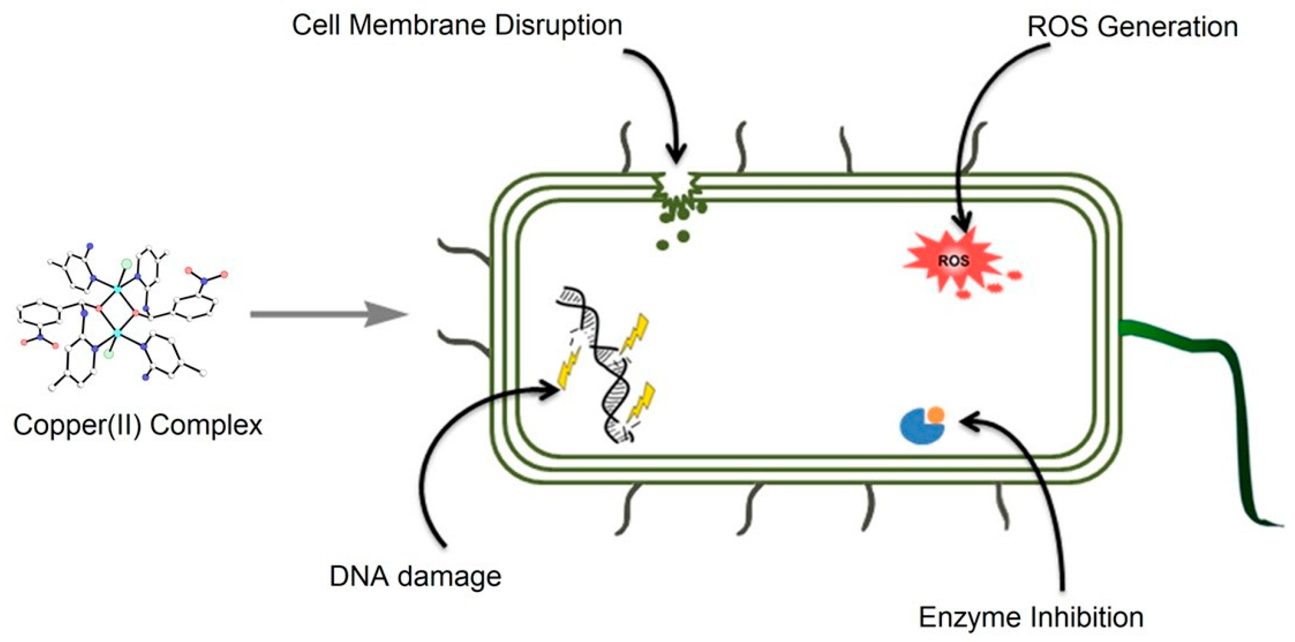
The broad-spectrum antimicrobial activity of 3nb2a4mpCu is likely attributable to the metal complex’s ability to disrupt microbial cell membranes, interfere with enzymatic functions, and induce oxidative stress [106], further augmented by additional pathways such as alterations of the cell wall or membrane, DNA damage, inhibition of key enzymes, and the generation of reactive oxygen species (ROS) (see Figure 9). The significant inhibition zones observed in this study suggest that this compound could be a promising candidate for further development in antimicrobial applications. Future research should focus on elucidating the exact mechanisms of action and evaluating the cytotoxicity of 3nb2a4mpCu in mammalian cells to ensure its safety for potential clinical use.

4. Conclusions

An efficient, one-pot synthesis of a coordination copper compound is presented. From a chemical point of view, this complex is interesting as it proves the stabilization of a reactive intermediate (hemiaminal) through the formation of coordination bonds.
The 3nb2a4mpCu complex exhibited the largest inhibition zones against S. epidermidis (35.2 mm) and C. albicans (32.3 mm), followed by Gram-negative strains such as K. pneumoniae (22.6 mm) and Y. enterocolitica (22.2 mm), confirming its broad-spectrum antimicrobial potential. These findings suggest that the antimicrobial efficacy of 3nb2a4mpCu may stem from its ability to compromise microbial membrane integrity and induce oxidative stress responses. Further research is needed to clarify its mode of action and evaluate cytotoxicity for possible biomedical applications.
Quantum chemical calculations provide valuable insights into the electronic structure and reactivity of the investigated compound. The calculated LUMO distribution and energy further support the compound’s potential involvement in biological activities, including antibacterial action. The computed descriptors, such as electronegativity, electrophilicity index, and chemical potential, confirm the compound’s significant reactivity and potential applicability in biologically relevant environments.

Author Contributions

Conceptualization, A.K.; methodology, A.K.; investigation, A.K., T.J., T.M., A.P., and Ż.C.; resources, A.K., T.J., T.M., A.P., and Ż.C.; writing—original draft preparation, A.K., T.J., T.M., and Ż.C.; writing—review and editing, A.K.; visualization, A.K., T.J., T.M., and Ż.C.; supervision, A.K.; project administration, A.K.; funding acquisition, Ż.C. All authors have read and agreed to the published version of the manuscript.

Funding

Synthesis, advanced structural studies, and reactivity calculations were funded by Wroclaw Medical University, grant number SUBZ.D290.25.066. Preliminary structural study was funded by University of Wroclaw, grant number 2266/M/WCH/12.

Institutional Review Board Statement

Not applicable.

Informed Consent Statement

Not applicable.

Data Availability Statement

The original contributions presented in this study are included in the article. Further inquiries can be directed to the corresponding authors.

Acknowledgments

The author (A.K.) would like to extend special thanks to Zbigniew Ciunik for his supervision and assistance at the early stage of this study. The authors gratefully acknowledge the allotment of the CPU time for calculations in the Wroclaw Center of Networking and Supercomputing (WCSS).

Conflicts of Interest

The authors declare no conflicts of interest.

Abbreviations

The following abbreviations are used in this manuscript:
2a4mp4-methylpyridin-2-amine
3nb3-nitrobenzaldehyde
3nb2a4mphemiaminal anion [(4-methylpyridin-2-yl)amino](3-nitrophenyl)methanolate
3nb2a4mpCua coordination compound of copper(II) with a chloride ion, 2a4mp and a hemiaminal 3nb2a4mp
Bu3Ntributylamine

References

  1. Hemiaminals. In The IUPAC Compendium of Chemical Terminology; International Union of Pure and Applied Chemistry (IUPAC): Research Triangle Park, NC, USA, 2014.
  2. Yufit, D.S.; Howard, J.A.K. Structure of Hemiaminal Intermediate of the Reaction of Diethylamine with Cyclobutanone. J. Mol. Struct. 2010, 984, 182–185. [Google Scholar] [CrossRef]
  3. Erdtman, E.; Bushnell, E.A.C.; Gauld, J.W.; Eriksson, L.A. Computational Studies on Schiff-Base Formation: Implications for the Catalytic Mechanism of Porphobilinogen Synthase. Comput. Theor. Chem. 2011, 963, 479–489. [Google Scholar] [CrossRef]
  4. Salvà, A.; Donoso, J.; Frau, J.; Muñoz, F. Theoretical Studies on Schiff Base Formation of Vitamin B6 Analogues. J. Mol. Struct. THEOCHEM 2002, 577, 229–238. [Google Scholar] [CrossRef]
  5. Salvà, A.; Donoso, J.; Frau, J.; Muñoz, F. DFT Studies on Schiff Base Formation of Vitamin B 6 Analogues. J. Phys. Chem. A 2003, 107, 9409–9414. [Google Scholar] [CrossRef]
  6. Hall, N.E.; Smith, B.J. High-Level Ab Initio Molecular Orbital Calculations of Imine Formation. J. Phys. Chem. A 1998, 102, 4930–4938. [Google Scholar] [CrossRef]
  7. Baymak, M.S.; Zuman, P. Equilibria of Formation and Dehydration of the Carbinolamine Intermediate in the Reaction of Benzaldehyde with Hydrazine. Tetrahedron 2007, 63, 5450–5454. [Google Scholar] [CrossRef]
  8. Evans, D.A.; Borg, G.; Scheidt, K.A. Remarkably Stable Tetrahedral Intermediates: Carbinols from Nucleophilic Additions to N-Acylpyrroles. Angew. Chem. Int. Ed. 2002, 41, 3188–3191. [Google Scholar] [CrossRef]
  9. Zhang, T.; Zhou, W.; Jin, W.; Jin, Q.; Chen, H. Direct Detection of Aromatic Amines and Observation of Intermediates of Schiff-Base Reactions by Reactive Desorption Electrospray Ionization Mass Spectrometry. Microchem. J. 2013, 108, 18–23. [Google Scholar] [CrossRef]
  10. Namli, H.; Turhan, O. Simultaneous Observation of Reagent Consumption and Product Formation with the Kinetics of Benzaldehyde and Aniline Reaction in FTIR Liquid Cell. Vib. Spectrosc. 2007, 43, 274–283. [Google Scholar] [CrossRef]
  11. Dolotko, O.; Wiench, J.W.; Dennis, K.W.; Pecharsky, V.K.; Balema, V.P. Mechanically Induced Reactions in Organic Solids: Liquid Eutectics or Solid-State Processes? New J. Chem. 2010, 34, 25–28. [Google Scholar] [CrossRef]
  12. Hooley, R.J.; Iwasawa, T.; Rebek, J. Detection of Reactive Tetrahedral Intermediates in a Deep Cavitand with an Introverted Functionality. J. Am. Chem. Soc. 2007, 129, 15330–15339. [Google Scholar] [CrossRef] [PubMed]
  13. Iwasawa, T.; Hooley, R.J.; Rebek, J. Stabilization of Labile Carbonyl Addition Intermediates by a Synthetic Receptor. Science 2007, 317, 493–496. [Google Scholar] [CrossRef] [PubMed]
  14. Xu, L.; Hua, S.; Li, S. Insight into the Reaction between a Primary Amine and a Cavitand with an Introverted Aldehyde Group: An Enzyme-like Mechanism. Chem. Commun. 2013, 49, 1542. [Google Scholar] [CrossRef] [PubMed]
  15. Li, M.; Dong, Y.; Quan, M.; Jiang, W. Stabilization of Imines and Hemiaminals in Water by an Endo-Functionalized Container Molecule. Angew. Chem. Int. Ed. 2022, 61, e202208508. [Google Scholar] [CrossRef]
  16. Kawamichi, T.; Haneda, T.; Kawano, M.; Fujita, M. X-Ray Observation of a Transient Hemiaminal Trapped in a Porous Network. Nature 2009, 461, 633–635. [Google Scholar] [CrossRef]
  17. Morris, W.; Doonan, C.J.; Yaghi, O.M. Postsynthetic Modification of a Metal–Organic Framework for Stabilization of a Hemiaminal and Ammonia Uptake. Inorg. Chem. 2011, 50, 6853–6855. [Google Scholar] [CrossRef]
  18. García, J.M.; Jones, G.O.; Virwani, K.; McCloskey, B.D.; Boday, D.J.; ter Huurne, G.M.; Horn, H.W.; Coady, D.J.; Bintaleb, A.M.; Alabdulrahman, A.M.S.; et al. Recyclable, Strong Thermosets and Organogels via Paraformaldehyde Condensation with Diamines. Science 2014, 344, 732–735. [Google Scholar] [CrossRef]
  19. Fox, C.H.; ter Hurrne, G.M.; Wojtecki, R.J.; Jones, G.O.; Horn, H.W.; Meijer, E.W.; Frank, C.W.; Hedrick, J.L.; García, J.M. Supramolecular Motifs in Dynamic Covalent PEG-Hemiaminal Organogels. Nat. Commun. 2015, 6, 7417. [Google Scholar] [CrossRef]
  20. Velázquez, M.; Salgado-Zamora, H.; Pérez, C.; Campos-A, M.E.; Mendoza, P.; Jiménez, H.; Jiménez, R. Intramolecular Hydrogen Bond Stabilization of Hemiaminal Structures, Precursors of Imidazo[1,2-a]Pyridine. J. Mol. Struct. 2010, 979, 56–61. [Google Scholar] [CrossRef]
  21. Suni, V.; Kurup, M.R.P.; Nethaji, M. Unusual Isolation of a Hemiaminal Product from 4-Cyclohexyl-3-Thiosemicarbazide and Di-2-Pyridyl Ketone: Structural and Spectral Investigations. J. Mol. Struct. 2005, 749, 177–182. [Google Scholar] [CrossRef]
  22. Barys, M.; Ciunik, Z.; Drabent, K.; Kwiecień, A. Stable Hemiaminals Containing a Triazole Ring. New J. Chem. 2010, 34, 2605–2611. [Google Scholar] [CrossRef]
  23. Kwiecień, A.; Barys, M.; Ciunik, Z. Stable Hemiaminals with a Cyano Group and a Triazole Ring. Molecules 2014, 19, 11160–11177. [Google Scholar] [CrossRef] [PubMed]
  24. Wajda-Hermanowicz, K.; Pieniążczak, D.; Zatajska, A.; Wróbel, R.; Drabent, K.; Ciunik, Z. A Study on the Condensation Reaction of 4-Amino-3,5-Dimethyl-1,2,4-Triazole with Benzaldehydes: Structure and Spectroscopic Properties of Some New Stable Hemiaminals. Molecules 2015, 20, 17109–17131. [Google Scholar] [CrossRef] [PubMed]
  25. Kwiecień, A.; Ciunik, Z. Stable Hemiaminals: 2-Aminopyrimidine Derivatives. Molecules 2015, 20, 14365–14376. [Google Scholar] [CrossRef]
  26. Berski, S.; Ciunik, L.Z. The Mechanism of the Formation of the Hemiaminal and Schiff Base from the Benzaldehyde and Triazole Studied by Means of the Topological Analysis of Electron Localisation Function and Catastrophe Theory. Mol. Phys. 2015, 113, 765–781. [Google Scholar] [CrossRef]
  27. Berski, S.; Gordon, A.J.; Ciunik, L.Z. The DFT Study on the Reaction between Benzaldehyde and 4-Amine-4H-1,2,4-Triazole and Their Derivatives as a Source of Stable Hemiaminals and Schiff Bases. Effect of Substitution and Solvation on the Reaction Mechanism. J. Mol. Model. 2015, 21, 57. [Google Scholar] [CrossRef]
  28. Groom, C.R.; Bruno, I.J.; Lightfoot, M.P.; Ward, S.C. The Cambridge Structural Database. Acta Crystallogr. Sect. B Struct. Sci. Cryst. Eng. Mater. 2016, 72, 171–179. [Google Scholar] [CrossRef]
  29. Liu, Z.; Kooijman, H.; Spek, A.L.; Bouwman, E. New Manganese-Based Catalyst Systems for Alkyd Paint Drying. Prog. Org. Coat. 2007, 60, 343–349. [Google Scholar] [CrossRef][Green Version]
  30. Liu, Z.; Liang, S.; Di, X.; Zhang, J. A Manganese(III) Complex That Exhibits Spin Crossover Behavior. Inorg. Chem. Commun. 2008, 11, 783–786. [Google Scholar] [CrossRef]
  31. Ding, S.; Xia, C.; Ji, Y.; Liu, Z.; Ding, Y. Bis[(2-Pyridyl)(2-Pyridylamino)Methanolato]Manganese(III) Nitrate. Acta Crystallogr. Sect. E Struct. Rep. Online 2011, 67, m622–m623. [Google Scholar] [CrossRef]
  32. Arulsamy, N.; Hodgson, D.J. Metal-Assisted Ligand Oxidation: Synthesis and Characterization of Novel Manganese(III), Iron(III), and Nickel(II) Complexes. Inorg. Chem. 1994, 33, 4531–4536. [Google Scholar] [CrossRef]
  33. Blake, A.B.; Sinn, E.; Yavari, A.; Moubaraki, B.; Murray, K.S. Magnetic Properties of Binuclear Manganese(II), Cobalt(II), Nickel(II) and Copper(II) Complexes of a Macrocyclic Ligand Derived from Pyridine-N-Oxide, and Crystal Structure of the Nickel Complex. Inorganica Chim. Acta 1995, 229, 281–290. [Google Scholar] [CrossRef]
  34. Eleder, M.; McKenzie, E.D. A Carbinolamine Bonded to Copper(II): Crystal and Molecular Structure (X-Ray Analysis) of Dibromo-[2-Picolyl-(2-Pyridylmethanol)Methylamine] Copper(II) Monohydrate. Inorganica Chim. Acta 1978, 31, 211–215. [Google Scholar] [CrossRef]
  35. Felcman, J.; Howie, R.A.; de Miranda, J.L.; Skakle, J.M.S.; Wardell, J.L. The Nitrate Dihydrate of an Aquadicopper(II) Complex Cation with Guanidinoacetic Acid and a Novel Trianionic Disubstituted Guanidine as Ligands at 120 K. Acta Crystallogr. Sect. C Cryst. Struct. Commun. 2003, 59, m103–m106. [Google Scholar] [CrossRef]
  36. Herrera, A.M.; Kalayda, G.V.; Disch, J.S.; Wikstrom, J.P.; Korendovych, I.V.; Staples, R.J.; Campana, C.F.; Nazarenko, A.Y.; Haas, T.E.; Rybak-Akimova, E.V. Reactions at the Azomethine C=N Bonds in the Nickel(II) and Copper(II) Complexes of Pyridine-Containing Schiff-Base Macrocyclic Ligands. Dalt. Trans. 2003, 4482–4492. [Google Scholar] [CrossRef]
  37. Cruz, C.; Audebrand, N.; Páez-Hernández, D.; Paredes-García, V. Novel Linear Trinuclear CuII Compound with Trapped Chiral Hemiaminal Ligand: Magnetostructural Study. Magnetochemistry 2023, 9, 175. [Google Scholar] [CrossRef]
  38. Adams, H.; Shongwe, M.S.; Al-Bahri, I.; Al-Busaidi, E.; Morris, M.J. Bis[(2-Pyridyl)(2-Pyridylamino)Methanolato]Cobalt(III) Perchlorate: A Consequence of Cobalt Ion-Assisted Oxidative Deamination of a Tris(Pyridyl)Aminal Ligand. Acta Crystallogr. C 2005, 61, m497–m500. [Google Scholar] [CrossRef]
  39. Browne, J.M.W.; Wikaira, J.; Hartshorn, R.M. Polydentate Ligand Construction: A Re-Examination of an Intramolecular Condensation Reaction. J. Chem. Soc. Dalt. Trans. 2001, 3513–3519. [Google Scholar] [CrossRef]
  40. Padhi, S.K.; Sahu, R.; Manivannan, V. Syntheses and Structures of Cobalt(III) Alcoholate Complexes Formed by Addition of a Water Molecule across 2-Pyridyl Substituted Imine Function. Inorganica Chim. Acta 2011, 367, 57–63. [Google Scholar] [CrossRef]
  41. Harrowfield, J.M.; Mocerino, M.; Skelton, B.W.; Wei, W.; White, A.H. Metal Complexes of Carbohydrates: Isolation and Characterisation of Cobalt(III) Complexes Containing N-Substituted Glycosylamine Ligands Derived from Ethane-1,2-Diamine and Glucosamine. J. Chem. Soc. Dalt. Trans. 1995, 783. [Google Scholar] [CrossRef]
  42. Haque, Z.P.; McPartlin, M.; Tasker, P.A. Macrocyclic Ligand Synthesis. Isolation of a Dicarbinolamine Complex from a Zinc(II)-Promoted Cyclization Reaction. Inorg. Chem. 1979, 18, 2920–2921. [Google Scholar] [CrossRef]
  43. Ulrich, S.; Lehn, J.-M. Adaptation to Shape Switching by Component Selection in a Constitutional Dynamic System. J. Am. Chem. Soc. 2009, 131, 5546–5559. [Google Scholar] [CrossRef] [PubMed]
  44. Gou, S.H.; You, X.Z.; Xu, Z.; Liu, S.X.; Lin, C.C. Structure of 2,2’-[5-(2-Aminoethyl)-1-Hydroxy-2,5,8-Triazanon-3-Ene-1,9-Diyl]Bis(Pyridine N-Oxide)Nickel Hexafluorophosphate Monohydrate, [Ni(C18H26N6O3)]2+.2PF6.H2O: A Novel Tripodal Nickel(II) Complex with Three Inequivalent Arms. Acta Crystallogr. Sect. C Cryst. Struct. Commun. 1991, 47, 708–711. [Google Scholar] [CrossRef]
  45. Ahmed, M.; Schwendt, P.; Sivá, M.; Marek, J. Synthesis and Characterization of Vanadium(V) Complexes with α-Hydroxyhippuric Acid. The X-Ray Crystal Structure of (NBu 4)2[V2O2(O2)2(R-α-Hhip) (S-α-Hhip)]·5H2O,[α-Hhip = α-Hydroxyhippurato(2-)]. Transit. Met. Chem. 2004, 29, 675–680. [Google Scholar] [CrossRef]
  46. Schwendt, P.; Ahmed, M.; Marek, J. Peroxovanadium(V) Complexes of α-Hydroxyhippuric Acid. The X-Ray Crystal Structure of (NPr4)2[V2O2(O2)2(α-Hhip)2] · 5H2O, [α-Hhip=α-Hydroxyhippurato (2–) − C9H7NO4 (2–)]. Inorg. Chem. Commun. 2004, 7, 631–633. [Google Scholar] [CrossRef]
  47. El Mail, R.; Garralda, M.A.; Hernández, R.; Ibarlucea, L.; Pinilla, E.; Torres, M.R. Hydroxyalkyl Complexes and Hemiaminal Formation in the Reaction of o -Diphenylphosphinobenzaldehyde with Rhodium(I) Dihydrazone Complexes. Organometallics 2000, 19, 5310–5317. [Google Scholar] [CrossRef]
  48. Aileen Bouffard, F.; Hammond, M.L.; Arison, B.H. Pneumocandin Bo Acid Degradate. Tetrahedron Lett. 1995, 36, 1405–1408. [Google Scholar] [CrossRef]
  49. Lal, B.; Gund, V.G. Approaches towards the Stabilization of Hemiaminal Function at Ornithine Unit of Mulundocandin. Bioorg. Med. Chem. Lett. 2004, 14, 1123–1128. [Google Scholar] [CrossRef]
  50. LaLonde, R.T.; Wong, C. Hemiaminal Derivatives of Neothiobinupharidine. J. Org. Chem. 1976, 41, 291–294. [Google Scholar] [CrossRef]
  51. Graf von Stosch, A. Aspartate Aminotransferase Complexed with Erythro -β-Hydroxyaspartate: Crystallographic and Spectroscopic Identification of the Carbinolamine Intermediate. Biochemistry 1996, 35, 15260–15268. [Google Scholar] [CrossRef]
  52. Liu, X.; Banister, S.D.; Christie, M.J.; Banati, R.; Meikle, S.; Coster, M.J.; Kassiou, M. Trishomocubanes: Novel σ Ligands Modulate Cocaine-Induced Behavioural Effects. Eur. J. Pharmacol. 2007, 555, 37–42. [Google Scholar] [CrossRef] [PubMed]
  53. Wilkes, D.K.; de Vries, A.; Oliver, D.W.; Malan, S.F. Nitric Oxide Synthase Inhibition by Pentacycloundecane Conjugates of Aminoguanidine and Tryptamine. Arch. Pharm. 2009, 342, 73–79. [Google Scholar] [CrossRef] [PubMed]
  54. Prins, L.H.A.; Preez, J.L.d.; van Dyk, S.; Malan, S.F. Polycyclic Cage Structures as Carrier Molecules for Neuroprotective Non-Steroidal Anti-Inflammatory Drugs. Eur. J. Med. Chem. 2009, 44, 2577–2582. [Google Scholar] [CrossRef] [PubMed]
  55. Olczak, T.; Śmiga, M.; Kwiecień, A.; Bielecki, M.; Wróbel, R.; Olczak, M.; Ciunik, Z. Antimicrobial Activity of Stable Hemiaminals against Porphyromonas Gingivalis. Anaerobe 2017, 44, 27–33. [Google Scholar] [CrossRef]
  56. Kabir, E.; Noyon, M.R.O.K.; Hossain, M.A. Synthesis, Biological and Medicinal Impacts of Metallodrugs: A Study. Results Chem. 2023, 5, 100935. [Google Scholar] [CrossRef]
  57. Alderden, R.A.; Hall, M.D.; Hambley, T.W. The Discovery and Development of Cisplatin. J. Chem. Educ. 2006, 83, 728. [Google Scholar] [CrossRef]
  58. Adhikari, S.; Nath, P.; Das, A.; Datta, A.; Baildya, N.; Duttaroy, A.K.; Pathak, S. A Review on Metal Complexes and Its Anti-Cancer Activities: Recent Updates from in Vivo Studies. Biomed. Pharmacother. 2024, 171, 116211. [Google Scholar] [CrossRef]
  59. Zehra, S.; Tabassum, S.; Arjmand, F. Biochemical Pathways of Copper Complexes: Progress over the Past 5 Years. Drug Discov. Today 2021, 26, 1086–1096. [Google Scholar] [CrossRef]
  60. Wang, Y.; Chen, Y.; Zhang, J.; Yang, Y.; Fleishman, J.S.; Wang, Y.; Wang, J.; Chen, J.; Li, Y.; Wang, H. Cuproptosis: A Novel Therapeutic Target for Overcoming Cancer Drug Resistance. Drug Resist. Updat. 2024, 72, 101018. [Google Scholar] [CrossRef]
  61. Jomova, K.; Makova, M.; Alomar, S.Y.; Alwasel, S.H.; Nepovimova, E.; Kuca, K.; Rhodes, C.J.; Valko, M. Essential Metals in Health and Disease. Chem. Biol. Interact. 2022, 367, 110173. [Google Scholar] [CrossRef]
  62. da Silva, D.A.; De Luca, A.; Squitti, R.; Rongioletti, M.; Rossi, L.; Machado, C.M.L.; Cerchiaro, G. Copper in Tumors and the Use of Copper-Based Compounds in Cancer Treatment. J. Inorg. Biochem. 2022, 226, 111634. [Google Scholar] [CrossRef] [PubMed]
  63. Ngece, K.; Khwaza, V.; Paca, A.M.; Aderibigbe, B.A. The Antimicrobial Efficacy of Copper Complexes: A Review. Antibiotics 2025, 14, 516. [Google Scholar] [CrossRef] [PubMed]
  64. Qi, J.; Wang, X.; Liu, T.; Kandawa-Schulz, M.; Wang, Y.; Zheng, X. Synthesis, Antiproliferative Activity and Mechanism of Copper(II)-Thiosemicarbazone Complexes as Potential Anticancer and Antimicrobial Agents. J. Coord. Chem. 2020, 73, 1208–1221. [Google Scholar] [CrossRef]
  65. Santiago, P.H.O.; Santiago, M.B.; Martins, C.H.G.; Gatto, C.C. Copper(II) and Zinc(II) Complexes with Hydrazone: Synthesis, Crystal Structure, Hirshfeld Surface and Antibacterial Activity. Inorganica Chim. Acta 2020, 508, 119632. [Google Scholar] [CrossRef]
  66. Beyene, B.B.; Wassie, G.A. Antibacterial Activity of Cu(II) and Co(II) Porphyrins: Role of Ligand Modification. BMC Chem. 2020, 14, 51. [Google Scholar] [CrossRef]
  67. Jyothi, N.; Ganji, N.; Daravath, S.; Shivaraj. Mononuclear Cobalt(II), Nickel(II) and Copper(II) Complexes: Synthesis, Spectral Characterization and Interaction Study with Nucleotide by in Vitro Biochemical Analysis. J. Mol. Struct. 2020, 1207, 127799. [Google Scholar] [CrossRef]
  68. Ferreira, L.; Andrade, J.; Lima, W.; Souza, J.; Chagas, R.; Leonel, C.; Cunha, L.; Herrera, K.; Braga, A.; Soares, L.; et al. Synthesis, Characterization and Antimicrobial Activity of New Cu(II), Co(II) and Sn(II) Complexes with the Sodium Hydrotris(2-Mercaptothiazolyl)Borate Ligand. Quim. Nova 2020, 43, 593–598. [Google Scholar] [CrossRef]
  69. Zhao, Y.-M.; Tang, G.-M.; Wang, Y.-T.; Cui, Y.-Z.; Ng, S.W. Copper-Based Metal Coordination Complexes with Voriconazole Ligand: Syntheses, Structures and Antimicrobial Properties. J. Solid State Chem. 2018, 259, 19–27. [Google Scholar] [CrossRef]
  70. Senthilkumar, G.S.; Sankarganesh, M.; Dhaveethu Raja, J.; Adwin Jose, P.R.; Sakthivel, A.; Christopher Jeyakumar, T.; Nandini Asha, R. Water Soluble Cu(II) and Zn(II) Complexes of Bidentate-Morpholine Based Ligand: Synthesis, Spectral, DFT Calculation, Biological Activities and Molecular Docking Studies. J. Biomol. Struct. Dyn. 2022, 40, 1074–1083. [Google Scholar] [CrossRef]
  71. Kargar, H.; Aghaei-Meybodi, F.; Behjatmanesh-Ardakani, R.; Elahifard, M.R.; Torabi, V.; Fallah-Mehrjardi, M.; Tahir, M.N.; Ashfaq, M.; Munawar, K.S. Synthesis, Crystal Structure, Theoretical Calculation, Spectroscopic and Antibacterial Activity Studies of Copper(II) Complexes Bearing Bidentate Schiff Base Ligands Derived from 4-Aminoantipyrine: Influence of Substitutions on Antibacterial Activity. J. Mol. Struct. 2021, 1230, 129908. [Google Scholar] [CrossRef]
  72. Ghanghas, P.; Choudhary, A.; Kumar, D.; Poonia, K. Coordination Metal Complexes with Schiff Bases: Useful Pharmacophores with Comprehensive Biological Applications. Inorg. Chem. Commun. 2021, 130, 108710. [Google Scholar] [CrossRef]
  73. Thakur, S.; Bhalla, A. Sustainable Synthetic Endeavors of Pharmaceutically Active Schiff Bases and Their Metal Complexes: A Review on Recent Reports. Tetrahedron 2024, 153, 133836. [Google Scholar] [CrossRef]
  74. Boulechfar, C.; Ferkous, H.; Delimi, A.; Djedouani, A.; Kahlouche, A.; Boublia, A.; Darwish, A.S.; Lemaoui, T.; Verma, R.; Benguerba, Y. Schiff Bases and Their Metal Complexes: A Review on the History, Synthesis, and Applications. Inorg. Chem. Commun. 2023, 150, 110451. [Google Scholar] [CrossRef]
  75. Kumar, R.; Singh, A.A.; Kumar, U.; Jain, P.; Sharma, A.K.; Kant, C.; Haque Faizi, M.S. Recent Advances in Synthesis of Heterocyclic Schiff Base Transition Metal Complexes and Their Antimicrobial Activities Especially Antibacterial and Antifungal. J. Mol. Struct. 2023, 1294, 136346. [Google Scholar] [CrossRef]
  76. Richa; Kumar, V.; Kataria, R. Phenanthroline and Schiff Base Associated Cu(II)-Coordinated Compounds Containing N, O as Donor Atoms for Potent Anticancer Activity. J. Inorg. Biochem. 2024, 251, 112440. [Google Scholar] [CrossRef]
  77. Podolski-Renić, A.; Čipak Gašparović, A.; Valente, A.; López, Ó.; Bormio Nunes, J.H.; Kowol, C.R.; Heffeter, P.; Filipović, N.R. Schiff Bases and Their Metal Complexes to Target and Overcome (Multidrug) Resistance in Cancer. Eur. J. Med. Chem. 2024, 270, 116363. [Google Scholar] [CrossRef]
  78. Mahdi, S.H.; Abdul Karem, L.K. Synthesis, Characterization, Anticancer and Antimicrobial Studies of Metal Nanoparticles Derived from Schiff Base Complexes. Inorg. Chem. Commun. 2024, 165, 112524. [Google Scholar] [CrossRef]
  79. Ashraf, T.; Ali, B.; Qayyum, H.; Haroone, M.S.; Shabbir, G. Pharmacological Aspects of Schiff Base Metal Complexes: A Critical Review. Inorg. Chem. Commun. 2023, 150, 110449. [Google Scholar] [CrossRef]
  80. Jiang, M.; Su, X.; Zhong, X.; Lan, Y.; Yang, F.; Qin, Y.; Jiang, C. Recent Development of Schiff-Base Metal Complexes as Therapeutic Agents for Lung Cancer. J. Mol. Struct. 2024, 1318, 139403. [Google Scholar] [CrossRef]
  81. More, M.S.; Joshi, P.G.; Mishra, Y.K.; Khanna, P.K. Metal Complexes Driven from Schiff Bases and Semicarbazones for Biomedical and Allied Applications: A Review. Mater. Today Chem. 2019, 14, 100195. [Google Scholar] [CrossRef]
  82. Kumari, P.; Choudhary, M.; Kumar, A.; Yadav, P.; Singh, B.; Kataria, R.; Kumar, V. Copper(II) Schiff Base Complexes: Synthetic and Medicinal Perspective. Inorg. Chem. Commun. 2023, 158, 111409. [Google Scholar] [CrossRef]
  83. Wrońska, M.; Wrzesinski, J.; Jeżowska-Bojczuk, M.; Szczepanik, W.; Starosta, R.; Barys, M.; Ciunik, Z.; Ciesiołka, J. The Impact of Isomers of Hemiaminal-1,2,4-Triazole Conjugates Differently Substituted in the Phenyl Ring and Their Cu2+ Complexes on the Catalytic Activity of the Antigenomic Delta Ribozyme. J. Inorg. Biochem. 2012, 108, 62–68. [Google Scholar] [CrossRef] [PubMed]
  84. Nikovskii, I.A.; Chistyakov, E.M.; Tupikov, A.S. Phosphazene-Containing Ligands and Complexes on Their Base. Russ. J. Gen. Chem. 2018, 88, 474–494. [Google Scholar] [CrossRef]
  85. Kolhe, N.H.; Tamboli, Y.; Bawazir, H.A.; Parihar, V.S. Advances in Mixed Ligand Transition Metal Complexes: Chromone Derivatives and N, O Donor Ligands—A Comprehensive Review. Polyhedron 2026, 284, 117900. [Google Scholar] [CrossRef]
  86. Repac Antić, D.; Parčina, M.; Gobin, I.; Petković Didović, M. Chelation in Antibacterial Drugs: From Nitroxoline to Cefiderocol and Beyond. Antibiotics 2022, 11, 1105. [Google Scholar] [CrossRef]
  87. Ismael, M.; Abdel-Mawgoud, A.-M.M.; Rabia, M.K.; Abdou, A. Synthesis, Characterization, Molecular Modeling and Preliminary Biochemical Evaluation of New Copper (II) Mixed-Ligand Complexes. J. Mol. Struct. 2021, 1227, 129695. [Google Scholar] [CrossRef]
  88. Azam, M.; Mohammad Wabaidur, S.; Alam, M.; Trzesowska-Kruszynska, A.; Kruszynski, R.; Al-Resayes, S.I.; Fahhad Alqahtani, F.; Rizwan Khan, M.; Rajendra. Design, Structural Investigations and Antimicrobial Activity of Pyrazole Nucleating Copper and Zinc Complexes. Polyhedron 2021, 195, 114991. [Google Scholar] [CrossRef]
  89. Ramesh, G.; Daravath, S.; Ganji, N.; Rambabu, A.; Venkateswarlu, K.; Shivaraj. Facile Synthesis, Structural Characterization, DNA Binding, Incision Evaluation, Antioxidant and Antimicrobial Activity Studies of Cobalt(II), Nickle(II) and Copper(II) Complexes of 3-Amino-5-(4-Fluorophenyl)Isoxazole Derivatives. J. Mol. Struct. 2020, 1202, 127338. [Google Scholar] [CrossRef]
  90. Yadav, M.; Sharma, S.; Devi, J. Designing, Spectroscopic Characterization, Biological Screening and Antioxidant Activity of Mononuclear Transition Metal Complexes of Bidentate Schiff Base Hydrazones. J. Chem. Sci. 2021, 133, 21. [Google Scholar] [CrossRef]
  91. Seppälä, P.; Sillanpää, R.; Lehtonen, A. Structural Diversity of Copper(II) Amino Alcoholate Complexes. Coord. Chem. Rev. 2017, 347, 98–114. [Google Scholar] [CrossRef]
  92. Sheldrick, G.M. Crystal Structure Refinement with SHELXL. Acta Crystallogr. Sect. C Struct. Chem. 2015, 71, 3–8. [Google Scholar] [CrossRef]
  93. Frisch, M.J.; Trucks, G.W.; Schlegel, H.B.; Scuseria, G.E.; Robb, M.a.; Cheeseman, J.R.; Scalmani, G.; Barone, V.; Petersson, G.a.; Nakatsuji, H.; et al. G16_C01 2016, Gaussian 16, Revision C.01; Gaussian, Inc.: Wallingford, CT, USA, 2016. [Google Scholar]
  94. Tomasi, J.; Mennucci, B.; Cammi, R. Quantum Mechanical Continuum Solvation Models. Chem. Rev. 2005, 105, 2999–3094. [Google Scholar] [CrossRef]
  95. Hanwell, M.D.; Curtis, D.E.; Lonie, D.C.; Vandermeersch, T.; Zurek, E.; Hutchison, G.R. Avogadro: An Advanced Semantic Chemical Editor, Visualization, and Analysis Platform. J. Cheminform. 2012, 4, 17. [Google Scholar] [CrossRef] [PubMed]
  96. Spackman, P.R.; Turner, M.J.; McKinnon, J.J.; Wolff, S.K.; Grimwood, D.J.; Jayatilaka, D.; Spackman, M.A. CrystalExplorer: A Program for Hirshfeld Surface Analysis, Visualization and Quantitative Analysis of Molecular Crystals. J. Appl. Crystallogr. 2021, 54, 1006–1011. [Google Scholar] [CrossRef] [PubMed]
  97. Biegler–König, F.; Schönbohm, J.; Bayles, D. AIM2000—A Program to Analyze and Visualize Atoms in Molecules. J. Comput. Chem. 2001, 22, 545–559. [Google Scholar] [CrossRef]
  98. Daoui, S.; Parlak, C.; Direkel, Ş.; Ramasami, P.; Abuelizz, H.A.; Oulmidi, A.; Benchat, N.; Karrouchi, K. Synthesis, Spectroscopic Characterization, Antibacterial Activity, Reactivity Parameters, and Molecular Docking Studies of Some Pyridazine Derivatives. ChemistrySelect 2024, 9, e202402996. [Google Scholar] [CrossRef]
  99. Kawakami, J.; Kakinami, H.; Matsushima, N.; Nakane, A.; Kitahara, H.; Nagaki, M.; Ito, S. Structure–Activity Relationship Analysis for Antimicrobial Activities of Tryptanthrin Derivatives Using Quantum Chemical Calculations. J. Comput. Chem. Jpn. 2013, 12, 109–112. [Google Scholar] [CrossRef]
  100. Koch, U.; Popelier, P.L.A. Characterization of C-H-O Hydrogen Bonds on the Basis of the Charge Density. J. Phys. Chem. 1995, 99, 9747–9754. [Google Scholar] [CrossRef]
  101. Effah, C.Y.; Sun, T.; Liu, S.; Wu, Y. Klebsiella Pneumoniae: An Increasing Threat to Public Health. Ann. Clin. Microbiol. Antimicrob. 2020, 19, 1. [Google Scholar] [CrossRef]
  102. Poole, K. Pseudomonas Aeruginosa: Resistance to the Max. Front. Microbiol. 2011, 2, 65. [Google Scholar] [CrossRef]
  103. Kaul, L.; Abdo, A.I.; Coenye, T.; Krom, B.P.; Hoogenkamp, M.A.; Zannettino, A.C.W.; Süss, R.; Richter, K. The Combination of Diethyldithiocarbamate and Copper Ions Is Active against Staphylococcus Aureus and Staphylococcus Epidermidis Biofilms in Vitro and in Vivo. Front. Microbiol. 2022, 13, 999893. [Google Scholar] [CrossRef]
  104. Sharma, B.; Shukla, S.; Rattan, R.; Fatima, M.; Goel, M.; Bhat, M.; Dutta, S.; Ranjan, R.K.; Sharma, M. Antimicrobial Agents Based on Metal Complexes: Present Situation and Future Prospects. Int. J. Biomater. 2022, 2022, 6819080. [Google Scholar] [CrossRef]
  105. Liang, J.; Sun, D.; Yang, Y.; Li, M.; Li, H.; Chen, L. Discovery of Metal-Based Complexes as Promising Antimicrobial Agents. Eur. J. Med. Chem. 2021, 224, 113696. [Google Scholar] [CrossRef]
  106. Lemire, J.A.; Harrison, J.J.; Turner, R.J. Antimicrobial Activity of Metals: Mechanisms, Molecular Targets and Applications. Nat. Rev. Microbiol. 2013, 11, 371–384. [Google Scholar] [CrossRef]

Scheme 1. One-pot synthesis of 3nb2a4mpCu.
Applsci 16 00136 sch001
Figure 1. The asymmetric part of the unit cell, with atom numbering scheme, shows a thermal ellipsoid for the non-hydrogen atoms drawn at a 30% probability level; for the disordered atoms, only one/major part is shown.
Applsci 16 00136 g001
Figure 2. Molecular structure of 3nb2a4mpCu with hydrogen atoms and disordered fragments omitted for better clarity.
Applsci 16 00136 g002
Figure 3. Coordination sphere around Cu(II) cations in 3nb2a4mpCu, symmetry code: (i) 1 − x, 1 − y, 1 − z.
Applsci 16 00136 g003
Figure 4. The bidentate coordination mode of 3nb2a4mp.
Applsci 16 00136 g004
Figure 5. Crystal packing of 3nb2a4mpCu, viewed along axis c.
Applsci 16 00136 g005
Figure 6. PXRD patterns of 3nba4mpCu: (a) calculated from single crystal structure (black line); (b) experimental for bulk sample (red line).
Applsci 16 00136 g006
Figure 7. (a) Three-dimensional (3D) Hirshfeld surface of the complex plotted over dnorm (dnorm range −0.4773 (red) to 1.5246 (blue) au) and (b) corresponding 2D fingerprint plot.
Applsci 16 00136 g007
Figure 8. Antimicrobial agar plate methods on 3nb2a4mpCu against (A) E. coli ATCC 10536; (B) K pneumoniae ATCC 13886; (C) P. aeruginosa ATCC 15442; (D) Y. enterocolitica DSM 23248; (E) P. mirabilis ATCC 21100; (F) B. cereus ATCC 10876; (G) S. epidermidis KCTC 1917; (H) E. hirae ATCC 10541; (I) S. aureus ATCC 25923; (J) C. albicans ATCC 10231.
Applsci 16 00136 g008
Figure 9. Illustration of the antimicrobial mechanism of the Cu(II) complex. Figure adapted and modified from Ngece et al. [63].
Applsci 16 00136 g009
Table 1. Selected geometrical parameters for 3nb2a4mpCu.
Bond Lengths/Interatomic Distances (Å)Valence Angles ()
Cu1–Cu1i3.0364 (7)O112–Cu1–N2192.01 (8)
Cu1–Cl12.487 (5)O112–Cu1– O112i77.40 (8)
Cu1–Cl1A2.663 (15)Cu1–O112–Cu1i102.60 (8)
Cu1–O1121.9336 (17)C11–C112–O112 110.9 (2)
Cu1–O112i1.9570 (17)C11–C112–N120 110.9 (2)
Cu1–N211.998 (2)O112–C112–N120 111.8 (2)
Cu1–N312.017 (2)C112–N120–C22 118.7 (2)
C11–C1121.527 (4)Torsion angles ()
C112–O1121.398 (3)
C112–N1201.492 (4)C16–C11–C112–O112 −170.9 (3)
C22–N1201.376 (3)C16–C11–C112–N120 64.3 (4)
C11–C112–N120–C22 56.2 (3)
O112–C112–N120–C22 −68.1 (3)
N21–C22–N120–C11251.1 (3)
Symmetry code: (i) 1 − x, 1 − y, 1 − z.
Table 2. Proposed hydrogen bond and noncovalent interactions in 3nb2a4mpCu crystal lattice.
D–H···AD–H (Å)H···A (Å)D···A (Å)<(D–H···A) (°)
N120–H120···Cl1i0.80 (3)2.57 (3)3.366 (6)170 (3)
N120–H120···Cl1Ai0.80 (3)2.31 (4)3.103 (17)172 (3)
N32–H32B···Cl1ii0.882.313.189 (7)170
N32–H32B···Cl1Aii0.882.333.197 (18)174
N32A–H32A···Cl10.882.383.251 (7)171
N32A–H32A···Cl1A0.882.223.097 (17)177
C12–H12···O1120.952.412.751 (3)100
C14–H14···O3iii0.952.423.197 (5)138
C33–H33···O2Aii0.952.603.247 (12)126
C37–H37···O2Aii0.982.573.437 (12)147
Symmetry codes: (i) −1/4 + y, 3/4−x, −1/4 + z, (ii) −1/4 + y, 5/4 − x, 5/4 − z, (iii) 3/4 − y, −1/4 + x, 7/4 − z.
Table 3. The HOMO/LUMO (highest occupied molecular orbital/lowest unoccupied molecular orbital) electron density maps, HOMO/LUMO energies, and other reactivity parameters.
Applsci 16 00136 i001Reactivity Parameter
electron affinity (A)6.173
ionization potential (I)6.846
chemical hardness (η)0.337
chemical softness (s)2.972
chemical potential (μ)−6.510
electronegativity (χ)6.510
electrophilicity index (ω)21.187
A = −ELUMO; I = −EHOMO; η = I     A 2 ; s =   1 2 η ; μ =   I   +   A 2 ; χ = I + A 2 ; ω = μ 2 2 η
Table 4. Summary of the topological parameters.
rDH···A (Å)<DH···A (˚)ρ(rBCP) (a.u.) 2 ρ
(rBCP) (a.u.)
G(rBCP) (a.u.)V(rBCP) (a.u.)H(rBCP) (a.u.)
Intramolecular HB
NH···Cl3.214172.990.01860.05950.0165−0.00160.0149
Intermolecular HB
NH···Cl3.189175.660.01940.06520.0164−0.01660.0002
CH···O3.194139.00.00540.02420.0049−0.00380.0011
Table 5. Inhibition zone (mean diameter of inhibition in mm) as a criterion of antibacterial and anti-Candida activities of the 3nb2a4mpCu.
MicroorganismDiameter of Inhibition Zone (mm)
Gram-negative bacteria
Escherichia coli ATCC 1053619.5
Klebsiella pneumoniae ATCC 1388622.6
Pseudomonas aeruginosa ATCC 1544218.0
Yersinia enterocolitica DSM 23248 22.2
Proteus mirabilis ATCC 2110018.2
Gram-positive bacteria
Bacillus cereus ATCC 1087621.2
Staphylococcus epidermidis KCTC 191735.2
Enterococcus hirae ATCC 1054118.1
Staphylococcus aureus ATCC 2592323.1
Yeast
Candida albicans ATCC 1023132.3
Disclaimer/Publisher’s Note: The statements, opinions and data contained in all publications are solely those of the individual author(s) and contributor(s) and not of MDPI and/or the editor(s). MDPI and/or the editor(s) disclaim responsibility for any injury to people or property resulting from any ideas, methods, instructions or products referred to in the content.