You are currently viewing a new version of our website. To view the old version click .
Applied Sciences
  • Article
  • Open Access

15 November 2025

Impact of Physician Height and Experience on Eye Lens Dose in Interventional Cardiology: An Initial Study

,
,
,
,
,
,
,
and
1
Department of Radiological Technology, Tohoku University Graduate School of Medicine, 2-1 Seiryo, Aoba-ku, Sendai 980-8575, Miyagi, Japan
2
Department of Radiology, Sendai Kosei Hospital, 1-20 Tsutsumidoriamamiyamachi, Aoba-ku, Sendai 981-0914, Miyagi, Japan
3
Department of Cardiovascular Medicine, Sendai Kosei Hospital, 1-20 Tsutsumidoriamamiyamachi, Aoba-ku, Sendai 981-0914, Miyagi, Japan
4
Department of Radiation Disaster Medicine, International Research Institute of Disaster Science, Tohoku University, 468-1 Aramaki Aza-Aoba, Aoba-ku, Sendai 980-0845, Miyagi, Japan

Abstract

In 2011, the International Commission on Radiological Protection (ICRP) recommended reducing the annual equivalent dose limit to the eye lens. This study investigated the effects of physician height and years of experience on lens radiation exposure in a clinical setting. The lens dose was measured using the DOSIRIS dosimeter, which quantified Hp(3) while accounting for the shielding effect of lead glasses, and a neck dosimeter for Hp(0.07). A significant negative correlation was found between physician height and both Hp(0.07) (R = −0.642) and Hp(3) (R = −0.728), suggesting that taller physicians received lower lens doses because of their greater distance from the scatter source. A positive correlation was observed between years of experience and Hp(0.07) (R = 0.650). Two-group comparisons showed that physicians shorter than 170 cm had a 2.77-fold higher median Hp(3) than those ≥170 cm (p < 0.05). As experienced physicians may be exposed to higher radiation levels, regular review of protective practices and continued radiation safety education are essential, regardless of clinical experience. This is the first clinical study to simultaneously evaluate the effects of physician height and experience on lens dose in interventional cardiology. Regular review of protective practices remains essential regardless of operator height or experience.

1. Introduction

Because interventional radiology (IR) is minimally invasive and place less physical strain on patients, the number of IR procedures has continued to increase [,,,]. These procedures are performed under fluoroscopic X-ray guidance, which raises concerns regarding patient radiation exposure [,,,,,]. Furthermore, both patients and medical staff are at risk of radiation exposure owing to scattered radiation from the patients [,,,]. In addition to the growing number of cases, recent advances in medical technology have made individual IR procedures more complex, often resulting in longer fluoroscopy times [,]. Several studies have examined radiation exposure among medical staff during IR procedures [,,]. Among these, the lens of the eye (hereafter referred to as the lens) is particularly sensitive to radiation, making it susceptible to radiation-induced cataracts and other radiation-related injuries [,,,]. In 1988, Vano et al. reported lens opacities in physicians and nurses involved in IR []. The International Commission on Radiological Protection (ICRP) cited this study in its 2000 recommendations (Publication 85), emphasizing the importance of managing and protecting against lens radiation exposure []. However, subsequent epidemiological studies have suggested that the threshold dose for cataract formation may be lower than that specified in ICRP Publication 60 [,,]. In response, the ICRP conducted a re-evaluation of the effects of radiation on the lens and significantly lowered the equivalent dose limit to the lens in its 2011 recommendations, reducing the limit from 150 mSv per year to 100 mSv over 5 years and 50 mSv per year []. Because of this dose limit reduction, cases involving physicians exceeding the dose limits have been reported []. Consequently, the evaluation and mitigation of lens radiation exposure have become critical, leading to numerous reports on this topic [,,,,]. However, studies investigating the relationship between the eye lens dose (hereafter referred to simply as lens dose) and physician height or years of experience among interventional radiologists are limited.
The ICRP recommends that the lens dose be evaluated based on Hp(3) (personal dose IC at 3 mm depth) []. The International Atomic Energy Agency (IAEA) also recommends that for an accurate assessment of the lens dose, measurements should be performed as close to the eye as possible []. However, in hospital radiation management and previous studies, either Hp(10) (personal dose equivalent at 1 cm depth) or Hp(0.07) (personal dose equivalent at 70 μm depth), whichever is higher, has been used, or Hp(3) has been estimated through calculations [,,,]. These values may not provide an accurate assessment of lens dose, as they are typically measured using neck dosimeters, which do not account for the shielding effects of lead glasses [,,,,]. Although the use of Hp(3) has been recommended, Hp(0.07) is commonly used because of the limited availability of appropriate detectors in the market [,]. However, in situations where the lens of the eye is at an increased risk of high radiation exposure, such as during IR procedures, it is important to evaluate the dose using Hp(3), which provides a more accurate assessment of the lens dose [,]. Therefore, we employed a dedicated lens dosimeter (DOSIRISTM, a registered trademark of IRSN [International number 1293046]) developed by the Institut de Radioprotection et de Sûreté Nucléaire (IRSN, France). The DOSIRIS directly measures Hp(3) by incorporating a dosimeter within a 3 mm-thick polypropylene capsule. This device measures the radiation dose in close proximity to the eye, inside the lead glasses, allowing for a more precise assessment of the lens radiation dose compared to conventional personal dosimetry methods [,,].
Several factors contribute to an increased lens dose among interventional cardiologists. This study focused on two physician-specific factors: height and years of experience. These variables are hypothesized to be associated with the level of lens exposure [,,,]. While previous studies have investigated the relationship between radiation dose and these factors, they have primarily relied on phantom models or Monte Carlo simulations, or have been conducted in contexts other than interventional cardiology (IC) procedures. To the best of our knowledge, this is the first clinical study to investigate the association between the lens equivalent dose, measured as Hp(3), using lens dosimeters to account for the protective effect of lead glasses, and physician characteristics, such as height and clinical experience, specifically among interventional cardiologists.
Previous studies using phantom models and Monte Carlo simulations have investigated the relationship between physician height and lens dose during fluoroscopic procedures [,,,]. Gangl et al. used a phantom to investigate this relationship during pelvic vascular interventions []. They found that when the dose was measured outside the lead glasses, the lens dose Hp(10) decreased as physician height increased. However, they also reported that as the physician height increased, the angle of incidence of scattered radiation decreased, allowing more radiation to pass through the gaps in the lead glasses, thereby reducing their protective effect.
Education on radiation protection is also a key factor in reducing physicians’ radiation exposure [,]. Few studies have investigated the relationship between physician experience and lens dose, and none have examined this relationship in real-world clinical settings involving IC procedures. Cheriachan et al. measured Hp(3) near the eyes of physicians during orthopedic procedures and reported that the radiation doses to consultant physicians and orthopedic trainees were similar [].
Several studies have used phantom models and Monte Carlo simulations to investigate the relationship between physician height and lens dose. However, few studies have directly measured Hp(3) in clinical settings to examine this relationship. Furthermore, studies investigating the impact of the years of experience as an IC physician on the lens dose remain lacking. The purpose of this study was to assess the influence of physician height and years of experience on eye lens radiation dose, under the hypothesis that these factors may affect individual exposure levels. Hp(3) was measured during IC procedures using a lens dosimeter (DOSIRIS), which accounts for the shielding effect of lead glasses.

2. Materials and Methods

2.1. Equipment

Six flat-panel detector-equipped under-table angiography systems were used: Infinix CeleveTM-I Series INFX-8000C, INFX-8000F Biplane, INFX-8000H, INFX-8000V, and two INFX-8000V Biplane units (Toshiba Medical Systems Corporation, 1385 Shimoishigami, Otawara-shi, Tochigi, 324-8550, Japan). Pulsed fluoroscopy was conducted at 7.5 fps, while radiography was performed at 10 fps. The size of the irradiation field was primarily set to 15 cm. Ceiling-suspended protective shields (700 mm × 650 mm, 0.5 mm lead equivalent) were used. All physicians wore apron-type radiation protection gowns (0.25 mm lead equivalent on the front side) manufactured by Maeda, Japan. In addition, all physicians wore lead glasses: either the Panorama ShieldTM Ultra-Light (0.07 mm lead equivalent on the front side) or the Panorama ShieldTM Extra-Wide (0.07 mm lead equivalent on the front side), both manufactured by Toray Medical, 19-21 Nihonbashi-Hakozakicho, Chuo-ku, Tokyo, 103-0015, Japan.

2.2. Dose Evaluation

The lens dosimeter DOSIRISTM (IRSN, 31 Avenue de la Division Leclerc, BP 17, 92260 Fontenay-aux-Roses, France) measured Hp(3), and the radio-photoluminescence dosimeter Glass BadgeTM (Chiyoda Technol, 1-7-12 Yushima, Bunkyo-ku, Tokyo, 113-8681, Japan) measured Hp(0.07). DOSIRIS is a thermoluminescence dosimeter (TLD) with a LiF:Mg,Ti element and a 3 mm polypropylene cover, enabling direct measurement of Hp(3). The detector is lightweight (only 3 g) and can be mounted on a headset to measure the dose inside the lead glasses. The measurable dose range is 0.1 mSv to 1 Sv, while the detectable energy range for X-rays and γ-rays spans from 24 keV to 1.25 MeV. Both the DOSIRIS and Glass Badge dosimeters are sent monthly to Chiyoda Technol for dose reading and calibration and are fully outsourced to the company. Chiyoda Technol is accredited by the Japan Accreditation Board for its personal radiation dosimetry service (ISO/IEC 17025, Radiation Monitoring).
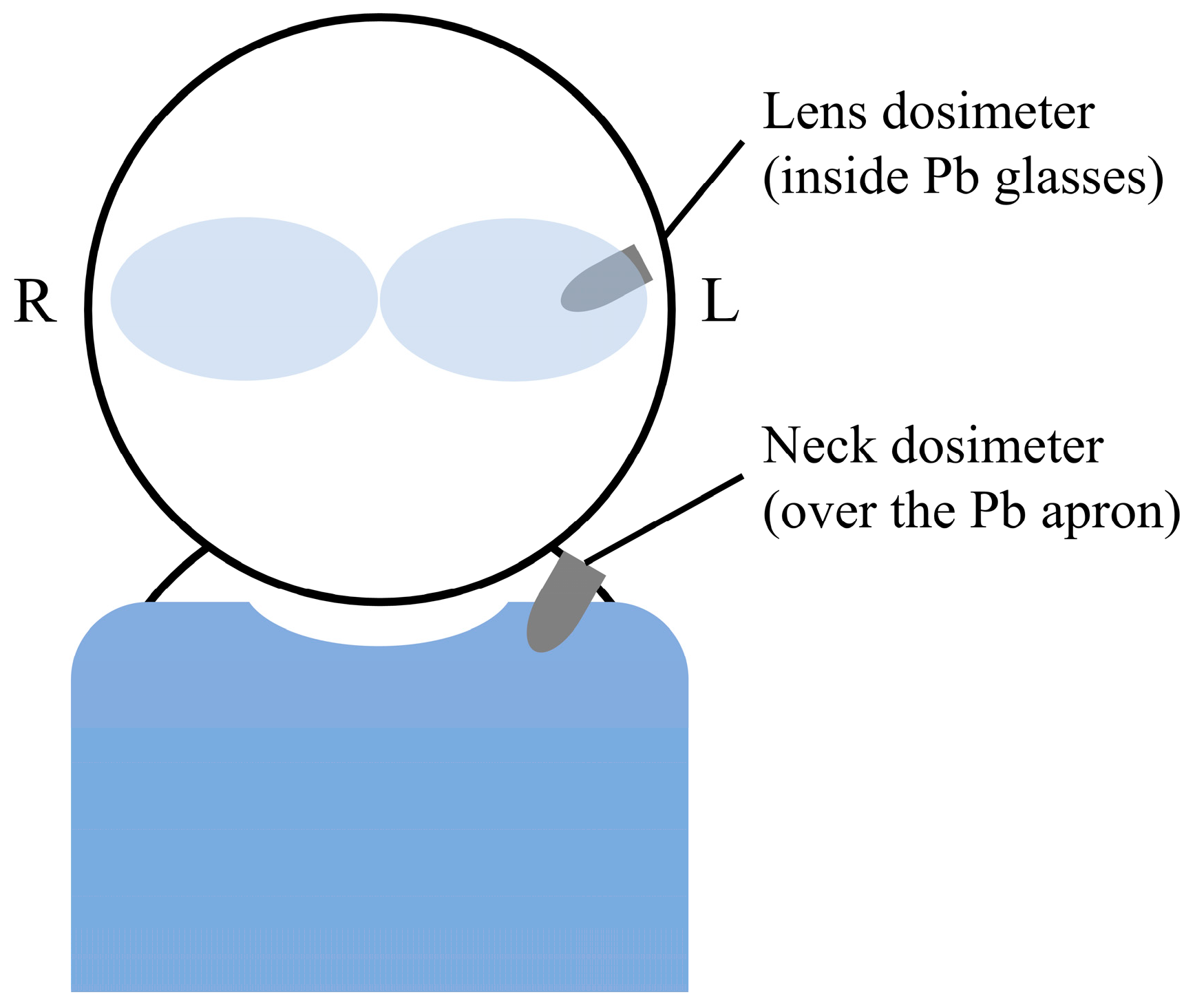
This study examined the lens dose of 10 physicians involved in IC at Sendai Kosei hospital. The physicians’ lens doses were measured using a lens dosimeter (DOSIRIS) for Hp(3) and a neck dosimeter (glass badge) for Hp(0.07). As shown in Figure 1, the lens dosimeter was positioned near the left eye, inside the lead glasses, whereas the neck dosimeter was placed outside the collar of the protective apron. To minimize inter-operator variability, the lens dosimeter was securely fixed inside the protective glasses, and the neck dosimeter was consistently attached to the same position on the collar of the lead apron for each procedure. Measurements were conducted for 1 year, from 1 April 2021, to 31 March 2022. Additionally, the physicians’ heights and years of experience were recorded. All procedures were conducted following the recommendations of the JCS/JSCVS 2018 Guideline, ensuring adherence to standardized clinical practice []. The study was conducted in accordance with the principles of the Declaration of Helsinki. Approval was granted by the Ethics Committee of Sendai Kosei hospital on 1 June 2021 (Approval Number: 30-19).
Figure 1. Positions of the dosimeters. A lens dosimeter (DOSIRIS), accounting for the shielding effect of lead glasses, was worn inside the lead glasses, while a neck dosimeter (glass badge) was worn on the outer part of the collar of the lead apron.

2.3. Statistics

Regression analyses were performed to examine the relationships between radiation dose, physician height, and years of experience. A post hoc power analysis was conducted for the correlation tests using G*Power 3.1. Based on the observed correlation coefficients, a significance level of α = 0.05, and the sample sizes of the present study, the statistical power was estimated. Two-group comparisons were also conducted. Physicians were divided into two groups based on height: ≥170 cm (n = 5) and <170 cm (n = 5), and lens doses were compared between these groups. The cutoff value of 170 cm was chosen because it approximates the average height of adult males in Japan, providing a clinically reasonable reference for group comparison []. For years of experience, physicians were categorized as having ≥10 years (n = 6) or <10 years (n = 4) of experience, and comparisons were made for both radiation dose and fluoroscopy time per procedure, with the latter serving as an indicator of procedural complexity. Because few previous studies have examined the relationship between operator experience and radiation exposure, the threshold for experience (10 years) was set based on a previous study that defined physicians with ≤10 years in practice as being in the early-career stage []. The Wilcoxon signed-rank test, a non-parametric test for comparing paired groups based on median values, was used to compare Hp(0.07) and Hp(3) for each procedure. The Wilcoxon rank-sum test, a nonparametric test for unpaired data, was used for comparisons between groups based on height and years of experience. In all analyses, the significance level was set at α = 0.05.

3. Results

Table 1 presents the annual number of procedures, fluoroscopy time per case, total accumulated doses of Hp(0.07) and Hp(3), and doses per procedure for the 10 IC physicians (physicians A–J) who participated in the study. Hp(0.07) represents the dose measured using a neck dosimeter, which does not account for the shielding effect of lead glasses, whereas Hp(3) represents the dose measured using a lens dosimeter, accounting for this shielding effect. The dose per procedure was calculated by dividing the total accumulated annual dose by the number of procedures performed annually. Table 2 summarizes the physicians’ height and years of experience since obtaining their medical qualifications.
Table 1. Number of procedures per year, fluoroscopy time per case, annual accumulated dose, and dose per procedure.
Table 2. Physician height and years of experience.
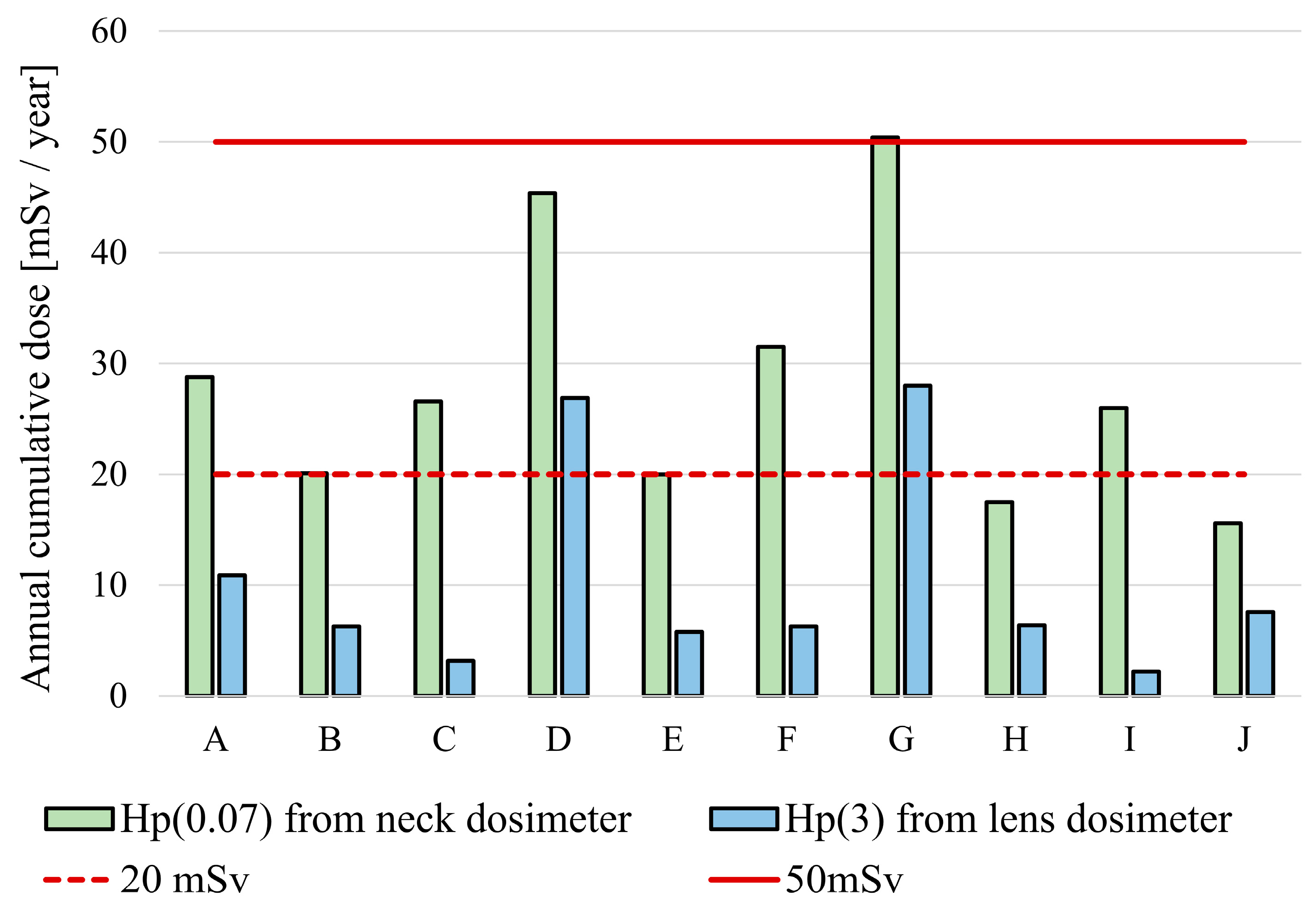
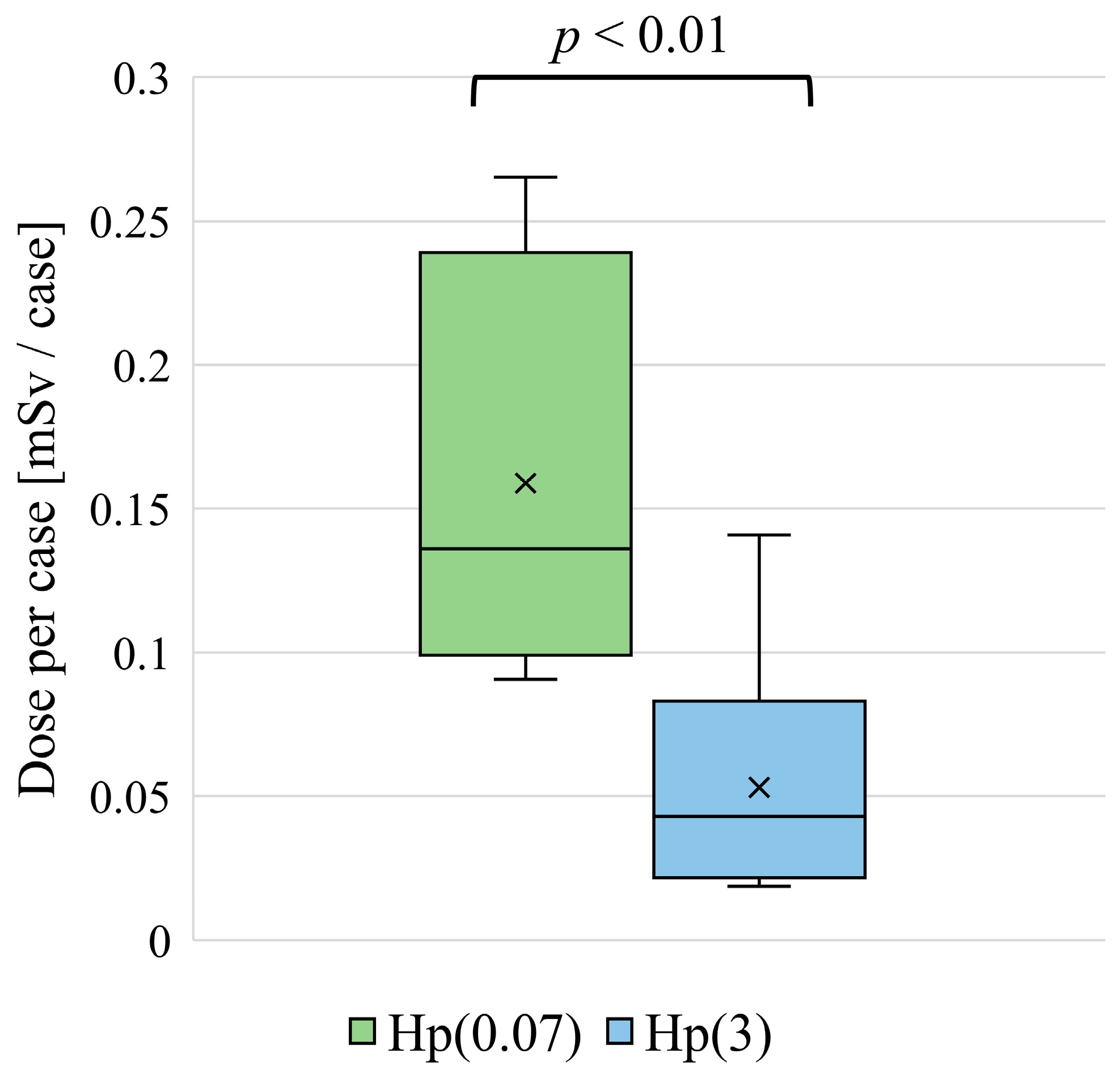
The number of procedures performed ranged from 98 (for physician who performed the fewest procedures) to 338 (for physician who performed the most procedures), with a median of 169.5 procedures. The fluoroscopy time per procedure ranged from 8.07 to 27.6 min, with a median of 15.9 min. Figure 2 shows the neck Hp(0.07) and lens Hp(3) values for the 10 physicians. Regarding Hp(0.07), 8 of 10 physicians exceeded 20 mSv/year, with one physician surpassing 50 mSv/year. In contrast, for Hp(3), 2 of 10 physicians exceeded 20 mSv/year. The neck Hp(0.07) significantly overestimated the lens dose. Figure 3 shows the neck Hp(0.07) and lens Hp(3) values for each procedure. The doses for each were expressed as median (mean ± standard deviation), with neck Hp(0.07) being 0.135 (0.155 ± 0.0621) mSv and lens Hp(3) being 0.0423 (0.0482 ± 0.0386) mSv, with a statistically significant difference (p < 0.01). The neck dosimeter tended to overestimate the lens dose. To account for differences in the number of procedures performed by each physician, the dose per procedure was used to analyze height and years of experience.
Figure 2. Annual accumulated dose for 10 physicians. The annual cumulative neck Hp(0.07) and lens Hp(3) values for the 10 physicians are shown. Based on the lens dose equivalent limit of 100 mSv over 5 years and 50 mSv per year, the red dashed line represents the annual average of 20 mSv/year, whereas the red solid line indicates the 50 mSv/year threshold.
Figure 3. Dose per case. The dose per case was calculated by dividing the annual accumulated dose by the number of procedures performed per year. Statistical significance was assessed using the Wilcoxon signed-rank test, a non-parametric test used to compare medians between two related groups.

3.1. Height and Lens Dose

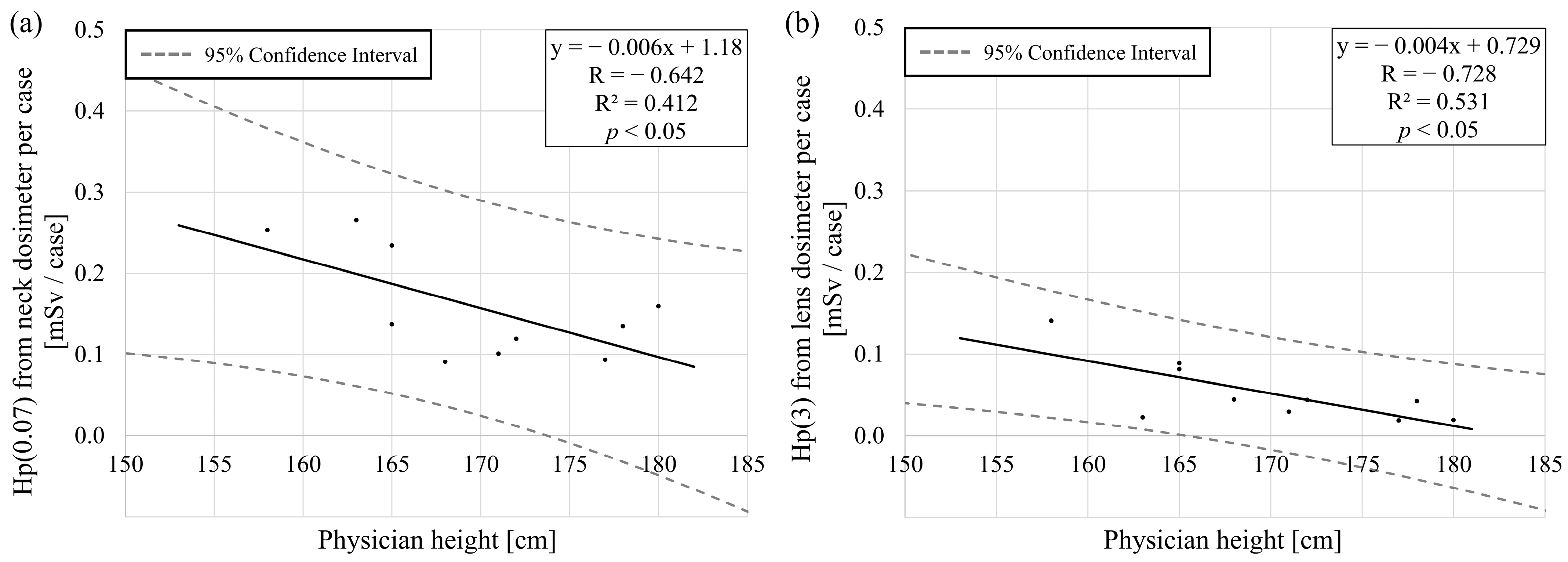
Figure 4a shows the correlation between the physician height and Hp(0.07) per procedure, and Figure 4b shows the correlation between the physician height and Hp(3) per procedure. Height and Hp(0.07) were y = −0.006x + 1.18 [x: physician height, y: Hp(0.07) from neck dosimeter per case], R = −0.642 (the 95% confidence interval: −0.906 to −0.021), R2 = 0.412, and p < 0.05. The post hoc statistical power for this correlation was 56.4%. Height and Hp(3) were y = −0.004x + 0.729 [x: physician height, y: Hp(3) from lens dosimeter per case], R = −0.728 (the 95% confidence interval: −0.931 to −0.182), R2 = 0.531, and p < 0.05. The post hoc statistical power for this correlation was 73.6%. Both Hp(0.07) and Hp(3) were correlated with physician height. The correlation coefficient for Hp(3) was larger than that for Hp(0.07), but the dose range was 0–0.141 mSv, and the difference in doses according to height was small.
Figure 4. Correlation between lens dose and physician height. (a) Correlation between Hp(0.07) from neck dosimeter per case and physician height. (b) Correlation between Hp(3) from lens dosimeter per case and physician height. The dashed line represents the 95% confidence interval.
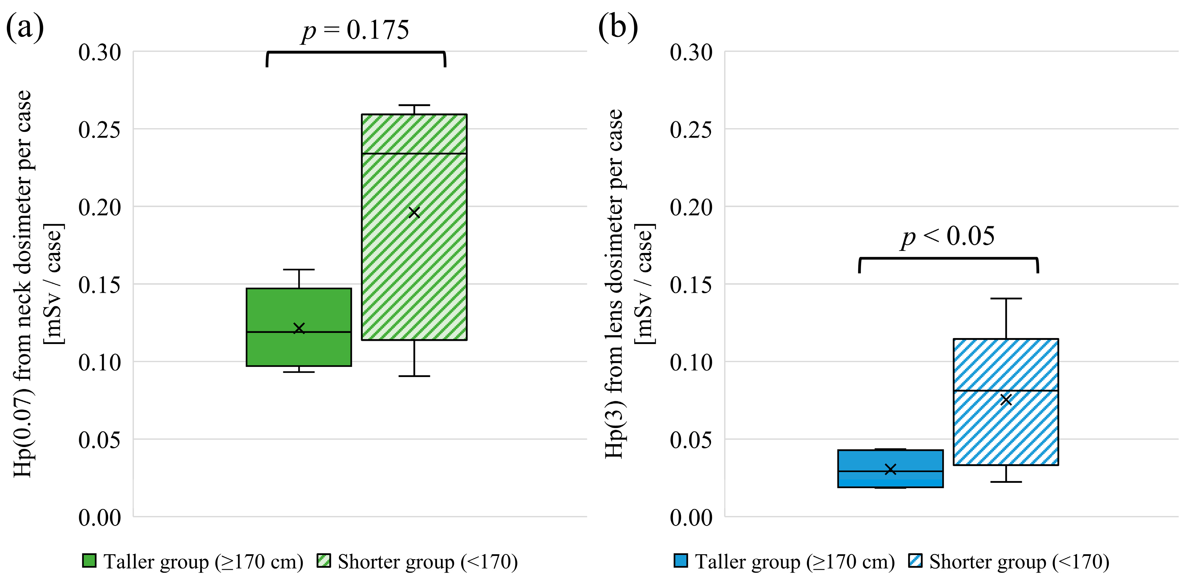
Figure 5 presents the results of a two-group comparison based on physician height: one group comprising physicians ≥ 170 cm (n = 5) and the other <170 cm (n = 5). Figure 5a shows a comparison for Hp(0.07), and Figure 5b shows a comparison for Hp(3). The radiation doses, expressed as median values (mean ± standard deviation), showed that the taller group received 0.119 mSv (0.121 ± 0.0238 mSv) for Hp(0.07), whereas the shorter group received 0.234 mSv (0.196 ± 0.0694 mSv). For Hp(3), the taller group received 0.0293 mSv (0.0306 ± 0.0108 mSv), while the shorter group received 0.0813 mSv (0.0754 ± 0.0406 mSv). A significant difference was observed in Hp(3), which was higher in the shorter group (p < 0.05).
Figure 5. Two-group comparison based on height. The physicians were divided into two groups based on height: a tall group (≥170 cm, n = 5) and short group (<170 cm, n = 5). (a) shows the comparison for Hp(0.07), and (b) shows the comparison for Hp(3). The Wilcoxon rank-sum test, a non-parametric test used to compare the medians of two independent groups, was used for analysis.

3.2. Experience as a Physician and Lens Dose

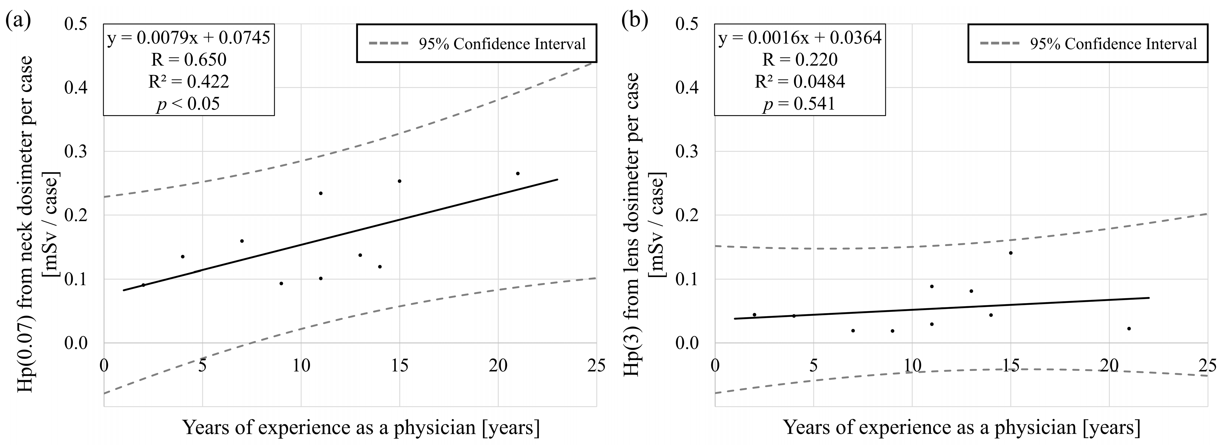
Figure 6a shows the correlation between the physician’s years of experience as a physician and Hp(0.07) per procedure, while Figure 6b shows the correlation between years of experience as a physician and Hp(3) per procedure. Years of experience and Hp(0.07) were y = 0.0079x + 0.0745 [x: years of experience as a physician, y: Hp(0.07) from neck dosimeter per case], R = 0.650 (the 95% confidence interval: 0.0345 to 0.908), R2 = 0.422, and p < 0.05. The post hoc statistical power for this correlation was 58.0%. Years of experience and Hp(3) were y = 0.0016x + 0.0364 [x: years of experience as a physician, y: Hp(3) from lens dosimeter per case], R = 0.220 (the 95% confidence interval: −0.475 to 0.746), R2 = 0.0484, and p = 0.541. The post hoc statistical power for this correlation was 9.21%. A significant correlation was found between years of experience and Hp(0.07); however, no such correlation was observed for Hp(3).
Figure 6. Correlation between lens dose and years of experience as a physician. (a) Correlation between Hp(0.07) from neck dosimeter per case and years of experience as a physician. (b) Correlation between Hp(3) from lens dosimeter per case and years of experience as a physician. The dashed line represents the 95% confidence interval.
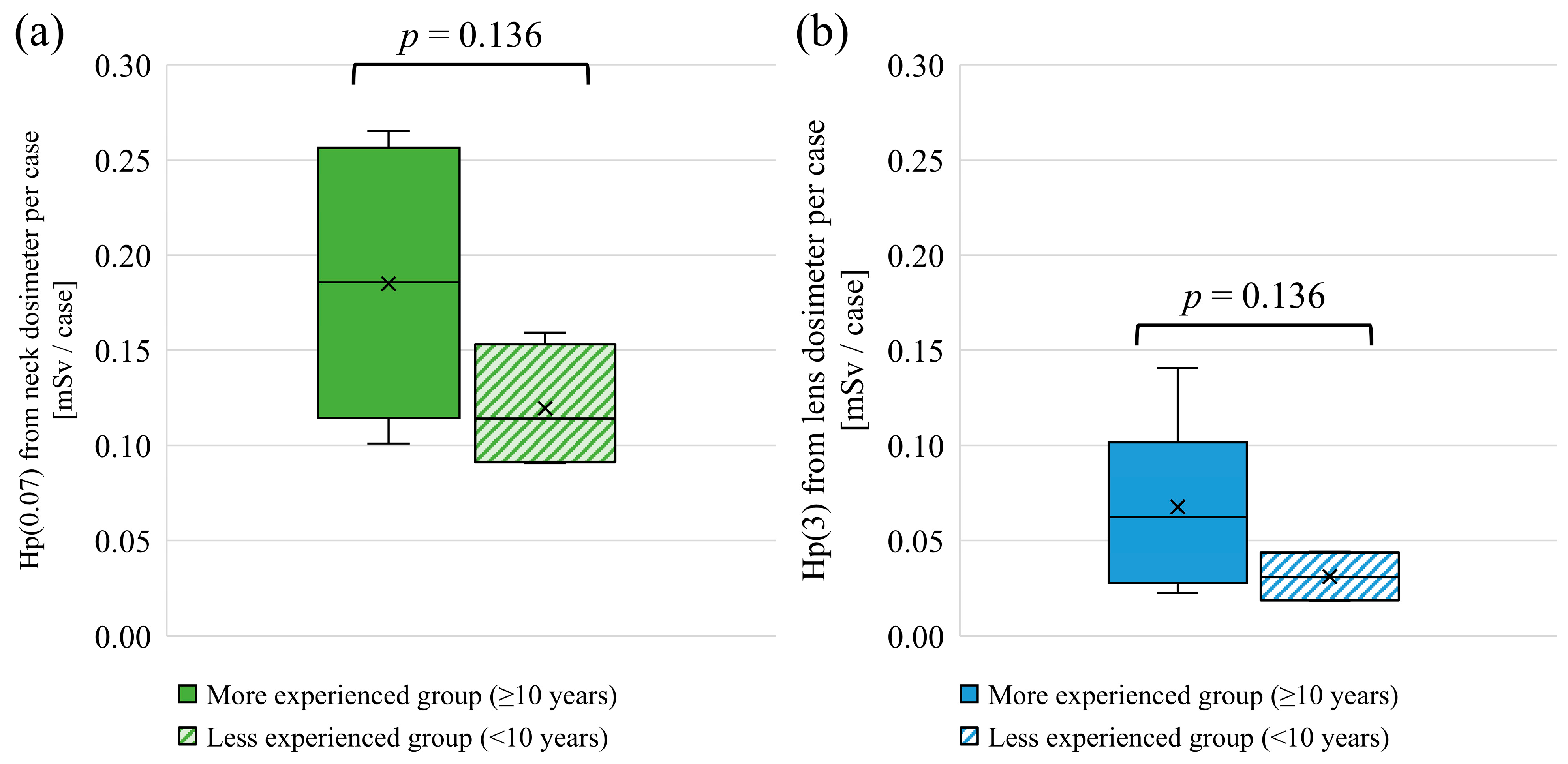
Figure 7 presents the results of a two-group comparison between physicians with ≥10 years of experience (n = 6) and those with <10 years of experience (n = 4). Figure 7a shows a comparison for Hp(0.07), and Figure 7b shows a comparison for Hp(3). Results are presented as median values, along with the mean ± standard deviation. For neck Hp(0.07), the experienced group had a median of 0.186 mSv (0.185 ± 0.0674 mSv), while the less experienced group had a median of 0.114 mSv (0.120 ± 0.0289 mSv). For lens Hp(3), the experienced group showed a median of 0.0624 mSv (0.0676 ± 0.0409 mSv), and the less experienced group had 0.0307 mSv (0.0311 ± 0.0122 mSv). Although the mean and median values of Hp(0.07) and Hp(3) were higher in the experienced group, the differences were not statistically significant.
Figure 7. Two-group comparison based on experience. The physicians were divided into two groups based on years of experience: a more experienced group (≥10 years, n = 6) and a less experienced group (<10 years, n = 4). (a) illustrates the comparison for Hp(0.07), and (b) for Hp(3). The Wilcoxon rank-sum test was used for the analysis, which is a non-parametric test used to compare the medians of two independent groups.
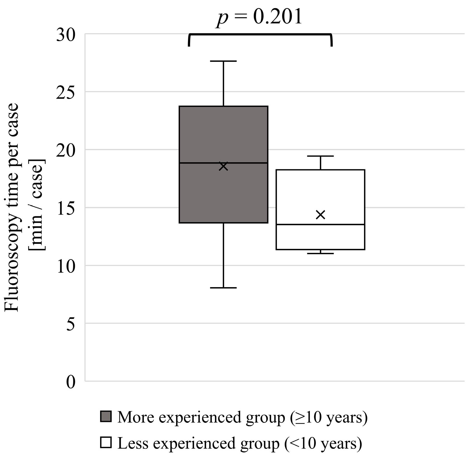
A similar comparison was conducted for the fluoroscopy time per procedure because experienced physicians are more likely to perform higher-complexity procedures that require longer fluoroscopy durations. The results are shown in Figure 8. The median fluoroscopy time per procedure (mean ± standard deviation) was 18.8 min (18.6 ± 6.2 min) in the experienced group and 13.5 min (14.4 ± 3.2 min) in the less experienced group. Although both the mean and median fluoroscopy times per procedure were longer in the experienced group, the differences were not significant.
Figure 8. Two-group comparison based on experience (fluoroscopy time per procedure). The physicians were divided into two groups based on years of experience: a more experienced group (≥10 years, n = 6) and a less experienced group (<10 years, n = 4). The Wilcoxon rank-sum test was used for the analysis, which is a non-parametric test used to compare the medians of two independent groups.

4. Discussion

This study addresses an important topic regarding occupational doses in IC procedures. The investigation of the relationship between physician height, years of experience, and lens Hp(3) is novel and clinically relevant, particularly given the updated ICRP dose limits. The use of a lens dosimeter to measure Hp(3) directly by accounting for the shielding effect of the lead glasses was a strength of this study. The reduction in the lens dose limit has raised concerns about physicians exceeding the dose limits. Numerous studies have been conducted on lens radiation protection [,,,]. New protective equipment is being developed [,,,,]. To reduce the lens dose more effectively, it is important to investigate the factors that contribute to dose increase. A few studies have examined the lens dose and physician height, but these are phantom or Monte Carlo simulation studies, and there are few clinical studies with physicians involved in IR procedures. In addition, few studies have examined the relationship between lens dose and years of experience as a physician. To our knowledge, this is the first study to examine the relationship between the lens dose and years of experience of physicians involved in IC procedures.
Some physicians were found to exceed an annual cumulative dose of 20 mSv even when measured with Hp(3), accounting for the shielding effect of lead glasses. This indicates that the current protective measures are insufficient, and a review of protection strategies is warranted. Comparison between neck Hp(0.07) and lens Hp(3) confirmed that neck Hp(0.07) values were significantly higher, suggesting an overestimation of the lens dose. However, these dosimeters use different sensing elements, and the glass badges used for neck Hp(0.07) measurements may exhibit directional dependence, which could have influenced the recorded values.
A power analysis for the correlation tests was conducted, indicating that a sample size of 12 is required to achieve 80% statistical power for a correlation coefficient of 0.728, and 16 for coefficients of 0.642 and 0.650. Although the sample size in the present study is close to these thresholds, it may still be insufficient. Moreover, no multiple-comparison correction was applied because the analyses were exploratory. Therefore, the results should be interpreted with caution, considering both the limited sample size and the potential inflation of Type I error.

4.1. Height and Lens Dose

Physician height negatively correlated with both Hp(0.07) and Hp(3). The 95% confidence intervals did not include zero, indicating a statistically significant association; however, the relatively wide intervals suggest that the estimates may be imprecise. The two-group comparison showed a significantly higher Hp(3) in shorter physicians than in taller ones (p < 0.05). Based on the median values, the shorter group exhibited 1.97 times higher Hp(0.07) and 2.77 times higher Hp(3) than the taller group. This can be attributed to the taller physicians having a greater distance between the lens and source of scattered radiation. The scattered radiation generated by the patient weakened according to the inverse square law. It is possible that taller physicians are subjected to greater attenuation of scattered radiation before it reaches the lens. The difference in the strength of the correlations between height and Hp(0.07) and Hp(3) may have been influenced by the use of ceiling-suspended protective plates and lead glasses. However, these specific dose-reduction effects were not verified in the present study. Further research is needed to analyze physician behavior to investigate these potential effects.
These findings suggest that shorter physicians may be at higher risk of receiving greater lens radiation exposure than taller physicians. Therefore, additional protective measures may be necessary for shorter physicians [,]. For example, it may be necessary to increase the lead equivalent of the lead glasses. However, this could potentially reduce the operability. It is recommended that differences in lens dose related to physician height be considered in future updates of protective equipment guidelines.
Gangl et al. investigated the impact of physician height on the occupational lens dose during pelvic vascular interventions by varying the eye height of the head phantom to 100, 157, and 176 cm []. Similar to our study, they reported that as the vertical height of the head phantom increased, the dose measured without lead glasses (using a dosimeter outside the lead glasses) decreased. However, the dose reduction factor (the ratio of the dose to the unprotected and protected lens) decreased with increasing eye height. This finding suggests that the shielding effectiveness of lead glasses may diminish as physician height increases. Our study measured lens doses during IC procedures and examined the relationship between physician height and lens dose in clinical settings. The results demonstrated a negative correlation not only for the neck dosimeter Hp(0.07) but also for the lens dosimeter Hp(3) inside the protective glasses. In clinical practice, the reduction in the shielding effect of protective glasses due to increased height may be outweighed by the attenuation effect resulting from the greater distance from the source of the scattered radiation. Both Hp(0.07) and Hp(3) were negatively correlated with physician height; however, the slope of the regression line was shallower for Hp(3). As mentioned earlier, one possible explanation is that the shielding effectiveness of the lead glasses decreases with increasing physician height. Additionally, the previous phantom study did not incorporate ceiling-suspended protective shields, which distinguished it from the present study. In contrast, our study utilized ceiling-suspended protective shields, which more accurately reflected clinical lens exposure conditions. To further investigate the relationship between height and the shielding effectiveness of protective glasses, future studies should measure Hp(3) outside the protective glasses to evaluate the shielding rate. Furthermore, it is hypothesized that the posture may differ between taller and shorter physicians, specifically in whether they “look up” or “look down” at the monitor during procedures. Since head angle may influence radiation exposure, analyzing procedural behavior is also essential. This study demonstrated that, even in clinical settings, shorter physicians are at a higher risk of receiving greater eye lens radiation doses.
Principi et al. investigated the relationship between the physician height and lens dose during IR and IC procedures using Monte Carlo simulations []. They simulated physician heights of 158, 168, 178, and 188 cm. Similar to our study, they reported that the lens dose Hp(3) decreased as the physician height increased. Principi et al. further reported that an increase in physician height by 10 cm resulted in a reduction of up to 50% in the dose to the left eye (comparing heights of 168 cm and 178 cm). In our study, when heights of 168 and 178 cm were applied to the correlation between height and Hp(0.07) and Hp(3), we observed the following reductions: 0.652 times for Hp(0.07) and 0.293 times for Hp(3). The results of our study may be more clinically relevant because the Monte Carlo simulations did not account for the effects of ceiling-suspended protective shields. Conducting a Monte Carlo simulation incorporating a ceiling-suspended protective shield may enable a clearer understanding of its impact by allowing absolute numerical comparisons across studies.
Albayati et al. measured eye-level radiation doses for three physicians performing thoracoabdominal aortic aneurysm procedures and examined the relationship between dose and height []. The physicians’ heights were 170, 186, and 193 cm. In that study, only three physicians were evaluated, and the dose was measured outside the lead glasses. In contrast, our study assessed the lens doses inside the lead glasses of 10 physicians, potentially providing a more accurate representation of the actual lens dose experienced by physicians. Consistent with our findings, their study also reported a negative correlation between height and radiation dose to the head.

4.2. Experience as a Physician and Lens Dose

A correlation between physician experience and Hp(0.07) was observed; however, no correlation was found between physician experience and Hp(3). The lack of correlation between the years of experience and Hp(3), as shown in Figure 5b, may be attributed to the shielding effects of lead glasses and the implementation of more effective radiation protection measures by experienced physicians. Although no statistically significant differences were observed in the two-group comparison based on years of experience, the mean and median values appeared to indicate a possible tendency of the more experienced group to receive higher radiation doses for both neck Hp(0.07) and lens Hp(3). The median Hp(0.07) was 0.186 mSv in the more experienced group compared to 0.114 mSv in the less experienced group (1.63-fold higher), and the median Hp(3) was 0.0624 mSv versus 0.0307 mSv (2.03-fold higher), respectively. Similarly, the median fluoroscopy time per procedure was 18.8 min in the more experienced group and 13.5 min in the less experienced group, representing a 1.39-fold difference. It was initially expected that physicians with more years of experience would have lower lens doses owing to their greater familiarity with radiation protection methods. However, Hp(3), which more accurately reflects the lens dose, showed no significant correlation with years of experience, and no significant difference was observed in the comparison between the two groups. These findings underscore the importance of regularly reviewing radiation protection practices and providing ongoing radiation safety education, regardless of clinical experience. Future studies may need to analyze the procedural behavior of both less and more experienced physicians to identify the specific actions associated with reduced radiation exposure.
Most of the physicians had over five years of experience, and the number of younger physicians, including residents with approximately 1–3 years of experience, was limited. Expanding the sample size to include a greater number of less experienced physicians may reveal clearer differences in radiation dose related to experience. Given the importance of radiation protection, continuous education and training are essential to minimize radiation exposure [,,]. For a more detailed analysis, future studies should consider procedural complexity and include larger samples of younger physicians with fewer years of experience, as well as a wider range of physician ages. In addition, incorporating analyses of ergonomic and behavioral factors, as well as simulation-based approaches that replicate realistic procedural setups, could further validate and expand upon the observed trends.
Cheriachan et al. measured physicians’ Hp(3) during orthopedic procedures and reported that the radiation doses among consultant physicians and orthopedic trainees were comparable []. A direct comparison between our study and theirs is challenging, as orthopedic procedures typically employ an over-table fluoroscopy system, whereas IC procedures commonly use an under-table fluoroscopy system. Over-table fluoroscopy systems may expose physicians to higher radiation doses than under-table fluoroscopy systems [].

4.3. Limitation

The study involved a small sample size and had limited representation of younger and less experienced physicians. Future research should expand the sample size and include more young physicians. In addition, potential sources of bias, such as variations in procedural complexity across cases, should be acknowledged and controlled in future studies.

5. Conclusions

This study investigated the relationship between lens dose and physician height and IC experience. Although several studies have explored the relationship between the physician height and lens dose, most of them relied on phantom experiments or Monte Carlo simulations. To the best of our knowledge, this is the first study to examine the relationship between lens dose and physician height in clinical practice involving IC physicians. Furthermore, few studies have examined the relationship between physician experience and lens dose. This study is the first to investigate the correlation between the years of experience and lens dose among IC physicians. In this study, physicians who exceeded the annual dose of 20 mSv were identified for both lens Hp(3) and neck Hp(0.07). A negative correlation was found between each dose and physician height, suggesting that shorter physicians may be at higher risk of an increased lens dose. In the two-group comparison, the Hp(3) values were significantly higher for shorter physicians than for taller physicians (p < 0.05). Based on the median values, the shorter group exhibited 1.97 times higher Hp(0.07) and 2.77 times higher Hp(3) than the taller group. Therefore, physicians with shorter heights may need to consider additional protective measures. A correlation was observed between experience and Hp(0.07); however, no correlation was found between experience and Hp(3). The results of this study indicate that Hp(3) does not strongly depend on years of experience. These findings highlight the importance of regularly reviewing radiation protection practices and providing refresher education on radiation safety, regardless of experience level. Future studies should investigate the specific behaviors associated with reduced radiation exposure. We believe that this work is relevant to the field of IR, given the lack of studies that discuss the occupational setup of the room and radioprotection equipment in relation to the doses collected.

Author Contributions

Conceptualization, K.S. and K.C.; methodology, Y.H. and T.K.; validation, K.S., M.A.; formal analysis, K.S.; investigation, K.S., Y.H., T.K., S.T., M.S. and N.T.; resources, Y.K. and K.C.; data curation, K.S., Y.H., T.K., S.T. and M.S.; writing—original draft preparation, K.S.; writing—review and editing, K.C.; visualization, K.S.; supervision, M.A. and K.C.; project administration, K.C.; funding acquisition, K.C. All authors have read and agreed to the published version of the manuscript.

Funding

This work was supported in part by a Grant-in-Aid for Scientific Research from the Japan Society for the Promotion of Science (23H03537); and the Industrial Disease Clinical Research Grant (240401-02), Japan.

Institutional Review Board Statement

The study was conducted in accordance with the Declaration of Helsinki, and approved by the Ethics Committee of Sendai Kosei Hospital on 1 June 2021. (approval number: 30-19).

Data Availability Statement

The data presented in this study are available on request from the corresponding author due to privacy and ethical restrictions.

Acknowledgments

We thank Taku Sato, Mako Tanabe and Mio Nakamura from Tohoku University Graduate School of Medicine, Japan, for their technical assistance.

Conflicts of Interest

The authors declare no conflicts of interest.

Abbreviations

The following abbreviations are used in this manuscript:
ICRPInternational Commission on Radiological Protection
IAEAInternational Atomic Energy Agency
IRInterventional Radiology
ICInterventional Cardiology
Hp(10)personal dose equivalent at 1 cm depth
Hp(0.07)personal dose equivalent at 70 μm depth
Hp(3)personal dose equivalent at 3 mm depth

References

  1. ICRP. Radiological Protection in Cardiology. ICRP Publication 120; Ann. ICRP: Ottawa, CA, USA, 2013. [Google Scholar]
  2. Elfandi, A.; Safirstein, J.G. Transradial PCI and Same Day Discharge. Curr. Treat. Options Cardiovasc. Med. 2018, 20, 10. [Google Scholar] [CrossRef]
  3. Sandhu, K.; Nadar, S.K. Percutaneous Coronary Intervention in the Elderly. Int. J. Cardiol. 2015, 199, 342–355. [Google Scholar] [CrossRef]
  4. Gianoli, M.; de Jong, A.R.; Jacob, K.A.; Namba, H.F.; van der Kaaij, N.P.; van der Harst, P.; Suyker, W.J. Minimally Invasive Surgery or Stenting for Left Anterior Descending Artery Disease—Meta-Analysis. Int. J. Cardiol. Heart Vasc. 2022, 40, 101046. [Google Scholar] [CrossRef]
  5. Kato, M.; Chida, K.; Nakamura, M.; Toyoshima, H.; Terata, K.; Abe, Y. New Real-Time Patient Radiation Dosimeter for Use in Radiofrequency Catheter Ablation. J. Radiat. Res. 2019, 60, 215–220. [Google Scholar] [CrossRef]
  6. Chida, K.; Fuda, K.; Saito, H.; Takai, Y.; Takahashi, S.; Yamada, S.; Kohzuki, M.; Zuguchi, M. Patient Skin Dose in Cardiac Interventional Procedures: Conventional Fluoroscopy versus Pulsed Fluoroscopy. Catheter. Cardiovasc. Interv. 2007, 69, 115–121. [Google Scholar] [CrossRef]
  7. Srimahachota, S.; Udayachalerm, W.; Kupharang, T.; Sukwijit, K.; Krisanachinda, A.; Rehani, M. Radiation Skin Injury Caused by Percutaneous Coronary Intervention, Report of 3 Cases. Int. J. Cardiol. 2012, 154, e31–e33. [Google Scholar] [CrossRef]
  8. Wagner, L.K.; McNeese, M.D.; Marx, M.V.; Siegel, E.L. Severe Skin Reactions from Interventional Fluoroscopy: Case Report and Review of the Literature. Radiology 1999, 213, 773–776. [Google Scholar] [CrossRef]
  9. Koenig, T.R.; Wolff, D.; Mettler, F.A.; Wagner, L.K. Skin Injuries from Fluoroscopically Guided Procedures. Am. J. Roentgenol. 2001, 177, 3–11. [Google Scholar] [CrossRef]
  10. Inaba, Y.; Chida, K.; Murabayashi, Y.; Endo, M.; Otomo, K.; Zuguchi, M. An Initial Investigation of a Wireless Patient Radiation Dosimeter for Use in Interventional Radiology. Radiol. Phys. Technol. 2020, 13, 321–326. [Google Scholar] [CrossRef]
  11. Roguin, A.; Goldstein, J.; Bar, O.; Goldstein, J.A. Brain and Neck Tumors Among Physicians Performing Interventional Procedures. Am. J. Cardiol. 2013, 111, 1368–1372. [Google Scholar] [CrossRef]
  12. Eagan, J.T., Jr.; Jones, C.T. Cutaneous Cancers in an Interventional Cardiologist: A Cautionary Tale. J. Interv. Cardiol. 2011, 24, 49–55. [Google Scholar] [CrossRef]
  13. Vano, E.; Kleiman, N.J.; Duran, A.; Romano-Miller, M.; Rehani, M.M. Radiation-Associated Lens Opacities in Catheterization Personnel: Results of a Survey and Direct Assessments. J. Vasc. Interv. Radiol. 2013, 24, 197–204. [Google Scholar] [CrossRef]
  14. Kato, M.; Chida, K.; Ishida, T.; Sasaki, F.; Toyoshima, H.; Oosaka, H.; Terata, K.; Abe, Y.; Kinoshita, T. Occupational Radiation Exposure Dose of The Eye in Department of Cardiac Arrhythmia Physician. Radiat. Prot. Dosim. 2019, 187, 361–368. [Google Scholar] [CrossRef]
  15. Werner, N.; Nickenig, G.; Sinning, J.-M. Complex PCI Procedures: Challenges for the Interventional Cardiologist. Clin. Res. Cardiol. 2018, 107, 64–73. [Google Scholar] [CrossRef]
  16. Kheifets, M.; Vons, S.A.; Bental, T.; Vaknin-Assa, H.; Greenberg, G.; Samara, A.; Codner, P.; Wittberg, G.; Talmor Barkan, Y.; Perl, L.; et al. Temporal Trends in Complex Percutaneous Coronary Interventions. Front. Cardiovasc. Med. 2022, 9, 913588. [Google Scholar] [CrossRef]
  17. Ali, A.; Al-Nuaimi, M.; Khan, B.; Burki, S.; Nishtar, T.; Yaseen, M.; Shah, S.S.A.; Irfan, M. Assessment of Occupational Radiation Doses to Eye Lens during Interventional Radiology Procedures. Radiat. Phys. Chem. 2024, 225, 112131. [Google Scholar] [CrossRef]
  18. Bellamy, M.B.; Miodownik, D.; Quinn, B.; Dauer, L. Occupational Eye Lens Dose Over Six Years in The Staff of A Us High-Volume Cancer Center. Radiat. Prot. Dosim. 2020, 192, 321–327. [Google Scholar] [CrossRef]
  19. Merrachi, N.-A.; Bouchard-Bellavance, R.; Perreault, P.; Gilbert, P.; Soulez, G.; Bouchard, L.; Oliva, V.L.; Giroux, M.-F.; Normandeau, L.; Therasse, E. Eye Lens Dosimetry in Interventional Radiology: Assessment with Dedicated Hp(3) Dosimeters. Can. Assoc. Radiol. J. 2021, 72, 317–323. [Google Scholar] [CrossRef]
  20. Ainsbury, E.A.; Barnard, S.G.R. Sensitivity and Latency of Ionising Radiation-Induced Cataract. Exp. Eye Res. 2021, 212, 108772. [Google Scholar] [CrossRef]
  21. Durchschlag, H.; Fochler, C.; Abraham, K.; Kulawik, B. Radiation Effects on Eye Components. Radiat. Phys. Chem. 1999, 55, 691–697. [Google Scholar] [CrossRef]
  22. Hamada, N.; Azizova, T.V.; Little, M.P. An Update on Effects of Ionizing Radiation Exposure on the Eye. Br. J. Radiol. 2020, 93, 20190829. [Google Scholar] [CrossRef] [PubMed]
  23. Matsubara, K.; Lertsuwunseri, V.; Srimahachota, S.; Krisanachinda, A.; Tulvatana, W.; Khambhiphant, B.; Sudchai, W.; Rehani, M. Eye Lens Dosimetry and the Study on Radiation Cataract in Interventional Cardiologists. Phys. Medica 2017, 44, 232–235. [Google Scholar] [CrossRef]
  24. Vañó, E.; González, L.; Guibelalde, E.; Fernández, J.M.; Ten, J.I. Radiation Exposure to Medical Staff in Interventional and Cardiac Radiology. Br. J. Radiol. 1998, 71, 954–960. [Google Scholar] [CrossRef] [PubMed]
  25. ICRP. Avoidance of Radiation Injuries from Medical Interventional Procedures; ICRP Publication 85; Ann. ICRP: Ottawa, CA, USA, 2000. [Google Scholar]
  26. Nakashima, E.; Neriishi, K.; Minamoto, A. A reanalysis of atomic-bomb cataract data, 2000–2002: A threshold analysis. Health Phys. 2006, 90, 154. [Google Scholar] [CrossRef] [PubMed]
  27. Worgul, B.V.; Kundiyev, Y.I.; Sergiyenko, N.M.; Chumak, V.V.; Vitte, P.M.; Medvedovsky, C.; Bakhanova, E.V.; Junk, A.K.; Kyrychenko, O.Y.; Musijachenko, N.V.; et al. Cataracts among Chernobyl Clean-up Workers: Implications Regarding Permissible Eye Exposures. Radiat. Res. 2007, 167, 233–243. [Google Scholar] [CrossRef]
  28. Neriishi, K.; Nakashima, E.; Minamoto, A.; Fujiwara, S.; Akahoshi, M.; Mishima, H.K.; Kitaoka, T.; Shore, R.E. Postoperative Cataract Cases among Atomic Bomb Survivors: Radiation Dose Response and Threshold. Radiat. Res. 2007, 168, 404–408. [Google Scholar] [CrossRef]
  29. ICRP. ICRP Statement on Tissue Reactions/Early and Late Effects of Radiation in Normal Tissues and Organs—Threshold Doses for Tissue Reactions in a Radiation Protection Context; ICRP Publication 118; Ann. ICRP: Ottawa, CA, USA, 2012. [Google Scholar]
  30. Sagehashi, K.; Haga, Y.; Takahira, S.; Tanabe, M.; Nakamura, M.; Sota, M.; Kaga, Y.; Abe, M.; Tada, N.; Chida, K. Evaluation of Radiation Dose to the Lens in Interventional Cardiology Physicians before and after Dose Limit Regulation Changes. J. Radiol. Prot. 2024, 44, 031512. [Google Scholar] [CrossRef]
  31. Haga, Y.; Chida, K.; Kaga, Y.; Sota, M.; Meguro, T.; Zuguchi, M. Occupational Eye Dose in Interventional Cardiology Procedures. Sci. Rep. 2017, 7, 569. [Google Scholar] [CrossRef]
  32. Ohno, S.; Konta, S.; Shindo, R.; Yamamoto, K.; Isobe, R.; Inaba, Y.; Suzuki, M.; Zuguchi, M.; Chida, K. Effect of Backscatter Radiation on the Occupational Eye-Lens Dose. J. Radiat. Res. 2024, 65, 450–458. [Google Scholar] [CrossRef]
  33. Yamada, A.; Haga, Y.; Sota, M.; Abe, M.; Kaga, Y.; Inaba, Y.; Suzuki, M.; Tada, N.; Zuguchi, M.; Chida, K. Eye Lens Radiation Dose to Nurses during Cardiac Interventional Radiology: An Initial Study. Diagnostics 2023, 13, 3003. [Google Scholar] [CrossRef]
  34. Hijikata, Y.; Yamashita, K.; Hatsusaka, N.; Nagata, T.; Kitamura, H.; Morota, K.; Matsuzaki, S.; Nakagami, K.; Hitomi, G.; Kuriyama, T.; et al. Prevalence of Cataractous Changes in the Eyes and Chronic Inflammatory Changes in the Hands Among Spine Surgeons. J. Bone Jt. Surg. 2025, 107, e25. [Google Scholar] [CrossRef] [PubMed]
  35. Kuriyama, T.; Moritake, T.; Nakagami, K.; Morota, K.; Hitomi, G.; Kitamura, H. Background Factors Affecting the Radiation Exposure of the Lens of the Eye among Nurses in Interventional Radiology: A Quantitative Observational Study. Nurs. Rep. 2024, 14, 413–427. [Google Scholar] [CrossRef] [PubMed]
  36. ICRP. The 2007 Recommendations of the International Commission on Radiological Protection; ICRP Publication 103; Ann. ICRP: Ottawa, CA, USA, 2007. [Google Scholar]
  37. IAEA. International Atomic Energy Agency, Implications for Occupational Radiation Protection of the New Dose Limit for the Lens of the Eye; IAEA TECDOC; IAEA: Vienna, Austria, 2013. [Google Scholar]
  38. Al-Haj, A.N.; Lobriguito, A.M.; Al-Gain, I. Staff Eye Doses in a Large Medical Centre in Saudi Arabia: Are They Meeting the New ICRP Recommendations? Radiat. Prot. Dosim. 2015, 165, 294–298. [Google Scholar] [CrossRef]
  39. Jacob, S.; Donadille, L.; Maccia, C.; Bar, O.; Boveda, S.; Laurier, D.; Bernier, M.-O. Eye Lens Radiation Exposure to Interventional Cardiologists: A Retrospective Assessment of Cumulative Doses. Radiat. Prot. Dosim. 2013, 153, 282–293. [Google Scholar] [CrossRef]
  40. Martin, C.J.; Magee, J.S. Assessment of Eye and Body Dose for Interventional Radiologists, Cardiologists, and Other Interventional Staff. J. Radiol. Prot. 2013, 33, 445. [Google Scholar] [CrossRef]
  41. Meijer, E.J.; van Zandvoort, D.W.H.; Loos, M.J.A.; Tseng, C.M.E.S.N.; van Pul, C. The Eye Lens Dose of the Interventionalist: Measurement in Practice. Phys. Medica 2022, 100, 1–5. [Google Scholar] [CrossRef]
  42. Bohari, A.; Hashim, S.; Ahmad, N.E.; Ghoshal, S.K.; Mohd Mustafa, S.N. Fluoroscopy-Guided Intervention Procedure Norms for Occupational Eye Radiation Dose: An Overall Evaluation. Radiat. Phys. Chem. 2021, 178, 108909. [Google Scholar] [CrossRef]
  43. Domienik, J.; Bissinger, A.; Grabowicz, W.; Jankowski, Ł.; Kręcki, R.; Makowski, M.; Masiarek, K.; Plewka, M.; Lubiński, A.; Peruga, J.Z. The Impact of Various Protective Tools on the Dose Reduction in the Eye Lens in an Interventional Cardiology—Clinical Study. J. Radiol. Prot. 2016, 36, 309. [Google Scholar] [CrossRef]
  44. Moriarty, H.K.; Clements, W.; Phan, T.; Wang, S.; Goh, G.S. Occupational Radiation Exposure to the Lens of the Eye in Interventional Radiology. J. Med. Imaging Radiat. Oncol. 2022, 66, 34–40. [Google Scholar] [CrossRef]
  45. Shindo, R.; Ohno, S.; Yamamoto, K.; Konta, S.; Inaba, Y.; Suzuki, M.; Zuguchi, M.; Chida, K. Comparison of Shielding Effects of Over-Glasses-Type and Regular Eyewear in Terms of Occupational Eye Dose Reduction. J. Radiol. Prot. 2024, 44, 023501. [Google Scholar] [CrossRef] [PubMed]
  46. van Rooijen, B.D.; de Haan, M.W.; Das, M.; Arnoldussen, C.W.K.P.; de Graaf, R.; van Zwam, W.H.; Backes, W.H.; Jeukens, C.R.L.P.N. Efficacy of Radiation Safety Glasses in Interventional Radiology. Cardiovasc. Interv. Radiol. 2014, 37, 1149–1155. [Google Scholar] [CrossRef] [PubMed]
  47. ICRU Report 60. Fundamental Quantities and Units for Ionizing Radiation. J. ICRU 1998. Available online: https://www.icru.org/report/fundamental-quantities-and-units-for-ionizing-radiation-report-60/ (accessed on 1 October 2025).
  48. Ishii, H.; Haga, Y.; Sota, M.; Inaba, Y.; Chida, K. Performance of the DOSIRISTM Eye Lens Dosimeter. J. Radiol. Prot. 2019, 39, N19. [Google Scholar] [CrossRef]
  49. Mori, Y.; Isobe, T.; Takei, H.; Miyazaki, S.; Kamizawa, S.; Tomita, T.; Kobayashi, D.; Sakurai, H.; Sakae, T. Evaluation of Basic Characteristics of 3-Mm Dose Equivalent Measuring Instrument for Evaluating Lens Exposure Dose in Radiotherapy. J. Med. Radiat. Sci. 2023, 70, 154–160. [Google Scholar] [CrossRef]
  50. Suliman, I.I. Review of Various Methods for Occupational Eye Lens Dosimetry in X-Ray Interventional Procedures. Radiat. Phys. Chem. 2025, 236, 112936. [Google Scholar] [CrossRef]
  51. Gangl, A.; Deutschmann, H.A.; Portugaller, R.H.; Stücklschweiger, G. Influence of Safety Glasses, Body Height and Magnification on the Occupational Eye Lens Dose during Pelvic Vascular Interventions: A Phantom Study. Eur. Radiol. 2022, 32, 1688–1696. [Google Scholar] [CrossRef]
  52. Koenig, A.M.; Etzel, R.; Greger, W.; Viniol, S.; Fiebich, M.; Thomas, R.P.; Mahnken, A.H. Protective Efficacy of Different Ocular Radiation Protection Devices: A Phantom Study. Cardiovasc. Interv. Radiol. 2020, 43, 127–134. [Google Scholar] [CrossRef] [PubMed]
  53. Mao, L.; Liu, T.; Caracappa, P.F.; Lin, H.; Gao, Y.; Dauer, L.T.; Xu, X.G. Influences of Operator Head Posture and Protective Eyewear on Eye Lens Doses in Interventional Radiology: A Monte Carlo Study. Med. Phys. 2019, 46, 2744–2751. [Google Scholar] [CrossRef]
  54. Principi, S.; Farah, J.; Ferrari, P.; Carinou, E.; Clairand, I.; Ginjaume, M. The Influence of Operator Position, Height and Body Orientation on Eye Lens Dose in Interventional Radiology and Cardiology: Monte Carlo Simulations versus Realistic Clinical Measurements. Phys. Medica 2016, 32, 1111–1117. [Google Scholar] [CrossRef]
  55. ICRP. Education and Training in Radiological Protection for Diagnostic and Interventional Procedures; ICRP Publication 113; Ann. ICRP: Ottawa, CA, USA, 2009. [Google Scholar]
  56. Chida, K. What Are Useful Methods to Reduce Occupational Radiation Exposure among Radiological Medical Workers, Especially for Interventional Radiology Personnel? Radiol. Phys. Technol. 2022, 15, 101–115. [Google Scholar] [CrossRef]
  57. Cheriachan, D.; Hughes, A.M.; du Moulin, W.S.M.; Williams, C.; Molnar, R. Ionizing Radiation Doses Detected at the Eye Level of the Primary Surgeon During Orthopaedic Procedures. J. Orthop. Trauma 2016, 30, e230. [Google Scholar] [CrossRef]
  58. Nakamura, M.; Yaku, H.; Ako, J.; Arai, H.; Asai, T.; Chikamori, T.; Daida, H.; Doi, K.; Fukui, T.; Ito, T.; et al. JCS/JSCVS 2018 Guideline on Revascularization of Stable Coronary Artery Disease. Circ. J. 2022, 86, 477–588. [Google Scholar] [CrossRef]
  59. The Ministry of Health, Labour and Welfare. Ministry of Health, Labour and Welfare National Health and Nutrition Survey 2012—Report of the Physical Status Survey; Ministry of Health, Labour and Welfare: Tokyo, Japan, 2014. [Google Scholar]
  60. Dyrbye, L.N.; Varkey, P.; Boone, S.L.; Satele, D.V.; Sloan, J.A.; Shanafelt, T.D. Physician Satisfaction and Burnout at Different Career Stages. Mayo Clin. Proc. 2013, 88, 1358–1367. [Google Scholar] [CrossRef] [PubMed]
  61. Fujisawa, M.; Haga, Y.; Sota, M.; Abe, M.; Kaga, Y.; Inaba, Y.; Suzuki, M.; Meguro, T.; Hosoi, Y.; Chida, K. Evaluation of Lens Doses among Medical Staff Involved in Nuclear Medicine: Current Eye Radiation Exposure among Nuclear-Medicine Staff. Appl. Sci. 2023, 13, 9182. [Google Scholar] [CrossRef]
  62. Inaba, Y.; Jingu, K.; Fujisawa, M.; Otomo, K.; Ishii, H.; Kato, T.; Murabayashi, Y.; Suzuki, M.; Zuguchi, M.; Chida, K. Evaluation of Radiation Doses Received by Physicians during Permanent 198Au Grain Implant Brachytherapy for Oral Cancer. Appl. Sci. 2024, 14, 6010. [Google Scholar] [CrossRef]
  63. Inaba, Y.; Hitachi, S.; Watanuki, M.; Chida, K. Occupational Radiation Dose to Eye Lenses in CT-Guided Interventions Using MDCT-Fluoroscopy. Diagnostics 2021, 11, 646. [Google Scholar] [CrossRef]
  64. Fujibuchi, T.; Nakashima, M.; Arakawa, H.; Miyazaki, H.; Anam, C. Evaluation of Radiation Protection Effectivity in a Cardiac Angiography Room Using Visualized Scattered Radiation Distribution. J. Radiol. Prot. 2024, 44, 031510. [Google Scholar] [CrossRef]
  65. Fukuda, A.; Lin, P.-J.P. Covering the Patient’s Arm Support in Lead Reduced the Radiation Dose Rate to the Cardiologists During Percutaneous Coronary Interventions: A Phantom Study. Radiat. Prot. Dosim. 2020, 188, 340–349. [Google Scholar] [CrossRef] [PubMed]
  66. Kato, M.; Chida, K.; Munehisa, M.; Sato, T.; Inaba, Y.; Suzuki, M.; Zuguchi, M. Non-Lead Protective Aprons for the Protection of Interventional Radiology Physicians from Radiation Exposure in Clinical Settings: An Initial Study. Diagnostics 2021, 11, 1613. [Google Scholar] [CrossRef]
  67. Zanca, F.; Dabin, J.; Collard, C.; Alexandre, N.; De Groote, A.; Salembier, J.P.; Henry, M.; Rombaut, E.; Sghaier, S.; Massart, P.-E. Evaluation of a Suspended Radiation Protection System to Reduce Operator Exposure in Cardiology Interventional Procedures. Catheter. Cardiovasc. Interv. 2021, 98, E687–E694. [Google Scholar] [CrossRef]
  68. McCaffrey, J.P.; Tessier, F.; Shen, H. Radiation Shielding Materials and Radiation Scatter Effects for Interventional Radiology (IR) Physicians. Med. Phys. 2012, 39, 4537–4546. [Google Scholar] [CrossRef]
  69. Endo, M.; Haga, Y.; Sota, M.; Tanaka, A.; Otomo, K.; Murabayashi, Y.; Abe, M.; Kaga, Y.; Inaba, Y.; Suzuki, M.; et al. Evaluation of Novel X-Ray Protective Eyewear in Reducing the Eye Dose to Interventional Radiology Physicians. J. Radiat. Res. 2021, 62, 414–419. [Google Scholar] [CrossRef]
  70. ICRP. The Optimisation of Radiological Protection–Broadening the Process; ICRP Publication 101b; Ann. ICRP: Ottawa, CA, USA, 2006. [Google Scholar]
  71. Albayati, M.A.; Kelly, S.; Gallagher, D.; Dourado, R.; Patel, A.S.; Saha, P.; Bajwa, A.; El-Sayed, T.; Salter, R.; Gkoutzios, P.; et al. Editor’s Choice—Angulation of the C-Arm During Complex Endovascular Aortic Procedures Increases Radiation Exposure to the Head. Eur. J. Vasc. Endovasc. Surg. 2015, 49, 396–402. [Google Scholar] [CrossRef] [PubMed]
  72. Sato, T.; Eguchi, Y.; Yamazaki, C.; Hino, T.; Saida, T.; Chida, K. Development of a New Radiation Shield for the Face and Neck of IVR Physicians. Bioengineering 2022, 9, 354. [Google Scholar] [CrossRef] [PubMed]
  73. Daring, D.L.; Sun, Z. Investigation of the Clinical Value of Three-Dimensional-Printed Personalised Vascular Models for the Education and Training of Clinicians When Performing Interventional Endovascular Procedures. Appl. Sci. 2025, 15, 5695. [Google Scholar] [CrossRef]
Disclaimer/Publisher’s Note: The statements, opinions and data contained in all publications are solely those of the individual author(s) and contributor(s) and not of MDPI and/or the editor(s). MDPI and/or the editor(s) disclaim responsibility for any injury to people or property resulting from any ideas, methods, instructions or products referred to in the content.

Article Metrics

Citations

Article Access Statistics

Multiple requests from the same IP address are counted as one view.