Article

Simplified Diagnosis of Mandibular Asymmetry in Panoramic Radiographs Through Digital Processing and Its Prospective Integration with Artificial Intelligence: A Pilot Study

by
Paulina Agurto-Sanhueza
1,
Karla Roco
2,
Pablo Navarro
1,3,
Andrés Neyem
4,5,
Nicolás I. Sumonte
4,5 and
Nicolás E. Ottone
1,6,7,8,*
1
Doctoral Program in Morphological Sciences, Universidad de La Frontera, Temuco 4780000, Chile
2
Escuela de Informática y Telecomunicaciones, Instituto Profesional Duoc UC, Santiago 7500550, Chile
3
Facultad de Ciencias de la Salud, Universidad Autónoma de Chile, Temuco 4920000, Chile
4
Department of Computer Science, School of Engineering, Pontificia Universidad Católica de Chile, Santiago 7820436, Chile
5
National Center for Artificial Intelligence (CENIA), National Research and Development Agency (ANID), Santiago 8320000, Chile
6
Laboratory of Plastination and Anatomical Techniques, Universidad de La Frontera, Temuco 4780000, Chile
7
Center of Excellence in Morphological and Surgical Studies (CEMyQ), Universidad de La Frontera, Temuco 4780000, Chile
8
Adults Integral Dentistry Department, Center for Research in Dental Sciences (CICO), Faculty of Dentistry, Universidad de La Frontera, Temuco 4780000, Chile
*
Author to whom correspondence should be addressed.
Appl. Sci. 2025, 15(19), 10802; https://doi.org/10.3390/app151910802
Submission received: 2 September 2025 / Revised: 5 October 2025 / Accepted: 6 October 2025 / Published: 8 October 2025
(This article belongs to the Special Issue Recent Advances in Orthodontic Diagnosis and Treatment)

Abstract

Background/Objectives: Mandibular asymmetry is a common morphological alteration in orthodontics and orthognathic surgery, generally diagnosed with panoramic radiographs despite their limitations. Automated processing systems offer a promising alternative for improving its detection and analysis. The aim of this study was to develop a pilot computational model to detect and measure mandibular asymmetry in the body and ramus by analyzing anatomical distances in digital panoramic radiographs of adults. Methods: This was a descriptive observational pilot study, carried out on 30 digital panoramic radiographs of young adult patients (15 men, 15 women). Three craniometric points (Condylion, Gonion and Gnathion) were used as references landmarks. An algorithm was implemented in Python® (v3.12) with OpenCV to extract anatomical coordinates and calculate Euclidean distances (Go-Gn, Co-Go) from pixels to millimeters. Data were statistically analyzed in SPSS (v23.0) using normality tests, paired t-tests, Wilcoxon tests, and Mann–Whitney U tests (p < 0.05). Results: No significant differences were observed in mandibular lengths by sex, with men having greater lengths in both the body (80.63 mm vs. 73.86 mm) and the ramus (55.82 mm vs. 49.15 mm). In addition, significant differences were found in total mandibular ramus measurements (p = 0.023). A classification of asymmetry by severity was proposed (mild: ≤3 mm, moderate: 3–6 mm, severe: >6 mm), with mild asymmetries being the most frequently found. The model showed reliable processing capacity. Conclusions: This pilot study shows the feasibility of using Python for automated measurement of mandibular asymmetry in panoramic radiographs and highlights its future potential for neural network integration and diagnostic-epidemiological use.

1. Introduction

In the field of orthodontics and surgery, facial asymmetry is of utmost clinical importance. At the lower third, it is highly influenced by mandibular asymmetry, which is defined as a discrepancy in length, shape, and size when comparing both sides of the jawbone. This asymmetry can be caused by multiple factors, including growth disturbances, functional disorders, pathological conditions, and trauma, the effects of which can manifest in both bone and underlying soft tissues [1,2,3].
According to its classification and characteristics, mandibular asymmetry can be subdivided based on to the specific anatomical regions it affects, such as the mandibular ramus, mandibular body, and chin. This helps to comprehensively assess the morphology of the mandible and the consequent diagnosis and treatment planning [4,5]. Prevalence rates in adults, mostly calculated at the level of the mandibular ramus, show values of 39.5%, with condylar and ramus asymmetries being even more prevalent, at 81.4% and 48.6%, respectively [6]. Likewise, Class III malocclusion is the most frequently associated with these asymmetries, with a prevalence ranging from 22.93% to 78% [7].
The main images used in the diagnosis of mandibular asymmetries, to measure the ramus and body of the mandible (according to the level of precision), are posteroanterior cephalometries, with low to moderate precision levels [8,9], panoramic radiographs (orthopantomography) with moderate ranges, which may present magnification and distortion errors [10,11,12] and CBCT, which allow a more precise three-dimensional evaluation [13,14,15]. However, panoramic radiographs remain the most useful in screening, due to their widespread and massive use and the lower radiation doses to which patients are exposed [10,11,16], reasons that were considered for the development of this study.
In the literature, the analysis of mandibular asymmetries on panoramic radiographs has mainly relied on traditional indices such as those of Habets [17] and Kjellberg [18]. While these indices provide a useful initial framework, they are limited in that they do not yield absolute millimetric values and do not facilitate the automation of the diagnostic process. Moreover, only a few studies have explored the incorporation of automated processing techniques in panoramic radiographs. Within this context, the present work is conceived as an exploratory step towards the integration of artificial intelligence in the assessment of mandibular asymmetry. Rather than attempting to compete with advanced three-dimensional techniques such as CBCT, the aim is to provide a preliminary, accessible, and low-cost tool that can serve as a foundation for future developments in automated detection.
Diagnosis is always a challenge for clinicians, which is why the use of current tools such as artificial intelligence is a promising field to explore. In fact, initial automation studies using convolutional neural networks (CNNs) show considerable improvements in the detection of mandibular asymmetries in panoramic radiographs, increasing the accuracy and reliability of measurements and consequently reducing human error [19,20].
The use of Python could represent a versatile programming language in addition to playing a crucial role in the development of Artificial Intelligence models, due to its high accuracy and efficiency, comparable to that of qualified orthodontists, in the development of task optimization systems that save time and therefore improve efficiency in orthodontic clinical practice [21,22,23].
The objective of this study was to develop a proof-of-concept model using computational design tools in Python to validate the concordance between radiographic measurements and clinical reality, as a preliminary step toward automating the process through convolutional neural networks (CNNs). This pilot will subsequently be applied to a representative sample with the aim of simplifying and standardizing the assessment of mandibular asymmetry, by establishing a morphological classification based on severity, differentiating between the mandibular body (horizontal asymmetry) and ramus (vertical asymmetry), and incorporating epidemiological data stratified by age and sex.

2. Materials and Methods

A descriptive observational pilot study was conducted with 30 digital panoramic radiographs (orthopantomographies), in JPG and PDF formats, of young adult patients between 18 and 45 years of age. The sample consisted of 15 males and 15 females from the private clinic of one of the authors. The images were randomly selected and anonymized to ensure patient confidentiality. All participants provided informed consent for their use, and the study was approved by the Ethics and Scientific Committee of Universidad de La Frontera (protocol code 020_25, dated 17 March 2025). Radiographs from three different radiology centers were used to evaluate variability in the model’s capabilities. The distribution and respective equipment are detailed below: 23 taken on an Orthopantomography (OPG) INSTRUMENTARIUM model OC-200 D (2008) (Instrumentarium Corp., Tuusula, Finland); 5 with an OPG NEWTOM model Giano HR (2022) (New-Tom, Imola, Italy) and 2 from the OPG VATECH, model PAX-400C (2011) (Vatech Company Limited, Hwaseong-si, Republic of Korea).
This sample included patients with a complete permanent dentition or with at most four missing teeth (excluding third molars), most of whom were in the pre-orthodontic stage of treatment. Three subjects were wearing fixed orthodontic appliances (conventional metal brackets), and one was in the retention stage with fixed retainers. Images of patients with a history of trauma, tumors, joint pathology, malformations, or those who were candidates for orthognathic surgery were excluded.
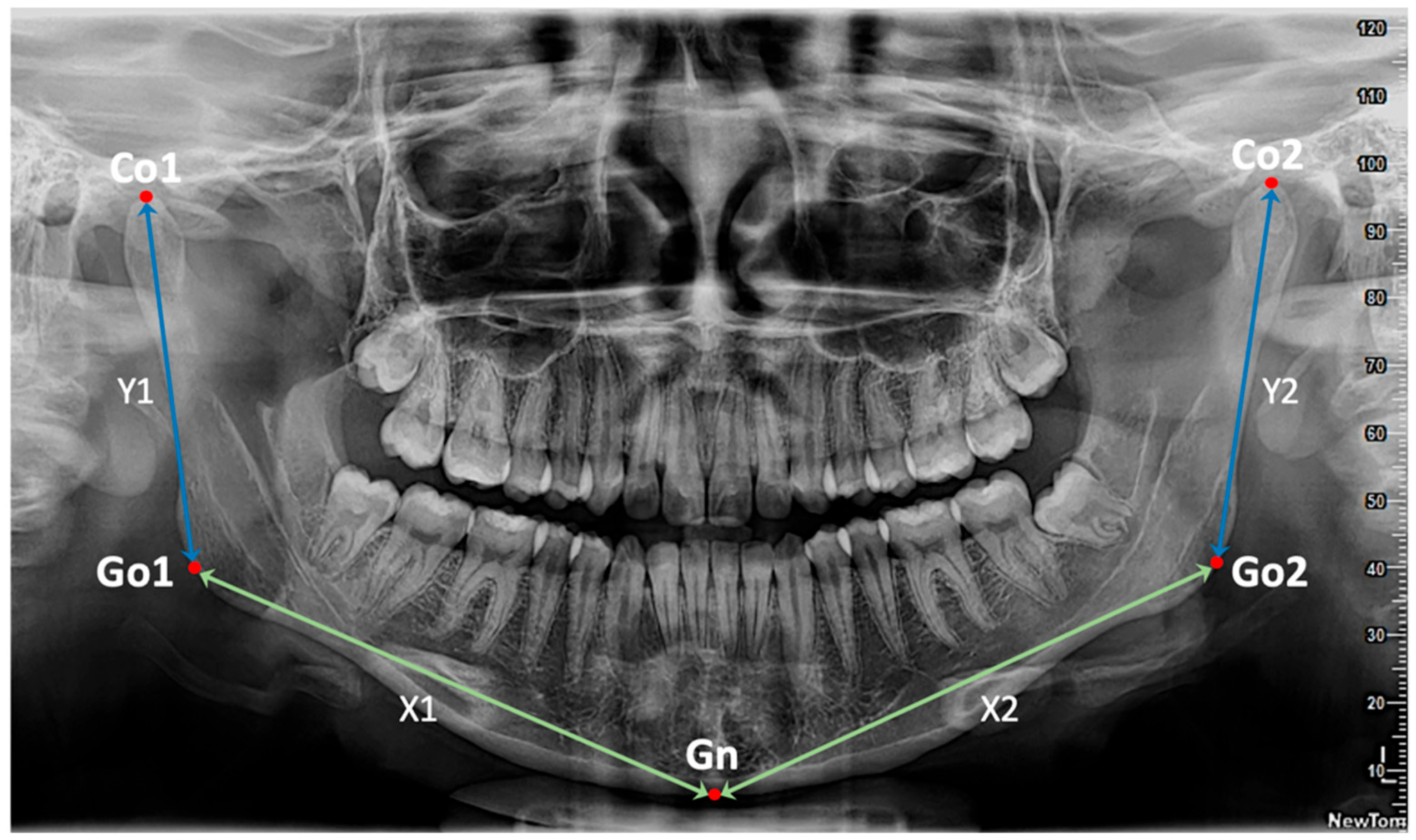
The Python programming language version 3.12 was used to develop the pilot model, and the analysis focused on the detection and measurement of distances between key anatomical points to assess asymmetry at the level of the mandibular body and ramus. The craniometric points used were Condylion (Co), the most superior and medial point on the head of the condyle [18]; Gonion (Go), the point located at the lowest, posterior, and lateral part of the external angle of the mandible; and Gnathion (Gn), the most caudal point on the lower border of the mandible, in the median plane, at the level of the mandibular symphysis [24].
Each radiograph was clinically validated: it was initially labeled by the orthodontist responsible for the study, and subsequently reviewed through an inter-observer concordance analysis conducted by another orthodontic specialist. In addition, intra-observer reliability was assessed by repeating the labeling process after a two-week interval. The reproducibility in the identification of craniofacial landmarks was evaluated using the intraclass correlation coefficient (ICC) for subsequent linear measurements in millimeters, considering a mixed-effects model and absolute agreement. The ICC = 0.90 (95% CI: 0.81–0.92), which reflects a high inter-observer concordance. Although the initial labeling was performed manually, all subsequent stages of the workflow (reading, coordinate extraction, pixel-to-millimeter conversion, and distance calculation) were fully automated through a Python-based pipeline. This ensured standardized processing conditions for all radiographs and minimized variability associated with human intervention. The high ICC value obtained confirms that the automated pipeline consistently operates on robustly defined anatomical landmarks, strengthening the reliability of the results.
Once the tagged images have been entered into the program, their processing can be divided into 3 stages (Table 1):
(1)
Extraction of anatomical coordinates: A computer vision algorithm was implemented to automatically identify the anatomical points of interest marked in red on the digital panoramic radiographs. To do this, the OpenCV library was used, working in the HSV color space to isolate the pixels of this hue. Subsequently, morphological operations were applied to eliminate noise, and the relevant contours were located. The centers of mass of these contours were recorded as (X, Y) coordinates of the anatomical points, and the results were exported to a CSV file for further analysis.
(2)
Conversion and calculation of distances: the obtained coordinates were processed from the 5 anatomical landmarks (Co1, Go1, Gn, Go2, Co2) and representative linear measurements were extracted for the calculation of the 4 key Euclidean distances defined as, X1: distance between Go1 and Gn (right half of the mandibular body); X2: distance between Gn and Go2 (left half of the mandibular body); Y1: distance between Co1 and Go1 (right mandibular ramus); Y2: distance between Go2 and Co2 (left mandibular ramus). All obtained distances were converted from pixels to millimeters using a conversion factor specific to the analyzed images (0.10585 mm/pixel) (Figure 1).
(3)
Storage and analysis of results: The coordinate and distance data were organized in Excel spreadsheets using Pandas, which allowed for comparative descriptive analyses between the right and left sides of the mandible, evaluating the presence and magnitude of asymmetry in each radiographic image.
Table 1. Detailed description of the three-stage processing pipeline applied to panoramic radiographs. Each stage includes the computational procedures performed and the outputs generated, ensuring methodological reproducibility and transparency for subsequent validation phases.
StageDescription
Stage 1: Extraction of anatomical coordinatesDetection of anatomical landmarks from panoramic radiographs using HSV masking; noise removal with morphological operations; contour filtering and centroid computation (X,Y).
Stage 2: Conversion & Distance CalculationConversion of pixel values to millimeters (scale factor 0.10585 mm/pixel); calculation of Euclidean distances (X1, X2, Y1, Y2); computation of differences (DIF X1–X2, DIF Y1–Y2); classification of asymmetry severity (≤3 mm, 3–6 mm, >6 mm).
Stage 3: Storage & Statistical AnalysisStorage of results in CSV/XLSX format; descriptive statistics; normality testing (Shapiro–Wilk); inferential statistics (paired t-test, Wilcoxon signed-rank test, Mann–Whitney U test).
Figure 1. Digital panoramic radiograph, where the labeling of the craniometric points Condylion (Co), Gonion (Go) and Gnathion (Gn) can be observed, for the measurement of mandibular asymmetry through the coordinate lines X (Go-Gn) for the body of the mandible and Y (Co-Go) for the ramus of the mandible.
Applsci 15 10802 g001
The development tools and environment were run in a Jupyter Notebook 10.0, as mentioned above, using the Python program. The following open-source libraries were also used: cv2, numpy, pandas, and pdf2image. The images were processed locally using a Windows 10 operating system, and the results were exported in CSV and Excel (.xlsx) formats. Although the present version of the pipeline depends on manual pre-labeling, it has been designed to be scalable. In future phases, convolutional neural networks (CNNs) could be incorporated for automatic landmark detection, reducing the need for manual intervention and enabling faster and broader clinical applications.
Statistical Analysis
Data collection was recorded in a Microsoft Office Excel spreadsheet, and a descriptive analysis of the data was subsequently performed, determining the mean and its respective standard deviation, the Shapiro–Wilk normality test, the t-test for related samples, the Wilcoxon test (Wt), and the Mann–Whitney U test.
Data analysis was performed using SPSS Statistics for Windows (version 23.0, IBM). A p-value < 0.05 was chosen as the threshold for statistical significance.

3. Results

The programming language was able to read the markings of the labeled craniometric points in all the images, using the process described above, and the Euclidean measurements were converted from pixels to millimeters. These obtained distances allowed for the following analyses.
Size differences by gender were evaluated by comparing the distances along the X and Y coordinates.
In females, the mean total mandibular body size (Go–Gn) was 73.86 mm.
  • Right side (X1): mean of 73.94 mm (range: 67.93 to 81.64 mm).
  • Left side (X2): mean of 73.79 mm (range: 66.82 to 80.86 mm).
Normality was confirmed by the Shapiro–Wilk test (X1, p = 0.504; X2, p = 0.973), and no significant difference was found between sides using a paired t-test (p = 0.874).
In males, the mean mandibular body size (Go–Gn) was larger at 80.63 mm.
  • Right side (X1): range 66.81 to 105.86 mm.
  • Left side (X2): range 70.87 to 106.47 mm.
Normality was not fully met (Shapiro–Wilk test: X1, p = 0.040; X2, p = 0.005), so the Wilcoxon signed-rank test was applied, showing no significant difference between sides (p = 0.069).
These results indicate that, although men exhibited greater mandibular body size and broader variability, no significant asymmetry was observed between the right and left sides in either sex.
Similarly, size differences in the Y coordinate (Co–Go), corresponding to the mandibular ramus, were assessed.
In females, the mean total size of the mandibular ramus was 49.15 mm.
  • Right side (Y1): values ranged from 44.25 to 55.11 mm.
  • Left side (Y2): values ranged from 43.73 to 55.44 mm.
  • Normality was confirmed by the Shapiro–Wilk test (Y1, p = 0.597; Y2, p = 0.238), and no significant difference between sides was found using a paired t-test (p = 0.058).
In males, the mean total size of the mandibular ramus was larger at 55.82 mm.
  • Right side (Y1): ranged from 46.30 to 82.31 mm.
  • Left side (Y2): ranged from 45.20 to 74.79 mm.
Normality was not fully met (Shapiro–Wilk test: Y1, p = 0.006; Y2, p = 0.040), so the Wilcoxon signed-rank test was applied, showing no significant difference between sides (p = 0.211).
These results indicate that side-to-side distributions were statistically equivalent, with no evidence of asymmetry in median values. Consistent with findings for the mandibular body, males exhibited larger mandibular ramus measurements and greater variability compared to females.
When analyzing the combined data for both sexes, the total X1 and X2 coordinates, representing the length of the mandibular body, did not follow a normal distribution (Shapiro–Wilk test: X1, p = 0.001; X2, p = 0.002).
The Wilcoxon signed-rank test indicated no statistically significant difference between the right and left sides (p = 0.082), suggesting symmetric distributions with no evidence of median asymmetry. However, greater value dispersion was observed in the mandibular body compared to the ramus.
In contrast, the total Y1 and Y2 coordinates—corresponding to the length of the mandibular ramus—also failed the normality test (Shapiro–Wilk: Y1, p = 0.001; Y2, p = 0.005), but the Wilcoxon signed-rank test revealed a statistically significant difference between sides (p = 0.023) (Figure 2).
These findings suggest that, while the mandibular body tends to maintain bilateral symmetry, the ramus shows significant side-to-side variation, reflecting greater morphological variability in this region.
Furthermore, a classification was created to measure asymmetry with 3 mm interval ranges, in order to allow the automated model to provide valid results and to generate a proposal for a clinical classification. By calculating the coordinate differences (DIF) to assess mandibular asymmetry, specifically in the mandibular body and ramus, it was possible to obtain clear and quantifiable asymmetry measurements by sex and by anatomical segment, expressed in millimeters. This digital processing approach enhances comparability and reproducibility in future measurements.
These results indicate that, in both females and males, mild asymmetries (≤3 mm) were the most frequently observed in both the mandibular ramus and body. Moderate asymmetries followed in frequency, while severe asymmetries represented the smallest proportion in both regions. Notably, severe asymmetries were more frequently observed in males.
When comparing coordinate differences, no statistically significant variation was found between groups (p > 0.05).
  • For DIF X1–X2: Shapiro–Wilk test showed normality for X1 (p = 0.480), but not for X2 (p = 0.005); the Mann–Whitney U test yielded a non-significant result (p = 0.074).
  • For DIF Y1–Y2: normality was borderline or not met (Y1, p = 0.060; Y2, p = 0.033); the Mann–Whitney U test also indicated no significant difference (p = 0.233).
These findings suggest that side-to-side differences in mandibular measurements do not significantly differ between sexes. However, further studies with larger and more representative samples are needed to confirm these results and to inform the development of a clinical classification system based on the severity of asymmetry.
The total results are detailed in Table 2, and their graphical distribution can be seen in Figure 3.
Beyond the numerical findings, this pilot study allowed us to establish a clinical classification framework for mandibular asymmetry based on three categories (mild: ≤3 mm, moderate: 3–6 mm, and severe: >6 mm). This approach provides absolute millimetric ranges for both the body and ramus of the mandible, which, to our knowledge, has not been systematically reported in studies using panoramic radiographs. While previous works have frequently employed proportional indices such as those of Habets [17] or Kjellberg [18], these methods do not offer a direct millimetric scale applicable to clinical decision-making. The classification proposed in this study therefore represents an initial step toward a more standardized and clinically useful interpretation of asymmetry severity when relying on panoramic radiographs.

4. Discussion

In this preliminary pilot study, we aimed to measure mandibular asymmetry by analyzing specific craniometric points such as Condylion (Co), Gonion (Go), and Gnathion (Gn) in digital panoramic radiographs, using Python as an automated analysis tool. The use of only three specific points is intended to simplify measurements and improve specificity in the use of the program. Other authors [25,26,27,28] have used these points; however, they used Menton instead of Gnathion, which raises doubts about its use from the perspective of its location and anatomical definition.
On the other hand, studies such as Azhari et al. (2019) [25] and Lemos et al. (2014) [28] consider the total length of the body of the mandible as a complete measurement, without considering that there are differences between one side and the other. However, Alfaro et al. (2016) [27] differentiated between the right and left sides of the mandibular body, which was also observed in our study, where we found considerable differences between both sides, in the same individual, which may be relevant in the diagnosis and the implications in orthodontic and surgical treatment.
In panoramic radiographs of adult patients, Alfaro et al. (2016) [27] found average measurements for the mandibular ramus (Co-Go) of 56.6 mm for women and 54.8 mm for men and for the mandibular body (Go-Me) of 77.4 mm in women and 76.7 mm in men. This contrasts with various studies in the adult population, which show that the length and other dimensions of the mandibular ramus are usually greater in men than in women, as is also the case with the diagonal and horizontal length of the body of the mandible, when comparing both sexes. In our study, we found average measurements for the body of the mandible (Go-Gn) 73.86 mm in women and 80.63 mm in men, and for the ramus of the mandible (Co-Go) 49.15 mm in women and 55.82 mm in men, measurements similar to those reported in other studies conducted with panoramic radiographs [29,30,31] and studies based on direct measurements in bone samples [32,33,34].
It should be noted that there are multiple studies on panoramic radiographs on mandibular asymmetry, which only consider linear measurements of the ramus of the mandible. In these studies, this asymmetry is classified as vertical mandibular asymmetry, where the percentage differences between both sides are calculated. These studies use the Habets indices [35,36,37,38,39,40], according to which if the threshold is greater than 3%, a significant vertical asymmetry is considered, similar to what is proposed in Kjellberg index [41,42,43,44]. In our opinion, these indices show the presence and percentage severity of vertical asymmetry. However, when applied to clinical practice, we believe the amount of asymmetry in millimeters is much more important in order to more accurately assess the correction needs according to the treatment to be performed.
Because mandibular asymmetry is a frequent clinical finding, it can be classified into different types according to the etiology, anatomical location, and degree of deviation from the median plane [45,46,47]. The findings in our study confirmed the existence of clear differences in the length of the mandibular ramus and body between the right and left sides in the analyzed sample, which is why we consider it important to make a clear classification for both structures, belonging to the mandibular bone. This proposal is expressed in differences of 3 mm intervals, a categorization similar to that proposed by Thiesen et al. (2018) [48], who developed a classification of relative mandibular asymmetry, moderate and severe, with 2 mm intervals. However, this classification was based on the measurement of the displacement of the Gnathion with respect to the median sagittal plane in Cone Beam Computed Tomography (CBCT) in a population with similar age ranges. Accordingly, our findings primarily align with mild to moderate structural asymmetries, identified through differential measurement of bilateral distances. Nevertheless, the frequencies found are unrepresentative due to the small size of the pilot sample.
Compared with traditional indices such as those of Habets [17] and Kjellberg [18], which rely on relative percentages [36,37,38,39,40,41], the classification proposed in our study provides absolute values in millimeters. This distinction is clinically relevant because orthodontic and surgical treatment planning is often guided by absolute discrepancies rather than proportional differences. By offering a millimetric scale, our model contributes to a clearer, more standardized interpretation of asymmetry severity, which could help clinicians determine when a panoramic radiograph is sufficient for initial evaluation and when complementary CBCT imaging is warranted [8,10,11]. In this way, the model not only addresses methodological reproducibility but also responds to immediate clinical needs.
Although panoramic radiographs have inherent limitations, such as distortion and overlapping structures, their use remains common in orthodontics due to their accessibility and low cost. Several previous studies have shown that, with proper calibration and standardization, these images can be useful for the initial detection of asymmetries. Currently, other researchers such as Pertek et al. (2024) [49], have used panoramic radiographs with AI for morphometric measurements in gender determination and mandibular asymmetry [20]. The latter study found consistency in measurements and reproducibility when compared with manual measurements.
Our work should therefore be considered exploratory and pilot in nature, representing a necessary step prior to the development of fully automated AI-based detection systems [19,20,21]. By establishing a reproducible computational pipeline for the analysis of mandibular asymmetry, our study lays the methodological foundation for subsequent integration of neural networks, which could enable direct detection of craniofacial landmarks without manual labeling. In this sense, the present research occupies an intermediate stage between purely manual cephalometric analyses and the more advanced, fully automated approaches that are currently under development [21,22].
Another important aspect of this study is the use of open-source tools, specifically Python and OpenCV. The transparency and accessibility of these resources make the pipeline replicable in different academic and clinical environments, fostering its use for both research and teaching purposes. This openness has the potential to democratize access to computational methods, particularly in contexts where access to expensive software or advanced imaging technologies is limited. Furthermore, by being adaptable and customizable, the system can serve as a basis for future enhancements, including the incorporation of convolutional neural networks (CNNs) to reduce manual intervention and to enable broader applications in large-scale clinical and epidemiological studies [20,21,23,49,50].
The use of Python enabled efficient automation of the measurement and analysis process, representing a significant advance over manual methods, particularly in terms of reducing human error and improving reproducibility. This study also highlights the need to implement automated models for the diagnosis of mandibular asymmetries in orthodontics. Artificial intelligence and computational analysis have the potential to become key tools for clinicians, facilitating the early identification of deviations that may require orthodontic or surgical treatment. Python-based computational models offer an additional advantage by being open-source and customizable, allowing their integration into different clinical and academic workflows [50].
In line with previous findings, such as those reported by Qu et al. (2025) [20], where no significant differences were observed between manual and AI-based methods when comparing ICC values, the present study also demonstrated high reproducibility. The inter-observer agreement yielded an intraclass correlation coefficient (ICC) of 0.90 (95% CI: 0.81–0.92), indicating good to excellent reliability in the identification of craniofacial landmarks. Nevertheless, a systematic comparison between automated and traditional manual measurements was not performed, which will be essential in future studies to establish the diagnostic accuracy of the method.
Although the applied conversion factor (0.10585 mm/pixel) was consistent across all processed images, variability among the radiographic equipment used (INSTRUMENTARIUM OC-200 D, NEWTOM Giano HR, and VATECH PAX-400C) may have influenced measurement accuracy. This heterogeneity constitutes a potential limitation; nevertheless, it also highlights the robustness of the algorithm in processing images acquired from different sources. Differences in image resolution, acquisition parameters, and device-specific processing algorithms can introduce systematic bias. Therefore, for future external validation, the implementation of inter-device calibration protocols or image normalization techniques is recommended to minimize these effects and improve both the reproducibility and the generalizability of the results.
It is important to acknowledge that this pilot study represents a pioneering step toward the automated processing of panoramic radiographs. While the algorithm successfully processed all 30 radiographs analyzed, detecting the labeled anatomical landmarks in 100% of the cases, the absence of formal validation must be recognized as a limitation. However, this limitation is inherent to the exploratory purpose of pilot studies, which are designed to test feasibility and establish proof of concept on a smaller scale [51]. Importantly, the sample size adopted here aligns with recommendations from previous methodological research [52,53], reinforcing the adequacy of this initial stage and highlighting its role in paving the way for future large-scale validation. The limited sample size of this study is consistent with the exploratory nature of pilot research, serving to refine methodology and generate preliminary data; broader validation with a representative population (including adult men and women aged 18 to 60 years) is already in progress as part of the next project phase. A further limitation is the absence of validation against a gold standard such as CBCT or direct clinical measurements. While this phase focused on feasibility, the next stage of the project includes validation with CBCT in a representative sample to confirm diagnostic accuracy and clinical reliability.
Clinical Implications and Future Directions
The development of a standardized classification of mandibular asymmetry based on its severity and anatomical location, providing parameters for both the mandibular body and the ramus, will allow for a better clinical approach, both in orthodontic treatment and orthognathic surgery. As mentioned above, mandibular asymmetry is usually assessed using indices or by evaluating the degree of deviation from the midplane. This preliminary assessment using panoramic radiographs makes them a useful tool for rapid and reliable diagnosis by orthodontists, allowing them to make more accurate decisions about treatment approaches. Depending on the severity observed and considering other parameters such as soft tissue characteristics and the individual needs of each patient, it will facilitate the most accurate indication of a complementary diagnostic study with CBCT and the eventual sub-sequent referral to orthognathic surgery.
In order to provide rigor and clinical and technical viability to the processes, it was decided to carry out this pilot study, in which, although the inherent limitations of this type of study must be recognized (such as the small sample size and the consequent low statistical power, which makes it difficult to extrapolate to generalized clinical parameters), it enabled us to obtain preliminary measurements and was very useful in improving and correcting errors in digital programming. In addition, it ensured the establishment of the methodological bases for the subsequent stages of the project, which include, as future directions, firstly, automated diagnosis with AI of mandibular asymmetry and a clear classification proposal in millimeters based on panoramic radiographs, and later, a final stage in CBCT images, with the incorporation of other useful measurements in craniofacial diagnosis.

5. Conclusions

Based on the sample analyzed in this pilot study, the findings support the technical feasibility of implementing an automated system for assessing mandibular asymmetry through digital processing of panoramic radiographs, using Python and OpenCV, with applicability to both the mandibular body and the ramus.
The algorithm developed was able to:
  • Successfully process radiographs from three different devices, suggesting adaptability to diverse image sources.
  • Consistently calculate key anatomical distances (Co-Go and Go-Gn) bilaterally.
  • Classify asymmetries according to severity in millimeters.
This work establishes the methodological foundation for the future development of a more sophisticated system that could incorporate:
  • Automatic detection of anatomical landmarks through convolutional neural networks.
  • Multicenter validation with larger samples.
  • Integration with radiological image management systems (PACS).
  • Development of population-specific normative values.
This pilot study, despite the inherent limitations of a reduced sample size, served as a small-scale investigation that enabled us to evaluate the feasibility and functionality of our project. The results obtained suggest a high potential for future integration and refinement through the application of neural networks, which could optimize the accuracy and efficiency of diagnostic measurements in orthodontics and maxillofacial surgery. In addition to providing valuable clinical, epidemiological, and morphological parameters, this approach may also hold promise for broader applications across other fields of dentistry and medicine. Future integration of convolutional neural networks (CNNs) and larger datasets will enhance diagnostic accuracy and enable real-time clinical application.

Author Contributions

Conceptualization, P.A.-S. and N.E.O.; methodology, P.A.-S. and K.R.; software, K.R.; validation, P.A.-S., N.E.O. and K.R.; formal analysis, P.A.-S., N.E.O., A.N. and N.I.S.; investigation, P.A.-S., K.R. and N.E.O.; resources, P.A.-S. and N.E.O.; data curation, P.A.-S., K.R. and P.N.; writing—original draft preparation, P.A.-S., N.E.O. and K.R.; writing—review and editing, P.A.-S., N.E.O., K.R., P.N., A.N. and N.I.S.; visualization, P.A.-S.; supervision, N.E.O.; project administration, N.E.O.; funding acquisition, P.A.-S. and N.E.O. All authors have read and agreed to the published version of the manuscript.

Funding

This research was partially funded by Dirección de Investigación, Universidad de La Frontera, Apoyo PP25-0031.

Institutional Review Board Statement

The study was conducted in accordance with the Declaration of Helsinki, and approved by the Ethics and Scientific Committee of Universidad de La Frontera (protocol code 020_25, dated 17 March 2025). All procedures were conducted in strict accordance with the ethical regulations of Chile for research involving human participants. Informed consent was obtained from all patients, explicitly authorizing the use of their anonymized radiographic images for research and publication purposes. Confidentiality and data protection standards were rigorously observed throughout the study.

Informed Consent Statement

Informed consent was obtained from all subjects involved in the study. Written informed consent has been obtained from the patient(s) to publish this paper.

Data Availability Statement

Dataset available on request from the authors.

Conflicts of Interest

The authors declare no conflicts of interest.

References

  1. Cohen, M. Perspectives on craniofacial asymmetry. III. Common and/or well-known causes of asymmetry. Int. J. Oral Maxillofac. Surg. 1995, 24, 127–133. [Google Scholar] [CrossRef]
  2. Solem, R.; Ruellas, A.; Miller, A.; Kelly, K.; Ricks-Oddie, J.; Cevidanes, L. Congenital and acquired mandibular asymmetry: Mapping growth and remodeling in 3 dimensions. Am. J. Orthod. Dentofac. Orthop. 2016, 150, 238–251. [Google Scholar] [CrossRef]
  3. Kumar, V.; Udupa, H. Mandibular asymmetry: Case discussion and literature review. Oral Surg. Oral Med. Oral Pathol. Oral Radiol. 2019, 127, 39–40. [Google Scholar] [CrossRef]
  4. Edward, C.; Loy, C.; Richard, J. Classification and management of mandibular asymmetry. Am. J. Surg. 1960, 100, 825–834. [Google Scholar] [CrossRef] [PubMed]
  5. Leung, M.; Leung, Y. Three-dimensional evaluation of mandibular asymmetry: A new classification and three-dimensional cephalometric analysis. Int. J. Oral. Maxillofac. Surg. 2018, 47, 1043–1051. [Google Scholar] [CrossRef]
  6. Pinto, S.; Arriola, L. Prevalence of mandibular, condylar and ramus asymmetry in panoramic radiographs of adult individuals. A cross-sectional study. J. Clin. Exp. Dent. 2024, 16, e1332–e1338. [Google Scholar] [CrossRef] [PubMed]
  7. Evangelista, K.; Teodoro, A.; Bianchi, J.; Soares, L.; de Oliveira-Ruellas, A.; Garcia-Silva, M.; Valladares-Neto, J. Prevalence of mandibular asymmetry in different skeletal sagittal patterns: A systematic review. Angle Orthod. 2021, 92, 118–126. [Google Scholar] [CrossRef]
  8. Damstra, J.; Fourie, Z.; Ren, Y. Evaluation and comparison of postero-anterior cephalograms and cone-beam computed tomography images for the detection of mandibular asymmetry. Eur. J. Orthod. 2013, 35, 45–50. [Google Scholar] [CrossRef]
  9. Edler, R.; Wertheim, D.; Greenhill, D. Comparison of radiographic and photographic measurement of mandibular asymmetry. Am. J. Orthod. Dentofac. Orthop. 2003, 123, 167–174. [Google Scholar] [CrossRef]
  10. Van Elslande, D.; Russett, S.; Major, P.; Flores-Mir, C. Mandibular asymmetry diagnosis with panoramic imaging. Am. J. Orthod. Dentofac. Orthop. 2008, 134, 183–192. [Google Scholar] [CrossRef]
  11. La Rosa, S.; Leonardi, R.; Ronsivalle, V.; Cicciù, M.; Lo Giudice, A. Radiographic and diagnostic approaches for mandibular asymmetries in orthodontic practice: A narrative review. J. Clin. Pediatr. Dent. 2024, 48, 1–15. [Google Scholar] [CrossRef]
  12. Molteni, R. The way we were (and how we got here): Fifty years of technology changes in dental and maxillofacial radiology. Dentomaxillofac. Radiol. 2021, 50, 20200133. [Google Scholar] [CrossRef] [PubMed]
  13. Chen, Y.; Baan, F.; Bruggink, R.; Bronkhorst, E.; Liao, Y.; Ongkosuwito, E. Three-dimensional characterization of mandibular asymmetry in craniofacial microsomia. Clin. Oral Investig. 2020, 24, 4363–4372. [Google Scholar] [CrossRef]
  14. Mangal, U.; Han, A.; Huisoo, K.; Jung, Y.; Lee, K.; Yu, H.; Hwang, J.; Choi, S. Tomographic similarity scan with a computed modified absolute mandibular midsagittal plane for precise and objective localization of mandibular asymmetry. Comput. Biol. Med. 2021, 134, 104465. [Google Scholar] [CrossRef]
  15. Fan, Y.; Zhang, Y.; Chen, G.; He, W.; Song, G.; Matthews, H.; Claes, P.; Pei, Y.; Zha, H.; Penington, A.; et al. Automated assessment of mandibular shape asymmetry in 3 dimensions. Am. J. Orthod. Dentofac. Orthop. 2022, 161, 698–707. [Google Scholar] [CrossRef]
  16. Martins, L.; Brasil, D.; Forner, L.; Viccari, C.; Haiter-Neto, F.; Freitas, D.; Oliveira, M. Does dose optimisation in digital panoramic radiography affect diagnostic performance? Clin. Oral Investig. 2021, 25, 637–643. [Google Scholar] [CrossRef]
  17. Habets, L.L.; Bezuur, J.N.; Naeiji, M.; Hansson, T.L. The Orthopantomogram, an aid in diagnosis of temporomandibular joint problems. II. The vertical symmetry. J. Oral Rehabil. 1988, 15, 465–471. [Google Scholar] [CrossRef]
  18. Kjellberg, H.; Ekestubbe, A.; Kiliaridis, S.; Thilander, B. Condylar height on panoramic radiographs: A methodologic study with a clinical application. Acta Odontol. Scand. 1994, 52, 43–50. [Google Scholar] [CrossRef] [PubMed]
  19. Niu, J.; Cheng, J.; Chen, Y.; Xie, Y.; Liu, H.; Lin, Y. Automated Assessment of Mandibular Asymmetry in Panoramic Radiography. In Proceedings of the IEEE 5th International Conference on Pattern Recognition and Machine Learning (PRML), Chongqing, China, 19–21 July 2024; pp. 421–428. [Google Scholar]
  20. Qu, W.; Qiu, Z.; Lam, K.C.; Sakaran, K.G.; Chen, H.; Lin, Y. Artificial intelligence-assisted identification and assessment of mandibular asymmetry on panoramic radiography. Am. J. Orthod. Dentofac. Orthop. 2025, 168, 14–23.e2. [Google Scholar] [CrossRef] [PubMed]
  21. Schwendicke, F.; Golla, T.; Dreher, M.; Krois, J. Convolutional neural networks for dental image diagnostics: A scoping review. J. Dent. 2019, 91, 103226. [Google Scholar] [CrossRef]
  22. Khanagar, S.; Al-Ehaideb, A.; Vishwanathaiah, S.; Maganur, P.; Patil, S.; Naik, S.; Baeshen, H.; Sarode, S. Scope and performance of artificial intelligence technology in orthodontic diagnosis, treatment planning, and clinical decision-making: A systematic review. J. Dent. Sci. 2020, 16, 482–492. [Google Scholar] [CrossRef]
  23. Bichu, Y.M.; Hansa, I.; Bichu, A.Y.; Premjani, P.; Flores-Mir, C.; Vaid, N.R. Applications of artificial intelligence and machine learning in orthodontics: A scoping review. Prog. Orthod. 2021, 22, 18. [Google Scholar] [CrossRef]
  24. Moore-Jansen, P.H.; Ousley, S.D.; Jantz, R.L. Data Collection Procedures for Forensic Skeletal Material; University of Tennessee: Knoxville, TN, USA, 1994. [Google Scholar]
  25. Azhari, A.; Pramatika, B.; Epsilawati, L. Differences between male and female mandibular length growth according to panoramic radiograph. Maj. Kedokt. Gigi Indones. 2019, 1, 43. [Google Scholar] [CrossRef]
  26. Faryal, A.; Shaikh, A. Reliability of orthopantomogram in comparison to lateral cephalogram for linear mandibular measurements. J. Ayub Med. Coll. Abbottabad JAMC 2022, 34, 957–963. [Google Scholar] [CrossRef]
  27. Alfaro, C.; Ayala, R.; Barrientos, S.; Rodríguez, A. Mandibular Asymmetries Prevalence in Panoramic Radiographies in a Bogotá Colombia Population. Int. J. Morphol. 2016, 34, 1203–1206. [Google Scholar] [CrossRef]
  28. Lemos, A.; Katz, C.; Heimer, M.; Rosenblatt, A. Mandibular asymmetry: A proposal of radiographic analysis with public domain software. Dent. Press J. Orthod. 2014, 19, 52–58. [Google Scholar] [CrossRef]
  29. Al-Gunaid, T.H. Sex-related variation in the dimensions of the mandibular ramus and its relationship with lower third molar impaction. J. Taibah Univ. Med. Sci. 2020, 15, 298–304. [Google Scholar] [CrossRef]
  30. Mostafa, R.; El-Fotouh, M. Mandibular morphometric analysis for sex assessment: A retrospective radiographic study. J. Indian Acad. Oral Med. Radiol. 2020, 32, 266–270. [Google Scholar] [CrossRef]
  31. Kaur, R.; Pallagatti, S.; Aggarwal, A.; Mittal, P.G.; Singh, M.; Patel, M.L. Mandibular ramus as a strong expressor of sex determinations: A digital radiographic study. J. Pharm. Bioallied Sci. 2021, 13, 421–424. [Google Scholar] [CrossRef] [PubMed]
  32. Fekonja, A.; Čretnik, A. Gender and age differences in mandibular ramus and body measurements: A radiographic study. J. Hard Tissue Biol. 2022, 31, 9–14. [Google Scholar] [CrossRef]
  33. Stăncioiu, A.; Motofelea, A.; Hușanu, A.; Vasica, L.; Nagib, R.; Popa, A.; Szuhanek, C. Associations of digital measurements: Analysis of orthopantomography versus lateral cephalograms for evaluation of facial asymmetry. J. Clin. Med. 2025, 14, 1296. [Google Scholar] [CrossRef]
  34. Pereira, J.G.D.; Lima, K.F.; Da Silva, R.H.A. Mandibular measurements for sex and age estimation in Brazilian sampling. Acta Stomatol. Croat. 2020, 54, 294–301. [Google Scholar] [CrossRef] [PubMed]
  35. Cappella, A.; Gibelli, D.; Vitale, A.; Zago, M.; Dolci, C.; Sforza, C.; Cattaneo, C. Preliminary study on sexual dimorphism of metric traits of cranium and mandible in a modern Italian skeletal population and review of population literature. Leg. Med. 2020, 44, 101695. [Google Scholar] [CrossRef]
  36. Bento, M.I.C.; Crosato, E.M.; Santiago, B.M.; Soriano, E.P.; Carvalho, M.V.D.; Rabello, P.M.; Francesquini Júnior, L.; Almeida, A.C.; Campello, R.I.C. Quantitative analysis of the mandible for sex estimation. Res. Soc. Dev. 2021, 10, e45910414284. [Google Scholar] [CrossRef]
  37. Jena, A.K.; Singh, S.P.; Utreja, A. Effects of sagittal maxillary growth hypoplasia severity on mandibular asymmetry in unilateral cleft lip and palate subjects. Angle Orthod. 2011, 81, 872–877. [Google Scholar] [CrossRef]
  38. Ferro, F.; Spinella, P.; Lama, N. Transverse maxillary arch form and mandibular asymmetry in patients with posterior unilateral crossbite. Am. J. Orthod. Dentofac. Orthop. 2011, 140, 828–838. [Google Scholar] [CrossRef]
  39. Abad-Santamaría, L.; López-de-Andrés, A.; Jiménez-Trujillo, I.; Ruíz, C.; Romero, M. Effect of unilateral posterior crossbite and unilateral cleft lip and palate on vertical mandibular asymmetry. Ir. J. Med. Sci. 2014, 183, 357–362. [Google Scholar] [CrossRef] [PubMed]
  40. Lim, Y.S.; Chung, D.H.; Lee, J.W.; Lee, S.M. Reliability and validity of mandibular posterior vertical asymmetry index in panoramic radiography compared with cone-beam computed tomography. Am. J. Orthod. Dentofac. Orthop. 2018, 153, 558–567. [Google Scholar] [CrossRef]
  41. Bal, B.; Dikbas, I.; Malkondu, O.; Oral, K. Radiological study on mandibular ramus asymmetry in young population. Folia Morphol. 2018, 77, 724–729. [Google Scholar] [CrossRef]
  42. Hlatcu, A.; Galan, E.; Milicescu, S.; Teodorescu, E.; Ionescu, E. An evaluation of the ramus mandibular asymmetry on panoramic radiography. Appl. Sci. 2023, 13, 7645. [Google Scholar] [CrossRef]
  43. Laster, W.S.; Ludlow, J.B.; Bailey, L.J.; Hershey, H.G. Accuracy of measurements of mandibular anatomy and prediction of asymmetry in panoramic radiographic images. Dentomaxillofac. Radiol. 2005, 34, 343–349. [Google Scholar] [CrossRef] [PubMed]
  44. Fuentes, R.; Arellano-Villalón, M.; Soto-Faúndez, N.; Dias, F.; Navarro, P.; Arias, A. Condylar and mandibular symmetry indexes through digital panoramic radiographs in a sample of Chilean patients. Int. J. Morphol. 2018, 36, 854–858. [Google Scholar] [CrossRef]
  45. Klenke, D.; Quast, A.; Prelog, M.; Holl-Wieden, A.; Riekert, M.; Stellzig-Eisenhauer, A.; Meyer-Marcotty, P. TMJ pathomorphology in patients with JIA: Radiographic parameters for early diagnosis. Head Face Med. 2018, 14, 15. [Google Scholar] [CrossRef]
  46. La Rosa, J.; Restrepo, M.; Torres, E.; Rodríguez, M. Asimetría mandibular en radiografías de niños no sindrómicos con y sin hendidura labio palatina en Bucaramanga, Colombia. Rev. Estomatol. Hered. 2021, 31, 73–80. [Google Scholar] [CrossRef]
  47. Severt, T.R.; Proffit, W.R. The prevalence of facial asymmetry in the dentofacial deformities population. Am. J. Orthod. Dentofac. Orthop. 1997, 112, 170–177. [Google Scholar]
  48. Thiesen, G.; Gribel, B.F.; Freitas, M.P.M.; Oliver, D.R.; Kim, K.B. Mandibular asymmetries and associated factors in orthodontic and orthognathic surgery patients. Angle Orthod. 2018, 88, 545–551. [Google Scholar] [CrossRef] [PubMed]
  49. Pertek, H.; Kamaşak, M.; Kotan, S.; Hatipoğlu, F.P.; Hatipoğlu, Ö.; Köse, T.E. Comparison of mandibular morphometric parameters in digital panoramic radiography in gender determination using machine learning. Oral Radiol. 2024, 40, 415–423. [Google Scholar] [CrossRef]
  50. Wang, H.; Wang, Y.; Du, Y.; Liu, B. Artificial intelligence applications in facial asymmetry diagnosis: A systematic review. J. Dent. Res. 2020, 99, 1345–1353. [Google Scholar]
  51. Leon, A.C.; Davis, L.L.; Kraemer, H.C. The role and interpretation of pilot studies in clinical research. J. Psychiatr. Res. 2011, 45, 626–629. [Google Scholar] [CrossRef]
  52. Lancaster, G.A.; Dodd, S.; Williamson, P.R. Design and analysis of pilot studies: Recommendations for good practice. J. Eval. Clin. Pract. 2004, 10, 307e12. [Google Scholar] [CrossRef] [PubMed]
  53. Sim, J.; Lewis, M. The size of a pilot study for a clinical trial should be calculated in relation to considerations of precision and efficiency. J. Clin. Epidemiol. 2012, 65, 301–308. [Google Scholar] [CrossRef] [PubMed]
Figure 2. Size ranges in millimeters for the X and Y coordinates (body and ramus of the mandible). All relationships showed a p > 0.05 except for Y1–Y2 total p = 0.023.
Applsci 15 10802 g002
Figure 3. Graphic distribution of mandibular asymmetry of the ramus and body of the mandible, comparative between men and women, in the pilot sample.
Applsci 15 10802 g003
Table 2. Frequencies obtained from the pilot sample according to severity of mandibular asymmetry, where DIF X1X2 refers to asymmetry of the body of the mandible and DIF Y1Y2 to the ramus of the mandible. No statistical significance was found when comparing the groups (p > 0.05).
Asymmetry (mm)Women DIF X1–X2n (%)Women DIF
Y1–Y2n (%)
Men DIF
X1–X2n (%)
Men DIF
Y1–Y2n (%)
Total DIF
X1–X2n (%)
Total DIF
Y1–Y2n (%)
Total n (%)
≤3 (Mild)10 (33.3%)9 (30.0%)6 (20.0%)9 (30.0%)16 (26.7%)18 (30.0%)34 (56.7%)
(3, 6] (Moderate)3 (10.0%)4 (13.3%)5 (16.7%)3 (10.0%)8 (13.3%)7 (11.7%)15 (25.0%)
>6 (Severe)2 (6.7%)2 (6.7%)4 (13.3%)3 (10.0%)6 (10.0%)5 (8.3%)11 (18.3%)
Disclaimer/Publisher’s Note: The statements, opinions and data contained in all publications are solely those of the individual author(s) and contributor(s) and not of MDPI and/or the editor(s). MDPI and/or the editor(s) disclaim responsibility for any injury to people or property resulting from any ideas, methods, instructions or products referred to in the content.