Abstract
The healthy properties of a functional food not only depend on its content in bioactive compounds—such as curcumin—but also on the changes that it undergoes during the digestive process that affect its bioaccessibility and bioavailability. This research aims to study oral in vitro bioaccessibility and bioavailability as key design variables for the rational design of three novel delivery systems (oleogel vs. Og/W simple emulsion vs. W1/Og/W2 multiple emulsion) with the dual purpose of facilitating the transport and controlled release of curcumin and simultaneously encapsulating and safeguarding the carried lipid phase (ω-3 PUFAs) against oxidation processes (the latter was previously optimized). To this end, a dynamic in vitro simulating system (SimuGIT) was used to mimic the release and absorption mechanisms throughout the gastrointestinal tract, including the oral (2 min), gastric (30 min) and intestinal phases (180 min). The oleogelified (not emulsified) system turned out to be the least bioaccessible and bioavailable, although the most promising strategy in terms of efficiency once released, with 41.8 ± 1.8% of the bioaccessible curcumin after the digestion phase being bioavailable at the end of the gastrointestinal tract. On the other hand, both emulsified systems, Og/W and W1/Og/W2, showed similar final bioavailability up to the colonic simulated stage of around 20.2 ± 2.5%, 1.7 times higher than that of the oleogel (p < 0.05) and 2.5 greater as compared to other in vitro values reported in the literature for free curcumin. Surprisingly, the curcumin in the W1/Og/W2 multiple emulsion was absorbed faster than the one vectorized in the Og/W system; thus, in terms of net values, both Og/W and W1/Og/W2 emulsions provided the same bioavailable curcumin. However, in terms of controlled release, the multiple emulsion would be the most suitable encapsulation system for rapid delivery, and the single emulsion for longer-term release applications. Thus, information obtained from this study could be useful in designing functional foods for the controlled delivery of lipophilic bioactive compounds.
1. Introduction
Nowadays, consumers increasingly demand foods that, in addition to their nutritional value, offer a beneficial effect on the body and reduce the risk of suffering from certain diseases (cholesterol, obesity, celiac disease, diabetes, etc.) [,,]. This beneficial effect depends not only on the food’s content of bioactive compounds but also on the changes that these undergo during the digestive process and that impact their bioaccessibility and bioavailability. For example, in relation to curcumin, its low bioavailability is one of its main technological limitations for its application in food products [,,,,,]. In fact, some estimates suggest that only 2% of curcumin taken orally is transported to tissues through the blood. As the pH varies throughout the digestive tract, it begins to crystallize, considerably inhibiting its absorption and incorporation into the bloodstream. The curcumin that manages to escape this unfortunate process is decomposed by the liver through biotransformation. Thus, Holder et al. (1978) [] identified the major biliary metabolites of curcumin by mass spectrometry, after having collected them from the bile of rats receiving 50 mg/kg intravenous curcumin. Between 50–60% of the administered dose was excreted in the bile within 5 h. The main bile metabolites were glucuronides of tetrahydrocurcumin and hexahydrocurcumin (52% and 42% of bile metabolites, respectively); dihydroferulic acid and ferulic acid were minor components. This demonstrates that most of the curcumin is endogenously reduced and subsequently glucuronidated by UDP-glucuronyl transferase. Therefore, curcumin in free form is first biotransformed by reduction reactions into dihydrocurcumin, tetrahydrocurcumin, hexahydrocurcumin and hexahydrocurcuminol, and subsequently these compounds are converted by conjugation reactions into sulfate and glucuronide conjugates []. Pan et al. (1999) [] showed that 99% of curcumin in the bloodstream is conjugated curcumin. This conjugated curcumin has significantly lower bioactivity than free curcumin and is easily excreted by the kidneys [,]. Its rapid elimination through the intestine further reduces its bioavailability, since its half-life is less than 2 h, with little curcumin previously absorbed into the bloodstream []. Wahlström & Blennow (1978) [] determined that when oral doses of 1 g/kg of radioactively labeled curcumin are administered to rats, approximately 75% is excreted in the feces and only trace amounts appear in the urine (approximately, 6%). Similar results were obtained with intraperitoneal administration of curcumin: 11% was excreted in the urine after 72 h []. In short, bioaccessibility and bioavailability are concepts that often go unnoticed but are fundamental and should be considered key design variables in the development of delivery systems rich in bioactive ingredients.
The first step for a biologically active or functional compound to be bioavailable is its release from the food system or matrix that contains it and its conversion into a chemical form that can bind and enter the cells of the intestine or even pass through them, that is, be bioaccessible. The concept of bioavailability, for its part, refers to the amount or proportion of a biologically active compound that our bodies absorb from food and which passes into the bloodstream [,]. Bioactive compounds are made bioavailable through the processes of chewing and initial enzymatic digestion of food in the mouth, their combination with acids and other enzymes in the gastric juices of the stomach and finally their release in the small intestine, the main site of absorption of nutrients. Once here, bile, along with other enzymes from pancreatic juices and intestinal juices, continue to break down the food matrix. Therefore, during this entire digestive transit, the functional compounds—many of them highly sensitive—are exposed to different more or less aggressive conditions: different types of enzymes, residence times and pH [], as shown in Figure 1. Without a doubt, all these conditions will affect the stability of the bioactive compounds transported, which may be altered or even partially or totally degraded before reaching the small intestine and, therefore, before being absorbed and used by the body; in that case, their ingestion would be completely useless and they would never exert any biological activity [].
Currently, several in vitro gastrointestinal methods have been used to determine the bioaccessibility and simulated bioavailability (hereinafter referred to as “bioavailability”) of different inorganic and organic compounds. Most of the in vitro methods can be classified as static methods, since they generally use a single set of initial conditions for each phase of digestion and do not consider the evolution of parameters over time nor the dynamic conditions that food experiences in the digestive system. However, as digestion is a dynamic process, factors such as pH changes, peristaltic movements, gastric emptying, concentration profiles and secretion flow rates make dynamic models more similar to the in vivo conditions of the human digestive system []. Consequently, research using different dynamic models for in vitro digestion has been developed in the last decade, to better mimic the physiological conditions of the human digestive tract []. For this reason, this research paper analyzes in vitro bioaccessibility and simulated bioavailability as key design variables in the design of three novel oral matrices containing curcumin (oleogel vs. oil gelled-in-water (Og/W) simple emulsion vs. water-in-oil gelled-in-water (W1/Og/W2) multiple emulsion) with a view to deploy different controlled-release profiles.
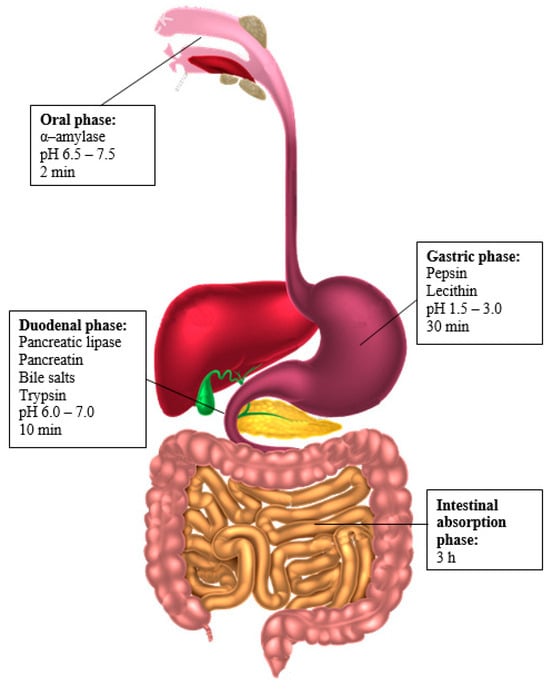
Figure 1.
Chemical–enzymatic conditions and residence times in the different stages of digestion [].
2. Experimental Section
2.1. Materials
Special fully hydrogenated triglycerides from rapeseed oil (Palsgaard® 6111) and a deodorized omega-3 concentrate (PronovaPure, 36.0% EPA and 24.0% DHA as triglyceride; BASF, Ludwigshafen, Germany) were used as gelling agent and lipid phase, respectively, for the preparation of each delivery system. A 85% curcumin extract from Solutex (Alcobendas, Spain) was used.
Sodium benzoate (Emerald Kalama Chemical, Kalama, WA, USA) and potassium sorbate (Serproquim Food, Sevilla, Spain) were used to prevent emulsions from becoming contaminated. All of the solutions were prepared using deionized water. Polyglycerol polyricinoleate (PGPR) (Danisco, Copenhagen, Denmark) and Tween 20 (Merck, Darmstadt, Germany) were used as hydrophobic and hydrophilic emulsifiers to stabilize W1/Og and Og/W2 interfaces, respectively. Xanthan gum (XG) was used as a thickening agent (HELM Iberica S.A., Madrid, Spain).
Absolute ethanol was used as solvent for curcumin concentration measurements. Other reagents, salts and enzymes used for in vitro simulation of the human gastrointestinal tract were hydrochloric acid, sodium hydroxide, calcium chloride dihydrate, magnesium chloride hexahydrate, sodium chloride, sodium dodecyl sulfate (SDS) and potassium dihydrogen phosphate, all purchased from PanReac AppliChem (Darmstadt, Germany); ammonium carbonate and potassium chloride from Merck; and sodium hydrogencarbonate, acquired from Sigma-Aldrich (Darmstadt, Germany). All of these were used to prepare the simulated salivary fluid (SSF) and the simulated gastric fluid (SGF) according to the composition and concentrations preestablished by Brodkorb et al. (2019) []. α-amylase from human saliva (300–1500 units/mg protein), lipase from porcine pancreas (100–500 units/mg protein), pancreatin from porcine pancreas (4 × USP specifications), pepsin from porcine gastric mucosa (250 units/mg protein) and trypsin from bovine pancreas (≥7500 BAEE units/mg solid) were all bought from Sigma-Aldrich. Finally, bile salts were purchased from Sigma-Aldrich and Lipoid P 45 (oil-free soy lecithin with 45% phosphatidylcholine) was supplied by Lipoid GmbH (Ludwigshafen am Rhein, Germany). All of the materials used were of analytical grade without any purification or modification of their properties.
2.2. Methods
2.2.1. Preparation of Oleogel
Oleogel was prepared as follows: the gelling agent and curcumin extract were premixed using a magnetic stirrer for 2 min at 1600 rpm []. The mixture was quickly heated to 66.5 °C to minimize curcumin and fish oil degradation by a bain-marie under mild agitation (400 rpm). Finally, the formation of the oleogel was completed after cooling up to 23 °C while stirring with a magnetic agitator at 1600 rpm for 3 min.
The oleogel composition used for testing and the preparation of all emulsions was the optimal one for minimizing lipid oxidation: curcumin concentration equal to 0.1500 wt.%, gelling agent concentration equal to 4.6483 wt.% and manufacturing temperature equal to 66.5 °C [].
2.2.2. Preparation of Oil Gelled-in-Water (Og/W) Simple Emulsion
Og/W emulsion was prepared at 25 °C and manufactured according to the procedure detailed by Vellido-Perez et al. (2021) []. In short, accurately weighed hydrophilic emulsifier was added to deionized water containing 0.06 wt.% sodium benzoate and 0.06 wt.% potassium sorbate to prevent the microbiological contamination of the emulsion and the resulting aqueous solution was energetically blended using a vortex for 30 s until completely dissolved. The oleogel described in Section 2.2.1. was then added to the previous aqueous solution and the Og/W emulsion was formed using a rotor–stator homogenizer (Ultra-Turrax® T 25, IKA, Staufen, Germany) for 5 min at 12,000 rpm. Finally, XG was added to the Og/W emulsion via soft homogenization at 3400 rpm for 3 min.
The Og/W emulsion composition used was 14.04 wt.% of dispersed phase, 5.00 wt.% of emulsifier, 0.49 wt.% of thickener and 12,000 rpm as homogenization speed (these values are the optimal ones for minimizing both average droplet size and oxidation processes of the oleogelified matrix) [].
2.2.3. Preparation of Water-in-Oil Gelled-in-Water (W1/Og/W2) Multiple Emulsion
The multiple emulsion was prepared according to Vellido-Perez et al. (2025) []. To this end, the corresponding W1/Og emulsion was created first. The dispersed phase of the W1/Og emulsion, W1, was carried out by adding the hydrophilic emulsifier and thickener to deionized water already containing 0.06 wt.% of sodium benzoate and potassium sorbate, and then strongly blending using a vortex for 30 s until completely dissolved. Additionally, the hydrophobic emulsifier was added to the oleogel detailed in Section 2.2.1. and the obtained oily solution (continuous phase of the W1/Og emulsion, Og) was also vigorously blended using a vortex for 30 s until completely dissolved. Finally, the aqueous solution described above was added to the previous oily solution and the W1/Og emulsion was formed using a rotor–stator homogenizer (Ultra-Turrax® T 25, IKA) for 5 min at 19,600 rpm. The W1/Og emulsion thus prepared was used in the dispersed phase of the final W1/Og/W2 emulsion.
Afterwards, accurately weighed hydrophilic emulsifier was added to deionized water containing 0.06 wt.% sodium benzoate and 0.06 wt.% potassium sorbate and the resulting aqueous solution (continuous phase of the W1/Og/W2 emulsion, W2) was energetically blended using a vortex for 30 s until completely dissolved. The W1/Og emulsion described above was then added to the previous aqueous solution, and the W1/Og/W2 emulsion was formed using a rotor–stator homogenizer (Ultra-Turrax® T 25, IKA) for 5 min at 6400 rpm. Finally, XG was added to the W1/Og/W2 emulsion via soft homogenization at 3400 rpm for 3 min.
The W1/Og/W2 emulsion composition used was 40 wt.% of W1/Og ratio in the primary emulsion, 0.31 wt.% of thickener in the external aqueous phase, 5.1 wt.% of oleogel which determines the dispersed phase fraction in the multiple emulsion, 8.1 wt.% of total emulsifier concentration in the primary emulsion, 89.8 wt.% of a mixture of hydrophobic emulsifier/hydrophilic emulsifier in the primary emulsion, 9.6 wt.% of hydrophilic emulsifier concentration in the multiple emulsion and a homogenization speed in the primary emulsion of 19,600 rpm and 6400 rpm for the multiple emulsion (these values are the optimal ones for minimizing both average droplet size and lipid oxidation) [].
2.2.4. In Vitro Bioaccessibility and Bioavailability Assays
The in vitro bioaccessibility and bioavailability assays were carried out in a dynamic in vitro simulator of the human gastrointestinal tract (SimuGIT) previously developed and validated [,].
Equipment Overview
The SimuGIT, shown schematically in Figure 2, consists of a continuous stirred-tank reactor (CSTR) and a continuous plug flow tubular reactor (PFR) equipped with a single-channel tubular ceramic membrane of a given cut-off size, both connected in series [,]. Each of its parts is described in detail below.
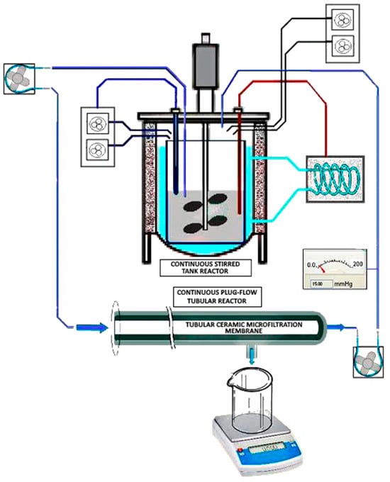
Figure 2.
Invitro gastrointestinal tract simulator system (SimuGIT) scheme.
The CSTR, where the gastric and duodenal simulations take place, consists of a Biostat B fermenter from the company Braun Biotech International GmbH (Kronberg, Germany) consisting of a control unit and a jacketed glass bioreactor of 2 L capacity. The control unit has an RS-422 interface that allows communication, control and supervision of the equipment from a desktop computer, as well as the recording of parameters and variables automatically for subsequent analysis. For its part, the reactor vessel has a turbine stirring system (two Rushton-type disc turbines with four blades each) powered by a 180 W servomotor that operates at a constant and adjustable speed (between 50 and 1200 rpm) to move the agitator shaft using an elastic coupling. Likewise, it has temperature, level, foam, dissolved oxygen and pH controls, all with their corresponding sensors, and includes several inputs for adding reagents and taking samples. It is equipped with a temperature stabilization system whose objective is to maintain the interior of the bioreactor in thermal conditions similar to those of the human body; for this purpose, it has a closed circulation system with a pump and a connection line to the reactor cooling/heating jacket, an electrical resistance and a cold-water circuit. The temperature is measured using a Pt-100 type digital sensor (Braun Biotech International GmbH, Kronberg, Germany) immersed inside the vessel in direct contact with the contents of the reactor, and its control is carried out using a PID regulation loop that ensures precise and constant temperature control (Tset point ± 0.1 °C). It is also provided with a pH regulation system containing a glass electrode (Hamilton, Reno, NV, USA, model Easyferm Plus K8) immersed inside the tank and in contact with the contents of the reactor as a sensing element, together with its own control system based on a module i-7011 data acquisition system that records the pH of the medium. The latter acts on a set of peristaltic pumps (Eyela, New York, NY, USA, model MP-3) that add acid (HCl, 1 M) or base (NaHCO3, 1 M) depending on the preset set point (pHset point ± 0.2), thus simulating the acid secretions of the gastric glands and the action of pancreatic juices.
To simulate the remaining small intestine (jejunum and ileum), where absorption processes primarily predominate, the contents of the CSTR are passed continuously and in a controlled manner through the interior of the PFR, a modular filtration system containing a single-channel tubular membrane of a determined cut-off size depending on the biological barrier to simulate. The integration of the tubular device and the CSTR into a single global simulation system requires the implementation of a set of chemically resistant polyethylene tubes and connections that make up the hydraulic supply and return circuits of the simulator.
The PFR to which we referred previously consists of a cylindrical stainless steel casing (Prozesstechnik GmbH, Basel, Switzerland) that contains the appropriate devices to collect the flows of permeate (bioavailable fraction) and retentate (non-bioavailable fraction or colonic residue). Inside, it houses the single-channel porous tubular ceramic membrane that acts as a filtering surface and allows simulation of diffusion through the intestinal epithelium. The membrane used in this research was a single-channel inorganic ultrafiltration membrane of TiO2 on an α–Al2O3 porous support provided by Atech Innovations GmbH (Gladbeck, Germany), model UF 50 N. The dimensions of the membrane were a length of 1000 mm, a duct diameter of 6 ± 0.5 mm and a thickness of 2 ± 0.5 mm, with an average pore diameter equal to 0.05 μm and a molecular cut-off size of 50 kDa. These types of membranes have a whole series of advantages: a high mechanical resistance and thermal stability (they withstand pressures of up to 10 bar and can be sterilized with steam at 121 °C), a high chemical resistance against highly corrosive chemicals and a stability against pH (from 0 to 14). They are also easy to wash and allow backwashing, exhibit high performance and selectivity and have a long service life.
To regulate the flow and pressure in the impulsion and return hydraulic circuits, we used our own control system which acts on a set of peristaltic pumps, in such a way that by varying the work cycle of these pumps that operate with constant rotational movement, it is possible to control the pressure within the hydraulic circuits and the filtration speed of the permeate. The operating pressure can also be precisely adjusted (Pset point ± 10 mmHg) using a spring-loaded pressure regulating valve (Swagelok, Solon, OH, USA, model SS–R4512MM–SP) and can be measured using a digital pressure gauge (Endress+Hauser, Reinach, Switzerland, model Ceraphant PTC31) located at the end where the retentate is collected. A precision electronic scale with USB connectivity (Sartorius, Gotinga, Germany, model Quintix 5102) continuously recorded permeate mass over time. Once the test is completed, the concentrated or retained product that remains in the CSTR, which acts as a reservoir, constitutes the colonic residue that would pass to the large intestine; however, this last phase of the simulation that would involve incorporating bacterial flora cultures was not included in this study.
Essay Overview
To compare the in vitro bioaccessibility and bioavailability of curcumin between the three delivery systems (oleogel vs. Og/W emulsion vs. W1/Og/W2 emulsion), all tests were carried out by supplying the same initial dose of curcumin: 1.527 mg (equivalent to 6.018 g of oleogel). This value is within the typical oral emulsion curcumin concentrations that vary depending on the specific formulation and desired effect but generally range from 0.0001–5% of the emulsion weight. In addition, this oleogel amount allowed us to provide 2.063 g of EPA and 1.375 g of DHA, which is approximately double the daily amount of EPA and DHA recommended by the International Society for the Study of Fatty Acids and Lipids (ISSFAL) for the general population []. The combination of ω-3 PUFAs and curcumin presents a polypharmacological approach, showcasing potential synergies for enhanced therapeutic impact. Therefore, the amount of sample—oleogel (6.018 g), Og/W emulsion (12.251 g) or W1/Og/W2 emulsion (25.000 g)—supplied in each test has been calculated based on the percentage of curcumin (or oleogel) which that delivery system contained, so that the same initial amount of curcumin was always fed to the simulator.
To perform each test, the simulator first needed to be brought to the physiological conditions of the human gastrointestinal system. To do this, a CSTR internal temperature of 37.5 ± 1.0 °C was established throughout the process [] and a stirring speed of 100 rpm was used to simulate the continuous peristaltic movements of the stomach walls []. Next, 800 mL of SGF was prepared according to the composition and concentrations preestablished by Brodkorb et al. (2019) [], and the pH was adjusted to 1.6 to simulate an empty stomach []. This SGF prepared in this way was introduced into the CSTR and allowed to heat up to the preset temperature; while this was happening, the simulation of the oral phase was performed simultaneously.
- Simulation of the oral phase
Firstly, the corresponding amount of food was weighed, which in our case was the delivery system to be studied (6.018 g of oleogel, 12.251 g of Og/W emulsion or 25.000 g of W1/Og/W2 emulsion). In parallel, 26.25 mL of SSF was prepared according to the composition and concentrations preestablished by Brodkorb et al. (2019) [], and the pH was adjusted to 7.0. Next, 17.5 mL of this SSF was taken and mixed with 1.25 mL of an α-amylase solution (75 U/mL), 125 μL of a CaCl2·2H2O solution (0.3 M) and 6.125 mL of distilled water, making a total volume of 25 mL. Finally, the already weighed food was added to the corresponding amount of this SSF (1:1, wt./wt.) and placed in a magnetic stirrer at 1200 rpm for 2 min to simulate chewing and mixing of the food with saliva. After the oral phase, 10 mL of sample was taken for subsequent analysis, and the rest went to the next phase.
- Simulation of the gastric phase
The simulation of the gastric phase began by adding the resulting mixture of the oral phase to the CSTR at 37.5 °C, along with 10 mL of a pepsin solution (2000 U/mL) and 50 mg of soy lecithin (Lipoid P 45) []. Next, the pH of the CSTR content was adjusted to 3.0 with the automatic and controlled addition of a 1 M NaHCO3 solution [] and the system was maintained under these conditions for 30 min. Once the gastric phase was completed, 10 mL of sample was taken for subsequent analysis, and the rest went to the next phase.
- Simulation of the duodenal phase
The simulation of the duodenal phase was carried out in the CSTR itself, now reproducing the chemical conditions of the duodenum (pancreatic juices and bile juices). To do this, the pH control system was activated to dose a 1 M NaHCO3 solution (at 4.5 mL/min) until reaching a pH in the reactor of 6.5 [], thus simulating the action of pancreatic juices when they neutralize gastric juices. Next, 10 mL of pancreatic lipase solution (2000 U/mL), 10 mL of pancreatin solution (10% wt./v), 1.63 g of bile salts (to achieve a final concentration of bile salts of 5 mM) and 1 mL of trypsin solution (50 mg/assay) [] were added, maintaining the system under these conditions for 10 min. Once the duodenal phase was completed, 10 mL of sample was taken for subsequent analysis, and the rest went to the next phase.
- Simulation of the intestinal absorption phase
To simulate the intestinal absorption phase, the contents of the CSTR were pumped through the modular filtration system described above. The molecular cut-off size of the membrane (50 kDa) was chosen based on our previous experience and the permeability of curcumin, which depends on its molecular weight (368.38 g/mol). The upper overpressure limit of the system was set at 150 ± 10 mmHg, so that the diffusion of curcumin through the membrane was only due to passive transport. Once the PFR and all the circuits (both impulsion and return) were primed, the intestinal absorption phase was considered to have begun, which lasted for 180 min. At 30, 60, 90, 120, 150 and 180 min, samples (10 mL) of permeate (fluid absorbed through the membrane) and retained (unabsorbed fluid that is part of the colonic residue) were taken for subsequent analysis.
In summary, each trial had a total duration of approximately 240 min: oral phase (2 min), gastric phase (30 min), duodenal phase (10 min) and, finally, intestinal absorption phase (180 min). In each experiment, our own developed equipment control software (BioDigestor v1) generated a data file with the record of all the processes and events that took place during the simulation. This file was conveniently processed to obtain the test information.
Description of the Membrane Cleaning Protocol
Once the test was completed, thorough and exhaustive cleaning of the simulator components was carried out. Due to its capital importance in the reproducibility and maintenance of the equipment, the cleaning of the membrane deserves special attention. To this end, a cleaning protocol was designed including water rinses and physical and chemical cleaning stages; all of them were carried out in laboratory-scale membrane equipment (Prozesstechnik GmbH). The protocol used is detailed below.
- Physical cleaning.
Once the test was completed, a first physical cleaning was carried out to eliminate substances weakly adhered to the membrane. This first cleaning consisted of washing with hot water (50 °C) at the minimum operating pressure offered by the equipment (2.5 bar), since higher pressures can induce compaction of the fouling on the membrane (increased resistance, clogging and blocking pores), making its removal more difficult. The duration of this first cleaning was 20 min.
- Chemical cleaning
Afterwards, a second cleaning was carried out, in this case chemical, to remove any traces of dirt that may have remained after the previous physical cleaning. To do this, chemical agents were introduced that reacted with the deposits, weakening the cohesive forces between them and between the deposits and the surface of the membrane. This second cleaning consisted of a wash at 50 °C and 2.5 bar pressure, combining two cleaning agents: NaOH (20 g/L) and SDS (2 g/L). The duration of this second wash was 30 min.
- Washes or rinses.
Finally, after the cleaning treatment was carried out, a series of washes were carried out to eliminate any possible residues that may have remained on the membrane. These washes or rinses consisted of 3 washes with hot water (50 °C), followed by a fourth with water at room temperature (25 °C) and a fifth with distilled water (25 °C), all lasting 15 min.
2.2.5. Curcumin Concentration Measurements
After each trial, the curcumin concentration was measured at the end of the oral, gastric and intestinal phases (colonic residue), as well as in permeate samples from the intestinal absorption phase obtained every 30 min for a total of 3 h via fluorescence spectroscopy (Cary Eclipse Fluorescence Spectrophotometer; Agilent Technologies, Santa Clara, CA, USA). To carry out the determination, first the fluorescence of the solvent used (ethanol) was measured and the fluorescence value zero was assigned. Next, the sample was diluted in ethanol to an appropriate concentration to be analyzed by a fluorescence spectrophotometer at excitation and emission wavelengths of 488 nm and 525 nm, respectively. Finally, the curcumin concentration was calculated from the emitted fluorescence using a calibration curve and taking into account the dilution factor.
To obtain the calibration curve, solutions of different concentrations of curcumin in ethanol (between 0 and 3 mg/L) were prepared, determining for each of them the fluorescence emitted (excitation wavelength 488 nm, emission wavelength 525 nm). These values were represented on the ordinate axis (Y axis) and the concentration values on the abscissa (X axis). Finally, the calibration curve (R2 = 99.93%) was used to calculate the concentration of curcumin in each sample. From these concentrations and the volumes involved in each phase, it was possible to calculate the amount of curcumin present in each stage of the digestion and, later, its bioaccessibility in the mouth, stomach and large intestine, and its bioavailability after the simulated in vitro digestion, according to Equations (1) and (2) (where m denotes mass). Accordingly, a macroscopic mass balance approach was always considered, applying the law of conservation of mass when tracking the curcumin intake and outputs (absorbed or as quantification sample).
2.2.6. Statistical Analysis
Statistical analysis was performed using Statgraphics Centurion XVI, (Statgraphics Technologies Inc., Fauquier County, VA, USA). Values are expressed as mean ± standard deviation (SD). Differences between groups were assessed by unpaired Student’s t-tests. p-values ≤ 0.05 were considered statistically significant.
3. Results and Discussion
Figure 3 comparatively represents the bioaccessibility percentages of vectorized curcumin among the three different delivery systems after the oral, gastric and intestinal phases (colonic residue). As can be seen, these percentages were nearly of the same order of magnitude for both emulsified systems (Og/W emulsion and W1/Og/W2 emulsion) and significantly different between them and the pure oleogel system.
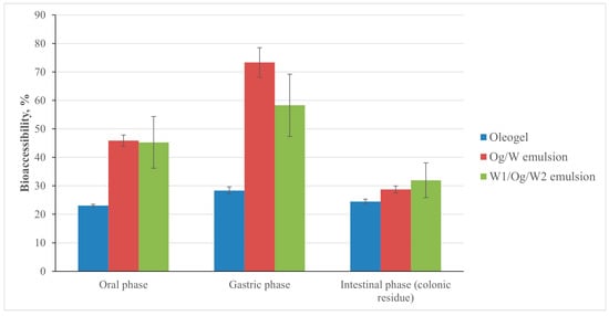
Figure 3.
Bioaccesibility of vectorized curcumin for each delivery system after the simulated oral, gastric and intestinal (colonic residue) phases (bars represent standard deviation of the mean).
The bioaccessibility percentage of the pure oleogel after the oral and gastric phases, e.g., curcumin delivered in the salivary or gastric media, was the lowest of the three delivery systems studied (23.1 ± 0.4% and 28.4 ± 1.3%, respectively). This means that curcumin was efficiently embedded inside the three-dimensional network created by the self-assembled structuring agent, a network that resisted the salivary and gastric conditions. However, the curcumin incorporated into the Og/W emulsion was the most bioaccessible in both phases, being twice as bioaccessible as that vectorized in the pure oleogel in the oral phase and 2.5 times more bioaccessible in the gastric phase (p < 0.05). Because of the softer structure of the Og/W emulsion, the gel particles in the emulsion were broken down much faster and the oil droplets were liberated from the matrix more readily than for the pure oleogel system during both phases. The Tween 20 surfactant molecules also helped this process, since they could be displaced from the emulsion interface and would participate in the formation of mixed micelles that would help to solubilize the released curcumin molecules []. Finally, the curcumin encapsulated in the W1/Og/W2 emulsion showed a bioaccessibility percentage very similar to that of the Og/W emulsion in the oral phase (45.3 ± 9.1% vs. 45.9 ± 2.0%) but moderately lower than the Og/W emulsion in the gastric phase (58.3 ± 10.9% vs. 73.3 ± 5.2%) (p < 0.05). This different behavior was probably due to the multi-compartmentalized structure of the multiple emulsion and the viscosity increase due to swelling. Somes authors have hinted at PGPR playing an important role in water transport between the two water phases in double emulsions [], inducing structural changes in the oil layer at the interphase that strongly facilitated the spontaneous formation of tiny water droplets in the oil phase, a process that led to swelling and thus higher stability after the gastric phase as compared to the simple Og/W emulsion.
Besides that, it is also evident that, regardless of the delivery system used, the percentage of bioaccessible curcumin increased from the oral phase to the gastric phase by a percentage between 22.8% in the case of the pure oleogel and 59.9% in the case of the Og/W emulsion. This increase in the percentage of bioaccessible curcumin when passing from the oral phase to the gastric phase reveals its stability against the chemical–enzymatic conditions of the stomach, that is, against the action of proteases (pepsin) and acidic conditions (pH 1.5–3.0). It is well known that at acidic pHs, curcumin is relatively stable thanks to the diene that is formed, although it is poorly soluble in water; at neutral and basic pHs, the formation of the phenyl anion causes curcumin to become unstable and rapidly decompose into compounds such as trans-6-(4′-hydroxy-3′-methoxyphenyl)-2,4-dioxo-5-hexenal, ferulic acid, feruloylmethane and vanillin []. This was confirmed by Wang et al. (1997) [], who studied the kinetics of curcumin degradation with pH as well as its stability under physiological conditions in vitro. These authors observed the decomposition of 90% of the curcumin after 30 min at pH 7.2 following an apparent first-order kinetics. Therefore, the chemical instability of curcumin under physiological conditions is more than proven: although it is stable under stomach conditions (pH 1.0–2.0), it is no longer so in the small intestine (pH 6.5), blood (pH 7.35–7.45) or inside cells (pH 7.2) [].
However, the percentage of bioaccessible curcumin after the small intestine (colonic residue) was reasonably acceptable for the three studied systems and very similar between them, varying between 24.5 ± 0.8% of the pure oleogel and 32.0 ± 6.1% of the W1/Og/W2 emulsion (p < 0.05), although obviously lower (between 1.8 and 2.6 times) than in the gastric phase as explained above. Apart from the innate instability of curcumin at above neutral pH values, the swelling process previously described in the multiple emulsion would be reverted after the incorporation of the pancreatic and bile juices during the intestinal phase that changed the osmotic pressure difference between the inner and outer phases and made it more bioaccessible for the transported curcumin.
Another of the main factors that determines the bioaccessibility of bioactive compounds is their solubility/dispersibility in aqueous media which, in the case of curcumin, is quite limited both at acidic and neutral pHs (11 ng/mL) []; in aqueous systems at a basic pH, the phenol group of curcumin donates its hydrogen, forming the phenolate ion, which allows curcumin to be slightly soluble under these conditions [,]. Thus, for the curcumin molecule to be bioaccessible, it must be dispersed and solubilized in the intestinal fluids, which are aqueous in nature, and to do so it must be in intimate contact with them. In the case of the pure oleogel system, its low bioaccessibility responds to the system’s own lipophilic nature, which means that the degree of mixing with intestinal fluids is not adequate, and to the physical trapping of curcumin within the three-dimensional and semi-solid structure of the oleogel, which limits its mobility within the matrix. However, in the case of the Og/W and W1/Og/W2 emulsified systems, the aqueous nature of the continuous phase, the enormous contact surface between the oleogel and water and the presence of hydrophilic emulsifier capable of forming colloidal structures (normally, micelles) favors the solubilization of curcumin in intestinal fluids and, therefore, the increase in its bioaccessibility [,]. Based on the above, the higher percentage of bioaccessibility of the curcumin vectorized in the Og/W emulsion could also find its explanation in the average droplet size, which for this type of emulsion was 8.424 µm (dmode = 3.248 µm), while for the W1/Og/W2 emulsion it was 27.521 µm (dmode = 4.826 µm). This difference in the average droplet size and the most frequent droplet size (dmode) of the Og/W and W1/Og/W2 emulsions is justified by their respective homogenization speeds, which is the independent variable that most significantly influences the average droplet size of an Og/W emulsion []; in fact, the Og/W emulsion was homogenized at 12,000 rpm while the W1/Og/W2 emulsion was homogenized at 6400 rpm to avoid, as much as possible, the shear forces from breaking the primary W1/Og emulsion that forms the dispersed phase of the multiple. Therefore, the lower homogenization speed inevitably influences the droplet size. On the other hand, it is worth remembering that, in a multiple emulsion, each drop of dispersed phase contains, in turn, smaller, equally dispersed droplets that need larger drops to contain them and, in addition, since there is an emulsion within another, the breakage mechanisms multiply and innumerable core-rupture processes may appear []. Thus, a smaller average droplet size implies a larger contact surface between the oleogel and water which, as mentioned, favors the solubilization of curcumin. This same effect was already observed by Pinheiro et al. (2013) [], who reported a significant increase (almost 10-fold) in the percentage of bioaccessibility of curcumin incorporated in nanoemulsions stabilized with Tween 20 with respect to the percentage of bioaccessibility in nanoemulsions stabilized with dodecyltrimethylammonium bromide (DTAB). This significantly higher bioaccessibility percentage in Tween 20-stabilized nanoemulsions was associated with the smaller average droplet size (between 100 and 310 nm) compared to the average droplet size of DTAB-stabilized nanoemulsions (between 80 and 890 nm).
On the other hand, Figure 4 comparatively represents the curcumin bioavailability percentages at different times (between 30 and 180 min) for each delivery system during the intestinal absorption phase. Based on that, we can observe how the bioavailability percentage of the curcumin vectorized in the pure oleogel matrix was the lowest regardless of the time considered, although it is true that said percentage increased from 2.6 ± 0.4% (30 min) up to 11.9 ± 1.8% (180 min). Additionally, it can be seen how the absorption rate of curcumin was somewhat slower especially during the first hour. However, the encapsulated curcumin in the Og/W and W1/Og/W2 emulsified systems showed very similar bioavailability percentages at the end of the intestinal phase (180 min, around 20.2 ± 2.5%) and much higher percentages than those of the oleogel (1.7 times higher) (p < 0.05). Comparing both emulsified systems during the first 90 min of the intestinal absorption phase (early absorption phase), the W1/Og/W2 emulsion reached slightly higher percentages of bioavailable curcumin (between 1.1 and 1.3 percentage points) than those of the Og/W emulsion; however, between 120 and 180 min (late absorption), the bioavailability percentages were very similar. This fact means that the curcumin encapsulated in the multiple emulsion was absorbed more quickly than the curcumin vectorized in the simple emulsion, whose greater absorption occurs after a longer amount of time. This could be related to the greater or lesser physical stability of both delivery systems under physiological conditions and the fact that, as already described, a hetero-dispersed structure has many more breakdown mechanisms (swelling, shrinkage, etc.) than a simple emulsion. Thus, as previously mentioned, the composition of the intestinal fluids (remember that intestinal fluids are composed of a large amount of electrolytes) contributes to increasing the osmotic pressure of the external aqueous phase of the multiple emulsion, giving rise to an imbalance between the osmotic pressures of the internal and external aqueous phases and, consequently, accelerating the physical destabilization of the W1/Og/W2 emulsion and the release of the dispersed phase contained therein with respect to the Og/W emulsion. This fact explains the difference observed between the percentage of bioavailability of curcumin vectorized in both emulsions over time. In any case, it can be seen that the final bioavailability values reached (11.9 ± 1.8% for the pure oleogelified system and 20.2 ± 2.5% for the emulsified ones, p < 0.05) were always higher than the 7.8% (greater by a factor of 2.5) reported in the literature for free curcumin dissolved in corn oil using a different simulated gastrointestinal model [].
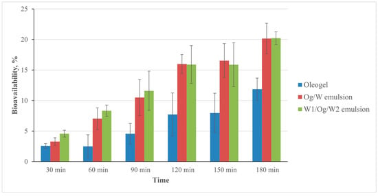
Figure 4.
Bioavailability of vectorized curcumin at different times for each delivery system during the simulated intestinal absorption phase (bars represent standard deviation of the mean).
Finally, Figure 5 comparatively represents the ratio between the amount of bioavailable and bioaccessible curcumin after the digestion stage during the intestinal absorption phase for each of the delivery systems at different times (between 30 and 180 min). This figure represents the efficiency of each delivery system studied to convert the bioaccesible curcumin (potentially bioavailable curcumin) into real and detectable bioavailable curcumin, coming within reach of its intended biological destination.
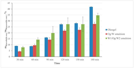
Figure 5.
Ratio bioavailable vs. bioaccessible curcumin after the digestion stage at different times during the simulated intestinal absorption phase (bars represent standard deviation of the mean).
According to Figure 5, the oleogelified system has turned out to be the most efficient delivery system regarding the bioaccesible/bioavailable ratio, with 41.8 ± 0.3% of the bioaccessible curcumin (released after the digestion stage) being bioavailable at 180 min. However, it is important to remind ourselves that the bioaccessibility percentage of the pure oleogel delivered into the simulated gastric media was the lowest of the three delivery systems studied, which makes it less interesting in absolute numbers. The continuous increase of this ratio with time (from 30 to 180 min) was due to the progressive collapse of the oleogel matrix containing the lipid-soluble gelator; this is a common structure in the three carriers that initially hindered the action of lipases and micelle formation and that was breaking down along the simulated intestinal phase.
With regard to the W1/Og/W2 emulsion, its efficiency (34.7 ± 1.8%) followed that of the oleogel, although at some times (60, 90 and 120 min) it was even higher, most probably due to its higher protection against chemical degradation by virtue of its multicompartmental structure. Finally, the Og/W emulsion turned out to be the least efficient concerning the bioavailable/bioaccesible conversion regardless of the time considered (27.5 ± 1.5% at 180 min) (p < 0.05). It should be pointed out that the softer structure of the Og/W emulsion, as explained above, helped to solubilize the released curcumin molecules that could be exposed to progressive degradation processes along the intestinal phase.
4. Conclusions
When studying the therapeutical potential of an organic compound such us curcumin, the determination of its bioaccessibility and bioavailability with time seems of utmost importance as indicators of its bioactivity evolution under the influence of the subsequent digestion stages. In summary, the pure oleogelified matrix and both the simple and multiple oleogelified emulsions (Og/W and W1/Og/W2) could effectively enhance the dynamic bioavailability of curcumin by a factor of up to 2.5. After the oral and gastric phases, the bioaccessibility percentage of the pure oleogel was the lowest of the three delivery systems (23.1 ± 0.4% and 28.4 ± 1.3%, respectively), whereas the emulsified carriers, Og/W and W1/Og/W2, were approximately twice as bioaccessible as that vectorized in the pure oleogel in the oral phase and between 2–2.5 times more bioaccessible in the gastric phase (58.3 ± 10.9% and 73.3 ± 5.2%, respectively) (p < 0.05). With respect to curcumin bioavailability, the Og/W and W1/Og/W2 systems showed values at the end of the simulated gastrointestinal tract of around 20.2 ± 2.5%, 1.7 times higher on average than that of the pure oleogel matrix (p < 0.05). The curcumin transported into the W1/Og/W2 emulsion was absorbed faster than the curcumin vectorized in the Og/W emulsion; therefore, the former is presented as a rapid-release curcumin encapsulation system, releasing most of the active ingredient in the first 90 min of the intestinal simulated phase, while the latter is proposed as a longer-term release curcumin encapsulation system (90 to 180 min). Additionally, although the curcumin incorporated in the pure oleogel is the least bioaccessible, this oleogelified system is the most efficient of all at protecting it against potential degradation reactions once released, with 41.8 ± 0.3% of the bioaccessible curcumin being bioavailable after 180 min. However, in vivo assessments need to be conducted to further confirm the in vitro–in vivo correlations. Overall, the results from this study could provide valuable information that can be used to design oleogelified delivery systems for controlled release of curcumin.
Author Contributions
Conceptualization, J.A.V.-P. and A.M.-F.; data curation, A.M.-F.; formal analysis, J.A.V.-P. and A.M.-F.; funding acquisition, A.M.-F.; investigation, J.A.V.-P.; methodology, J.A.V.-P. and A.M.-F.; project administration, A.M.-F.; resources, A.M.-F.; supervision, A.M.-F.; validation, J.A.V.-P. and A.M.-F.; visualization, J.A.V.-P. and A.M.-F.; writing—original draft, J.A.V.-P.; writing—review and editing, A.M.-F. All authors have read and agreed to the published version of the manuscript.
Funding
This research was funded by the Spanish Ministry of Education, Culture and Sport, through the grant FPU17/03005 of Vellido-Perez, and the University of Granada through the program “Precompetitive Research Projects for Young Researchers 2018–Modality B” with the project entitled “Design and development of stable oleogelified structures for vehiculization and protection of curcumin” IB2018-24).
Data Availability Statement
The data are available upon request from the authors.
Acknowledgments
This study was supported by the TEP025 “Technologies for Chemical and Biochemical Processes” Research Group from the Chemical Engineering Department of the University of Granada.
Conflicts of Interest
The authors declare that there are no conflicts of interest.
References
- Diplock, A.T.; Aggett, P.J.; Ashwell, M.; Bornet, F.; Fern, E.B.; Roberfroid, M.B. Scientific Concepts of Functional Foods in Europe. Consensus Document. Br. J. Nutr. 1999, 81, S1–S27. [Google Scholar] [CrossRef]
- Gil Hernández, Á.; Sánchez de Medina Contreras, F.; Ruiz López, M.; Camarero González, E.; Álvarez Hernández, J. Tratado de Nutrición, 1st ed.; Acción Médica: Madrid, Spain, 2005; ISBN 978-84-88336-40-8. [Google Scholar]
- Granato, D.; Barba, F.J.; Bursać Kovačević, D.; Lorenzo, J.M.; Cruz, A.G.; Putnik, P. Functional Foods: Product Development, Technological Trends, Efficacy Testing, and Safety. Annu. Rev. Food Sci. Technol. 2020, 11, 93–118. [Google Scholar] [CrossRef] [PubMed]
- Anand, P.; Kunnumakkara, A.B.; Newman, R.A.; Aggarwal, B.B. Bioavailability of Curcumin: Problems and Promises. Mol. Pharm. 2007, 4, 807–818. [Google Scholar] [CrossRef] [PubMed]
- Dulbecco, P.; Savarino, V. Therapeutic Potential of Curcumin in Digestive Diseases. World J. Gastroenterol. 2013, 19, 9256–9270. [Google Scholar] [CrossRef] [PubMed]
- Epstein, J.; Sanderson, I.R.; MacDonald, T.T. Curcumin as a Therapeutic Agent: The Evidence from In Vitro, Animal and Human Studies. Br. J. Nutr. 2010, 103, 1545–1557. [Google Scholar] [CrossRef]
- Grynkiewicz, G.; Ślifirski, P. Curcumin and Curcuminoids in Quest for Medicinal Status. Acta Biochim. Pol. 2012, 59, 201–212. [Google Scholar] [CrossRef]
- Maheshwari, R.K.; Singh, A.K.; Gaddipati, J.; Srimal, R.C. Multiple Biological Activities of Curcumin: A Short Review. Life Sci. 2006, 78, 2081–2087. [Google Scholar] [CrossRef]
- Ravindranath, V.; Chandrasekhara, N. Absorption and Tissue Distribution of Curcumin in Rats. Toxicology 1982, 22, 337. [Google Scholar] [CrossRef]
- Holder, G.M.; Plummer, J.L.; Ryan, A.J. The Metabolism and Excretion of Curcumin (1,7-Bis-(4-Hydroxy-3-Methoxyphenyl)-1,6-Heptadiene-3,5-Dione) in the Rat. Xenobiotica Fate Foreign Compd. Biol. Syst. 1978, 8, 761–768. [Google Scholar] [CrossRef]
- Prasad, S.; Tyagi, A.K.; Aggarwal, B.B. Recent Developments in Delivery, Bioavailability, Absorption and Metabolism of Curcumin: The Golden Pigment from Golden Spice. Cancer Res. Treat. Off. J. Korean Cancer Assoc. 2014, 46, 2–18. [Google Scholar] [CrossRef]
- Pan, M.-H.; Huang, T.-M.; Lin, J.-K. Biotransformation of Curcumin through Reduction and Glucuronidation in Mice. Drug Metab. Dispos. 1999, 27, 486–494. [Google Scholar] [CrossRef]
- Ireson, C.; Orr, S.; Jones, D.J.; Verschoyle, R.; Lim, C.K.; Luo, J.L.; Howells, L.; Plummer, S.; Jukes, R.; Williams, M.; et al. Characterization of Metabolites of the Chemopreventive Agent Curcumin in Human and Rat Hepatocytes and in the Rat in Vivo, and Evaluation of Their Ability to Inhibit Phorbol Ester-Induced Prostaglandin E2 Production. Cancer Res. 2001, 61, 1058–1064. [Google Scholar]
- Pal, A.; Sung, B.; Bhanu Prasad, B.A.; Schuber, P.T.; Prasad, S.; Aggarwal, B.B.; Bornmann, W.G. Curcumin Glucuronides: Assessing the Proliferative Activity Against Human Cell Lines. Bioorg. Med. Chem. 2014, 22, 435–439. [Google Scholar] [CrossRef] [PubMed]
- Sharma, R.A.; McLelland, H.R.; Hill, K.A.; Ireson, C.R.; Euden, S.A.; Manson, M.M.; Pirmohamed, M.; Marnett, L.J.; Gescher, A.J.; Steward, W.P. Pharmacodynamic and Pharmacokinetic Study of Oral Curcuma Extract in Patients with Colorectal Cancer. Clin. Cancer Res. Off. J. Am. Assoc. Cancer Res. 2001, 7, 1894–1900. [Google Scholar]
- Wahlström, B.; Blennow, G. A Study on the Fate of Curcumin in the Rat. Acta Pharmacol. Toxicol. 1978, 43, 86–92. [Google Scholar] [CrossRef]
- Aggett, P.J. Population Reference Intakes and Micronutrient Bioavailability: A European Perspective. Am. J. Clin. Nutr. 2010, 91, 1433S–1437S. [Google Scholar] [CrossRef]
- Hurrell, R.; Egli, I. Iron Bioavailability and Dietary Reference Values. Am. J. Clin. Nutr. 2010, 91, 1461S–1467S. [Google Scholar] [CrossRef]
- Hur, S.J.; Lim, B.O.; Decker, E.A.; McClements, D.J. In Vitro Human Digestion Models for Food Applications. Food Chem. 2011, 125, 1–12. [Google Scholar] [CrossRef]
- Holst, B.; Williamson, G. Nutrients and Phytochemicals: From Bioavailability to Bioefficacy Beyond Antioxidants. Curr. Opin. Biotechnol. 2008, 19, 73–82. [Google Scholar] [CrossRef]
- Paixão Teixeira, J.L.; Lima Pallone, J.A.; Seiquer, I.; Morales-González, J.A.; Vellido-Pérez, J.A.; Martinez-Ferez, A. In Vitro Digestion Assays Using Dynamic Models for Essential Minerals in Brazilian Goat Cheeses. Food Anal. Methods 2022, 15, 2879–2889. [Google Scholar] [CrossRef]
- Rivas-Montoya, E.; Ochando-Pulido, J.M.; López-Romero, J.M.; Martinez-Ferez, A. Application of a novel gastrointestinal tract simulator system based on a membrane bioreactor (SimuGIT®) to study the stomach tolerance and effective delivery enhancement of nanoencapsulated macelignan. Chem. Eng. Sci. 2016, 140, 104–113. [Google Scholar] [CrossRef]
- Vellido Pérez, J.A. Diseño, Desarrollo y Optimización de Diferentes Sistemas de Liberación Modificada Para la Protección y Vehiculización de Ácidos Grasos Poliinsaturados Omega-3 y Curcumina. Ph.D. Thesis, University of Granada, Granada, Spain, 2021. [Google Scholar]
- Brodkorb, A.; Egger, L.; Alminger, M.; Alvito, P.; Assunção, R.; Ballance, S.; Bohn, T.; Bourlieu-Lacanal, C.; Boutrou, R.; Carrière, F.; et al. INFOGEST Static In Vitro Simulation of Gastrointestinal Food Digestion. Nat. Protoc. 2019, 14, 991–1014. [Google Scholar] [CrossRef]
- Vellido-Pérez, J.A.; Rodríguez-Remacho, C.; Rodríguez-Rodríguez, J.; Ochando-Pulido, J.M.; la Fuente, E.B.-D.; Martínez-Férez, A. Optimization of Oleogel Formulation for Curcumin Vehiculization and Lipid Oxidation Stability by Multi-Response Surface Methodology. Chem. Eng. Trans. 2019, 75, 427–432. [Google Scholar] [CrossRef]
- Vellido-Perez, J.A.; Ochando-Pulido, J.M.; Brito-de la Fuente, E.; Martinez-Ferez, A. Effect of Operating Parameters on the Physical and Chemical Stability of an Oil Gelled-in-Water Emulsified Curcumin Delivery System. J. Sci. Food Agric. 2021, 101, 6395–6406. [Google Scholar] [CrossRef]
- Vellido-Perez, J.A.; Ochando-Pulido, J.M.; Martinez-Ferez, A. Enhancing Physical and Chemical Stability of a Water-in-Oil Gelled-in-Water Multiple Emulsion Containing Omega-3 Polyunsaturated Fatty Acids and Curcumin by Draper-Lin Composite Design. LWT 2025, 215, 117265. [Google Scholar] [CrossRef]
- Godoy, V.; Martínez-Férez, A.; Martín-Lara, M.A.; Vellido-Pérez, J.A.; Calero, M.; Blázquez, G. Microplastics as Vectors of Chromium and Lead During Dynamic Simulation of the Human Gastrointestinal Tract. Sustainability 2020, 12, 4792. [Google Scholar] [CrossRef]
- Carrero, J.J.; Martín-Bautista, E.; Baró, L.; Fonollá, J.; Jiménez, J.; Boza, J.J.; López-Huertas, E. Efectos Cardiovasculares de Los Ácidos Grasos Omega-3 y Alternativas Para Incrementar Su Ingesta. Nutr. Hosp. 2005, 20, 63–69. [Google Scholar]
- Abad, P.; Arroyo-Manzanares, N.; Rivas-Montoya, E.; Ochando-Pulido, J.m.; Guillamon, E.; García-Campaña, A.M.; Martinez-Ferez, A. Effects of Different Vehiculization Strategies for the Allium Derivative Propyl Propane Thiosulfonate During Dynamic Simulation of the Pig Gastrointestinal Tract. Can. J. Anim. Sci. 2018, 99, 244–253. [Google Scholar] [CrossRef]
- Luo, N.; Ye, A.; Wolber, F.M.; Singh, H. Digestion behaviour of capsaicinoid-loaded emulsion gels and bioaccessibility of capsaicinoids: Effect of emulsifier type. Curr. Res. Food Sci. 2023, 6, 100473. [Google Scholar] [CrossRef]
- Eisinaite, V.; Duque Estrada, P.; Schroën, K.; Berton-Carabin, C.; Leskauskaite, D. Tayloring W/O/W emulsion composition for effective encapsulation: The role of PGPR in water transfer-induced swelling. Food Res. Int. 2018, 106, 722–728. [Google Scholar] [CrossRef]
- Lestari, M.L.A.D.; Indrayanto, G. Profiles of Drug Substances, Excipients and Related Methodology. Curcumin 2014, 39, 113–204. [Google Scholar] [CrossRef]
- Wang, Y.-J.; Pan, M.-H.; Cheng, A.-L.; Lin, L.-I.; Ho, Y.-S.; Hsieh, C.-Y.; Lin, J.-K. Stability of Curcumin in Buffer Solutions and Characterization of Its Degradation Products. J. Pharm. Biomed. Anal. 1997, 15, 1867–1876. [Google Scholar] [CrossRef]
- Tønnesen, H.H.; Karlsen, J. Studies on Curcumin and Curcuminoids—VI. Kinetics of Curcumin Degradation in Aqueous Solution. Z. Für Lebensm. Unters. Forsch. 1985, 180, 402–404. [Google Scholar] [CrossRef]
- Zhang, R.; McClements, D.J. Enhancing Nutraceutical Bioavailability by Controlling the Composition and Structure of Gastrointestinal Contents: Emulsion-Based Delivery and Excipient Systems. Food Struct. 2016, 10, 21–36. [Google Scholar] [CrossRef]
- Jagannathan, R.; Abraham, P.M.; Poddar, P. Temperature-Dependent Spectroscopic Evidences of Curcumin in Aqueous Medium: A Mechanistic Study of Its Solubility and Stability. J. Phys. Chem. B 2012, 116, 14533–14540. [Google Scholar] [CrossRef]
- Araiza-Calahorra, A.; Akhtar, M.; Sarkar, A. Recent Advances in Emulsion-Based Delivery Approaches for Curcumin: From Encapsulation to Bioaccessibility. Trends Food Sci. Technol. 2018, 71, 155–169. [Google Scholar] [CrossRef]
- Yao, M.; Xiao, H.; McClements, D.J. Delivery of Lipophilic Bioactives: Assembly, Disassembly, and Reassembly of Lipid Nanoparticles. Annu. Rev. Food Sci. Technol. 2014, 5, 53–81. [Google Scholar] [CrossRef]
- Jiao, J.; Rhodes, D.G.; Burgess, D.J. Multiple Emulsion Stability: Pressure Balance and Interfacial Film Strength. J. Colloid Interface Sci. 2002, 250, 444–450. [Google Scholar] [CrossRef]
- Pinheiro, A.C.; Lad, M.; Silva, H.D.; Coimbra, M.A.; Boland, M.; Vicente, A.A. Unravelling the Behaviour of Curcumin Nanoemulsions During In Vitro Digestion: Effect of the Surface Charge. Soft Matter 2013, 9, 3147–3154. [Google Scholar] [CrossRef]
- Lu, X.; Zhu, J.; Pana, Y.; Huang, Q. Assessment of dynamic bioaccessibility of curcumin encapsulated in milled starch particle stabilized Pickering emulsions using TNO’s gastrointestinal model. Food Funct. 2019, 10, 2583–2594. [Google Scholar] [CrossRef]
Disclaimer/Publisher’s Note: The statements, opinions and data contained in all publications are solely those of the individual author(s) and contributor(s) and not of MDPI and/or the editor(s). MDPI and/or the editor(s) disclaim responsibility for any injury to people or property resulting from any ideas, methods, instructions or products referred to in the content. |
© 2025 by the authors. Licensee MDPI, Basel, Switzerland. This article is an open access article distributed under the terms and conditions of the Creative Commons Attribution (CC BY) license (https://creativecommons.org/licenses/by/4.0/).