Abstract
The objective of this retrospective study was to compare the dento-alveolar effects of two different expansion protocols, Invisalign First (IF) and Leaf Expander (LE), in patients in mixed dentition with transversal upper maxillary deficiency. Materials and Methods: 30 patients were treated with IF, whereas 38 patients were treated with LE. For each sample 3D digital cast models were analyzed pre and post expansion and transversal diameter of the upper arch, molar rotation and inclination and arch perimeter were measured. Results: LE resulted in a more significant expansion of the molar width and the arch perimeter, with less effect on the expansion of deciduous canines and deciduous molars. IF allowed a more effective molar derotation, but with a greater buccal tipping movement than LE, which determines a more bodily movement of the molars: the expansion determined by IF seems to be more dental than skeletal. Conclusions: IF is a good alternative to LE in case of limited transversal maxillary contraction, particularly when there is a significant mesio-rotation of the first upper molars.
1. Introduction
The upper maxilla’s transversal discrepancy is considered one of the most common cranio-facial deformations dentists have to treat during their clinical practice [1], and its incidence has been reported to be 8–22% [2].
The treatment of this kind of discrepancy must undergo an accurate diagnosis which consists in an attentive analysis of medical history, intra and extra oral photos, cast models, bidimensional radiological exams such as OPG and Tele X-rays and, in some cases, even three-dimensional radiographies like CBCT [3].
During the last century, new devices, diagnostic instruments and operative protocols were developed in order to enlarge the upper arch perimeter starting from Biederman’s Hyrax RPE in 1968 [3] to Ricketts Quad Helix SPE of 1975 [3], to Lee’s and Suzuki’s Miniscrew assisted rapid palatal expansion (MARPE) [4,5] and eventually to the surgically assisted rapid palatal expansion (SARPE) [6].
Rapid Maxillary Expansion (RME) is the most consolidated technique used to correct maxillary deficiency: it determines an opening of the mid-palatine suture in growing patients using heavy and intermittent forces for a short time, and it is reached through teeth or tissues anchored devices [7].
On the contrary there are different appliances that determine a Slow Maxillary Expansion (SME), using lower and continuous forces applied for a longer period than RME [8].
The RME protocol consists in two activations a day, which correspond to 0.50 mm of transversal expansion a day. The patient must be monitored weekly, and the expansion must continue until an over-expansion is obtained: usually an edge-to-edge contact between the palatal cusps of the upper first molars and the vestibular cusps of the lower fist molars is expected [9].
Three months retention are also necessary for the bone to rearrange in the mid-palatal suture space. The aim of these kind of appliances is to expand the palatal suture the quickest way possible applying intense orthopedic forces (up to 9–10 kilos per half-palate) and to give more skeletal than dental expansion. This clinical approach is nowadays still well accepted and applied [9,10,11].
Nevertheless, it has been proven that RME has numerous side effects such as pain, relapse, molar uncontrolled buccal inclination, bone loss, gingival recession, ulcerations, necrosis of the palatal mucosa, root absorption, bite opening tendency, TMJ’s microtrauma [12,13].
By contrast, SME uses decidedly lighter forces (around 450 gr up to 900 gr) resulting in less discomfort for the patient, less resistance of the skeletal structures and better bone formation within the median palatal suture [14]. The activations should be done twice a week, the end of the activations is reached around the 10th–12th week. The perceived pain is less than in the RME devices [13,15].
In 2013 a new device for palatal expansion was designed: the Leaf Expander [16]. This device allows the maxilla’s expansion thanks to the application of slight and continues forces with specific intensity and direction, which cause a dento-alveolar remodeling [17].
The structure of the Leaf Expander resembles a common RME or SME, but it differs in the active components’ characteristics and the operating instructions. In fact, the screw doesn’t directly act on the supporting teeth, but it determines a measured expansion of the upper jaw by compressing a NiTi leaf-like coil [16].
The new device indeed encompasses several characteristics that can be deemed optimal for a fixed orthodontic expansion appliance, such as: a remarkably reduced number of intraoral reactivation sessions; easily executable activations; absence of pain even in the initial phases of expansion; controlled bodily vestibularization of middle and posterior teeth; high control over the progression of movement; impossibility of device deactivation due to occlusal forces; development of light, predetermined, and continuous forces; possibility to precisely graduate the extent of movement; absence of risk of overexpansion [18].
From a purely bio-mechanical point of view the Leaf Expander effects on the overall expansion of the palate has no statistically significant difference with a RME or traditional SME devices [17,19].
There are at present four types of Leaf Expander available on the market [16], each with different expansion amounts and different types of force generated.
The most common structural characteristics of the Leaf Expander involve the use of two bands typically positioned on second deciduous molars or first permanent molars, with possible variations and adjustments required by specific clinical situations [16].
When the canines are present in the arch, two well shaped extensions are made in order two guarantee a satisfying expansion effect on the anterior segment of the maxilla and a greater stability of the device [15].
In the last decades, an increasing number of adults patients are seeking orthodontic solutions that are more comfortable and more aesthetic than conventional fixed appliances: the term Clear Aligner Therapy (CAT) indicates a wide range of devices used to treat different malocclusions with different methods of construction and modes of action [20].
The aligners are realized using clear thermoformed plastic, and they cover many or all teeth, but each different system of aligners has some significant differences in treating the malocclusions for which they are designed [20,21].
At the beginning, CAT was limited to simple orthodontics cases with little irregularities of tooth position only, then gradually systems were developed that were also suitable for solving complex malocclusion [21].
Frequently, bonded resin attachments on teeth are necessary to realize orthodontic movements otherwise impossible to achieve with only CAT systems [22]. Additionally, auxiliary elements can be added as needed, such as elastic hooks and bite ramps. The aligners are changed weekly and allow better oral hygiene, becoming usable also by patients aged from 6 to 10 years old [23].
Published clinical evidence supporting such devices is either lacking or, for the most part, well short of high-level scientific evidence. This is also due to the rapid technological changes of aligner materials and design, which make very difficult to determine scientific assessment of CAT system efficacy.
In 2018, Align Technology Inc. (San Jose, CA, USA), the current leading company in the production of aligners and with the most scientific studies now, introduced the “Invisalign First Clear Aligners” system, specifically designed for managing patients in the mixed dentition phase. According to preliminary studies published in the literature [24], this protocol can be used to implement phase I of orthodontic treatment, including correction of contracted maxillary arches. According to some studies, the CAT system provides digital planning of the movements needed to obtain the expansion of the upper arch using the combination of two dental movements: buccal dental tipping and bodily translation of the posterior teeth. However, many authors have pointed out that the expansion occurs mainly by dental tipping, making the CAT system less predictable in the realization of more complex movements [25].
The aim of this study was to compare the effects of the Invisalign First with the Leaf Expander using a 6 mm screw and a force of 450 g in patients in mixed dentition.
2. Materials and Methods
This is a retrospective study including patients in mixed dentition with maxillary transverse deficiency. The patients enrolled in this study came from private practices, and have been informed that the data resulting from their treatment will be used for research. The parents or the legal guardians of each subject of the sample had to accept an informed written consent before the beginning of dental treatment. The study was conducted in agreement with the Helsinki declaration (version, 2008).
One sample was treated with Invisalign First and the other one was treated using a 6 mm screw and 450 g force Leaf Expander.
The Invisalign First treated patients were 30: 15 females and 15 males with an average age of 8.4 ± 1.2 years and a range of treatment between 8 and 12 months.
The expansion protocol performed on this group was as follows:
- (1)
- Mesial out derotation of the upper first molars with the control of root buccal torque, if needed;
- (2)
- Sequential expansion first of the upper permanent molars and then of all remaining teeth in the arch;
- (3)
- Each expansion stage did not exceed 3 mm;
- (4)
- The aligners were changed every week for a total use of indicatively 24 aligners, and the check up was every 6 weeks.
The Leaf Expander treated sample (tooth-borne expander anchored on the second deciduous molars with extension arms to canines) was composed by 38 patients: 19 females and 19 males with an average age of 8.2 ± 1.3 years and with a range of treatment of approximately 8 to 10 months.
The two samples have been paired so that at T0 there were no differences regarding age, maxillary discrepancy, molar and canine diameters.
In both samples, the inclusion criteria were:
- -
- Age between 6 and 10 years;
- -
- Mixed dentition;
- -
- First upper molars completely erupted;
- -
- Adequate data collection (photos, radiographs, study casts);
- -
- Transversal discrepancy less than 3 mm.
Exclusion criteria were:
- -
- no previews orthodontic treatment;
- -
- no cranio-facial district development anomalies.
For each patient, initial records were collected for the case study, as is normally routinely performed: medical history, intra ed extra oral photos, cast models, OPG and Tele X-rays.
In particular, for the Invisalign First group digital impressions were taken at T0 with an iTero intra-oral scanner (iTero; Align Technologies, San Jose, CA, USA), whereas for the Leaf Expander group a Trios 3Shape scanner was used (Trios® 3 Cart wired, 3Shape, Copenhagen, Denmark). The files, after a proper conversion, were subsequently uploaded on two specialized measurement software applications: “Maestro 3D Ortho Studio-version 6.00.00.20000” and “Mimics Innovation Suite 22”.
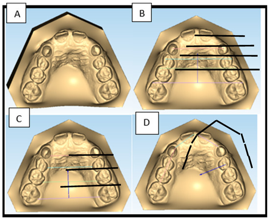
The following measurements were taken into consideration [26,27]:
- -
- Deciduous upper canines gingival width (53-63G): linear distance between the center of the upper deciduous canines palatal surface in contact with the gingival margin.
- -
- First deciduous upper molars gingival width (54-64G): linear distance between the center of the upper first deciduous molars palatal surface in contact with the gingival margin.
- -
- Second deciduous upper molars gingival width (55-65G): linear distance between the center of the upper second deciduous molars palatal surface in contact with the gingival margin.
- -
- First permanent upper molars gingival width (16-26G): distance (mm) between the inner part of the upper first permanent molars palatal surface.
- -
- Deciduous upper canines dental width (53-63): distance (mm) between the edges of the cusps of the upper deciduous canines.
- -
- First deciduous upper molars dental width (54-64): distance (mm) between the edges of vestibular cusps of the upper first deciduous molars.
- -
- Second deciduous upper molars dental width (55-65): distance (mm) between the edges of the mesio-vestibular cusps of the upper second deciduous molars.
- -
- First permanent upper molars dental width (16-26): linear distance between the edges of the mesio-vestibular cusps of the upper first permanent molars.
- -
- Arch perimeter: length of a line passing through the mesial surfaces of first upper permanent molar, deciduous canines and central incisors.
- -
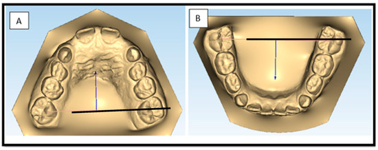
- First permanent upper molars dental width (bis): linear distance between the mesio-palatal cusps (MPC) of the first upper permanent molars.
- -
- First permanent lower molars dental width: linear distance between the central fossa (FC) of the first lower permanent molars.
- -
- Inclination of the first upper permanent molars: angle between the facial axis of the clinical crown (FACC) and a plane created by connecting the inter-incisors papilla to the points where the first permanent upper molars cross the palatal gingival margin at half of the two palatal cusps.
- -
- Rotation of the first upper permanent molars: angle between the extension of the axis passing through the disto-vestibular and mesio-palatal cusps of the upper first molars.
At the end of treatment (T1) patients were scanned again and the same measurements were done.
In Figure 1 and Figure 2 are represented some of the measurements that had been taken in this study.

Figure 1.
Measurements on Maestro 3D Ortho Studio’s digital impressions before and after the expansion; occlusal view (A), dental widths (B), gingival widths (C), arch perimeter (D).

Figure 2.
Measurements of first permanent upper molar dental width (bis) (A) and first permanent lower molar dental width (B).
Statistical Analysis
Method error: to standardize the measurements, the data were collected by the main operator (S.I.) and revised by an operator with experience with the used software (A.U.). To determine the method error, the measurements and the reference points were repeated on ten cases selected randomly. The method error according to Dahlberg formula (inter and intra operator) varied from 0.21 to 0.55 mm (no significant value). Besides, interclass correlation coefficient was calculated, and all the values resulted higher than 0.95. Overall, the error in the methodology can be considered negligible.
Shapiro-Wilks test demonstrated that the data were normally distributed (W = 0.93) so a parametric statistic was utilized. The sample size was calculated with a study’s statistic power higher than 0.85 based on the variable “maxillary molar expansion” (average values and standard deviations) found by Cozzani et al. [28] with alfa = 0.5 and beta = 2. Based on these parameters, the requested sample size resulted in a minimum 30 patients for each group.
The descriptive statistics (average and standard deviation) were calculated for the considered variables. All the analysis were conducted on the distinct differences at T0 and T1 identified in each patient. The difference between the two devices were tested using the parametric statistics using the T-Student test on all the found measurements. The significance of the values lower than 0.05 were accepted as significant in all the statistics.
3. Results
The results of the measurements are summarized in Table 1.

Table 1.
Mean results of the measurements done on both samples. Statistically significant values (p < 0.05) are marked with (*).
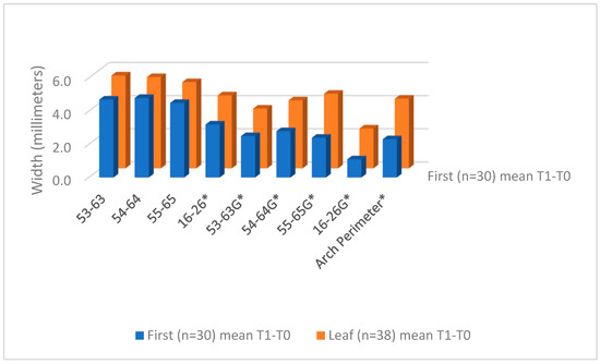
For what concerns the canines, a 4.7 ± 2.3 mm dental expansion was observed in the IF cases against the 5.6 ± 1.9 mm of the LE cases.
On the gingival level, the canine’s expansion was 2.5 ± 1.6 mm for the IF cases and 3.6 ± 1.7 mm for the LE cases.
For the first permanent molars it was observed a 3.2 ± 1.2 mm dental expansion for the IF cases and 4.4 ± 1.4 mm for the LE cases, whereas gingivally a 1.1 ± 1.3 mm expansion was noted for the IF cases and 2.4 ± 1.4 for the LE cases.
As for the first deciduous molars, a 4.8 ± 1.7 mm dental expansion was obtained in the IF cases and a 5.5 ± 1.8 mm dental expansion in the LE cases. Gingivally, a 2.8 ± 2.0 mm expansion for the IF cases and a 4.1 ± 1.5 mm for the LE cases were noted.
As for the second deciduous molars a 4.5 ± 1.6 mm dental expansion for the IF cases and 5.2 ± 1.5 mm for the LE cases was found. Gingivally it was observed a 2.4 ± 1.8 expansion for the IF cases and a 4.5 ± 1.6 mm for the LE cases.
These results are displayed in Figure 3.

Figure 3.
Comparison between mean expansion with Invisalign First and Leaf Expander. Statistically significant values are marked with (*).
The Leaf Expander expanded the arch perimeter for a mean of 4.2 mm whereas the First Invisalign expanded for a mean of 2.3 mm (Figure 4).

Figure 4.
Expansion Clear difference between Invisalign First and Leaf Expander. Statistically significant values are marked with (*).
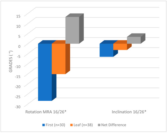
For what concerns the first upper permanent molars rotation, a 28.4 ± 3.2 mm grades of disto-rotation were observed in the IF cases against a 15.1 ± 1.0 grades of disto-rotation in the LE cases.
For the molars inclination’s variation, a 6.5 ± 3.8 grades of vestibular tipping was noted in the IF cases against a 3.1 ± 4.1 grades of vestibular tipping in the LE cases (Figure 5).

Figure 5.
Effects on inclination and rotation of the first upper molars in both Leaf Expander and Invisalign First. Statistically significant values are marked with (*).
4. Discussion
The success of the treatment of the maxillary contraction using slow or rapid palatal expanders is at this point well documented in the scientific literature [24,28,29,30,31].
It was reported in the literature that the range of obtainable expansion with both slow and rapid protocol is 2.49–3.58 mm for the inter-molar width and 2.27–2.64 mm for the inter-canine width [32].
Only few studies investigated the effects of the aligners on the maxillary transversal expansion [24,33,34,35] and only two studies on Invisalign First were published [23,24].
The correct management of phase 1 in orthodontics is very important to intercept malocclusion at an early stage, and the use of clear aligners, as well as representing a more aesthetic and comfortable alternative to traditional treatment, it also appears to be able to solve different issues at the same time, with movements on the three space dimensions simultaneously [24].
The expansion of the maxilla using aligners represents nowadays one the most debated topics in the orthodontics scientific community. In the permanent dentition, the expansion of the upper arch can be done with Invisalign, but the increase in width is obtained mainly by vestibular tipping of the crowns [25]. According to Houle et al. [34] the mean accuracy for the upper arch expansion movements is around 72.8%: 82.9% at the cusps level and 62.7% at the gingival margins level. Zhou and Guo [33] declared that the efficiency of the coronal expansion is between 68.31% and 79.75% whereas the efficiency of the bodily expansion movement of the first upper permanent molars is around 36.35%. Solano-Mendoza et al. [35] affirm that the amount of expansion at the end of treatment is not predictable because of the difference between the planned virtual set up and the final 3D digital models.
In both the protocols used in this study, the exerted forces are similar, with a range of 350–450 g. As for the deciduous canines, first and second deciduous molars, no substantial difference was observed between the two protocols. On the contrary, the expansion at the gingival level was significantly higher in the Leaf Expander cases for the deciduous canines and deciduous first and second molars.
Regarding the first permanent upper molars, the expansion difference was statistically significant for the LE cases both at dental and gingival level.
For what concerns the rotation of the first permanent molars, IF was found to be more effective in the derotation movements (28.4° ± 3.2° in IF sample vs. 15.1° ± 1.6° in LE sample): this is in agreement with Blevins’ article, in which the author underline the effectiveness of Invisalign First in derotating and distalyzing molars, resulting in much more significative stable molar class [23,36].
For what concerns the inclination of the permanent molars, IF gave a higher vestibular tipping of the crown then LE (6.5° ± 3.8° in IF vs. 3.1° ± 4.1° in LE), in accordance with what has already been observed in literature [25].
The interpretation of these data suggests that the transversal expansion in the molar area is more dental rather than skeletal in the Invisalign First sample whereas Leaf Expander give a more bodily vestibularization which makes this last device preferable and limits the use of Invisalign First for light transversal discrepancies (3 mm maximum) [24].
Since both protocols use light forces, a great advantage in both the approaches was the reduced discomfort for the patients in the first days and weeks right after the first activations [37,38].
The use of a slow expansion protocol leads to an extension of the total duration of the treatment which, in fact, goes from 8 to 10 months. Even though both Invisalign First aligners and Leaf Expander use light forces, these forces are applied in two totally different ways because of the use of different materials and designs. The Leaf Expander, thanks to the leaf-like NiTi coils with shape memory and the metallic extensions through all the arch length guarantees a more bodily expansion with uniform and constant forces application [16]. Like in the hyrax-designed rapid expander, the Leaf Expander too can give some phonation and swallowing problems in the first week after the application, but eventually disappear right after the adaptation of the tongue and oral muscles [38].
Another great advantage of the Leaf Expander, compared to the Invisalign First protocol, is that there is no need for patient’s compliance. Leaf Expander is cemented to patient’s teeth and directly activated by the orthodontist during the appointment [16], whereas for the aligners patient must remember to change them every time, even if some studies have underlined an unexpexted high level of compliance, even by children [23].
On the contrary, the clear aligners are removable, so the advantages include a better oral hygiene and a level of patient comfort higher than what typically experienced with fixed appliances [23]. Other positive aspects are represented by less discomfort during food eating and a higher chance of acceptance of the treatment [37,39,40,41,42,43,44]; since the aligners are made of plastic and are changed weekly, there are virtually no emergency appointments, and if the patient loses one aligner, he is instructed to wear the next aligner without the need for a clinical visit [23].
The results of the study must be assessed according to some limitations, including the limited number of the two samples; future studies will consider a larger sample, in order to make different subgroups depending on the severity of the maxillary contraction and also study how patient compliance, varying oral hygiene practices and individual responses can impact the treatment outcomes. Expanding the sample for the medium term and monitoring the patients of this study will allow us to have long term data that are missing in this study, in order to draw information on stability and therefore understand whether one protocol is better than the other in the long term.
5. Conclusions
For transversal discrepancies less than 3 mm, Invisalign First can may be considered a valid alternative in patients in mixed dentition. However, for higher dental discrepancies, the use of the Leaf Expander was found to be more efficient. The transversal expansion obtained on the upper arch with the aligners is mainly due to a vestibular tipping of the teeth whereas the Leaf Expander generates and orthopedic action on the maxillary bone.
The higher efficacy of the Invisalign First in the management of the molars derotation leads to a more stable molar class and occlusion during the expansion phase.
Therefore, considering the promising clinical results obtained using Invisalign First in 3 mm maximum discrepancies, new perspectives in the research field are opened in the use of the Invisalign. Next studies will focus on the possible skeletal effects of aligners on the maxillary structure to verify and validate their possible application in more than 3 mm maxillary discrepancies.
Author Contributions
F.S.-B. involved in writing and metodology; S.I. involved in investigation and writing the original draft; F.P. involved in resources and conceptualization; E.K. involved in data curation and software; A.A. involved in conceptualization and metodology; V.L. involved in review and editing and supervision; A.U. involved in formal analysis, reviewed and edited the manuscript. All authors have read and agreed to the published version of the manuscript.
Funding
This research received no external funding.
Institutional Review Board Statement
The study was conducted in agreement with the Helsinki declaration (version, 2008).
Informed Consent Statement
The patients enrolled in this study came from private practices, and have been informed that the data resulting from their treatment will be used for research. The parents or the legal guardians of each subject of the sample had to accept an informed written consent before the beginning of dental treatment.
Data Availability Statement
The raw data supporting the conclusions of this article will be made available by the authors on request.
Conflicts of Interest
The authors declare no conflict of interest.
References
- Bin Dakhil, N.; Bin, S.F. The Diagnosis Methods and Management Modalities of Maxillary Transverse Discrepancy. Cureus 2021, 13, e20482. [Google Scholar] [CrossRef] [PubMed]
- Kosrhavi, M.; Ugolini, A.; Miresmaeili, A.; Mirzaei, H.; Shahidi-Zandi, V.; Soheilifar, S.; Karami, M.; Mahmoudzadeh, M. Tooth-borne versus bone-borne rapid maxillary expansion for transverse maxillary deficiency: A systematic review. Int. Orthod. 2019, 17, 425–436. [Google Scholar] [CrossRef] [PubMed]
- Proffit, W.R. Contemporary Orthodontics, 6th ed.; Elsevier: Philadelphia, PA, USA, 2019; pp. 138–202. [Google Scholar]
- Lee, K.J.; Park, Y.C.; Park, J.Y.; Hwang, W.S. Miniscrew-assisted nonsurgical palatal expansion before orthognathic surgery for a patient with severe mandibular prognathism. Am. J. Orthod. Dentofac. Orthop. 2010, 137, 830–839. [Google Scholar] [CrossRef] [PubMed]
- Suzuki, H.; Moon, W.; Previdente, L.H.; Suzuki, S.S.; Garcez, A.S.; Consolaro, A. Miniscrew-assisted rapid palatal expander (MARPE): The quest for pure orthopedic movement. Dent. Press J. Orthod. 2016, 2, 17–23. [Google Scholar] [CrossRef]
- Suri, L.; Taneja, P. Surgically assisted rapid palatal expansion: A literature review. Am. J. Orthod. Dentofac. Orthop. 2008, 133, 290–302. [Google Scholar] [CrossRef]
- Bistaffa, A.G.I.; Belomo-Yamaguchi, L.; Almeida, M.R.; Conti, A.C.C.F.; Oltramari, P.V.P.; Fernandes, T.M.F. Immediate skeletal effects of rapid maxillary expansion at midpalatal suture opening with Differential, Hyrax and Haas expanders. Dent. Press J. Orthod. 2023, 27, e2220525. [Google Scholar] [CrossRef]
- Huynh, T.; Kennedy, D.B.; Joondeph, D.R.; Bollen, A.M. Treatment response and stability of slow maxillary expansion using Haas, hyrax, and quad-helix appliances: A retrospective study. Am. J. Orthod. Dentofac. Orthop. 2009, 136, 331–339. [Google Scholar] [CrossRef]
- Gill, D.; Naini, F.; McNally, M.; Jones, A. The management of transverse maxillary deficiency. Dent. Update 2004, 35, 521–523. [Google Scholar] [CrossRef]
- Bala, A.K.; Campbell, P.M.; Tadlock, L.P.; Schneiderman, E.D.; Buschang, P.H. Short-term skeletal and dentoalveolar effects of overexpansion. Angle Orthod. 2022, 92, 55–63. [Google Scholar] [CrossRef]
- Srivastava, S.C.; Mahida, K.; Agarwal, C.; Chavda, R.M.; Patel, H.A. Longitudinal Stability of Rapid and Slow Maxillary Expansion: A Systematic Review. J. Contemp. Dent. Pract. 2020, 21, 1068–1072. [Google Scholar] [CrossRef]
- Bucci, R.; D’Antò, V.; Rongo, R.; Valletta, R.; Martina, R.; Michelotti, A. Dental and skeletal effects of palatal expansion techniques: A systematic review of the current evidence from systematic reviews and meta-analyses. J. Oral. Rehabil. 2016, 43, 543–564. [Google Scholar] [CrossRef] [PubMed]
- Bastos, R.T.D.R.M.; Blagitz, M.N.; Aragón, M.L.S.C.; Maia, L.C.; Normando, D. Periodontal side effects of rapid and slow maxillary expansion: A systematic review. Angle Orthod. 2019, 89, 651–660. [Google Scholar] [CrossRef] [PubMed]
- Rabah, N.; Al-Ibrahim, H.M.; Hajeer, M.Y.; Ajaj, M.A. Evaluation of rapid versus slow maxillary expansion in early adolescent patients with skeletal maxillary constriction using cone-beam computed tomography: A short-term follow-up randomized controlled trial. Dent. Med. Probl. 2022, 59, 583–591. [Google Scholar] [CrossRef] [PubMed]
- Rutili, V.; Nieri, M.; Franceschi, D.; Pierleoni, F.; Giuntini, V.; Franchi, L. Comparison of rapid versus slow maxillary expansion on patient-reported outcome measures in growing patients: A systematic review and meta-analysis. Prog. Orthod. 2022, 23, 47. [Google Scholar] [CrossRef]
- Lanteri, C.; Beretta, M.; Lanteri, V.; Gianolio, A.; Cherchi, C.; Franchi, L. The Leaf Expander for Non-Compliance Treatment in the Mixed Dentition. J. Clin. Orthod. 2016, 50, 552–560. [Google Scholar]
- Lanteri, V.; Cossellu, G.; Gianolio, A.; Beretta, M.; Lanteri, C.; Cherchi, C.; Farronato, G. Comparison between RME, SME and Leaf Expander in growing patients: A retrospective postero-anterior cephalometric study. Eur. J. Paediatr. Dent. 2018, 19, 199–204. [Google Scholar]
- Benhamour, S.; Brezulier, D. Hyrax versus Leaf expander in growing patients, what about adverse dental effects? A retrospective study. Int. Orthod. 2022, 20, 100684. [Google Scholar] [CrossRef]
- Abate, A.; Ugolini, A.; Maspero, C.; Silvestrini-Biavati, F.; Caprioglio, A.; Lanteri, V. Comparison of the skeletal, dentoalveolar, and periodontal changes after Ni-Ti leaf spring expander and rapid maxillary expansion: A three-dimensional CBCT based evaluation. Clin. Oral. Investig. 2023, 27, 5249–5262. [Google Scholar] [CrossRef]
- Zheng, M.; Liu, R.; Ni, Z.; Yu, Z. Efficiency, effectiveness and treatment stability of clear aligners: A systematic review and meta-analysis. Orthod. Craniofac. Res. 2017, 20, 127–133. [Google Scholar] [CrossRef]
- Bichu, Y.M.; Alwafi, A.; Liu, X.; Andrews, J.; Ludwig, B.; Bichu, A.Y.; Zou, B. Advances in orthodontic clear aligner materials. Bioact. Mater. 2022, 22, 384–403. [Google Scholar] [CrossRef]
- Nucera, R.; Dolci, C.; Bellocchio, A.M.; Costa, S.; Barbera, S.; Rustico, L.; Farronato, M.; Militi, A.; Portelli, M. Effects of Composite Attachments on Orthodontic Clear Aligners Therapy: A Systematic Review. Materials 2022, 15, 533. [Google Scholar] [CrossRef] [PubMed]
- Blevins, R. Phase I orthodontic treatment using Invisalign First. J. Clin. Orthod. 2019, 53, 73–83. [Google Scholar] [PubMed]
- Levrini, L.; Carganico, A.; Abbate, L. Maxillary expansion with clear aligners in the mixed dentition: A preliminary study with Invisalign® First system. Eur. J. Paediatr. Dent. 2021, 22, 125–128. [Google Scholar] [PubMed]
- Lione, R.; Paoloni, V.; Bartolommei, L.; Gazzani, F.; Meuli, S.; Pavoni, C.; Cozza, P. Maxillary arch development with the Invisalign System. Angle Orthod. 2021, 91, 433–440. [Google Scholar] [CrossRef]
- Ugolini, A.; Cerruto, C.; Di Vece, L.; Ghislanzoni, L.H.; Sforza, C.; Doldo, T.; Silvestrini-Biavati, A.; Caprioglio, A. Dental arch response to Haas-type rapid maxillary expansion anchored to deciduous vs permanent molars: A multicentric randomized controlled trial. Angle Orthod. 2015, 85, 570–576. [Google Scholar] [CrossRef]
- Cerruto, C.; Ugolini, A.; Di Vece, L.; Doldo, T.; Caprioglio, A.; Silvestrini-Biavati, A. Cephalometric and dental arch changes to Haas-type rapid maxillary expander anchored to deciduous vs permanent molars: A multicenter, randomized controlled trial. J. Orofac. Orthop. 2017, 78, 385–393. [Google Scholar] [CrossRef]
- Cozzani, M.; Guiducci, A.; Mirenghi, S.; Mutinelli, S.; Siciliani, G. Arch width changes with a rapid maxillary expansion appliance anchored to the primary teeth. Angle Orthod. 2007, 77, 296–302. [Google Scholar] [CrossRef]
- Haas, A.J. The Treatment of maxillary deficiency by opening the midpalatal suture. Angle Orthod. 1965, 35, 200–217. [Google Scholar]
- Geran, R.G.; McNamara, J.A., Jr.; Baccetti, T.; Franchi, L.; Shapiro, L.M. A prospective long-term study on the effects of rapid maxillary expansion in the early mixed dentition. Am. J. Orthod. Dentofac. Orthop. 2006, 129, 631–640. [Google Scholar] [CrossRef]
- Corbridge, J.K.; Campbell, P.M.; Taylor, R.; Ceen, R.F.; Buschang, P.H. Transverse dentoalveolar changes after slow maxillary expansion. Am. J. Orthod. Dentofac. Orthop. 2011, 140, 317–325. [Google Scholar] [CrossRef]
- Zhou, Y.; Long, H.; Ye, N.; Xue, J.; Yang, X.; Liao, L.; Lai, W. The effectiveness of non-surgical maxillary expansion: A meta-analysis. Eur. J. Orthod. 2014, 36, 233–242. [Google Scholar] [CrossRef] [PubMed]
- Zhou, N.; Guo, J. Efficiency of upper arch expansion with the Invisalign system. Angle Orthod. 2020, 90, 23–30. [Google Scholar] [CrossRef] [PubMed]
- Houle, J.P.; Piedade, L.; Todescan, R., Jr.; Pinheiro, F.H. The predictability of transverse changes with Invisalign. Angle Orthod. 2017, 87, 19–24. [Google Scholar] [CrossRef] [PubMed]
- Solano-Mendoza, B.; Sonnemberg, B.; Solano-Reina, E.; Iglesias-Linares, A. How effective is the Invisalign® system in expansion movement with Ex30’ aligners? Clin. Oral Investig. 2017, 21, 1475–1484. [Google Scholar] [CrossRef] [PubMed]
- Upadhyay, M.; Arqub, S.A. Biomechanics of clear aligners: Hidden truths & first principles. J. World Fed. Orthod. 2022, 11, 12–21. [Google Scholar]
- Cardoso, P.C.; Espinosa, D.G.; Mecenas, P.; Flores-Mir, C.; Normando, D. Pain level between clear aligners and fixed appliances: A systematic review. Prog. Orthod. 2020, 21, 3. [Google Scholar] [CrossRef]
- Nieri, M.; Paoloni, V.; Lione, R.; Barone, V.; Marino Merlo, M.; Giuntini, V.; Cozza, P.; Franchi, L. Comparison between two screws for maxillary expansion: A multicenter randomized controlled trial on patient’s reported outcome measures. Eur. J. Orthod. 2021, 43, 293–300. [Google Scholar] [CrossRef]
- Yassir, Y.A.; Nabbat, S.A.; McIntyre, G.T.; Bearn, D.R. Clinical effectiveness of clear aligner treatment compared to fixed appliance treatment: An overview of systematic reviews. Clin. Oral. Investig. 2022, 26, 2353–2370. [Google Scholar] [CrossRef]
- Oikonomou, E.; Foros, P.; Tagkli, A.; Rahiotis, C.; Eliades, T.; Koletsi, D. Impact of Aligners and Fixed Appliances on Oral Health during Orthodontic Treatment: A Systematic Review and Meta-Analysis. Oral Health Prev. Dent. 2021, 19, 659–672. [Google Scholar]
- Putrino, A.; Barbato, E.; Galluccio, G. Clear Aligners: Between Evolution and Efficiency-A Scoping Review. Int. J. Environ. Res. Public Health 2021, 18, 2870. [Google Scholar] [CrossRef]
- Galluccio, G. Is the use of clear aligners a real critical change in oral health prevention and treatment? Clin. Ter. 2021, 172, 113–115. [Google Scholar] [PubMed]
- Marya, A.; Venugopal, A.; Vaid, N.; Alam, M.K.; Karobari, M.I. Essential Attributes of Clear Aligner Therapy in terms of Appliance Configuration, Hygiene, and Pain Levels during the Pandemic: A Brief Review. Pain Res. Manag. 2020, 2020, 6677929. [Google Scholar] [CrossRef] [PubMed]
- Zhang, B.; Huang, X.; Huo, S.; Zhang, C.; Zhao, S.; Cen, X.; Zhao, Z. Effect of clear aligners on oral health-related quality of life: A systematic review. Orthod. Craniofac. Res. 2020, 23, 363–370. [Google Scholar] [CrossRef] [PubMed]
Disclaimer/Publisher’s Note: The statements, opinions and data contained in all publications are solely those of the individual author(s) and contributor(s) and not of MDPI and/or the editor(s). MDPI and/or the editor(s) disclaim responsibility for any injury to people or property resulting from any ideas, methods, instructions or products referred to in the content. |
© 2024 by the authors. Licensee MDPI, Basel, Switzerland. This article is an open access article distributed under the terms and conditions of the Creative Commons Attribution (CC BY) license (https://creativecommons.org/licenses/by/4.0/).