Abstract
Owing to progressive aging in the population, there is an increase in patients with cognitive impairment. For the prevention of dementia, the use of plant remedies is relevant. Of particular interest is Klasea centauroides (L.) Cass. (Serratula centauroides L., Asteraceae), which has significant natural reserves, contains a wide range of biologically active substances, and is used in folk medicine to treat nervous system diseases. This study aimed to estimate the neuroprotective, energy-protective, and antioxidant effects of K. centauroides extract in cholinergic deficiency caused by long-term scopolamine administration. It has been established that K. centauroides extract accelerates passive avoidance-conditioned reflex development and ensures its preservation over a longer time period under cholinergic deficiency conditions. The K. centauroides extract increases the resistance of brain tissues to the toxic effects of scopolamine, reducing the number of neuron regressive forms in the cerebral cortex and hippocampus. The K. centauroides extract enhances the predominance of aerobic glycolysis over anaerobic glycolysis and enhances the NADH-dehydrogenase and succinate-dehydrogenase complexes activity, thus promoting more intensive ATP synthesis against this background, the introduction of scopolamine. The use of K. centauroides extracts reduces the malonic dialdehyde (MDA) content in the brain structures and increases the catalase (CAT) and antioxidant system glutathione unit activities.
1. Introduction
Currently, the world is experiencing population aging, and this process is significantly accelerating. In every country in the world, the number of older people is increasing, as well as their share in the population. According to the WHO, between 2015 and 2050, the proportion of the world’s population over 60 years of age will nearly double, amounting to 2.1 billion people, and the number of persons aged 80 years or older is expected to triple to reach 426 million [1]. Thus, the number of cerebral and neurodegenerative diseases will increase, and dementia and depression are among the most common diseases in older people; thus, it is important to search for rational approaches for the treatment of nervous system diseases [2,3,4].
Plant remedies deserve attention in the neurological disease prevention. Klasea centauroides (L.) Cass. (Serratula centauroides L.) from the Asteraceae family is of particular interest for the treatment of neurological diseases. This plant has significant raw material reserves in Russia, Mongolia, and China [5] and is used in traditional medicine in these countries for epilepsy, increased nervous excitability, insomnia, immunodeficiency states, and gastrointestinal tract diseases [5,6,7]. It is known that K. centauroides contains ecdysteroids, flavonoids, di- and triterpenoids, polysaccharides, amino acids, saturated and unsaturated fatty acids, vitamins, and microelements [8,9,10,11,12], but the plant extracts have not earlier been profiled using the high-performance liquid chromatography with mass spectrometric detection technique. Experimental studies have shown that K. centauroides leaf extract exhibits anxiolytic [13], antihypoxic [14], anticonvulsant, nootropic [15], immunocorrective [16], adaptogenic, and antioxidant [17] actions. Moreover, K. centauroides leaf extract has a neuroprotective action on hypoxia/reperfusion and ischemia against chronic stress [18,19]. The K. centauroides leaf extract reduces the malondialdehyde (MDA) content and increases the reduced glutathione (GSH) concentration, the catalase (CAT) activity, and superoxide dismutase (SOD) activity in animal blood serum against stressful situations [19].
Clinical and experimental studies have shown the pronounced therapeutic efficacy of medicinal plants (e.g., Ginkgo biloba, Withania somnifera, Panax ginseng, Baccopa monnieri, Daphne genkwa, Jatropha multifida, Galanthus nivalis, Lycium barbarum) and their secondary metabolites in neurodegenerative diseases (e.g., Parkinson’s disease, Alzheimer’s disease, Huntington’s disease, stroke, etc.) [20,21,22]. It is known that natural compounds (e.g., curcumin, lycopene, ginsenoside, vitexin, baicalin, and kaempferol) exhibit a pronounced neuroprotective effect against ischemic-induced injury by increasing neuron viability, tissue perfusion, and cerebral blood flow and reducing apoptosis; in neurodegenerative diseases, they have an antiamyloidogenic effect and reduce the loss of dopaminergic neurons in the brain [23,24,25]. There have also been positive results in the treatment of Alzheimer’s disease and age-related cognitive impairment using anthocyanin-rich extracts [26]. Alkaloids (e.g., isoquinoline, indole, pyrroloindole, oxindole, piperidine, pyridine, aporphine, periwinkle) play positive roles in ameliorating the pathophysiology of neurodegenerative diseases by functioning as muscarinic and adenosine receptor agonists, anti-oxidant, anti-amyloid and MAO inhibitors, acetylcholinesterase and butyrylcholinesterase inhibitors, inhibitors of α-synuclein aggregation, dopaminergic and nicotine agonists, and NMDA antagonists [27].
Scopolamine is a widely used model to study dementia-related conditions that occur with natural aging and Alzheimer’s disease [28,29,30,31]. Scopolamine is considered to be a non-selective muscarinic receptor antagonist that causes cognitive impairment and electrophysiological changes in the brain, similar to those that occur during natural aging and Alzheimer’s disease [29]. Scopolamine induces a number of cellular alterations, including an impaired antioxidative defense system, increased oxidative stress, mitochondrial dysfunction, apoptosis, and neuroinflammation [31].
Despite the existing plant arsenal with proven therapeutic efficacy in neurodegenerative diseases, the search for new species with neuroprotective activity continues. Medicinal plants (e.g., Eclipta alba, Nelumbo nucifera, Fenugreek seed, and Caesalpinia sappan) have been shown to prevent scopolamine-induced learning and memory deficits in the Morris maze and passive avoidance test by inhibiting acetylcholinesterase, reducing oxidative stress, and activating the antioxidant system [32,33,34,35]. Astragalus membranaceus roots are widely used in China for treating diseases of various types in the elderly. Astragalus membranaceus extract possesses significant nootropic activity in scopolamine-induced amnesia by reducing acetylcholinesterase activity [36]. The estrogen-containing plant Asparagus racemosus extract limits the death of hippocampal neurons that are induced by scopolamine [37].
As part of the ongoing studies of K. centauroides phytocomponents and their bioactivity, in this study, we aimed to estimate the chemical composition of K. centauroides leaf extract using chromatographic profiling and investigate the effect of the leaf extract on cognitive functions, brain antioxidants, and energy potential in cholinergic deficiency induced by scopolamine.
2. Materials and Methods
2.1. Plant Material and Chemicals
Leaves of Klasea centauroides were collected in the Republic of Buryatia (Ivolginsky District, 51°45′55.2″ N, 107°09′34.3″ E), dried in a ventilated oven (50 °C), and stored in a Plant Repository of the IGEB (at 4 °C). Before analysis leaves were ground using A11 basic analytical mill (IKA®-WerkeGmbh & Co. KG, Staufen, Germany) and sieved on an ERL-M1 sieving machine (Zernotekhnika, Moscow, Russia; particle size 0.5 mm). The commercial reference standards used: arbutin (≥98%; No. A4256), 3-O-caffeoylquinic acid (≥95%; No. C3878), 4-O-caffeoylquinic acid (≥98%; No. 65969), 5-O-caffeoylquinic acid (≥98%; No. 94419), 3,4-di-O-caffeoylquinic acid (≥90%; No. SMB00224), 3,5-di-O-caffeoylquinic acid, 4,5-di-O-caffeoylquinic acid (≥85%; No. SMB00221) (Sigma-Aldrich, St. Louis, MO, USA); methyl arbutin (≥98%; Scientific Laboratory Supplies Ltd., Wilford, UK, No. G5041); 20-hydroxyecdysone (≥95%; Selleck Chemicals, Houston, TX, USA, No. S2417), ecdysone (≥99%; AdooQ® Bioscience, Irvine, CA, USA), turkesterone (≥98%; MedKoo Biosciences, Inc., Morrisville, NC, USA, No. 462668), polypodine B (≥98%; BioCrick BioTech, Sichuan, China, No. BCN0474), apigenin 7-O-glucuronide (≥98%; No. CFN98500), luteolin 7-O-glucuronide (≥98%; No. CFN98512) (ChemFaces, Wuhan, China), chrysoeriol 7-O-glucuronide (≥99%; MedChemExpress, Monmouth Junction, NJ, USA, No. HY-N2376), diosmetin 7-O-glucuronide (≥95%; Apollo Scientific Ltd., Bredbury, UK, No. BICL5032). 20-Hydroxyecdysone 3-O-glucoside, integristerone A, 2-deoxy-20-hydroxyecdysone, 20-hydroxyecdysone 3-O-acetate, 2-deoxyecdysone were isolated and NMR-characterized in our laboratory [8,9,19]. Acetonitrile for HPLC (≥99.9%; No. 34851); formic acid (≥95%; No. F0507); (-)-scopolamine hydrochloride (≥99.9%; No. 37022); formalin (10%; No. 65346-M); cresyl violet acetate (≥70%; No. C5042); D-mannitol (≥98%; No. M4125); sucrose (≥99.5%; No. S7903); 3-(N-morpholino)propanesulfonic acid (MOPS) (≥99.5%; No. M5162); ethylene glycol-bis(2-aminoethylether)-N,N,N′,N′-tetraacetic acid (EGTA) (≥97%; No. E0396) were purchased from Sigma–Aldrich (St. Louis, MO, USA).
2.2. Plant Extracts Preparation and Chromatographic Analysis
Before HPLC analysis, the grounded plant sample (200 mg) was extracted three times with 70% ethanol (2 mL) [19] and sonicated for 30 min at 50 °C (ultrasound power 100 W, frequency 35 kHz). The resulting extracts were combined, centrifuged (6000× g, 10 min), filtered (0.22-μm syringe filters) into a 10 mL-measuring flask, and the final volume was filled up to 10 mL with 70% ethanol. Then, 2mL aliquots of ethanolic extract was diluted with 3 mL of bi-distilled water (BD-water) in a measuring flask (5 mL), and 2 mL of diluted extract was passed through a polyamide cartridge (1 g) pretreated with 20 mL of methanol and 30 mL of BD-water. The cartridge was eluted with 20 mL of water (fraction SPE-1) and 25 mL 0.5% NH3 in methanol (fraction SPE-2) at measuring flasks (25 mL) filled up to 25 mL by water and methanol, respectively. The final fractions SPE-1 and SPE-2 were stored at 2 °C before analysis.
Plant extracts with solvent variation were prepared using 150 g-portion of material sonicated thrice for 90 min at 50 °C (ultrasound power 100 W, frequency 35 kHz) with 1.5 L of 70% ethanol. The combined extract was filtered through a paper filter, concentrated in a vacuum and dried in a vacuum oven.
Chromatographic separation was performed by liquid chromatograph LC-20 Prominence paired with a photodiode array detector SPD-M30A, triple-quadrupole mass spectrometer LCMS 8050 and column GLC Mastro C18 (2.1 × 150 mm, 3 μm; all Shimadzu, Columbia, MD, USA) using the known method [19]. Identification criteria were the proximity of chromatographic (retention time) and spectral data (ultraviolet, mass spectral patterns) comparing with the reference standards and literature data.
2.3. Animal Study
Experiments were performed on 64 male Wistar rats that weighed 220–240 g. The Wistar rats were treated with standard conditions of the vivarium (t—20–22 °C, humidity—no more than 50%, air exchange (air intake/outlet)—8:10, light conditions (day/night)—1:1, free access to water). The Wistar rats were fed twice a day. The animals were placed 7 per plastic cage. The rat studies followed the ‘European Convention for the protection of vertebrate animals used for experimental and other scientific purposes’ ETS no. 123 dated 18.03.1986 (Strasburg, 1986). The rats were removed from the experiments by instantaneous decapitation under brief ether anesthesia. The research was approved by the Institute Ethics Committee (Protocol No. 4 dated 26.01.2017).
Animals were randomized according to age, sex, and weight, and divided into four groups: intact, control, and two experimental groups. Each group consisted of 14 animals. Scopolamine hydrochloride at a dose of 1 mg/kg was administered intraperitoneally to animal control and experimental groups for 21 days. Animals in the intact group were similarly injected with saline. Then, for 14 days, the animals in the I and II experimental groups were injected per os with K. centauroides extract at a dose of 100 mg/kg and Ginkgo biloba extract (EGB761, Hunan Warrant Pharmaceuticals, China) at a dose of 100 mg/kg, respectively. The animals in the intact and control groups received purified water in the equivalent volume according to the analogous scheme. On the tenth day, the animals developed a conditioned passive avoidance reflex, which was checked after 1, 24, and 72 h. On day 15, the animals were euthanized according to bioethical conventions. The brains of six animals from each group were removed and placed in 10% buffered formalin for histological studies; the brains of the remaining animals were immersed in a cold isolation medium, and we obtained a homogenate by the methods described below.
2.4. Passive Avoidance Learning Test
The passive avoidance learning test is the main model for assessing substance effect on the formation and reproduction of a memory trace in normal conditions and under conditions of impairment (amnesia). The setup consisted of dark and light adjacent compartments. The light compartment (20 × 20 × 30 cm) had an opening at the top and was brightly lit (90 lux). The floor of the dark chamber (20 × 20 × 30 cm) was made of metal wire (3 mm) spaced 1 cm apart. The floor of the dark chamber could be electrified using a shock generator. In the beginning, the rats were placed in the lit compartment of the apparatus facing away from the door. When moving to the dark compartment, the rats received electric pain stimulation with alternating current (20–30 mA, 1 s, 50 Hz). The retention test was performed 1, 24, and 72 h after the learning acquisition. The rats were placed in the lit chamber as in the passive avoidance learning acquisition training, and the number of animals with a developed reflex (animals that did not enter the dark compartment within 300 s) and the latent time of the first entry into the dark compartment were recorded.
2.5. Histological Studies
The brains were fixed in 10% buffered formalin, and the organs were passed through a standard alcohol battery, embedded in paraffin, and cut into sections (5 μm thick). Sections of the frontal cortex and hippocampus were stained with Nissl cresyl violet. To determine the degree of damage to the brain structures, a morphometric analysis was performed of the cerebral cortex II–V layers and their cellular composition and the CA1 region of the dorsal hippocampus (pyramidal cell layer). The number of different structural neurons was counted in layers II–V of the cortex: normochromic (Nissl’s substance is evenly distributed in the cytoplasm, euchromatin and nucleolus are visible in the nucleus), hyperchromic or pycnotic (the cell is reduced in size, lumps of Nissl’s substance approach each other, merge into a compact dark-colored mass), hypochromic (homogenization and pallor of the cytoplasm, the nucleus is preserved), and shadow cells (diffuse, total lysis of tigroid, karyolysis). The number of hyperchromic (pycnotic) neurons was counted in the hippocampus. At least 200 neurons were counted for each micropreparation. Morphological and morphometric studies were performed using an Axio LAB.A1 (Carl Zeiss Microscopy GmbH, Jena, Germany) light microscope with an AxioCam ERc5s digital camera with Axio Vision SE64 Rel.4.8.3 and ZEN 2012 ver. 2 image analysis software (Carl Zeiss Microscopy GmbH, Jena, Germany).
2.6. Biochemical Studies
2.6.1. Preparation of the Brain Tissue Homogenate
To estimate the activities of the enzymes and products of metabolism and lipid peroxidation, the brains of eight animals from each group were removed, and a homogenate preparation was used based on the protocol by Pollard et al. (2016) with some modifications. The brains were homogenized in mitochondrial extraction buffer consisting of 75 mM mannitol, 150 mM sucrose, 20 mM MOPS, pH 7.2, and 1 mM EGTA (1:4, w:v). The resulting homogenate was divided into 0.5-mL aliquots. All procedures were performed on ice at 4 °C. The aliquots were taken through five freeze-thaw cycles and used to determine the activities of NADH-dehydrogenase and succinate-dehydrogenase complexes, the contents and activities of the components of the pro- and antioxidant systems, and the energetic state of the brains.
2.6.2. Protein Content Determination
Mitochondrial protein concentration was determined according to the method by Bradford [38] using BSA as the standard.
2.6.3. Assessment of Energy Metabolism and the State of Pro- and Antioxidant Systems
The activities of NADH-dehydrogenase and succinate-dehydrogenase complexes were estimated by the methods presented by Pollard et al. [39] and Spinazzi et al. [40], respectively. The protein concentration for the determination of complexes I and II was 30 µg protein/mL in the reaction mixture. The energetic states of the brains were estimated taking into account the contents of adenosine triphosphate (ATP), lactic and pyruvic acids, and their ratio [41]. The states of pro- and antioxidant systems were evaluated according to the concentration of malonic dialdehyde (MDA) [42] and the activities of catalase [43], pyruvate kinase (PK) [44], glutathione peroxidase (GP), and glutathione reductase (GR) [45], as well as according to the content of reduced glutathione (GSH) [46] in the brain homogenate. The activity of enzymatic reactions was estimated at 36 °C.
2.7. Statistical Analysis
The distribution normality was evaluated by the Shapiro–Wilk test using BioStat 7.7. software (AnalystSoft Inc., Walnut, CA, USA). All data were presented as the mean SEM, and p < 0.05 was considered statistically significant. The statistical analysis between groups was used by one-way analysis of variance (ANOVA) followed by Bonferroni’s multiple comparison tests. ANOVA was used to evaluate the effects of the K. centauroides and G. biloba extracts the following parameters: latent period in the Passive Avoidance Learning Test, morphometric parameters of the cerebral cortex and hippocampus, ATP content, mitochondrial complexes I and II activity, glycolysis indicators, malonic dialdehyde level, catalase, glutathione reductase, glutathione peroxidase glutathione reductase actions and reduced glutathione level in the rats’ brain. A Fisher’s exact test was performed to compare the incidence of lesions in the comparison groups.
3. Results
3.1. LC-MS Profiling of K. centauroides Leaf Extract
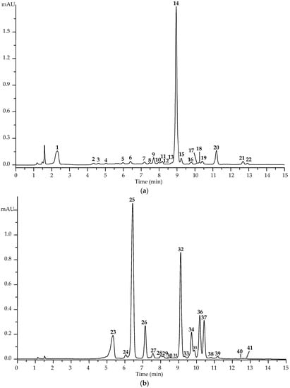
Successful separation and identification of 41 compounds in K. centauroides leaf extract were achieved after the polyamide solid phase extraction (SPE) procedure, followed by high-performance liquid chromatography with a photodiode and electrospray ionization triple quadrupole mass spectrometric detection. The application of SPE resulted in the isolation of the SPE-1 fraction enriched with non-absorbed compounds (e.g., phenols (1–4) and ecdysteroids (5–22)) and the SPE-2 fraction containing absorbed compounds such as caffeoylquinic acids (23–25, 33–35), flavonol glucuronides (26–32, 36–38), and acylated flavonol glycosides (39–41) identified after comparing retention times, ultraviolet and mass spectra with the reference standard and literature data [47,48,49,50,51,52,53,54] (Figure 1, Table 1).

Figure 1.
High-performance liquid chromatography with photodiode array detection chromatogram of SPE-1 ((a); 254 nm) and SPE-2 ((b); 330 nm) fractions of K. centauroides leaf extract. Compounds are numbered as listed in Table 1.

Table 1.
Chromatographic (t) and mass-spectrometric data of compounds 1–41 found in K. centauroides leaf extract.
Four phenol glucosides were found in the SPE-1 fraction, including the known compounds of arbutin (hydroquinone O-glucoside, 1) typical for Klasea plants [55] and the new Klasea chemical methyl arbutin (2) previously found in Ericaceae plants [56].
Tentative identification was achieved for two phenols in the mass spectrum by the deprotonated ion at m/z 255; they were phenol O-hexoside (3), whose analog phenyl glucoside was isolated from Gymnadenia conopsea (L.) R.Br. (Orchidaceae) [57], and hydroquinone O-desoxyhexoside (4); both compounds were unknown for the Asteraceae family. Ecdysteroids have specific mass-spectrometric patterns that have been described many times [58] and corresponded to the known K. centauroides compounds of integristerone A (9), 20-hydoxyecdysone (14), and 2-desoxy-20-hydroxyecdysone (20) found in herb [9], and the newly detected 20-hydroxyecdysone 3-O-glucoside (6), turkesterone (10), polypodine B (15), ecdysone (19), 20-hydroxyecdysone 3-O-acetate (21), and 2-deoxyecdysone (22). Previously, turkesterone was found in Serratula coronata subsp. coronata [59]; polypodine B in K. chinensis [60] and K. quinquefolia [61]; ecdysone in K. radiata [62]; 20-hydoxyecdysone 3-O-acetate in K. chinensis [63]; and 20-hydroxyecdysone 3-O-glucoside, ecdysone, and 2-deoxyecdysone in the Klasea genus for the first time. Compound 5 was tentatively described as 20-hydroxyecdysone O-hexoside for its similarity to 20-hydroxyecdysone 3-O-glucoside (6). Two compounds, 7 and 8, showed the loss of a pentose fragment (m/z 132), and compounds 11–13 lost the desoxyhexoside fragment (m/z 146) with a similar aglycone fragmentation close to 20-hydroxyecdysone, allowing annotation of the compounds as 20-hydroxyecdysone O-pentosides (7, 8) and 20-hydroxyecdysone O-desoxyhexosides (11–13). The structures of 16, 17, and 18 were described as 2-deoxy-20-hydroxyecdysone O-hexoside, 20-hydroxyecdysone O-acetyl-O-hexoside, and 2-deoxyecdysone O-hexoside, respectively, based on the same rules. The natural glycosylated 20-hydroxyecdysones with pentosyl and desoxyhexosyl fragments are still unknown, in contrast to 2-deoxy-20-hydroxyecdysone 3-O-glucoside from Paratinospora sagittata (Oliv.) Wei Wang (syn. Tinospora capillipes Gagnep., Menispermaceae) [64] and 2-deoxy-20-hydroxyecdysone 25-O-glucoside from Silene gigantea (L.) L. (Caryophyllaceae) [65], which are close to 16; 20-hydroxyecdysone 3-O-acetate 2-O-glucoside and 2-O-galactoside from K. chinensis [66] are close to 17; and 2-deoxyecdysone 22-O-glucoside from Silene praemixta Popov [67] is close to 18.
Fraction SPE-2 contained six caffeoylquinic acids, including 4-O- (23), 3-O- (24), 5-O- (25), 3,4-di-O- (33), 3,5-di-O- (34), and 4,5-di-O-caffeoylquinic acid (35); then, 23, 25, 34, 35 were found in K. centauroides [19], and 24 and 33 have not been described in Klasea. Flavonoids of K. centauroides leaves were acidic flavone O-glycosides; four 7-O-glucuronides of luteolin (32), apigenin (36), chrysoeriol (37), and diosmetin (38) were identified using reference standards, and 36 and 37 were found in the K. centauroides herb [19]. Flavones 32, 36, and 37 were described in Klasea [19,68], and 38 was found in the genus for the first time. Tentative identification was done for di-O-hexuronides (27, 30, 31), O-hexuronide-O-hexosides (26, 28, 29), and O-hexuronide-O-hexoside-O-caffeates (39–41) of luteolin, apigenin, and chrysoeriol, none of which have known analogs in Klasea flavonoids.
Thus, in the first chromatographic profiling of K. centauroides leaf extract, 41 compounds were discovered, including ten described previously and 31 found for the first time in the species.
3.2. Quantification of K. centauroides Leaf Extract
For the further characterization of K. centauroides leaves, quantification of six extracts obtained by various solvent extractions was performed using SPE-HPLC-MS assay, and the levels of ten compounds, including arbutin, two ecdysteroids (20-hydroxyecdysone, 2-deoxy-20-hydroxyecdysone), three caffeoylquinic acids (4-O-, 5-O-, 3,5-di-O-caffeoylquinic acid), and four flavonoids (luteolin 7-O-glucuronide, luteolin O-hexuronide-O-hexoside 26, apigenin 7-O-glucuronide, chrysoeriol 7-O-glucuronide) were determined (Table 2).

Table 2.
Yield and content of ten compounds in K. centauroides leaf extracts.
Water and water–ethanol mixtures were chosen as solvents due to their good extraction power for the isolation of phytocomponents with various polarities found in the Klasea species [9,19]. Finally, 40% ethanol extract showed the highest yield (32.1%) and total identified compound content (252.58 mg/g) with arbutin at 62.04 mg/g, ecdysteroids at 36.81 mg/g, caffeoylquinic acid at 57.08 mg/g, and flavonoids at 96.65 mg/g. The dominant compounds were arbutin, 5-O-caffeoylquinic acid at 45.73 mg/g, luteolin 7-O-glucuronide at 39.01 mg/g, and 20-hydroxyecdysone at 30.08 mg/g. The 40% ethanol extract of K. centauroides leaves showed a higher level of basic compounds than the extract of the K. centauroides herb [19].
3.3. Evaluation of the Neuroprotective and Antioxidant Potential of K. centauroides Leaf Extract
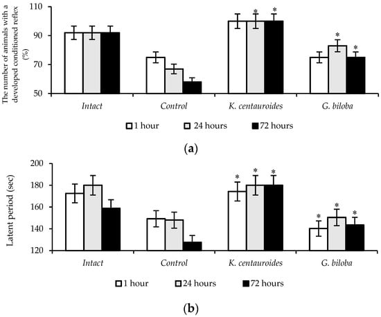
The scopolamine model was used to assess cognitive impairments that occur against the background of cholinergic deficiency. The study of cognitive functions revealed that, after 1 h, passive avoidance learning was formed in 75% of the animals and remained on the 3rd day only in 58% of the animals in the control group. The latent period of entry into the installation dark compartment in the control group for all periods of observation was significantly lower as compared to that in the intact group (Figure 2). The introduction of the K. centauroides extract, the passive avoidance reflex had manifested in 100% of the animals in 1 h and, in 75% of the animals receiving the G. biloba extract. When tested after 24 and 72 h, the passive avoidance reflex was preserved in 100% of animals (p ≤ 0.05) treated with K. centauroides extract, and the latent period was higher by an average of 1.3 and 1.5 times, as compared to the control. The extract of G. biloba after 24 and 72 h retained passive avoidance reflex in 83% and 75% of the animals, respectively (Figure 2).

Figure 2.
The number of animals with developed passive avoidance reflex (a) and the duration of the latent period (b) during prolonged scopolamine exposure in rats; intact (saline + dist. water), control (scopolamine + dist. water), K. centauroides (scopolamine + K. centauroides, 100 mg/kg), G. biloba (scopolamine + G. biloba, 100 mg/kg). The conventions are similar for Figure 3 and Table 3, Table 4 and Table 5. *—p < 0.05 vs. control group.
The histological studies showed that scopolamine intoxication involving structural changes developed in the cerebral cortex, characterized by an increase in the number of hyperchromic neurons (by 77%), “shadow cells” (by 48%) compared to the data from intact animals (Table 3).

Table 3.
Morphometric parameters of the rats’ cerebral cortex and hippocampus with prolonged scopolamine exposure.
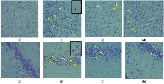
In the control group, neurons were observed that were reduced in size with dystrophic changes in the form of dendrite thinning, and there were indistinct nuclear components against the homogeneous cytoplasm background. Hyperchromic pycnotic cells were detected mainly in layers II and III, and “shadow cells” were located diffusely in all layers of the cerebral cortex (Figure 3).

Figure 3.
Cerebral cortex (a–d) and hippocampus (e–h) in rats with long-term scopolamine exposure: intact group (a,e), control group (b,f), K. centauroides (100 mg/kg; (c,g)) and G. biloba (100 mg/g; (d,h)). Stained with cresyl violet. Magnification ×200. Solid yellow arrow—pycnotic neurons; white arrow—cytolysis of neurons; yellow dotted arrow—satellite; in insets–thinning dendrites.
There was perivascular edema as well as the phenomena of neuronophagia and satellite disease. Single neuropil vacuolization, hyperchromatic and wrinkled nuclei, as well as an altered shape of the apical dendrites in a “corkscrew” form were observed in the neurons. In animals treated with K. centauroides extract, hyperchromic (pycnotic) neurons were much less common, in most cases, in layers III and V; “shadow cells” were located diffusely in all layers of the cerebral cortex (Figure 3). Single neurons were observed with neuropil vacuolization, hyperchromatic nuclei, and “corkscrew-like” altered apical dendrites. At the site of destructive neurons, satellitosis phenomena and neuronophagia were noted but much less frequently than in the control. K. centauroides extract reduced the number of hyperchromic neurons (by 55%), hypochromic neurons (by 17%), and “shadow cells” (by 11%) compared to the control, and G. biloba extract reduced the number of regressive forms of neurons by 50, 15, and 14%, respectively (Table 3).
Long-term scopolamine administration in the hippocampus of the animal control group showed rarefaction of neuronal layers and an increase in the number of hyperchromic neurons that were reduced in size with dystrophic changes in the form of fuzzy nuclear components and thinning of dendrites (Figure 3). Against the background of the K. centauroides extract, the number of pycnotic neurons with thinned dendrites was noticeably less as compared with control; chromatolysis was noted in pyramidal neurons and few foci of neuronal devastation (Figure 3). According to morphometric studies, extracts of K. centauroides and G. biloba reduced the number of hyperchromic neurons by 24 and 19%, respectively, relative to the control (Table 3).
Biochemical studies of the brain showed that the experimental animals injected with K. centauroides extract, showed the ATP content in the brain homogenate increased by 2.0 times, and for the G. biloba extract, it increased by 1.7 times compared to the control animals (Table 4). The increase in the ATP level in the brain compared to the studied drug was due to the correction of oxidative phosphorylation processes and glycolysis. In animals treated with K. centauroides, the activity of NADH-dehydrogenase and succinate-dehydrogenase complexes of brain mitochondria increased by 1.7 and 1.8, respectively, compared to those of the animal control group (Table 4). G. biloba extract increased these parameters by 1.9 and 1.2 times, respectively. The K. centauroides extract increased pyruvate kinase activity by 30% (Table 4). As a result, the pyruvate content increased by 36%, the lactate content decreased by 36% compared to the control, and the ratio of lactate/pyruvate corresponded to the intact group.

Table 4.
ATP content, of mitochondrial complexes I and II activity and of glycolysis indicators in the rats’ brain with prolonged scopolamine exposure.
The changes in the functioning of the electron transport chain of brain mitochondrial compared with those of scopolamine intoxication agreed with anaerobic glycolysis predominance and inhibition of the Krebs’s cycle reactions due to the activation of free radical oxidation processes of biomacromolecules and inhibiting the activity of the endogenic antioxidant system (Table 5).

Table 5.
Malonic dialdehyde level, catalase, glutathione reductase, glutathione peroxidase actions and reduced glutathione level in the rats’ brain with prolonged scopolamine exposure.
The K. centauroides extract promotes the inhibition of free radical oxidation reactions and increases the enzyme activities and contents of endogen antioxidant system peptides in white rat brains. With the introduction of the K. centauroides extract and G. biloba extract, the MDA concentration decreased by 22% and 30%, CAT activity increased by 34% and 67%, and GPx activity increased by 84% and 68%, respectively, in the brain homogenate compared with data for the animals control group, which indicates a positive effect of the studied phytochemicals on redox processes due to the destruction of toxic hydrogen peroxide. The K. centauroides and G. biloba extracts contributed to an increase in GR activity by 43% and 34%, respectively, and the tripeptide (GSH) content increased by 1.6 and 1.8 times, compared to those in control animals.
4. Discussion
It has been established that K. centauroides extract has an antianemic effect; specifically, it improves passive avoidance learning and its preservation over a longer period of cholinergic deficiency induced by scopolamine administration. K. centauroides extract increases the resistance of brain tissues to the toxic effects of scopolamine, limiting the number of neuron regressive forms (hyperchromic, hypochromic, and “shadow cells”) and increasing the number of functional neurons in the cerebral cortex and hippocampus. The neuroprotective effect of the K. centauroides extract is associated with its ability to influence the functional activity of mitochondrial respiratory chain NADH-dehydrogenase and succinate-dehydrogenase complexes, correct aerobic glycolysis processes, reduce the contents of the products of MDA, and increase the intensity of the endogenous antioxidant system by increasing the activities of enzymes (GPx, GR and CAT,) in the brain. The neuroprotective, antioxidant, and energy-protective effects of K. centauroides extract are comparable to those of the reference drug, G. biloba extract.
The revealed pharmacotherapeutic effect of K. centauroides extract is attributed to the presence of various compounds in it, among which phenylpropanoids, ecdysteroids, flavonoids, and arbutin dominate. The biologically active substances of K. centauroides extract contribute to the inhibition of dysfunction of the cholinergic system and oxidative stress. Caffeoylquinic acid reduces β-amyloid deposition by modulating its excretion pathways and thus reduces cognitive dysfunction and neuronal death in the hippocampus of an APP/PS2 transgenic mouse Alzheimer’s disease model [69]. Caffeoylquinic acid inhibited Aβ25-35-induced autophagy by modulating lysosomal function in SH-SY5Y cells and thus attenuated the loss of neurons and cognitive defects in APP/PS1 mice [70]. Caffeoylquinic acid is neuroprotective in cholinergic deficiency by improving the impairment of short-term or working memory induced by scopolamine in the passive avoidance, Y-maze, and Morris water maze tests in laboratory animals and is linked to inhibition of acetylcholinesterase activity and reducing MDA levels in the frontal cortex and hippocampus [71]. Caffeoylquinic acid also showed a pronounced therapeutic effect in mitochondria-mediated apoptotic aging of dopamine neurons in a Parkinson’s disease model. During MPTP induced neurotoxicity, this phenylpropanoid improves the motor function in mice and increases the activity of mitochondrial complexes I, IV, and V, superoxide dismutase, mitochondrial glutathione in the mice brain, against the background of inhibition of proapoptotic proteins. [72]. One of the mechanisms of the neuroprotective effect of caffeoylquinic acid in cerebral and neurodegenerative conditions is its ability to enhance the expression of brain-derived neurotrophic factor (BDNF) and nerve growth factor (NGF) and also inhibit neuron apoptosis in the hippocampus [73,74]. Numerous experimental results show that the neuroprotective effect of caffeoylquinic acid is based on pronounced antioxidant and anti-inflammatory effects [75,76,77].
According to various researchers, given that the flavonoids luteolin and apigenin have high antioxidant and anti-inflammatory activity, they are promising agents in the treatment of neurodegenerative diseases, such as Parkinson’s and Alzheimer’s disease [78,79,80]. Three months of oral treatment with apigenin to APP/PS1 mice improved learning and memory retention by inhibiting fibrillar amyloid deposits and reducing the concentration of insoluble β-amyloid peptide, both of which appear to play a critical role in the onset and progression of Alzheimer’s disease [81]. In addition, these authors showed that apigenin exhibited superoxide anion scavenging effects, improved antioxidative enzyme activity of GPx and SOD, and restored the neurotrophic ERK/CREB/BDNF pathway in the cerebral cortex. Apigenin exhibits a neuroprotective effect in amnesia caused by Aβ25-35, improving learning and memory, maintaining neurovascular unit integrity, modulating microvascular function, reducing neurovascular oxidative damage, increasing regional cerebral blood flow, and improving the cholinergic system involving the inhibition of acetylcholinesterase activity and modification of BNDF, TrkB, and phospho-CREB levels [82]. The introduction of this flavonoid improved cognitive functions in the novel objective recognition test, T-maze test, and Morris water maze test in mice with Alzheimer’s disease caused by the administration of scopolamine. In this experiment, apigenin attenuated scopolamine-induced lipid peroxidation in the brain, reduced the expression of apoptotic factors, such as B-cell lymphoma 2-associated X/B-cell lymphoma 2 (Bax/Bcl-2), cleaved caspase-3, and cleaved PARP, inhibited BACE, PS1, PS2, and RAGE protein expression, increased expression of BDNF and tropomyosin receptor kinase B (TrkB) [83]. According to [84], apigenin did not contribute to the preservation of the passive avoidance reflex in white rats after 24 h after a single scopolamine injection, but it had a more pronounced effect on the retention of a memory trace over a longer period (8 weeks). In a human pluripotent stem cell (iPSC) model of Alzheimer’s disease for familial and sporadic Alzheimer’s disease, apigenin reduced neuronal hyperexcitability and apoptosis and inhibited cytokine activation and NO production, protecting neurons from inflammation-induced stress and neurite retraction [85].
In animal experiments, 20E has been shown to be non-toxic and pharmacotherapeutically effective in the treatment of neuromuscular, cardio-metabolic, and respiratory diseases [86]. According to [87], 20E reduces stress, anxiety, and depression, while activating antioxidant enzymes (CAT, SOD, and GPx), promoting antioxidant activity (anti-oxidant capacity, sulfhydryl groups, and GSH), and reducing oxidative markers (lipid peroxidation). In addition, 20E increases the concentration of NO in the striatum, possibly improving memory function and antioxidant activity. 20-hydroxyecdysone protects dopaminergic neurons in the treatment of Parkinson’s disease by significantly suppressing motor deficits and bradykinesia in mice. The introduction of 20E for MPTP-induced neurotoxicity increased the level of markers of antioxidant protection (SOD, CAT, and GSH) and the concentration of dopamine in the striatum and reduced the expression of apoptotic factors (pERK1/2, caspase-7, HSP27, pJNK, IkBa, pp38, and TAK-1) [88]. The use of ecdysterone and its combination with high-intensity interval training in an Alzheimer’s disease model by β-amyloid (Aβ) injection showed a pronounced anamnestic effect, improving the formation and preservation of the passive avoidance reflex, limiting the loss of neurons in the hippocampus and stimulating the antioxidant defense enzyme activities (SOD, CAT, GRx) in the brain [89]. In a model of Alzheimer’s disease, β-ecdysone inhibited BACE1 activity and induced the open form to closed conformational transition of BACE1, thus blocking substrate binding and inhibiting Aβ aggregation [90].
5. Conclusions
Thus, the K. centauroides leaf extract, due to a wide range of biologically active substances, has a neuroprotective effect in cholinergic deficiency, improves cognitive functions, and limits neuron death in the cerebral cortex and hippocampus. The K. centauroides extract enhances the mitochondrial respiratory functional activity chain complexes, corrects the aerobic glycolysis processes, suppresses the free-radical oxidation of biomacromolecule reactions, and increases the endogenous antioxidant system intensity in the brain.
Author Contributions
Conceptualization, Y.G.R. and D.N.O.; methodology, Y.G.R. and D.N.O.; software, A.A.T. and D.N.O.; validation, Y.G.R., K.V.M., and N.I.K.; formal analysis, K.V.M., A.A.T., and N.I.K.; investigation, Y.G.R., K.V.M., A.A.T., N.I.K., and D.N.O.; resources, A.A.T. and D.N.O.; data curation, K.V.M.; writing—original draft preparation, Y.G.R. and N.I.K.; writing—review and editing, D.N.O.; visualization, A.A.T. and D.N.O.; supervision, Y.G.R.; project administration, D.N.O.; funding acquisition, D.N.O. All authors have read and agreed to the published version of the manuscript.
Funding
This research was funded by the Ministry of Education and Science of Russia, grant numbers 121030100227-7.
Institutional Review Board Statement
The study was conducted in accordance with the guidelines of the Declaration of Helsinki and approved by the Russian Health Ministry (protocol code 708H, 23 August 2010) and the Ethics Committee of the Institute of General and Experimental Biology (protocol code LM-0324, 26 January 2017).
Informed Consent Statement
Not applicable.
Data Availability Statement
Data are contained within the article.
Acknowledgments
The authors acknowledge the Buryat Research Resource Center for the technical support in chromatographic and mass-spectrometric research.
Conflicts of Interest
The authors declare no conflict of interest. The funders had no role in the design of the study; in the collection, analyses, or interpretation of data; in the writing of the manuscript; or in the decision to publish the results.
References
- World Health Organization. Ageing and Health. Available online: https://www.who.int/news-room/fact-sheets/detail/ageing-and-health (accessed on 29 November 2022).
- Pohl, F.; Kong Thoo Lin, P. The potential use of plant natural products and plant extracts with antioxidant properties for the prevention/treatment of neurodegenerative diseases: In vitro, in vivo and clinical trials. Molecules 2018, 23, 3283. [Google Scholar] [CrossRef] [PubMed]
- Rasool, M.; Malik, A.; Qureshi, M.S.; Manan, A.; Pushparaj, P.N.; Asif, M.; Qazi, M.H.; Qazi, A.M.; Kamal, M.A.; Gan, S.H.; et al. Recent updates in the treatment of neurodegenerative disorders using natural compounds. Evid. Based Complement. Altern. Med. 2014, 2014, 979730. [Google Scholar] [CrossRef] [PubMed]
- Rekatsina, M.; Paladini, A.; Piroli, A.; Zis, P.; Pergolizzi, J.V.; Varrassi, G. Pathophysiology and therapeutic perspectives of oxidative stress and neurodegenerative diseases: A narrative review. Adv. Ther. 2020, 37, 113–139. [Google Scholar] [CrossRef]
- Nikolaeva, G.G.; Shantanova, L.N.; Nikolaeva, I.G.; Radnaeva, L.D.; Garmaeva, L.L.; Tsybiktarova, L.P. Rhaponticum uniflorum (L.) and Serratula centauroides (L.) are promising ecdysteroid-containing plants. Acta Biomed. Sci. 2014, 3, 93–96. [Google Scholar]
- Abysheva, L.N.; Belenovskaya, L.M.; Bobyleva, N.S. Wild Useful Plants of Russia; SPKhFA: Saint Petersburg, Russia, 2001; pp. 123–124. [Google Scholar]
- Hammerman, A.F.; Semichev, B.V. Dictionary of Tibetan-Latin-Russian Names of Medicinal Plant Materials Used in Tibetan Medicine; AN SSSR: Ulan-Ude, Russia, 1963; pp. 80–82. [Google Scholar]
- Nikolaeva, I.G.; Tsybiktarova, L.P.; Garmaeva, L.L.; Nikolaeva, G.G.; Olennikov, D.N.; Matkhanov, I.E. Determination of ecdysteroids in Fornicium uniflorum (L.) and Serratula centauroides (L.) raw materials by chromatography-UV spectrophotometry. J. Anal. Chem. 2017, 72, 854–861. [Google Scholar] [CrossRef]
- Olennikov, D.N.; Kashchenko, N.I. Phytoecdysteroids of Serratula centauroides Herb from Cisbaikalia. Russ. J. Bioorg. Chem. 2019, 45, 913–919. [Google Scholar] [CrossRef]
- Tsybiktarova, L.P.; Nikolaeva, I.G.; Nikolaeva, G.G. Amino acids from Serratula centauroides. Chem. Nat. Compd. 2017, 53, 203–204. [Google Scholar] [CrossRef]
- Tsybiktarova, L.P.; Taraskin, V.V.; Nikolaeva, I.G.; Radnaeva, L.D.; Nikolaeva, G.G.; Garmaeva, L.L. Lipids from Serratula centauroides. Chem. Nat. Compd. 2016, 52, 294–295. [Google Scholar] [CrossRef]
- Tsybiktarova, L.P.; Taraskin, V.V.; Nikolaeva, I.G.; Radnaeva, L.D.; Gereltu, B.; Nikolaeva, G.G. Constituent composition of essential oil from Serratula centauroides. Chem. Nat. Compd. 2016, 52, 1123–1124. [Google Scholar] [CrossRef]
- Nikolaev, S.M.; Nikolaeva, I.G.; Razuvaeva, Y.G.; Matkhanov, I.E.; Tsybiktarova, L.P.; Shantanova, L.N.; Nikolaeva, G.G. Phenolic compounds of Serratula centauroides and anxiolytic effect. Farmacia 2019, 67, 504–510. [Google Scholar] [CrossRef]
- Sviridov, I.V.; Razuvaeva, Y.G.; Shantanova, L.N. Antihypoxic properties of the dry extract from Serratula centauroides. Acta Biomed. Sci. 2014, 6, 77–79. [Google Scholar]
- Sviridov, I.V.; Razuvaeva, Y.G.; Shantanova, L.N. Nootropic and anticonvulsant properties of the dry extract from Serratula centauroides roots. Acta Biomed. Sci. 2015, 2, 89–91. [Google Scholar]
- Sviridov, I.V.; Khobrakova, V.B.; Shantanova, L.N. Immunomodulating effect of the extract from Serratula centauroides. Sib. Med. Zh. 2015, 5, 120–122. [Google Scholar]
- Olennikov, D.N. Metabolites of Serratula L. and Klasea Cass. (Asteraceae): Diversity, separation methods, and bioactivity. Separations 2022, 9, 448. [Google Scholar] [CrossRef]
- Markova, K.V.; Razuvaeva, Y.G.; Toropova, A.A.; Olennikov, D.N. Morphological assessment of neuroprotective effects of Rhaponticum uniflorum and Serratula centauroides dry extracts in hypoxia/reoxygenation. J. Biomed. 2022, 18, 56–62. [Google Scholar] [CrossRef]
- Shantanova, L.N.; Olennikov, D.N.; Matkhanov, I.E.; Gulyaev, S.M.; Toropova, A.A.; Nikolaeva, I.G.; Nikolaev, S.M. Rhaponticum uniflorum and Serratula centauroides extracts attenuate emotional injury in acute and chronic emotional stress. Pharmaceuticals 2021, 14, 1186. [Google Scholar] [CrossRef]
- Gonzalez, M.P.; Benedi, J.; Bermejo-Bescos, P.; Martin-Aragon, S. Plants with evidence-based therapeutic effects against neurodegenerative diseases. Pharm. Pharmacol. Int. J. 2019, 7, 221–227. [Google Scholar] [CrossRef]
- Luthra, R.; Roy, A. Role of medicinal plants against neurodegenerative diseases. Curr. Pharm. Biotechnol. 2022, 23, 123–139. [Google Scholar] [CrossRef]
- Ratheesh, G.; Tian, L.; Venugopal, J.R.; Ezhilarasu, H.; Sadiq, A.; Fan, T.-P.; Ramakrishna, S. Role of medicinal plants in neurodegenerative diseases. Biomanuf. Rev. 2017, 2, 2. [Google Scholar] [CrossRef]
- Nejabati, H.R.; Roshangar, L. Kaempferol as a potential neuroprotector in Alzheimer’s disease. J. Food Biochem. 2022, 46, e14375. [Google Scholar] [CrossRef]
- Putteeraj, M.; Lim, W.L.; Teoh, S.L.; Yahaya, M.F. Flavonoids and its neuroprotective effects on brain ischemia and neurodegenerative diseases. Curr. Drug. Targets 2018, 19, 1710–1720. [Google Scholar] [CrossRef] [PubMed]
- Yavarpour-Bali, H.; Ghasemi-Kasman, M.; Pirzadeh, M. Curcumin-loaded nanoparticles: A novel therapeutic strategy in treatment of central nervous system disorders. Int. J. Nanomed. 2019, 14, 4449–4460. [Google Scholar] [CrossRef]
- Winter, A.N.; Bickford, P.C. Anthocyanins and their metabolites as therapeutic agents for neurodegenerative disease. Antioxidants 2019, 8, 333. [Google Scholar] [CrossRef] [PubMed]
- Hussain, G.; Rasul, A.; Anwar, H.; Aziz, N.; Razzaq, A.; Wei, W.; Ali, M.; Li, J.; Li, X. Role of plant derived alkaloids and their mechanism in neurodegenerative disorders. Int. J. Biol. Sci. 2018, 14, 341–357. [Google Scholar] [CrossRef] [PubMed]
- Arjan, B. Cholinergic models of memory impairment in animals and man: Scopolamine vs. biperiden. Behav. Pharmacol. 2022, 33, 231–237. [Google Scholar] [CrossRef]
- Bajo, R.; Pusil, S.; López, M.E.; Canuet, L.; Pereda, E.; Osipova, D.; Maestú, F.; Pekkonen, E. Scopolamine effects on functional brain connectivity: A pharmacological model of Alzheimer’s disease. Sci. Rep. 2015, 5, 9748. [Google Scholar] [CrossRef] [PubMed]
- Budzynska, B.; Boguszewska-Czubara, A.; Kruk-Slomka, M.; Skalicka-Wozniak, K.; Michalak, A.; Irena, M.; Biala, G. Effects of imperatorin on scopolamine-induced cognitive impairment and oxidative stress in mice. Psychopharmacology 2015, 232, 931–942. [Google Scholar] [CrossRef]
- Tang, K.S. The cellular and molecular processes associated with scopolamine-induced memory deficit: A model of Alzheimer’s biomarkers. Life Sci. 2019, 233, 116695. [Google Scholar] [CrossRef]
- Chahal, H.S.; Sharma, S. Effect of Eclipta alba on scopolamine induced amnesia in mice. J. Drud Deliv. Ther. 2018, 8, 162–168. Available online: http://www.jddtonline.info/index.php/jddt/article/view/1926 (accessed on 29 November 2022). [CrossRef]
- Helmi, H.; Fakhrudin, N.; Nurrochmad, A.; Ikawati, Z. Caesalpinia sappan L. ameliorates scopolamine-induced memory deficits in mice via the cAMP/PKA/CREB/BDNF pathway. Sci. Pharm. 2021, 89, 29. [Google Scholar] [CrossRef]
- Khan, R.A.; Rajput, M.A.; Assad, T. Effect of Nelumbo nucifera fruit on scopolamine induced memory deficits and motor coordination. Metab. Brain. Dis. 2019, 34, 87–92. [Google Scholar] [CrossRef] [PubMed]
- Mohamed Hussain, S.; Almutairi, N.; Alrakaf, F.; Aljameli, M.; Alshammari, M.; Alnasser, S. Nootropic effect of Fenugreek seed extract against scopolamine induced cognitive decline in experimental mice. Preprints 2020, 2020090675. [Google Scholar] [CrossRef]
- Pathak, L. Acute toxicity test and nootropic activity of ethanolic extract of Astragalus membranaceus Bunge on scopolamine-induced amnesia in rats. J. Drud Deliv. Ther. 2022, 12, 11–18. Available online: http://www.jddtonline.info/index.php/jddt/article/view/5230 (accessed on 29 November 2022). [CrossRef]
- Jagdish, P.; Maheep, B. Neuroprotective efficacy of Asparagus racemosus root extract on hippocampal neurons in scopolamine mouse model of Alzheimer’s disease. Asian J. Biol. Life Sci. 2021, 10, 391–399. [Google Scholar] [CrossRef]
- Bradford, M.M. A rapid and sensitive method for the quantitation of microgram quantities of protein utilizing the principle of protein-dye binding. Anal. Biochem. 1976, 72, 248–254. [Google Scholar] [CrossRef]
- Pollard, A.K.; Craig, E.L.; Chacrabarti, L. Mitochondrial complex I activity measured by spectrophotometry is reduced across all brain regions in ageing and more specifically in neurodegeneration. PLoS ONE 2016, 11, e0157405. [Google Scholar] [CrossRef]
- Spinazzi, M.; Casarin, A.; Pertegato, V.; Salviati, L.; Angelini, C. Assesment of mitochondrial respiratory chain enzymatic activities on tissues and cultured cells. Nat Protoc. 2012, 7, 1235–1246. [Google Scholar] [CrossRef]
- Patergnani, S.; Baldassari, F.; De Marchi, E.; Karkucinska-Wieckowska, A.; Wieckowski, M.R.; Pinton, P. Methods to monitor and compare mitochondrial and glycolytic ATP production. Methods Enzymol. 2014, 542, 313–332. [Google Scholar] [CrossRef]
- Kikugawa, K.; Kojima, T.; Yamaki, S.; Kosugi, H. Interpretation of the thiobarbituric acid reactivity of rat liver and brain homogenates in the presence of ferric ion and ethylenediaminetetraacetic acid. Anal Biochem. 1992, 202, 249–255. [Google Scholar] [CrossRef]
- Hamza, T.A.; Hadwan, M.H. New spectrophotometric method for the assessment of catalase enzyme activity in biological tissues. Curr. Anal. Chem. 2020, 16, 1054–1062. [Google Scholar] [CrossRef]
- Osterman, J.; Fritz, P.J.; Wuntch, T. Pyruvate kinase isozymes from rat tissues. J. Biol. Chem. 1973, 248, 1011–1018. [Google Scholar] [CrossRef]
- Pinto, R.E.; Bartley, W. The effect of age and sex on glutathione reductase and glutation peroxidase activities and aerobic glutathione oxidation in rat liver homogenates. Biochem. J. 1969, 112, 109–115. [Google Scholar] [CrossRef] [PubMed]
- Shaik, I.H.; Mehvar, R. Rapid determination of reduced and oxidized glutathione levels using a new thiol-masking reagent and the enzymatic recycling method: Application to the rat liver and bile samples. Anal. Bioanal. Chem. 2006, 385, 105–113. [Google Scholar] [CrossRef] [PubMed]
- Olennikov, D.N.; Shamilov, A.A. New compounds from Vaccinium vitis-idaea. Chem. Nat. Compd. 2022, 58, 240–244. [Google Scholar] [CrossRef]
- Olennikov, D.N.; Chekhirova, G.V. 6″-Galloylpicein and other phenolic compounds from Arctostaphylos uva-ursi. Chem. Nat. Compd. 2013, 49, 1–7. [Google Scholar] [CrossRef]
- Olennikov, D.N. Makisterone C-20,22-acetonide from Rhaponticum uniflorum. Chem. Nat. Compd. 2018, 54, 930–933. [Google Scholar] [CrossRef]
- Olennikov, D.N.; Kashchenko, N.I. New flavonoids and turkesterone-2-O-cinnamate from leaves of Rhaponticum uniflorum. Chem. Nat. Comp. 2019, 55, 256–264. [Google Scholar] [CrossRef]
- Olennikov, D.N. Triterpenic and phenolic compounds from Asparagus burjaticus. Chem. Nat. Comp. 2019, 55, 1192–1194. [Google Scholar] [CrossRef]
- Kashchenko, N.I.; Jafarova, G.S.; Isaev, J.I.; Olennikov, D.N.; Chirikova, N.K. Caucasian dragonheads: Phenolic compounds, polysaccharides, and bioactivity of Dracocephalum austriacum and Dracocephalum botryoides. Plants 2022, 11, 2126. [Google Scholar] [CrossRef]
- Olennikov, D.N.; Vasilieva, A.G.; Chirikova, N.K. Fragaria viridis fruit metabolites: Variation of LC-MS profile and antioxidant potential during ripening and storage. Pharmaceuticals 2020, 13, 262. [Google Scholar] [CrossRef]
- Olennikov, D.N.; Gadimli, A.I.; Isaev, J.I.; Kashchenko, N.I.; Prokopyev, A.S.; Katayeva, T.N.; Chirikova, N.K.; Vennos, C. Caucasian Gentiana species: Untargeted LC-MS metabolic profiling, antioxidant and digestive enzyme inhibiting activity of six plants. Metabolites 2019, 9, 271. [Google Scholar] [CrossRef] [PubMed]
- Nowak, G.; Nawrot, J.; Latowski, K. Arbutin in Serratula quinquefolia M.B. (Asteraceae). Acta Soc. Bot. Pol. 2009, 78, 137–140. [Google Scholar] [CrossRef]
- Shamilov, A.A.; Olennikov, D.N.; Pozdnyakov, D.I.; Bubenchikova, V.N.; Garsiya, E.R. Investigation of phenolic compounds at the leaves and shoots Arctostaphylos spp. and their antioxidant and antityrosinase activities. Nat. Prod. Res. 2022, 36, 6312. [Google Scholar] [CrossRef]
- Morikawa, T.; Xie, H.; Matsuda, H.; Wang, T.; Yoshikawa, M. Bioactive constituents from Chinese natural medicines. XVII. Constituents with radical scavenging effect and new glucosyloxybenzyl 2-isobutylmalates from Gymnadenia conopsea. Chem. Pharm. Bull. 2006, 54, 506–513. [Google Scholar] [CrossRef]
- Lafont, R.; Harmatha, J.; Marion-Poll, F.; Dinan, L.; Wilson, I.D. The Ecdysone Handbook, 3rd ed.; Available online: https://ecdybase.org (accessed on 29 November 2022).
- Hunyadi, A.; Gergely, A.; Simon, A.; Toth, G.; Veress, G.; Bathori, M. Preparative-scale chromatography of ecdysteroids of Serratula wolffii Andrae. J. Chromatogr. Sci. 2007, 45, 76–86. [Google Scholar] [CrossRef] [PubMed]
- Tang, H.-J.; Fan, C.-L.; Wang, G.-Y.; Wei, W.; Wang, Y.; Ye, W.-C. Chemical constituents from roots tubers of Serratula chinensis. Chin. Trad. Herb. Drugs. 2014, 45, 906–912. [Google Scholar] [CrossRef]
- Odinokov, V.N.; Galyautdinov, I.V.; Mel’nikova, D.A.; Muslimov, Z.S.; Khalilov, L.M.; Denisenko, O.N.; Mogilenko, T.G.; Zaripova, E.R.; Zakirova, L.M. Isolation and identification of phytoecdysteroids from juice of Serratula quinquefolia. Chem. Nat. Compd. 2013, 49, 392–394. [Google Scholar] [CrossRef]
- Abubakirov, N.K. Ecdysteroids of flowering plants (Angiospermae). Chem. Nat. Compd. 1981, 17, 489–503. [Google Scholar] [CrossRef]
- Ling, T.; Zhang, Z.; Xia, T.; Ling, W.; Wan, X. Phytoecdysteroids and other constituents from the roots of Klaseopsis chinensis. Biochem. Syst. Ecol. 2009, 37, 49–51. [Google Scholar] [CrossRef]
- Song, C.Q.; Xu, R.S. Phytoecdysones from the roots of Tinospora capillipes. Chin. Chem. Lett. 1991, 2, 13–14. [Google Scholar]
- Zibareva, L.; Yeriomina, V.I.; Munkhjargal, N.; Girault, J.-P.; Dinan, L.; Lafont, R. The phytoecdysteroid profiles of 7 species of Silene (Caryophyllaceae). Arch. Insect Biochem. Physiol. 2009, 72, 234–248. [Google Scholar] [CrossRef]
- Zhang, Z.-Y.; Yang, W.-Q.; Fan, C.-L.; Zhao, H.-N.; Huang, X.-J.; Wang, Y.; Ye, W.-C. New ecdysteroid and ecdysteroid glycosides from the roots of Serratula chinensis. J. Asian Nat. Prod. Res. 2016, 19, 208–214. [Google Scholar] [CrossRef] [PubMed]
- Agzamova, M.; Ogly Isaev, I.; Mamathanov, A.; Ogly Isaev, M.; Ibragimov, T. Phytoecdysteroids from Silene praemixta. Adv. Biol. Chem. 2014, 4, 42703. [Google Scholar] [CrossRef]
- Myagchilov, A.V.; Gorovoi, P.G.; Sokolova, L.I. Flavonoids from Serratula komarovii Iljin (the Asteraceae Family). Russ. J. Bioorg. Chem. 2021, 47, 1418–1423. [Google Scholar] [CrossRef]
- Ishida, K.; Misawa, K.; Nishimura, H.; Hirata, T.; Yamamoto, M.; Ota, N. 5-Caffeoylquinic acid ameliorates cognitive decline and reduces Aβ deposition by modulating Aβ clearance pathways in APP/PS2 transgenic mice. Nutrients 2020, 12, 494. [Google Scholar] [CrossRef] [PubMed]
- Gao, L.; Li, X.; Meng, S.; Ma, T.; Wan, L.; Xu, S. Chlorogenic acid alleviates Aβ25-35-induced autophagy and cognitive impairment via the mTOR/TFEB signaling pathway. Drug Des. Dev. Ther. 2020, 14, 1705–1716. [Google Scholar] [CrossRef]
- Kwon, S.-H.; Lee, H.-K.; Kim, J.-A.; Hong, S.-I.; Kim, H.-C.; Jo, T.-H.; Park, Y.-I.; Lee, C.-K.; Kim, Y.-B.; Lee, S.-Y.; et al. Neuroprotective effects of chlorogenic acid on scopolamine-induced amnesia via anti-acetylcholinesterase and anti-oxidative activities in mice. Eur. J. Pharmacol. 2010, 649, 210–217. [Google Scholar] [CrossRef]
- Singh, S.S.; Rai, S.N.; Birla, H.; Zahra, W.; Rathore, A.S.; Dilnashin, H.; Singh, R.; Singh, S.P. Neuroprotective effect of chlorogenic acid on mitochondrial dysfunction-mediated apoptotic death of DA neurons in a parkinsonian mouse model. Oxid. Med. Cell. Longev. 2020, 2020, 6571484. [Google Scholar] [CrossRef]
- Shi, M.; Sun, F.; Wang, Y.; Kang, J.; Zhang, S.; Li, H. CGA restrains the apoptosis of Aβ25-35-induced hippocampal neurons. Int. J. Neurosci. 2020, 130, 700–707. [Google Scholar] [CrossRef]
- Liu, D.; Wang, H.; Zhang, Y.; Zhang, Z. Protective effects of chlorogenic acid on cerebral ischemia/reperfusion injury rats by regulating oxidative stress-related Nrf2 pathway. Drug Des. Dev. Ther. 2020, 14, 51–60. [Google Scholar] [CrossRef]
- Heitman, E.; Ingram, D.K. Cognitive and neuroprotective effects of chlorogenic acid. Nutr. Neurosci. 2017, 20, 32–39. [Google Scholar] [CrossRef] [PubMed]
- Mitrea, D.R.; Malkey, R.; Florian, T.L.; Filip, A.; Clichici, S.; Bidian, C.; Moldovan, R.; Hoteiuc, O.A.; Toader, A.M.; Baldea, I. Daily oral administration of chlorogenic acid prevents the experimental carrageenan-induced oxidative stress. J. Physiol. Pharmacol. 2020, 71, 55–65. [Google Scholar] [CrossRef]
- Rebai, O.; Belkhir, M.; Sanchez-Gomez, M.V.; Matute, C.; Fattouch, S.; Amri, M. Differential molecular targets for neuroprotective effect of chlorogenic acid and its related compounds against glutamate induced excitotoxicity and oxidative stress in rat cortical neurons. Neurochem. Res. 2017, 42, 3559–3572. [Google Scholar] [CrossRef] [PubMed]
- Venigalla, M.; Gyengesim, E.; Munch, G. Curcumin and apigenin-Novel and promising therapeutics against chronic neuroinflammation in Alzheimer’s disease. Nat. Regen. Res. 2015, 10, 1181–1185. [Google Scholar] [CrossRef]
- Nabavi, S.F.; Khan, H.; D’onofrio, G.; Šamec, D.; Shirooie, S.; Dehpour, A.R.; Argüelles, S.; Habtemariam, S.; Sobarzo-Sanchez, E. Apigenin as neuroprotective agent: Of mice and men. Pharm. Res. 2018, 128, 359–365. [Google Scholar] [CrossRef]
- Salehi, B.; Venditti, A.; Sharifi-Rad, M.; Kręgiel, D.; Sharifi-Rad, J.; Durazzo, A.; Lucarini, M.; Santini, A.; Souto, E.B.; Novellino, E.; et al. The therapeutic potential of apigenin. Int. J. Mol. Sci. 2019, 20, 1305. [Google Scholar] [CrossRef]
- Zhao, L.; Wang, J.; Liu, R.; Li, X.X.; Li, J.; Zhang, L. Neuroprotective, anti-amyloidogenic and neurotrophic effects of apigenin in an Alzheimer’s disease mouse model. Molecules 2013, 18, 9949–9965. [Google Scholar] [CrossRef]
- Liu, R.; Zhang, T.; Yang, H.; Lan, X.; Ying, J.; Du, G. The flavonoid apigenin protects brain neurovascular c8upling against amyloid-beta(2) (5)(-)(3)(5)-induced toxicity in mice. J. Alzheimers Dis. 2011, 24, 85–100. [Google Scholar] [CrossRef]
- Kim, Y.; Kim, J.; He, M.; Lee, A.; Cho, E. Apigenin Ameliorates scopolamine-induced cognitive dysfunction and neuronal damage in mice. Molecules 2021, 26, 5192. [Google Scholar] [CrossRef]
- Popović, M.; Caballero-Bleda, M.; Benavente-García, O.; Castillo, J. The flavonoid apigenin delays forgetting of passive avoidance conditioning in rats. J. Psychopharmacol. 2014, 28, 498–501. [Google Scholar] [CrossRef]
- Balez, R.; Steiner, N.; Engel, M.; Muñoz, S.S.; Lum, J.S.; Wu, Y.; Wang, D.; Vallotton, P.; Sachdev, P.; O’Connor, M.; et al. Neuroprotective effects of apigenin against inflammation, neuronal excitability and apoptosis in an induced pluripotent stem cell model of Alzheimer’s disease. Sci. Rep. 2016, 6, 31450. [Google Scholar] [CrossRef] [PubMed]
- Dinan, L.; Dioh, W.; Veillet, S.; Lafont, R. 20-Hydroxyecdysone, from plant extracts to clinical use: Therapeutic potential for the treatment of neuromuscular, cardio-metabolic and respiratory diseases. Biomedicines 2021, 9, 492. [Google Scholar] [CrossRef] [PubMed]
- Franco, R.R.; de Almeida Takata, L.; Chagas, K.; Justino, A.B.; Saraiva, A.L.; Goulart, L.R.; de Melo Rodrigues Ávila, V.; Otoni, W.C.; Espindola, F.S.; da Silva, C.R. A 20-hydroxyecdysone-enriched fraction from Pfaffia glomerata (Spreng.) pedersen roots alleviates stress, anxiety, and depression in mice. J. Ethnopharmacol. 2021, 267, 113599. [Google Scholar] [CrossRef] [PubMed]
- Lim, H.-S.; Moon, B.C.; Lee, J.; Choi, G.; Park, G. The insect molting hormone 20-hydroxyecdysone protects dopaminergic neurons against MPTP-induced neurotoxicity in a mouse model of Parkinson’s disease. Free Radic. Biol. Med. 2020, 159, 23–36. [Google Scholar] [CrossRef]
- Gholipour, P.; Komaki, A.; Ramezani, M.; Parsa, H. Effects of the combination of high-intensity interval training and ecdysterone on learning and memory abilities, antioxidant enzyme activities, and neuronal population in an amyloid-beta-induced rat model of Alzheimer’s disease. Physiol. Behav. 2022, 251, 113817. [Google Scholar] [CrossRef]
- Chakraborty, S.; Basu, S. Dual inhibition of BACE1 and Aβ aggregation by β-ecdysone: Application of a phytoecdysteroid scaffold in Alzheimer’s disease therapeutics. Int. J. Biol. Macromol. 2017, 95, 281–287. [Google Scholar] [CrossRef]
Disclaimer/Publisher’s Note: The statements, opinions and data contained in all publications are solely those of the individual author(s) and contributor(s) and not of MDPI and/or the editor(s). MDPI and/or the editor(s) disclaim responsibility for any injury to people or property resulting from any ideas, methods, instructions or products referred to in the content. |
© 2023 by the authors. Licensee MDPI, Basel, Switzerland. This article is an open access article distributed under the terms and conditions of the Creative Commons Attribution (CC BY) license (https://creativecommons.org/licenses/by/4.0/).